Article

Staphylococcus epidermidis and Staphylococcus haemolyticus: Molecular Detection of Cytotoxin and Enterotoxin Genes

by
Luiza Pinheiro
,
Carla Ivo Brito
,
Adilson De Oliveira
,
Patrícia Yoshida Faccioli Martins
,
Valéria Cataneli Pereira
and
Maria De Lourdes Ribeiro de Souza Da Cunha
*
Departamento de Microbiologia e Imunologia, Instituto de Biociências, UNESP—Univ Estadual Paulista, Botucatu, SP 18618-970, Brasil
*
Author to whom correspondence should be addressed.
Deceased on 8 September 2013.
Toxins 2015, 7(9), 3688-3699; https://doi.org/10.3390/toxins7093688
Submission received: 6 May 2015 / Revised: 26 June 2015 / Accepted: 6 July 2015 / Published: 14 September 2015
(This article belongs to the Section Bacterial Toxins)

Abstract

:
Although opportunistic pathogens, coagulase-negative staphylococci (CoNS), including Staphylococcus epidermidis and Staphylococcus haemolyticus, have long been regarded as avirulent organisms. The role of toxins in the development of infections caused by CoNS is still controversial. The objective of this study was to characterize the presence of enterotoxin and cytotoxin genes in S. epidermidis and S. haemolyticus isolates obtained from blood cultures. Cytotoxin genes were detected by PCR using novel species-specific primers. Among the 85 S. epidermidis and 84 S. haemolyticus isolates, 95.3% and 79.8%, respectively, carried at least one enterotoxin gene. The most frequent enterotoxin genes were sea (53.3%), seg (64.5%) and sei (67.5%). The seg gene was positively associated with S. epidermidis (p = 0.02), and this species was more toxigenic than S. haemolyticus. The hla/yidD gene was detected in 92.9% of S. epidermidis and the hla gene in 91.7% of S. haemolyticus isolates; hlb was detected in 92.9% of the S. epidermidis isolates and hld in 95.3%. Nosocomial Staphylococcus epidermidis and S. haemolyticus isolates exhibited a high toxigenic potential, mainly containing the non-classical enterotoxin genes seg and sei. The previously unreported detection of hla/yidD and hlb in S. epidermidis and S. haemolyticus using species-specific primers showed that these hemolysin genes differ between CoNS species and that they are highly frequent in blood culture isolates.

1. Background

Coagulase-negative staphylococci (CoNS), including the clinically-significant species Staphylococcus epidermidis and Staphylococcus haemolyticus, have been well established as significant nosocomial agents of invasive medical device-associated infections [1]. Enterotoxins are well-characterized virulence factors in Staphylococcus aureus, and their genes and synthesis have been described in CoNS [2,3], including CoNS causing infections [4,5]. Enterotoxins are superantigens that stimulate the immune system to produce an exaggerated response, causing cytokine release, clonal expansion and clonal deletion of part of these lymphocytes via apoptosis [6]. The release of proinflammatory cytokines is responsible for the rapid onset of high fever, capillary leakage and multiorgan dysfunction. The suddenness and magnitude of cytokine release determine the severity and outcome of the patient [7].
Cytotoxins or hemolysins are important molecules involved in the pathogenesis of S. aureus, but their role in CoNS infections is still unknown. α-hemolysin exerts a hemolytic, dermonecrotic and neurotoxic effect [8], while β-toxin possesses phosphorylase activity and high affinity for the cell membrane of different types of cells, causing membrane instability [9]. δ-hemolysin causes lysis of a variety of mammalian cells, including erythrocytes and intracellular structures, such as organelles with an envelope [8]. The δ-toxin gene, hld, is located within the RNAIII locus, a transcript of the P3 operon, which acts as an effector of the agr quorum sensing system [10]. However, its specific role in the development of staphylococcal infections has not been clearly established. In S. aureus, δ-hemolysin is a polypeptide formed by 26 amino acids, while in S. epidermidis, it consists of 25 amino acids with high homology to the δ-toxin of S. aureus [11].
Few reports have described the presence of cytotoxin-encoding genes and their expression in CoNS [12]. Although there are reports of the presence of the hla and hld genes encoding α- and δ-hemolysin, respectively, in S. epidermidis [13], studies involving other CoNS species that exhibit weak or moderate hemolytic activity in human and bovine erythrocytes and sheep or rabbit blood, particularly S. haemolyticus, are sparse [14]. To our knowledge, there is no technique that can efficiently detect the genetic determinants of α- and β-toxins in S. epidermidis and S. haemolyticus.
The α-hemolysin gene was described in only one strain of S. epidermidis (S. epidermidis W23144 (GenBank:ACJC01000124.1) and then denoted as “yidD”. This gene encodes a protein with 82 amino acids, a membrane protein that possesses α-hemolysin activity [15,16]. Sixty-eight of these amino acids are identical to the hemolytic domain of a protein found in a S. epidermidis strain (NCBI: AIR83523.1), called “putative membrane protein insertion efficiency factor” [17].
Therefore, the objective of the present study was to characterize the presence of enterotoxin genes and of the cytotoxin-encoding genes hla, hlb and hld using species-specific primers in S. epidermidis and S. haemolyticus blood culture isolates.

2. Results

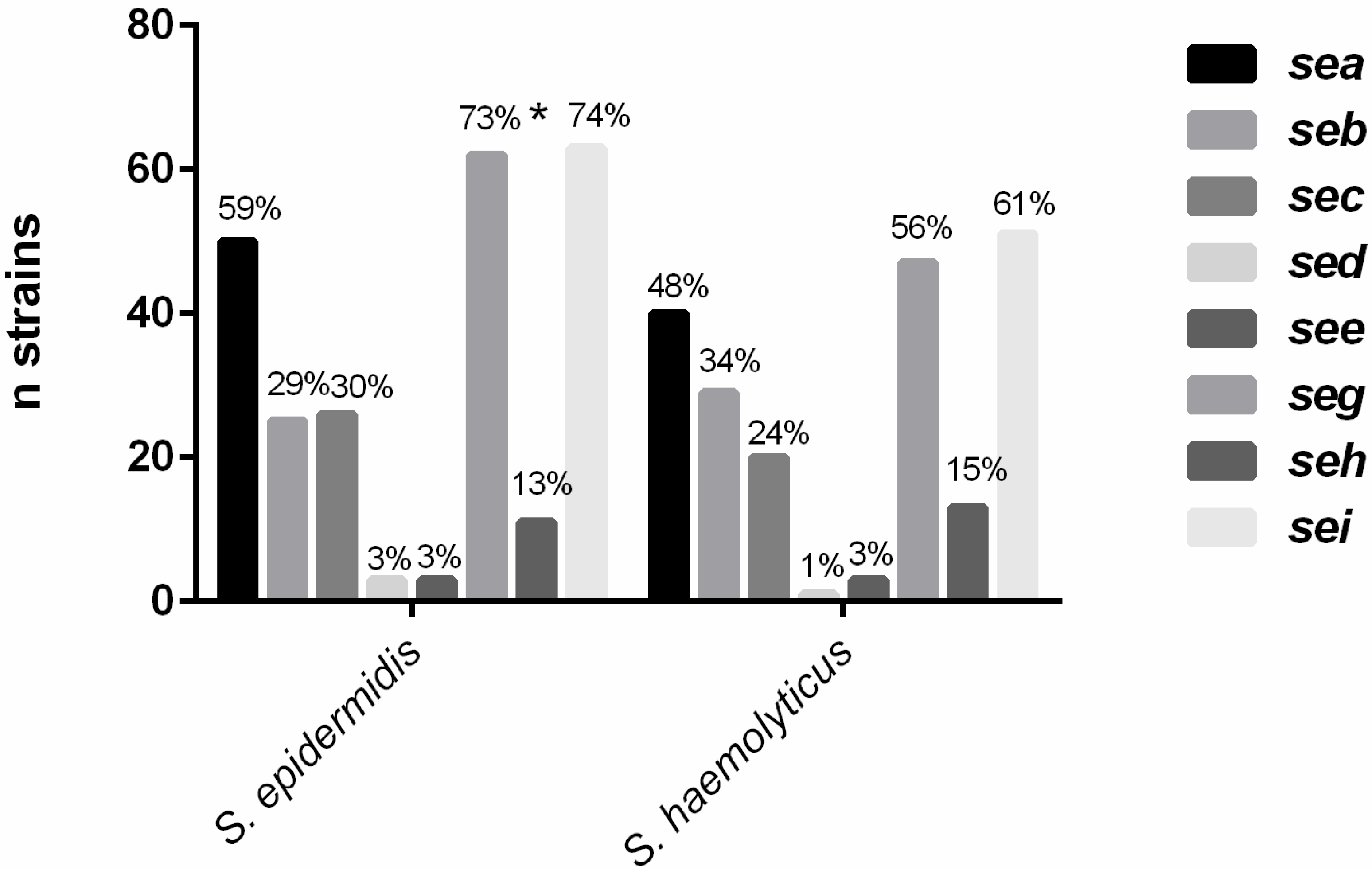
2.1. Detection of Enterotoxin Genes

A total of 169 isolates were studied, including 85 S. epidermidis and 84 S. haemolyticus.
Figure 1 illustrates the detection of enterotoxin genes in the S. haemolyticus and S. epidermidis isolates. The proportion of positive isolates was higher for the latter species, except for seb and seh (34% and 15%, respectively), which were more frequent in S. haemolyticus. The sed and see genes were rarely found (2% and 3%, respectively), while sei, seg and sea were the most frequent genes in both species. Detection of the seg gene was significantly associated with S. epidermidis (p = 0.02).
Figure 1. Detection of the enterotoxin genes seasei in S. epidermidis and S. haemolyticus isolates. * Significantly positive association with S. epidermidis.
Toxins 07 03688 g001
The sea and seb genes were concomitantly detected in 20% (n = 34) of the isolates, including 17.6% of S. epidermidis and 21.4% of S. haemolyticus, while seg and sei were concomitantly present in 52.7% (n = 89) (61.2% of S. epidermidis and 44% of S. haemolyticus). Among the strains studied, 87.6% (n = 148) carried at least one enterotoxin gene, including 95.3% (n = 81) of the S. epidermidis isolates and 79.8% (n = 67) of S. haemolyticus.

2.2. Molecular and Phenotypic Detection of Cytotoxins

The hemolysin genes hla/yidD, hlb and hld were detected in S. epidermidis using species-specific primers. The hlb and hld primers of S. epidermidis could not identify the β- and δ-toxin genes in S. haemolyticus. Novel primers were designed to identify the hla gene in S. haemolyticus. However, primers for the S. haemolyticus hlb and hld genes could not be designed, since these genes have not yet been described in that species.
The rate of detection of hla/yidD was similar in S. epidermidis and S. haemolyticus (92.9% and 91.7%, respectively). In S. epidermidis, hlb was detected in 92.9% of the isolates and hld in 95.3%. The hla and hlb genes were concomitantly present in 89.4% of the S. epidermidis isolates. β-toxin was found in 81% of the S. haemolyticus isolates and δ-toxin in 40.5%. Thirty percent of the S. haemolyticus isolates produced both β- and δ-toxin (Table 1).
Table 1. Detection of α-, β- and δ-cytotoxin genes and production.
Organisms (n)hla *hlbhldα-toxinβ-toxinδ-toxin
n%n%n%n%n%n%
S. epidermidis (85)7992.97992.98195.32428.22529.4--
S. haemolyticus (84)7791.7----7083.368813440.5
Total (169)15692.37992.98195.39455.693553440.5
* hla/yidD for S. epidermidis.
Table 2 shows the comparison between genotypic detection of hla/yidD and hlb and α- and β-toxin production. The results showed that, while 78% of the S. haemolyticus isolates carried hla and produced α-toxin, only 28% of the S. epidermidis isolates carried the hla/yidD and hlb genes and produced the respective cytotoxins. Discrepancies were observed in the case of five S. haemolyticus isolates, which were negative for hla, but showed phenotypic production, and one S. epidermidis isolate, which was negative for hlb and a producer of β-toxin.
Table 2. Comparison of the frequency of the hla and hlb genes and phenotypic production of α- and β-toxins.
Staphylococcus haemolyticusStaphylococcus epidermidis
GenesGenesGenes
Toxinhla+hlaTotalhla+ *hla− *TotalToxinhlb+hlbTotal
n(%)n (%)n (%)n (%)n (%)n (%)n (%)n (%)n (%)
α-Toxin +65 (78)5 (6)70 (84)24 (28)024 (28)β-Toxin +24(28)1 (1)25 (29)
α-Toxin −12 (14)2 (2)14 (16)55 (65)6 (7)61 (72)β-Toxin −55 (65)5 (6)60 (71)
Total77 (92)7 (8)84 (100)79 (93)6 (7)85 (100)Total79 (93)6 (7)85 (100)
+, positive; , negative. * hla/yidD.
The toxin gene profile and phenotypic toxin production of each isolate are shown in Supplemental Table S1.

3. Discussion

Staphylococcus epidermidis and S. haemolyticus are the main CoNS species colonizing the human nose [18], the most common species isolated from blood cultures [19] and are often related to catheter-associated bloodstream infections [20]. In addition to being the main cause of food poisoning, staphylococcal enterotoxins play an important role in pathological processes, such as sepsis, osteomyelitis and respiratory distress syndrome [21]. However, the enterotoxigenic potential of CoNS is controversial.
The present study showed a high frequency of enterotoxin genes in blood culture isolates, with 95.3% of S. epidermidis isolates and 79.8% of S. haemolyticus isolates carrying at least one toxin gene. The most common classical enterotoxin genes detected were sea, seb and sec, and seg and sei were the most prevalent among all enterotoxin genes. Another study [22] also showed a higher percentage of sea, seb and sec in CoNS isolated from bovine milk. Furthermore, enterotoxin a production by human CoNS isolates has also been reported [23]. The production of the classical enterotoxins SEA, SEB and SEC by clinical isolates of S. epidermidis and of SEC by S. haemolyticus has been described [5,24], with a high percentage of isolates producing a combination of two or more toxins [25]. Similar to the present study, the presence of the SEE, SEG, SEH and SEI genes and the production of these enterotoxins have also been reported [26], but studies showing the absence or a low frequency of these genes in CoNS predominate in the literature. These differences between studies may be related to bias in the method used and in the isolates studied, including the number, nature and geographic origin of the strains. Nosocomial isolates may be better equipped with virulence factors obtained by facilitated transfer through selective pressure.
In the present study, 20% of the strains were positive for both sea and seb. The frequent presence of these two genes in the same bacterium is explained by the fact that they occupy the same chromosome locus [27]. Furthermore, 61.2% of S. epidermidis and 44% of S. haemolyticus were found to be positive for both seg and sei. Several studies [28,29] have indicated a systematic association between seg and sei and a high frequency of these genes in S. aureus, which may also occur in CoNS. The concomitant presence of the seg and sei genes is expected, since these two genes are found in the egc cluster, which also contains genes encoding other staphylococcal enterotoxins [30].
Staphylococcus epidermidis has been indicated as the CoNS with the highest toxigenic potential in some studies [25]. In fact, this species showed a higher rate of enterotoxin genes compared to S. haemolyticus (95.3% vs. 79.8%, respectively). A pathogenicity island expressing several enterotoxin genes has been recently described in a clinical S. epidermidis isolate [31].
Data regarding the presence of hemolysins and hemolysin genes in CoNS are still sparse. Although 81% of S. haemolyticus isolates show β-hemolytic activity and 40% produce δ-toxin, genome sequencing was unable to identify the genes responsible for hemolysis in these species; only the α-hemolysin gene has been demonstrated. Hemolysin primers designed for S. aureus [32] and S. epidermidis, as well as hld primers designed from the hld sequence of S. simulans (GenBankAccession Number AJ223775.1; forward: AAGGGGGCAATACACATGRC; reverse: CCGAACGCTTCATTTCCGAT), could not detect these genes in S. haemolyticus. Huseby et al. [33] demonstrated species-specific differences in the β-toxin of S. schleiferi and S. epidermidis, whose proteins showed 72% and 52% homology with S. aureus β-toxin, respectively. These differences between β-hemolysins of different CoNS species may be the result of bacterial adaptation to a wide variety of potential hosts [34]. Since the hlb and hld primers for S. epidermidis could not identify these genes in S. haemolyticus and the hlb and hld genes have not yet been described in the latter species, although they are produced as demonstrated by a phenotypic detection method, considerable differences in their sequences may exist, suggesting that these toxins have a distinct structure and, consequently, different functions in CoNS species.
To our knowledge, this is the first study to detect the hla gene using specific primers for S. epidermidis and S. haemolyticus. The gene used for the primer design of strain S. epidermidis W23144 had been denoted in GenBank as “α-hemolysin” until June 2013. On that date, the authors altered the denotation of this gene to “yidD” and classified it as a membrane protein. According to previous studies, some members of the yidD family were annotated as hemolysins, which resulted from the unpublished observation reported in GenBank L36462 that the hlyA gene, which is homologous to yidD of Aeromonas hydrophila, possesses α-hemolysin activity [15,16]. Some databases show that yidD is orthologous to the proteins with hemolytic function SE1462 of S. epidermidis ATCC 12228 and SERP1356 of S. epidermidis RP62A (http://www.xbase.ac.uk/genome/buchnera-aphidicola-str-sg-schizaphis-graminum/NC_004061/BUsg015;yidD/super/orthologues).
The α-toxin/yidD-encoding gene was found in 92.9% of the S. epidermidis isolates and the hla gene in 91.7% of the S. haemolyticus isolates, while hlb was detected at the same frequency (92.9%) in S. epidermidis. On the other hand, another study [13] detected hla in only 20% of S. epidermidis isolates and the absence of hlb in all strains. β-toxin has been described in 75% of CoNS and α-hemolysis in 57% [35]. Nataro et al. [36] observed 61% of positivity for β-toxin in CoNS, while in the present study, β-hemolysin production was observed in 81% of the S. haemolyticus isolates. Moraveji et al. [37] observed double the frequency of hemolysin genes and production in human strains compared to animal strains. The importance of hlb and β-toxin is due to the ability of this protein to promote the escape of bacteria from the host immune system and to its involvement in nutrient uptake [33], permitting survival of the pathogen.
The divergence in the hld gene is so high among species that it cannot be amplified in some CoNS [12]. This diversity is demonstrated by the fact that the partial identity of this toxin gene between S. aureus and S. epidermidis is only 83% [12]. The same may apply to S. haemolyticus and may explain the lack of amplification of this gene by S. epidermidis primers in the present study. δ-hemolysin is encoded by regulatory RNAIII in S. aureus associated with the agr system [38], a system described in several staphylococcal species, including S. epidermidis and S. haemolyticus [39,40]. In the present study, the hld gene was detected in 95.3% of the S. epidermidis isolates, and δ-hemolysin was produced by 40.5% of the S. haemolyticus isolates. According to Gemmel [41], δ-hemolysin is more frequently expressed by CoNS isolated from clinically-important infections compared to inapparent human infections.
Despite the high frequency of the hla gene observed in the present study in S. epidermidis and S. haemolyticus, phenotypic production of the toxin encoded by hla seems to be more frequent in the latter species, with most hla-positive S. haemolyticus isolates (85%) expressing α-toxin. In contrast, despite the high frequency of hla/yidD and hlb in S. epidermidis, less than one-third (30%) of the isolates carrying these genes also expressed them. The absence of the gene and the presence of the toxin observed in five S. haemolyticus isolates and in one S. epidermidis isolate might be related to mutations in the sequences of these genes, such as insertion sequences that interfere with the amplification of the gene by PCR.
One limitation of the present study is the fact that the prevalence of the toxigenic genes is not equivalent to the prevalence of the expression of these genes. However, expression was demonstrated in this study by hemolysis on blood agar. Further studies using other methods to evaluate the expression of these genes, such as Western blotting, are needed. Furthermore, genome sequencing of some of these positive strains will be important to identify these genes in the genomes of S. epidermidis and S. haemolyticus.

4. Material and Methods

4.1. Isolates

The strains were isolated from blood cultures of inpatients admitted to the University Hospital of the Botucatu Medical School (Hospital das Clínicas, Faculdade de Medicina de Botucatu (HC-FMB)), Paulista State University (Universidade Estadual Paulista (UNESP)), Botucatu Campus, between 2000 and 2011. Only one isolate per patient was included in the study. The strains were isolated as described by Koneman et al. [42].

4.2. Species Identification

The genus Staphylococcus was identified as described by Koneman et al. [42]. Staphylococcus epidermidis and S. haemolyticus were identified by the simplified method proposed by Cunha et al. [43]. Species identification was genetically confirmed by PCR amplification of the 16S-23S internal transcribed spacer (ITS) region as described by Couto et al. [44] after DNA extraction with the Illustra kit (GE Healthcare, Little Chalfont, Buckinghamshire, UK). The following international reference strains were used as controls: S. epidermidis (ATCC 12228), S. epidermidis (ATCC 35983) and S. haemolyticus (ATCC 29970).

4.3. Detection of Enterotoxin Genes

PCR for the detection of enterotoxin genes was performed using the primers and parameters described by Johnson et al. [45] and Cunha et al. [4]. International reference strains were included in all reactions as positive (S. aureus American Type Culture Collection—ATCC 13565 (sea), ATCC 14458 (seb), ATCC 19095 (sec), ATCC 23235 (sed), ATCC 27664 (see), ATCC 51811 (seh), S. aureus Food Research Institute—FRI 361 (seg and sei)) and negative (S. xylosus ATCC 29971) controls. The primer sequences are shown in Table 3.
Table 3. Sequence of primers and amplicon size.
NameProductSequenceReferenceAmplicon Size (bp)
sea-1Enterotoxin ATTGGAAACGGTTAAAACGAA[29]120
sea-2GAACCTTCCCATCAAAAACA
seb-1Enterotoxin BTCGCATCAAACTGACAAACG[29]478
seb-2GCAGGTACTCTATAAGTGCC
sec-1Enterotoxin CGACATAAAAGCTAGGAATTT[29]257
sec-2AAATCGGATTAACATTATCC
sed-1Enterotoxin DCTAGTTTGGTAATATCTCCT[29]317
sed-2TAATGCTATATCTTATAGGG
see-1Enterotoxin ECAAAGAAATGCTTTAAGCAATCTTAGGCCAC[29]170
see-2CTTACCGCCAAAGCTG
seg-1Enterotoxin GAATTATGTGAATGCTCAACCCGATC[36]642
seg-2AAACTTATATGGAACAAAAGGTACTAGTTC
sei-1Enterotoxin HCAATCACATCATATGCGAAAGCAG[36]376
seh-2CATCTACCCAAACATTAGCACC
sei-1Enterotoxin ICTCAAGGTGATATTGGTGTAGG[36]576
sei-2AAAAAACTTACAGGCAGTCCATCTC
hla/yidD_epid-1α-hemolysin/yidDTTTCKCCACTTACACCMCCThis study160
hla/yidD_epid-2GGAACAGGATCAAAGCCACCT
hlb_epid-1β-hemolysinTGGTGGCGTTGGTATTGTGAThis study541
hlb_epid-2ACCCCAAGATTTCACGGACC
hla_haem-1α-hemolysinTGGGCCATAAACTTCAATCGCThis study72
hla-haem-2ACGCCACCTACATGCAGATTT
hld-epid-1δ-hemolysinATGGCAGCAGATATCATTTC[30]444
hld-epid-2CGTGAGCTTGGGAGAGAC

4.4. Detection of Hemolysin Genes

The δ-hemolysin gene, hld, was detected using the primers and parameters described by Marconi et al. [46].
The hla/yidD gene was detected in S. epidermidis isolates using primers designed with NCBI-PrimerBlast, 2008 and sequences of the strain S. epidermidis W23144 (GenBank: ACJC01000124.1) (hla/yidD_epid). The hla gene was detected in S. haemolyticus isolates using primers designed with PrimerBlast and the sequence of the strain JCSC1435 (NCBI: NC_007168.1) (hla_haem). Primers for the hlb gene in S. epidermidis were designed using PrimerBlast and the sequence of S. epidermidis RP62A (NCBI:NC_002976.3) (hlb_epid). Primers for the hlb gene in S. haemolyticus could not be designed, since this gene has not been described in that species in the NCBI-GenBank database. The reaction mixture contained 2.0 U Taq polymerase, 1× PCR buffer containing 0.75 mM MgCl2, 100 µM triphosphate deoxyribonucleotides, 1 M of each primer and 150 ng nucleic acid. The PCR conditions were as follows: for hla, one step at 94 °C for 4 min; 35 cycles at 94 °C for 1 min, 60 °C for 1 min, 72 °C for 1 min and 72 °C for 5 min; for hlb, one step at 94 °C for 4 min, 35 cycles at 94 °C for 1 min, 60 °C for 1 min, 72 °C for 1.5 min and 72 °C for 6 min. Reference strains were included in all reactions: hla/yidD_epid: S. epidermidis ATCC 12228; hla_haem: S. haemolyticus ATCC 29970; hlb_epid: S. epidermidis ATCC 12228. The primer sequences are shown in Table 3.

4.5. Phenotypic Production of β-and δ-Cytotoxins

The production of α-toxin was determined on blood agar plates containing 5% rabbit blood incubated at 37 °C for 24 h. A positive result was indicated by the formation of hemolysis zones around the isolated colonies.
β- and δ-toxin production in S. haemolyticus isolates was detected as described by Hébert and Hancock [47]. β-hemolysis was observed by the presence of a zone with incomplete hemolysis on a sheep blood agar plate incubated at 37 °C for 24 h and then overnight at 4 °C [48]. The presence of δ-toxin was verified by the presence of synergism with β-hemolysin of S. aureus ATCC 25923. For this purpose, the isolate was streaked perpendicular to the S. aureus strain on a sheep blood agar plate. The plate was incubated at 37 °C for 24 h, and δ-toxin production was observed by the formation of an arrowhead-shaped zone of hemolysis [47].

4.6. Statistical Analysis

The chi-square test was used to verify the association between variables, adopting a level of significance <0.05.

5. Conclusions

The clinical isolates of S. epidermidis and S. haemolyticus exhibited a high toxigenic potential, producing especially enterotoxins G and I. The use of novel species-specific primers for hla/yidD and hlb of S. epidermidis and for hla of S. haemolyticus revealed a high frequency of these genes in nosocomial isolates of these species. The findings demonstrate an important role of these cytotoxin genes in the establishment of these species and possibly in the development of infections caused by CoNS.

Supplementary Materials

Supplementary materials can be accessed at: https://www.mdpi.com/2072-6651/7/9/3688/s1.

Acknowledgments

This work was supported by the state funding agency São Paulo Research Foundation (FAPESP—Fundação de Amparo à Pesquisa do Estado de São Paulo; Grant 2011/15396-1) and National Council for Technological and Scientific Development (CNPq—Conselho Nacional de Desenvolvimento Científico e Tecnológico; Grant 304729/2014-0).

Author Contributions

L.P.: conceived of the study, participated in its design, carried out the experiments, analyzed the data and drafted the manuscript. C.I.B.: participated in the conception of the study and in the laboratory experiments. A.O.: contributed to the laboratory experiments. P.Y.F.M.: participated in the primers’ design. V.C.P.: contributed in the molecular experiments. M.L.R.S.C.: conceived of the study, participated in its design and coordination and revised the manuscript. All authors read and approved the final manuscript.

Conflicts of interest

The authors declare no conflict of interest.

References

  1. Vuong, C.; Otto, M. Staphylococcus epidermidisinfections. Microbes Infect. 2002, 4, 481–489. [Google Scholar] [CrossRef]
  2. Irlinger, F. Safety assessment of dairy microorganisms: Coagulase-negative staphylococci. Int. J. Food Microbiol. 2008, 126, 302–310. [Google Scholar] [CrossRef] [PubMed]
  3. Podkowik, M.; Bystroń, J.; Bania, J. Genotypes, antibiotic resistance, and virulence factors of staphylococci from ready-to-eat food. Foodborne Pathog. Dis. 2012, 9, 91–93. [Google Scholar] [CrossRef] [PubMed]
  4. Cunha, M.L.R.S.; Calsolari, R.A.O.; Araújo-Júnior, J.P. Detection of enterotoxin and toxic shock syndrome toxin 1 genes in Staphylococcus, with emphasis on coagulase-negative staphylococci. Microbiol. Immunol. 2007, 51, 381–390. [Google Scholar] [CrossRef] [PubMed]
  5. Barretti, P.; Montelli, A.C.; Batalha, J.E.; Caramori, J.C.; Cunha, M.L.R.S. The role of virulence factors in the outcome of staphylococcal peritonitis in CAPD patients. BMC Infect. Dis. 2009, 9, 212. [Google Scholar] [CrossRef] [PubMed]
  6. Taylor, A.L.; Llewelyn, M.J. Superantigen-induced proliferation of human CD4+CD25− T cells is followed by a switch to a functional regulatory phenotype. J. Immunol. 2010, 185, 6591–6598. [Google Scholar] [CrossRef] [PubMed]
  7. Frase, J.D.; Proft, T. The bacterial superantigen and superantigen-like proteins. Immunol. Rev. 2008, 225, 226–243. [Google Scholar] [CrossRef] [PubMed]
  8. Dinges, M.M.; Orwin, P.M.; Schlievert, P.M. Exotoxins of Staphylococcus aureus. Clin. Microbiol. Rev. 2000, 13, 16–34. [Google Scholar] [CrossRef] [PubMed]
  9. Coelho, S.M.O.; Reinoso, E.; Pereira, I.A.; Soares, L.C.; Demo, M.; Bogni, C.; Souza, M.M.S. Virulence factors and antimicrobial resistence of Staphylococcus aureus isolated from bovine mastitis in Rio de Janeiro. Pesq. Vet. Bras. 2009, 29, 369–374. [Google Scholar] [CrossRef]
  10. Peng, H.L.; Novick, R.P.; Kreiswirth, B.; Kornblum, J.; Schlievert, P. Cloning, characterization, and sequencing of an accessory gene regulator (agr) in Staphylococcus aureus. J. Bacteriol. 1988, 170, 4365–4372. [Google Scholar] [PubMed]
  11. McKevitt, A.I.; Bjornson, G.L.; Mauracher, C.A.; Scheifele, D.W. Amino acid sequence of a deltalike toxin from Staphylococcus epidermidis. Infect. Immun. 1990, 58, 1473–1475. [Google Scholar] [PubMed]
  12. Even, S.; Leroy, S.; Charlier, C.; Zakour, N.B.; Chacornac, J.P.; Lebert, I.; Jamet, E.; Desmonts, M.H.; Coton, E.; Pochet, S.; et al. Low occurrence of safety hazards in coagulase negative staphylococci isolated from fermented food stuffs. Int. J. Food Microbiol. 2010, 139, 87–95. [Google Scholar] [CrossRef] [PubMed]
  13. Okee, M.S.; Joloba, M.L.; Okello, M.; Najjuka, F.C.; Katabazi, F.A.; Bwanga, F.; Nanteza, A.; Kateete, D.P. Prevalence of virulence determinants in Staphylococcus epidermidis from ICU patients in Kampala, Uganda. J. Infect. Dev. Ctries. 2012, 6, 242–250. [Google Scholar] [PubMed]
  14. Schleifer, K.H.; Kloos, W.E. Isolation and characterization of staphylococci from human skin. Int. J. Syst. Bacteriol. 1975, 25, 50–61. [Google Scholar] [CrossRef]
  15. Rudd, K.E.; Humphery-Smith, I.; Wasinger, V.C.; Bairoch, A. Low molecular weight proteins: A challenge for post-genomic research. Electrophoresis 1998, 19, 536–544. [Google Scholar] [CrossRef] [PubMed]
  16. Yu, Z.; Lavèn, M.; Klepsch, M.; de Gier, J.W.; Bitter, W.; van Ulsen, P.; Luirink, J. Role for Escherichia coli YidD in Membrane Protein Insertion. J. Bacteriol. 2011, 193, 5242–5251. [Google Scholar] [CrossRef] [PubMed]
  17. Davenport, K.W.; Daligault, H.E.; Minogue, T.D.; Bishop-Lilly, K.A.; Broomall, S.M.; Bruce, D.C.; Chain, P.S.; Coyne, S.R.; Frey, K.G.; Gibbons, H.S.; et al. Complete Genome Assembly of Staphylococcus epidermidis. Genome Announc. 2014, 2, e01059-14. [Google Scholar] [CrossRef] [PubMed]
  18. Costa, S.F.; Miceli, M.H.; Anaissie, E.J. Mucosa or skin as source of coagulase-negative staphylococcal bacteraemia? Lancet Infect. Dis. 2004, 4, 278–286. [Google Scholar] [CrossRef] [PubMed]
  19. Falcone, M.; Giannella, M.; Raponi, G.; Mancini, C.; Venditti, M. Teicoplanin use and emergence of Staphylococcus haemolyticus: Is there a link? Clin. Microbiol. Infect. 2006, 12, 96–97. [Google Scholar] [CrossRef] [PubMed]
  20. Mack, D.; Rohde, H.; Harris, L.G.; Davies, A.P.; Horstkotte, M.A.; Knobloch, J.K. Biofilm formation in medical device-related infection. Int. J. Artif. Organs 2006, 29, 343–359. [Google Scholar] [PubMed]
  21. Michelin, A.F.; Carlos, I.Z. Interaction of staphylococcal exterotoxins with the immmune system of the host. Rev. Ciênc. Farm 2003, 24, 83–95. [Google Scholar]
  22. De Freitas Guimarães, F.; Nóbrega, D.B.; Richini-Pereira, V.B.; Marson, P.M.; de Figueiredo Pantoja, J.C.; Langoni, H. Enterotoxin genes in coagulase-negative and coagulase-positive staphylococci isolated from bovine milk. J. Dairy Sci. 2013, 96, 2866–2872. [Google Scholar] [CrossRef] [PubMed]
  23. Rojas, M.B.; Antonelli, C.M.; Pereira Franchi, E.P.L.; Cunha, M.L.R.S. Detection of enterotoxin a in coagulase-negative staphylococci isolated from nutrition students. Arch. Clin. Microbiol. 2012, 3. Available online: http://imedpub.com/ojs/index.php/acmicrob/article/view/414 (accessed on 10 September 2014). [Google Scholar] [CrossRef]
  24. Crass, B.; Bergdoll, M.S. Involvement of coagulase-negative staphylococci in toxic shock syndrome. J. Clin. Microbiol. 1986, 23, 43–45. [Google Scholar] [PubMed]
  25. Cunha, M.L.R.S.; Rugolo, L.M.S.S.; Lopes, C.A.M. Study of virulence factors in coagulase-negative staphylococci isolated from newborns. Mem. Inst. Oswaldo Cruz 2006, 101, 661–668. [Google Scholar] [CrossRef] [PubMed]
  26. Vasconcelos, N.G.; Pereira, V.C.; Araújo-Júnior, J.P.; Cunha, M.L.R.S. Molecular detection of enterotoxins E, G, H and I in Staphylococcus aureus and coagulase-negative staphylococci isolated from clinical samples of newborns in Brazil. J. App. Microbiol. 2011, 111, 749–762. [Google Scholar] [CrossRef] [PubMed]
  27. Jett, M.; Ionin, B.; Das, R.; Neill, R. The staphylococcal enterotoxins. In Molecular Medical Microbiology; Sussman, M., Ed.; Academic Press: San Diego, CA, USA, 2001; pp. 1089–1116. [Google Scholar]
  28. Lammler, C.; Akineden, O.; Annemuller, C.; Wolter, W.; Zschock, M. Molecular analysis of virulence factors of Staphylococcus aureus isolated from bovine sub-clinical mastitis. In Proceedings of the Symposium on Immunology of Ruminant Mammary Gland, Stresa, Italy, 11–14 June 2002; pp. 226–330.
  29. Akineden, O.; Annemuller, C.; Hassan, A.A.; Lammler, C.; Wolter, W.; Zschock, M. Toxin genes and other characteristics of Staphylococcus aureus isolates from milk of cows with mastitis. Clin. Diagn. Lab. Immunol. 2001, 8, 959–964. [Google Scholar] [CrossRef] [PubMed]
  30. Rosec, J.P.; Gigaud, O. Staphylococcal enterotoxin genes of classical and new types detected by PCR in France. J. Food Microbiol. 2002, 77, 61–70. [Google Scholar] [CrossRef]
  31. Madhusoodanan, J.; Seo, K.S.; Remortel, B.; Park, J.Y.; Hwang, S.Y.; Fox, L.K.; Park, Y.H.; Deobald, C.F.; Wang, D.; Liu, S.; et al. An enterotoxin-bearing pathogenicity island in Staphylococcus epidermidis. J. Bacteriol. 2011, 193, 1854–1862. [Google Scholar] [CrossRef] [PubMed]
  32. Jarraud, S.; Mougel, C.; Thioulouse, J.; Lina, G.; Meugnier, H.; Forey, F.; Nesme, X.; Etienne, J.; Vandenesch, F. Relationships between Staphylococcus aureus genetic background, virulence factors, agr groups (alleles), and human disease. Infect. Immun. 2002, 70, 631–641. [Google Scholar] [CrossRef] [PubMed]
  33. Huseby, M.; Shi, K.; Brown, C.K.; Digre, J.; Mengistu, F.; Seo, K.S.; Bohach, G.A.; Schlievert, P.M.; Ohlendorf, D.H.; Earhart, C.A. Structure and biological activities of beta toxin from Staphylococcus aureus. J. Bacteriol. 2007, 189, 8719–8726. [Google Scholar] [CrossRef] [PubMed]
  34. Dziewanowska, K.; Edwards, V.M.; Deringer, J.R.; Bohach, G.A.; Guerra, D.J. Comparison of the β-toxins from Staphylococcus aureus and Staphylococcus intermedius. Arch. Biochem. Biophys. 1996, 335, 102–108. [Google Scholar] [CrossRef] [PubMed]
  35. Bedidi-Madani, N.; Greenland, T.; Richard, Y. Exoprotein and slime production by coagulase-negative staphylococci isolated from goats’ milk. Vet. Microbiol. 1998, 59, 139–145. [Google Scholar] [CrossRef]
  36. Nataro, J.P.; Corcoran, L.; Zirin, S.; Swink, S.; Taichman, N.; Goin, J.; Harris, M.C. Prospective analysis of coagulase-negative staphylococcal infection in hospitalized infants. J. Pediatr. 1994, 125, 798–804. [Google Scholar] [CrossRef]
  37. Moraveji, Z.; Tabatabaei, M.; Aski, S.H.; Khoshbakht, R. Characterization of hemolysins of Staphylococcus strains isolated from human and bovine, southern Iran. Iran. J. Vet. Res. 2014, 15, 326–330. [Google Scholar]
  38. Novick, R.P. Autoinduction and signal transduction in the regulation of staphylococcal virulence. Mol. Microbiol. 2003, 48, 1429–1449. [Google Scholar] [CrossRef] [PubMed]
  39. Dordet-Frisoni, E.; Dorchies, G.; De, A.C.; Talon, R.; Leroy, S. Genomic diversity in Staphylococcus xylosus. Appl. Environ. Microbiol. 2007, 73, 7199–7209. [Google Scholar] [CrossRef] [PubMed]
  40. Wuster, A.; Babu, M.M. Conservation and evolutionary dynamics of the agr cell-to-cell communication system across firmicutes. J. Bacteriol. 2008, 190, 743–746. [Google Scholar] [CrossRef] [PubMed]
  41. Gemmel, C.G. Coagulase-negative staphylococci. J. Med. Microbial. 1986, 22, 285–295. [Google Scholar] [CrossRef] [PubMed]
  42. Koneman, E.W.; Allen, S.D.; Janda, W.M.; Schreckenberger, P.C.; Winn, W.C., Jr. Color Atlas and Textbook of Diagnostic Microbiology, 5th ed.; Lippincott: Philadelphia, PA, USA, 1997. [Google Scholar]
  43. Cunha, M.L.R.S.; Sinzato, Y.K.; Silveira, L.V.A. Comparision of methods for identification of Coagulase-negative Staphylococci. Mem. Inst. Oswaldo Cruz 2004, 99, 855–860. [Google Scholar] [CrossRef]
  44. Couto, I.; Pereira, S.; Miragaia, M.; Sanches, I.S.; Lencastre, H. Identification of clinical staphylococcal isolates from humans by Internal Transcribed Spacer PCR. J. Clin. Microbiol. 2001, 39, 3099–3103. [Google Scholar] [CrossRef] [PubMed]
  45. Johnson, W.M.; Tyler, S.D.; Ewan, E.P.; Ashton, F.E.; Pollard, D.R.; Rozee, K.R. Detection of genes for enterotoxins, exfoliative toxins, and toxic shock syndrome toxin 1 in Staphylococcus aureus by the polymerase chain reaction. J. Clin. Microbiol. 1991, 29, 426–430. [Google Scholar] [PubMed]
  46. Marconi, C.; Cunha, M.L.R.S.; Araujo, J.P., Jr.; Rugolo, L.M.S.S. Standardization of the PCR technique for the detection of delta toxin in Staphylococcus spp. J. Venom. Anim. Toxins Incl. Trop. Dis. 2005, 11, 117–128. [Google Scholar] [CrossRef]
  47. Hébert, G.A.; Hancock, G.A. Synergistic hemolysis exhibited by species of Staphylococci. J. Clin. Microbiol. 1985, 22, 409–415. [Google Scholar] [PubMed]
  48. Freer, J.H.; Arbuthnott, J.P. Toxins of Staphylococcus aureus. Pharmacol. Ther. 1983, 19, 55–106. [Google Scholar] [CrossRef]

