Naturally Occurring Food Toxins

Abstract
:1. Introduction
2. Regulatory Accommodation
The term “food” means (1) articles used for food or drink for man or other animals, (2) chewing gum, and (3) articles used for components of any such article.
§402. A food shall be deemed to be adulterated—(a) (1) If it bears or contains any poisonous or deleterious substance which may render it injurious to health; but in case the substance is not an added substance such food shall not be considered adulterated under this clause if the quantity of such substance in such food does not ordinarily render it injurious to health…
§406 Any poisonous or deleterious substance added to any food, except where such substance is required in the production thereof or cannot be avoided by good manufacturing practice shall be deemed to be unsafe for purposes of the application of clause (2) (A) of section 402(a); but when such substance is so required or cannot be so avoided, the Secretary shall promulgate regulations limiting the quantity therein or thereon to such extent as he finds necessary for the protection of public health, and any quantity exceeding the limits so fixed shall also be deemed to be unsafe for purposes of the application of clause (2) (A) of section 402(a).
3. Factors Driving the Acceptance of Certain Foods

| Disease/Syndrome | Causative Food | Cause | Comment |
|---|---|---|---|
| Disaccharide intolerance | Sucrose, dextrins | Autosomal recessive trait characterized by the deficiency or absence of enzymes sucrase and isomaltase in the intestine. | Attacks characterized by bloating and diarrhea. |
| Favism | Broadbean (Vicia fava) | X-linked recessive trait resulting in low amounts of glucose-P-dehydrogenase. Several subtypes known. | Hemolytic anemia may result from consumption of offending foods. |
| Galactosemia | Galactose and lactose (dairy products) | Autosomal recessive trait with low levels of any one of three enzymes directly responsible for galactose metabolism. | High levels of galactose in the blood results in hepatomegaly, cirrhosis, and renal failure. Infant mortality is ~75%. |
| Gluten intolerance | Wheat, barley, gluten containing foods | Autoimmune disease | Sensitivity to storage protein (gliadin) in some grains. |
| Lactose intolerance | Dairy products | Inborn error of metabolism—low or no lactase enzyme in the intestine. | Lactase is required to cleave lactose (a disaccharide of galactose and glucose). Bloating and diarrhea may develop. |
| Ornithine transcarbamylase deficiency | Dietary nitrogen (primarily meat) | X-linked recessive disorder resulting in low production of hepatic ornithine transcarbamylase interrupting the urea cycle and leading to accumulation of ammonia. | Although usually first seen in neonates, there may be an adult onset. |
| Citrullinemia is another genetic disease affecting the urea cycle. | |||
| Phenylketonuria (PKU disease) | Phenylalanine in foods | Autosomal recessive trait characterized by inadequate hepatic phenylalanine hydroxylase. | Leads to accumulation of phenylpyruvate which may accumulate in the brain and lead to seizures, mental retardation, etc. Products containing phenylalaine must be labeled. |
| Refractory sprue | Wheat, barley and rye | Autoimmune disorder triggered by gliadin, a gluten storage protein. | Unlike common celiac sprue, adherence to a gluten-free diet may not cause symptoms to abate. |
| Trimethylaminuria | Fish | Autosomal recessive resulting in low production of flavin containing monoxygenase enzyme 3 (FMO3). | Fish odor syndrome. Failure to breakdown trimethylamine, a build of which results in a fish odor. |
| Very long chain Acyl CoA dehydrogenase deficiency (LCAD) | Very long chain fatty acids | Autosomal recessive trait resulting from a mutation in the HADHA gene. | Prevents mitochondrial metabolism of very long chain fatty acids. |
| Enzyme or Transporter | Food | Drug |
|---|---|---|
| CYP1A2 | Caffeine, theophylline, grapefruit juice (naringen and furanocourmarins bergmottin and dihydroxybergamotin), grape juice, cruciferous vegetables, apiaceous vegetables, cooked meat | Clozapine, fluvoxamine, imipramine |
| CYP2E1 | Watercress and possibly other isothiocyanate-containing cruciferous vegetables; polyunsaturated fatty acids (corn oil, menhaden oil) | Ethanol, halothane, enflurane |
| CYP3A4 | Grapefruit, orange juice, red wine, possibly other polyphenol-containing substances, St. Johns wort, garlic | Ketoconazole, cyclosporine, erythromycin, protease inhibitors, HMG-CoA reductase inhibitors |
| UGT and GST | Brussels sprouts, cabbage, watercress, broccoli | Acetaminophen, oxazepam, morphine, ibuprofen |
| P-glycopeptide and OATP | Vegetables, fruit juice, St. Johns wort | Digoxin, cyclosporine, parvastatin |
4. Toxin Incorporation during Growth, Storage or Processing
4.1. Environmental contaminants
4.1.1. Selenium in grain
4.1.2. Methyl mercury in seafood
4.2. Naturally formed substances
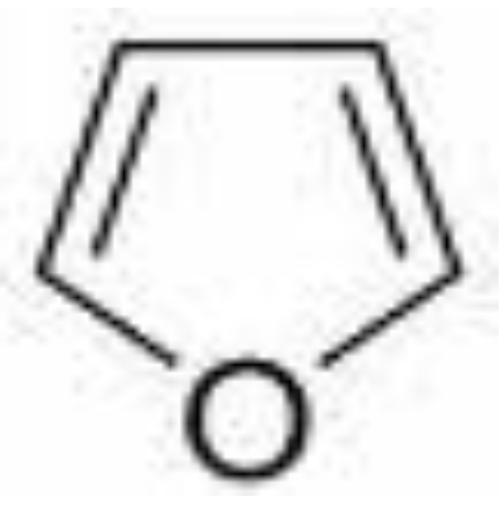
4.2.1. β-Thujone
4.2.2. Prussic acid in cherry, apple and peach pits
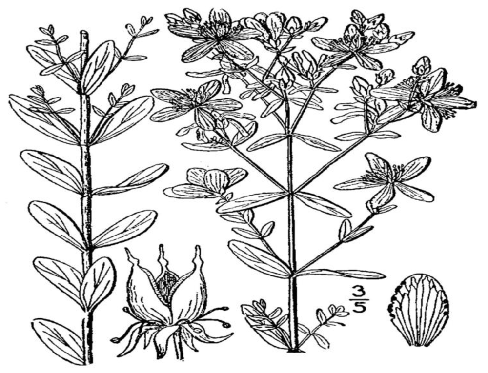
4.2.3. Hypericin in St. John’s wort

4.2.4. Goitrogens (glucosinolates) in Brassica spp.
4.2.5. Erucic acid in rape
4.2.6. Furocoumarins
4.2.7. Amylase inhibitors
4.2.8. Lectins in legumes
4.2.9. Anti-thiamine compounds
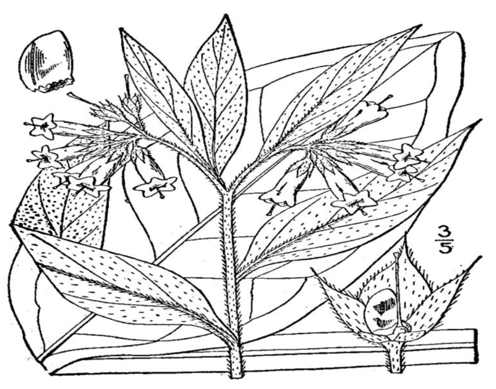
4.2.10. Pyrrolizidine alkaloids

4.2.11. Rhubarb and oxalic acid
4.2.12. Zucchini and cucurbitacins
4.2.13. Coumarins (tonka bean, woodruff, clover)
4.2.14. Phytates and phytic acid
4.2.15. Hypoglycin in Ackee
4.2.16. Safrole
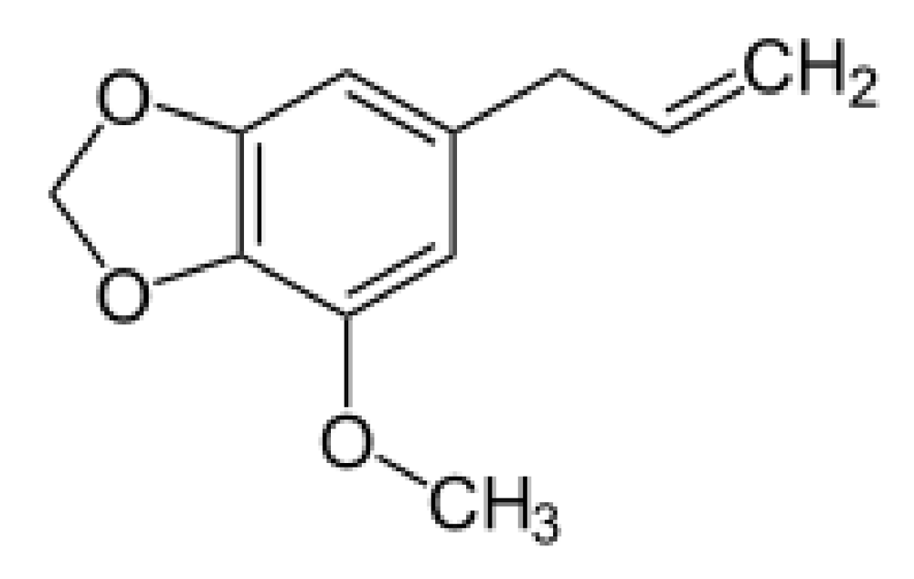
4.2.17. Myristicin

4.2.18. Tomatine in tomatoes
4.2.19. Japanese star anise
4.3. Substances formed as the result of product abuse
4.3.1. Glycoalkaloids (solanine and chaconine) in potatoes
4.3.2. Furocoumarin in parsnips
4.4. Substances formed as the result of processing
4.4.1. Heterocyclic aromatic amines
4.4.2. Polycyclic aromatic hydrocarbons
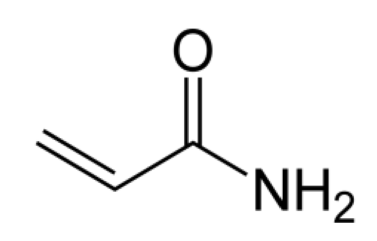
4.4.3. Acrylamide

4.4.4. Chloropropanols
4.4.5. Furan

4.4.6. Trans fatty acids
4.4.7. Nitrosamines formed during drying, curing and preserving
4.4.8. Biogenic amines
5. Substances Passed from Animals to Humans
5.1. Toxins in seafood
5.1.1. Toxins involving algae
5.1.1.1. Paralytic shellfish poisoning
5.1.1.2. Neurotoxic shellfish poisoning
5.1.1.3. Amnesic shellfish poisoning (Domoic acid)

5.1.1.4. Diarrhetic shellfish poisoning
5.1.1.5. Ciguatera poisoning
5.1.2. Toxins not involving algae
5.1.2.1. Gempylotoxin

5.1.2.2. Tetramine in whelks
5.1.2.3. Trimethylamine oxide
5.2. Toxins from animal, non-seafood sources passed on to humans
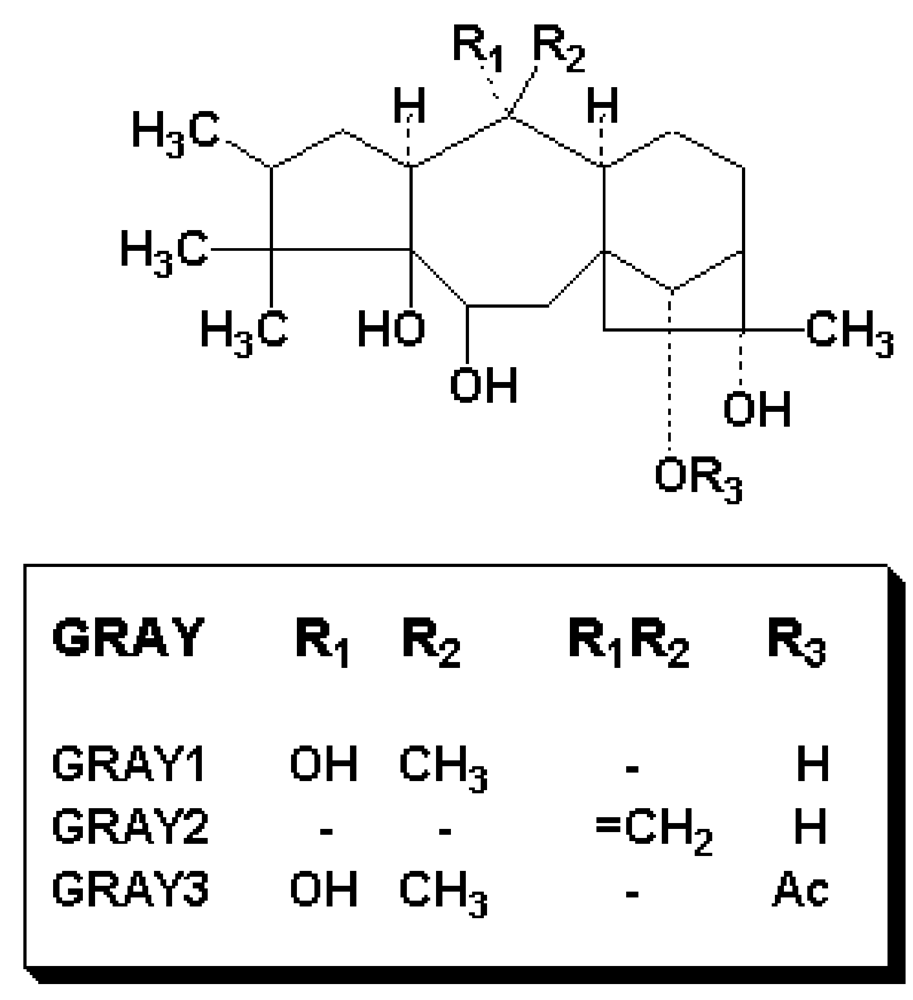
5.2.1. Grayanotoxins in honey and direct contact with food

5.2.2. Tremetol contamination of milk from white snakeroot
6. Conclusions
References
- Institute of Medicine (IOM). Dietary Reference Intakes for Vitamin A, Vitamin K, Arsenic, Boron, Chromium, Copper, Iodine, Iron, Manganese, Molybdenum, Nickel, Silicon, Vanadium and Zinc; National Academies Press: Washington, DC, USA, 2001. [Google Scholar]
- Burton, G.W.; Ingold, K.U. beta-Carotene: An unusual type of lipid antioxidant. Science 1984, 224, 569–573. [Google Scholar]
- Bannister, B.; Gibsburg, G.; Shneerson, T. Cardiac arrest due to liquoriceinduced hypokalaemia. Br. Med. J. 1977, 2, 738–739. [Google Scholar]
- Isbrucker, R.A.; Burdock, G.A. Risk and safety assessment on the consumption of licorice root (Glycyrrhiza sp.), its extract and powder as a food ingredient, with emphasis on the pharmacology and toxicology of glycyrrhizin. Regul. Toxicol. Pharmacol. 2006, 46, 167–192. [Google Scholar] [CrossRef] [PubMed]
- United States Government Accountability Office (GAO). Food safety. FDA should strengthen its oversight of food ingredients determined to be generally recognized as safe (GRAS). GAO-10-246. February 2010. Available online: http://www.gao.gov/new.items/d10246.pdf (accessed on 21 July 2010).
- Food and Drug Law Institute. Sec. 201. [321] Definitions. In FDCA Statutory Supplement Including FDA Amendments Act of 2007 and Related Sections of Additional Statutes; Food and Drug Law Institute: Washington, DC, USA, 2008; pp. 1–2. [Google Scholar]
- Food and Drug Law Institute. Sec. 402. [342] Adulterated Food. In FDCA Statutory Supplement Including FDA Amendments Act of 2007 and Related Sections of Additional Statutes; Food and Drug Law Institute: Washington, DC, USA, 2008; p. 31. [Google Scholar]
- Food and Drug Law Institute. Sec. 406. [346] Tolerances for Poisonous Ingredients in Food. In FDCA Statutory Supplement Including FDA Amendments Act of 2007 and Related Sections of Additional Statutes; Food and Drug Law Institute, 2008; Volume Washington, DC, USA, p. 31. [Google Scholar]
- Kracov, D.A. The regulation of foods and food additives. In A Practical Guide to Food and Drug Law Regulation, 2nd; Piña, K.R., Pines, W.L., Eds.; Food and Drug Law Institute: Washington, DC, USA, 2002; pp. 159–214. [Google Scholar]
- Tullo, A. Newscripts: Vile weed or essential ingredient? Chem. Eng. News 2010, 88, 72. [Google Scholar]
- Fischer, R.; Griffin, F.; Kaplan, A.R. Taste thresholds, cigarette smoking, and food dislikes. Med. Exp. Int. J. Exp. Med. 1963, 9, 151–167. [Google Scholar]
- Goff, S.A.; Klee, H.J. Plant volatile compounds: Sensory clues for health and nutritional value? Science 2006, 311, 815–819. [Google Scholar] [PubMed]
- National Organization for Rare Disorders (NORD). 2010. Available online: http://www.rarediseases.org (accessed on 21 July 2010).
- Carabin, I.G.; Magnuson, B.A. New Labeling Requirements for Food Allergens. Nutritional Outlook. April 2006. Available online: http://www.nutritionaloutlook.com/article.php?ArticleID=2096 (accessed on 21 July 2010).
- Kotsonis, F.N.; Burdock, G.A. Food Toxicology. In Casarett and Doull’s Toxicology: The Basic Science of Poisons, 7th; Klaassen, C.D., Ed.; McGraw-Hill: New York, NY, USA, 2008; pp. 1191–1236. [Google Scholar]
- Sors, T.G.; Ellis, D.R.; Salt, D.E. Selenium uptake, translocation, assimilation and metabolic fate in plants. Photosynth. Res. 2005, 86, 373–389. [Google Scholar]
- Yang, G.; Wang, S.; Zhou, R.; Sun, S. Endemic selenium intoxication of humans in China. Am. J. Clin. Nutr. 1983, 37, 872–881. [Google Scholar]
- Reilly, C. Selenium: Physiology, dietary sources and requirements. In Encyclopaedia of Human Nutrition; Sadler, M.J., Ed.; Academic: San Diego, CA, USA, 1998; pp. 1752–1758. [Google Scholar]
- United States Environmental Protection Agency (EPA). Selenium and compounds (CASRN 7782-49-2). 1 March 1991. Available online: http://www.epa.gov/iris/subst/0472.htm (accessed on 21 July 2010).
- Waldron, H.A. Did the Mad Hatter have mercury poisoning? Br. Med. J. 1983, 287, 1961. [Google Scholar] [CrossRef]
- Carrington, C.; Bolger, M. An Exposure Assessment for Methylmercury from Seafood for Consumers in the United States. Available online: http://www.fda.gov/downloads/Food/FoodSafety/Product-SpecificInformation/Seafood/FoodbornePathogensContaminants/Methylmercury/UCM114740.pdf (accessed on 21 July 2010).
- United States Food and Drug Administration (FDA). Chapter 10: Methyl Mercury. Fish and Fisheries Products Hazards and Controls Guidance, 3rd ed. June 2001. Available online: http://www.fda.gov/Food/GuidanceComplianceRegulatoryInformation/GuidanceDocuments/Seafood/ucm092041.htm (accessed on 21 July 2010).
- Hutt, P.B.; Merrill, R.A.; Grossman, L.W. Food and Drug Law, 3rd ed; Foundation Press: New York, NY, USA, 2007; p. 369. [Google Scholar]
- European Commission. Scientific Committee on Food. Opinion of the scientific committee on food on thujone. 6 February 2003. Available online: http://ec.europa.eu/food/fs/sc/scf/out162_en.pdf (accessed on 21 July 2010).
- United States Food and Drug Administration. Code of Federal Regulations (CFR) 21 § 172.510; U.S. Government Printing Office: Washington, DC, USA, 2006; pp. 55–57.
- Galli, C.L.; Galli, G.; Tragni, E.; Caruso, D.; Fiecchi, A. Quantitative analysis of alpha, beta-thujone, pulegone, safrole, coumarin and beta-asarone in alcoholic beverages by selected-ion monitoring. J. Appl. Toxicol. 1984, 4, 273–276. [Google Scholar] [CrossRef] [PubMed]
- Lawrence, B.M. Progress in essential oils. Sage oil. In Essential Oils: 2001–2004; Allured Publishing: Carol Stream, IL, USA, 2006; pp. 25–30. [Google Scholar]
- Ben Farhat, M.; Jordán, M.J.; Chaouech-Hamada, R.; Landoulsi, A.; Sotomayor, J.A. Variations in essential oil, phenolic compounds, and antioxidant activity of tunisian cultivated Salvia officinalis L. J. Agric. Food Chem. 2009, 57, 10349–10356. [Google Scholar] [PubMed]
- Patocka, J.; Plucar, B. Pharmacology and toxicology of absinthe. J. Appl. Biomed. 2003, 1, 199–205. [Google Scholar]
- Millet, Y.; Jouglard, J.; Steinmetz, M.D.; Tognetti, P.; Joanny, P.; Arditti, J. Toxicity of some essential plant oils. Clinical and experimental study. Clin. Toxicol. 1981, 18, 1485–1498. [Google Scholar] [CrossRef] [PubMed]
- Bonkovsky, H.L.; Cable, E.E.; Cable, J.W.; Donohue, S.E.; White, E.C.; Greene, Y.J.; Lambrecht, R.W.; Srivastava, K.K.; Arnold, W.N. Porphyrogenic properties of the terpenes camphor, pinene, and thujone. Biochem. Pharmacol. 1992, 43, 2359–2368. [Google Scholar]
- United States National Toxicology Program (NTP). Alpha-Thujone. 10 December 1997. Available online: http://ntp.niehs.nih.gov/index.cfm?objectid=03DB8C36-E7A1-9889-3BDF8436F2A8C51F (accessed on 21 July 2010).
- Hold, K.M.; Sirisoma, N.S.; Casida, J.E. Detoxification of alpha- and beta-thujones (the active ingredients of absinthe): Site specificity and species differences in cytochrome P450 oxidation in vivo and in vivo. Chem. Res. Toxicol. 2001, 14, 589–595. [Google Scholar]
- Perdue University, Cooperative Extension Service (Perdue). Indiana plants poisonous to livestock and pets. Available online: http://www.vet.purdue.edu/toxic/plant46.htm (accessed on 21 July 2010).
- Merck. Cyanide Poisoning: Introduction. The Merck Veterinary Manual. 2008. Available online: http://www.merckvetmanual.com/mvm/index.jsp?cfile=htm/bc/210800.htm&word=prussic%2cacid (accessed on 21 July 2010).
- Panter, K.E. Natural toxins of plant origin. In Toxins in Food; Dabrowski, W.M., Sikorski, Z.E., Eds.; CRC Press: Boca Raton, FL, USA, 2004; pp. 11–63. [Google Scholar]
- Wentworth, J.M.; Agostini, M.; Love, J.; Schwabe, J.W.; Chatterjee, V.K. St John's wort, a herbal antidepressant, activates the steroid X receptor. J. Endocrinol. 2000, 166, R11–R16. [Google Scholar]
- Karioti, A.; Bilia, A.R. Hypericins as potential leads for new therapeutics. Int. J. Mol. Sci. 2010, 11, 562–594. [Google Scholar]
- Hammerness, P.; Basch, E.; Ulbricht, C.; Barrette, E.P.; Foppa, I.; Basch, S.; Bent, S.; Boon, H.; Ernst, E. St. John’s Wort: A systematic review of adverse effects and drug interactions for the consultation psychiatrist. Psychosomatics 2003, 44, 271–282. [Google Scholar] [CrossRef] [PubMed]
- Britton, N.L.; Brown, A. Hypericum perforatum L. In An illustrated Flora of the Northern United States, Canada and the British Possessions; Charles Scribner's Sons: New York, NY, USA, 1913; Volume 2, p. 533, USDA-NRCS PLANTS Database. Available online: http://plants.usda.gov/java/profile?symbol=HYPE&photoID=hype_001_avd.tif (accessed on 31 August 2010). [Google Scholar]
- State of Victoria Department of Primary Industries (Victoria). Landcare notes. St. John’s wort. 2007. Available online: http://www.dpi.vic.gov.au/dpi/nreninf.nsf/93a98744f6ec41bd4a256c8e00013aa9/9f65b9c41bbc7aa5ca25737500119160/$FILE/LC0177_Sep07.pdf (accessed on 21 July 2010).
- Greer, M.A. Goitrogenic substances in food. Am. J. Clin. Nutr. 1957, 5, 440–444. [Google Scholar]
- Conn, E.E. Cyanogenetic Glycosides. In Toxicants Occurring Naturally in Foods, 2nd ed; Committee on Food Protection, Food and Nutrition Board, National Research Council, National Academy of Sciences: Washington, DC, USA, 1973; pp. 299–308. [Google Scholar]
- VenEtten, C.H.; Wolff, I.A. Natural sulfur compounds. In Toxicants Occurring Naturally in Foods, 2nd ed; Committee on Food Protection, Food and Nutrition Board, National Research Council, National Academy of Sciences: Washington, DC, USA, 1973; pp. 210–234. [Google Scholar]
- United States Department of Agriculture (USDA). Plants Profile: Brassica napus L. Available online: http://plants.usda.gov/java/profile?symbol=BRNA (accessed on 21 July 2010).
- Carroll, K.K. Erucic acid as the factor in rape oil affecting adrenal cholesterol in the rat. J. Biol. Chem. 1953, 200, 287–292. [Google Scholar]
- Chien, K.R.; Bellary, A.; Nicar, M.; Mukherjee, A.; Buja, L.M. Induction of a reversible cardiac lipidosis by a dietary long-chain fatty acid (erucic acid). Am. J. Pathol. 1983, 112, 68–77. [Google Scholar]
- Ratanasethkul, C.; Riddell, C.; Salmon, R.E.; O’Neil, J.B. Pathological changes in chickens, ducks and turkeys fed high levels of rapeseed oil. Can. J. Comp. Med. 1976, 40, 360–369. [Google Scholar]
- Mattson, F.H. Potential toxicity of food lipids. In Toxicants Occurring Naturally in Foods, 2nd ed; Committee on Food Protection, Food and Nutrition Board, National Research Council, National Academy of Sciences: Washington, DC, USA, 1973; pp. 189–209. [Google Scholar]
- Mori, H.; Tanaka, T.; Hirono, I. Toxicants in Food: Naturally Occurring. In Nutrition and Chemical Toxicity; Ioannides, C., Ed.; John Wiley & Sons: West Sussex, England, UK, 1998; pp. 1–27. [Google Scholar]
- Biotechnology Australia (Australian Government). "What is canola?" A problem with weeds—the canola story. Available online: http://www.biotechnologyonline.gov.au/foodag/weeds.html (accessed on 21 July 2010).
- Health Canada. Low Erucic Acid Rapeseed (Lear) Oil Derived From Canola-quality Brassica juncea (L.) CZERN. Lines PC 97-03, PC98-44 AND PC98-45. 27 March 2003. Available online: http://www.hc-sc.gc.ca/fn-an/gmf-agm/appro/low_erucic-faible_erucique-eng.php (accessed on 21 July 2010).
- Wagstaff, D. Dietary exposure to furocoumarins. Regul. Toxicol. Pharmacol. 1991, 14, 261–272. [Google Scholar]
- Ashwood-Smith, M.J.; Ceska, O.; Chaudhary, S.K.; Warrington, P.J.; Woodcock, P. Detection of furocoumarins in plants and plant products with an ultrasensitive biological photoassay employing a DNA-repair-deficient bacterium. J. Chem. Ecol. 1986, 12, 915–932. [Google Scholar]
- Zobel, A.M.; Brown, S.A. Dermatitis-inducing psoralens on the surfaces of seven medicinal plant species. J. Toxicol. Cutaneous Ocul. Toxicol. 1991, 10, 223–231. [Google Scholar]
- Dunnick, J.K. NTP Technical Report on the Toxicology and Carcinogenesis Studies of 8-Methoxypsoralen (CAS No. 298-81-7) in F344/N Rats. NIH Publication No. 89-2814; National Toxicology Program: Research Triangle Park, NC, USA, 1989. [Google Scholar]
- International Agency for Research on Cancer (IARC). Summaries & Evaluations, 8-Methoxypsoralen (Methoxsalen) plus ultraviolet radiation. IARC, 1987; 7 (Suppl.), p. 261. Available online: http://www.inchem.org/documents/iarc/suppl7/methoxypsoralen-8.html (accessed on 21 July 2010).
- Stern, R.S.; Nichols, K.T.; Vakeva, L.H. Malignant melanoma in patients treated for psoriasis with methoxsalen (psoralen) and ultraviolet A radiation (PUVA). The PUVA follow-up study. N. Engl. J. Med. 1997, 336, 1041–1045. [Google Scholar] [CrossRef] [PubMed]
- International Agency for Research on Cancer (IARC). Summaries & Evaluations, 5-Methoxypsoralen. IARC, 1986; 40, p. 327. Available online: http://www.inchem.org/documents/iarc/vol40/5-methoxypsoralen.html (accessed on 21 July 2010).
- Girennavar, B.; Poulose, S.M.; Jayaprakasha, G.K.; Bhat, N.G.; Patil, B.S. Furocoumarins from grapefruit juice and their effect on human CYP3A4 and CYP1B1 isoenzymes. Bioorg. Med. Chem. 2006, 14, 2606–2612. [Google Scholar]
- Bailey, D.G.; Malcom, J.; Arnold, O.; Spence, J.D. Grapefruit juice-drug interactions. Br. J. Clin. Pharmacol. 1998, 46, 101–110. [Google Scholar]
- Duke, J.A. Handbook of Phytochemical Constituents of GRAS Herbs and Other Economic Plants; CRC Press: Boca Raton, FL, USA, 1992; pp. 171, 174, 180, 183. [Google Scholar]
- Placzek, M.; Fromel, W.; Eberlein, B.; Gilbertz, K.P.; Przybilla, B. Evaluation of phototoxic properties of fragrances. Acta Derm. Venereol. 2007, 87, 312–316. [Google Scholar]
- Marzulli, F.N.; Maibach, H.I. Perfume phototoxicity. J. Soc. Cosmet. Chem. 1970, 21, 695–715. [Google Scholar]
- Coulumbe, R.A., Jr. Natural toxins and chemopreventives in plants. In Food Toxicology; Helferich, W., Winter, C.K., Eds.; CRC Press: Boca Raton, FL, USA, 2001; p. 152. [Google Scholar]
- Schlatter, J.; Zimmerli, B.; Dick, R.; Panizzon, R.; Schlatter, C. Dietary intake and risk assessment of phototoxic furocoumarins in humans. Food Chem. Toxicol. 1991, 29, 523–530. [Google Scholar]
- Deshpande, S.S. Food Additives. In Handbook of Food Toxicology; Marcel Dekker: New York, NY, USA, 2002; pp. 219–284. [Google Scholar]
- Nutrilab, Inc. v. S. Schweiker, 713 F.2d 335 (7th Cir. 1983). Available online: http://openjurist.org/713/f2d/335 (accessed on 21 July 2010).
- Franken, J.; Stephan, U.; Meyer, H.E.; Konig, W. Identification of alpha-amylase inhibitor as a major allergen of wheat flour. Int. Arch. Allergy Appl. Immunol. 1994, 104, 171–174. [Google Scholar]
- Moreno-Ancillo, A.; Dominguez-Noche, C.; Gil-Arados, A.C.; Cosmes, P.M. Bread eating induced oral angiodema due to a-amylase allergy. J. Investig. Allergol. Clin. Immunol. 2004, 14, 346–347. [Google Scholar]
- Granum, P.E. Studies on α-amylase in foods. Food Chem. 1979, 4, 173–178. [Google Scholar]
- Phadia, A.B. Available online: http://www.immunocapinvitrosight.com/ImmunoCAPDefault____23027.aspx (accessed on 14 September 2010).
- Jones, J.M.J. Food Safety; Eagan Press: St. Paul, MN, USA, 1995; pp. 71, 77, 84, 87. [Google Scholar]
- Shibamoto, T.; Bjeldanes, L.F. Natural toxins in plant foodstuffs. In Introduction to Food Toxicology; Academic Press: San Diego, CA, USA, 1993; pp. 78–79, 82–84. [Google Scholar]
- Omaye, S.T. Toxicity of Nutrients. In Food and Nutritional Toxicology; CRC Press: Boca Raton, FL, USA, 2004; pp. 205–213. [Google Scholar]
- Banwell, J.G.; Boldt, D.H.; Meyers, J.; Weber, F.L., Jr. Phytohemagglutinin derived from red kidney bean (Phaseolus vulgaris): A cause for intestinal malabsorption associated with bacterial overgrowth in the rat. Gastroenterology 1983, 84, 506–515. [Google Scholar]
- Dobbins, J.W.; Laurenson, J.P.; Gorelick, F.S.; Banwell, J.G. Phytohemagglutinin from red kidney bean (Phaseolus vulgaris) inhibits sodium and chloride absorption in the rabbit ileum. Gastroenterology 1986, 90, 1907–1913. [Google Scholar]
- United States Food and Drug Administration (FDA). Phytohaemagglutinin. Bad Bug Book. Foodborne Pathogenic Microorganisms and Natural Toxins Handbook. 14 May 2009. Available online: http://www.fda.gov/food/foodsafety/foodborneillness/foodborneillnessfoodbornepathogensnaturaltoxins/badbugbook/ucm071092.htm (accessed on 21 July 2010).
- Buhler, R. Eating raw, undercooked beans can be unpleasant. High Plains/Midwest AG Journal. Available online: http://www.hpj.com/archives/2004/nov04/nov15/Eatingrawundercookeddrybean.cfm (accessed on 21 July 2010).
- Cornell University. Plants poisonous to livestock. Thiaminases. Available online: http://www.ansci.cornell.edu/plants/toxicagents/thiaminase.html (accessed on 21 July 2010).
- Deshpande, S.S. Toxicants and antinutrients in plant foods. In Handbook of Food Toxicology; Marcel Dekker: New York, NY, USA, 2002; pp. 331–372. [Google Scholar]
- Prakash, A.S.; Pereira, T.N.; Reilly, P.E.B.; Seawright, A.A. Pyrrolizidine alkaloids in human diet. Mutat. Res. 1999, 443, 53–67. [Google Scholar]
- Britton, N.L.; Brown, A. Symphytum officinale L. In An Illustrated Flora of the Northern United States, Canada and the British Possessions; Charles Scribner's Sons: New York, NY, USA, 1913; Volume 2, p. 92, USDA-NRCS PLANTS Database. Available online: http://plants.usda.gov/java/profile?symbol=SYOF&photoID=syof_001_avd.tif (accessed on 31 August 2010). [Google Scholar]
- Dharmananda, S. Safety issues affecting herbs: Pyrollizidine alkaloids. November 2001. Available online: http://www.itmonline.org/arts/pas.htm (accessed on 21 July 2010).
- Lowry, N. Rhubarb and Oxalic Acid. Available online: http://helios.hampshire.edu/~nlNS/mompdfs/oxalicacid.pdf (accessed on 21 July 2010).
- Finkelstein, V.A.; Goldfarb, D.S. Strategies for preventing calcium oxalate stones. Can. Med. Assoc. J. 2006, 174 (10), 1407–1409. [Google Scholar]
- Subbiah, V. Method of isolating cucurbitacin. 20 July 1999. Available online: http://www.freepatentsonline.com/5925356.html (accessed on 21 July 2010).
- Martin, P.A.W.; Blackburn, M.; Schroder, R.F.W.; Matsuo, K.; Li, B.W. Stabilization of cucurbitacin E-glycocide, a feeding stimulant for diabroticite beetles, extracted from bitter Hawkesbury watermelon. J. Insect Sci. 2002, 2, 1–6. [Google Scholar]
- Feather, S. Growing zucchini. Why your garden zucchinis might taste bitter. Available online: http://www.donnan.com/Zucchini.htm (accessed on 21 July 2010).
- Browning, S.; Hodges, L. Bitterness in Zucchini Squash and Cucumber. 19 February 2010. Available online: http://cuke.hort.ncsu.edu/cucurbit/cuke/cukehndbk/cukebitterness.html (accessed on 21 July 2010).
- Burfield, T. Coumarin: The real story. January 2008. Available online: http://www.leffingwell.com/Coumarin%20-%20the%20real%20story%20update2.pdf (accessed on 21 July 2010).
- Cornell University. Plants poisonous to livestock. Coumarin Glycosides. Available online: http://www.ansci.cornell.edu/plants/toxicagents/coumarin.html (accessed on 21 July 2010).
- Lake, B.G. Coumarin metabolism, toxicity and carcinogenicity: Relevance for human risk assessment. Food Chem. Toxicol. 1999, 37, 423–453. [Google Scholar]
- Fallon, S.; Enig, M.G. Cinderella’s dark side. Available online: http://www.mercola.com/article/soy/avoid_soy.htm (accessed on 21 July 2010).
- Kumar, V.; Sinha, A.K.; Makkar, H.P.S.; Becker, K. Dietary roles of phytate and phytase in human nutrition: A review. Food Chem. 2010, 120, 945–959. [Google Scholar]
- Baruah, K.; Sahu, N.P.; Pal, A.K.; Debnath, D. Dietary phytase: An ideal approach for a cost effective and low-polluting aquafeed. NAGA, WorldFish Center Quarterly 2004, 27 (3 & 4), 15–19. [Google Scholar]
- Schecter, J.C.; Wiener, S.W. Plant Poisoning, Hypoglycemics. 16 December 2009. Available online: http://emedicine.medscape.com/article/817325-overview (accessed on 21 July 2010).
- Lancashire, R.J. Jamaican Ackee. 21 November 2008. Available online: http://wwwchem.uwimona.edu.jm/lectures/ackee.html (accessed on 21 July 2010).
- Sherratt, H.S.A. Hypoglycin, the famous toxin of the unripe Jamaican ackee fruit. Trends Pharmacol. Sci. 1986, 7, 186–191. [Google Scholar]
- United States Food and Drug Administration (FDA). Haitian ackee fruit. January 2010. Available online: http://www.fda.gov/Food/NewsEvents/WhatsNewinFood/ucm197850.htm (accessed on 21 July 2010).
- Henry, S.H.; Page, S.W.; Bolger, P.M. Hazard assessment of ackee fruit (Blighia sapida). Hum. Ecol. Risk Assess. 1998, 4, 1175–1187. [Google Scholar]
- Blake, O.A.; Jackson, J.C.; Jackson, M.A.; Gordon, C.L.A. Assessment of dietary exposure to the natural toxin hypoglycin in ackee (Blighia sapida) by Jamaican consumers. Food Res. Int. 2004, 37, 833–838. [Google Scholar]
- Blake, O.A.; Bennink, M.R.; Jackson, J.C. Ackee (Blighia sapida) hypoglycin A toxicity: Dose response assessment in laboratory rats. Food Chem. Toxicol. 2006, 44, 207–213. [Google Scholar]
- United States Food and Drug Administration (FDA). Detention without Physical Examination of Ackees. Import Alert 21–11. 3 June 2010. Available online: http://www.accessdata.fda.gov/cms_ia/importalert_64.html (accessed on 21 July 2010).
- McGuffin, M. American Herbal Product Association’s Botanical Safety Handbook; CRC Press: Boca Raton, FL, USA, 1997; pp. 149–152. [Google Scholar]
- Homburger, F.; Boger, E. The carcinogenicity of essential oils, flavors and spices: A review. Cancer Res. 1968, 28, 2372–2374. [Google Scholar]
- United States National Institute of Environmental Health Sciences (NIEHS). Substance Profiles: Safrole (CAS No. 94-59-7). Report on Carcinogens, 11th ed. 31 January 2005. Available online: http://ntp.niehs.nih.gov/ntp/roc/eleventh/profiles/s159safa.pdf (accessed on 21 July 2010).
- Wislocki, P.G.; Miller, E.C.; Miller, J.A.; McCoy, E.C.; Rosenkranz, H.S. Carcinogenic and mutagenic activities of safrole, 1’-hydroxysafrole, and some known or possible metabolites. Cancer Res. 1977, 37, 1883–1891. [Google Scholar]
- Burfield, T. Safrole: Human carcinogenicity risk over-stated? September 2009. Available online: http://www.cropwatch.org/Safrole%20human%20carcinogenicity.pdf (accessed on 21 July 2010).
- Hallstrom, H.; Thuvander, A. Toxicological evaluation of myristicin. Nat. Toxins 1997, 5, 186–192. [Google Scholar]
- Arneson, P.A.; Drubin, R.D. Studies on the mode of action of tomatine as a fungitoxic agent. Plant Physiol. 1968, 43, 683–686. [Google Scholar]
- Rick, C.M.; Uhlig, J.W.; Jones, A.D. High alpha-tomatine content in ripe fruit of Andean Lycopersicon esculentum var. cerasiforme: developmental and genetic aspects. Proc. Natl. Acad. Sci. USA 1994, 91, 12877–12881. [Google Scholar] [CrossRef]
- Friedman, M.; Levin, C.E. α-Tomatine content in tomato and tomato products determined HPLC with pulsed amperometric detection. J. Agric. Food Chem. 1995, 43, 1507–1511. [Google Scholar]
- Friedman, M.; Fitch, T.E.; Yokayama, W.E. Lowering of plasma LDL cholesterol in hamsters by the tomato glycoalkaloid tomatine. Food Chem. Toxicol. 2000, 38, 549–553. [Google Scholar]
- Ize-Ludlow, D.; Ragone, S.; Bruck, I.S.; Bernstein, J.N.; Duchowny, M.; Pena, M.G. Neurotoxicities in infants seen with consumption of star anise tea. Pediatrics 2004, 114, e653–e656. [Google Scholar]
- United States Food and Drug Administration (FDA). Inspections, Compliance, Enforcement and Criminal Investigations. Available online: http://www.fda.gov/ICECI/EnforcementActions/EnforcementStory/EnforcementStoryArchive/ucm095929.htm (accessed on 21 July 2010).
- Tice, R. α-Chaconine [20562-03-2] and α-Solanine [20562-02-1]. Review of toxicological literature. Prepared for Errol Zeiger, National Institute of Environmental Health Sciences. February 1998. Available online: http://ntp.niehs.nih.gov/ntp/htdocs/Chem_Background/ExSumPdf/ChaconineSolanine.pdf (accessed on 21 July 2010).
- Surak, J.G. Phytoalexins and human health—a review. FSHS Proc. 1978, 91, 256–258. [Google Scholar]
- United States Food and Drug Administration (FDA). FDA Poisonous Database. 1 January 2008. Available online: http://www.accessdata.fda.gov/scripts/Plantox/Detail.CFM?ID=6537 (accessed on 21 July 2010).
- Dinkins, C.L.P.; Peterson, R.K.D. A human dietary risk assessment associated with glycoalkaloid response of potato to Colorado potato beetle defoliation. Food Chem. Toxicol. 2008, 46, 2837–2840. [Google Scholar]
- Ceska, O.; Chaudhary, S.K.; Warrington, P.J.; Ashwood-Smith, M.J. Naturally-occurring crystals of photocarcinogenic furocoumarins on the surface of parsnip roots sold as food. Experentia 1986, 42, 1302–1304. [Google Scholar]
- Ostertag, E.; Becker, T.; Ammon, J.; Bauer-Aymanns, H.; Schrenk, D. Effects of storage conditions on furocoumarin levels in intact, chopped, or homogenized parsnips. J. Agric. Food Chem. 2002, 50, 2565–2570. [Google Scholar]
- Turesky, R.J. Heterocyclic Aromatic Amines (Part 2.3). In Process-Induced Food Toxicants. Occurrence, Formation, Mitigation, and Health Risks; Stadler, R.H., Lineback, D.R., Eds.; John Wiley & Sons: Hoboken, NJ, USA, 2009; pp. 75–115. [Google Scholar]
- United States National Institute of Environmental Health Sciences (NIEHS). Selected Heterocylclic Amines. Report on Carcinogens, 11th ed. 31 January 2005. Available online: http://ntp.niehs.nih.gov/ntp/roc/eleventh/profiles/s092vhca.pdf (accessed on 21 July 2010).
- Park, J.-H.; Penning, T.M. Polyaromatic Hydrocarbons (Part 2.8). In Process-Induced Food Toxicants. Occurrence, Formation, Mitigation, and Health Risks; Stadler, R.H., Lineback, D.R., Eds.; John Wiley & Sons: Hoboken, NJ, USA, 2009; pp. 243–282. [Google Scholar]
- Joint FAO/WHO Expert Committee on Food Additives (JECFA). Sixty-fourth meeting (64/SC). Section 2.6. Available online: http://www.who.int/ipcs/food/jecfa/summaries/summary_report_64_final.pdf (accessed on 21 July 2010).
- Mills, C.; Mottram, D.S.; Wedzicha, B.L. Acrylamide (Part 2.1). In Process-Induced Food Toxicants. Occurrence, Formation, Mitigation, and Health Risks; Stadler, R.H., Lineback, D.R., Eds.; John Wiley & Sons: Hoboken, NJ, USA, 2009; pp. 23–50. [Google Scholar]
- Exon, J.H. A review of the toxicology of acrylamide. J. Toxicol. Environ. Health 2006, 9, 397–412. [Google Scholar]
- Hamlet, C.G.; Sadd, P.A. Chloropropanols and Chloroesters (Part 2.6). In Process-Induced Food Toxicants. Occurrence, Formation, Mitigation, and Health Risks; Stadler, R.H., Lineback, D.R., Eds.; John Wiley & Sons: Hoboken, NJ, USA, 2009; pp. 175–214. [Google Scholar]
- Watkins, C. Chloroesters in foods: An emerging issue. April 2009. Available online: http://www.aocs.org/Membership/FreeCover.cfm?itemnumber=1084 (accessed on 21 July 2010).
- Directorate-General Health and Consumer Protection. Reports on tasks for scientific cooperation. Collection and collation of data on levels of 3-monochloropropanediol (3-MCPD) and related substances in foodstuffs. June 2004. Available online: http://ec.europa.eu/food/food/chemicalsafety/contaminants/scoop_3-2-9_final_report_chloropropanols_en.pdf (accessed on 21 July 2010).
- Food Standards Australia New Zealand (FSANZ). Chloropropanols in Food—an Analysis of Public Health Risk; Technical Report Series No. 15; Food Standards Australia New Zealand: Canberra, Australia, 2003. [Google Scholar]
- United States Food and Drug Administration (FDA). Sec. 500.500 Guidance levels for 3-MCPD (3-chloro-1,2-propanediol) in acid-hydrolyzed protein and asian-style sauces. March 2008. Available online: http://www.fda.gov/ICECI/ComplianceManuals/CompliancePolicyGuidanceManual/ucm074419.htm (accessed on 31 August 2010).
- Carthew, P.; DiNovi, M.; Setzer, R.W. Application of the margin of exposure (MoE) approach to substances in food that are genotoxic and carcinogenic. Example: Furan (CAS No. 110-00-9). Food Chem. Toxicol. 2010, 48, S69–S74. [Google Scholar] [CrossRef] [PubMed]
- Bolger, P.M.; Tao, S.; Dinovi, M. Hazards of Dietary Furan. In Process-Induced Food Toxicants. Occurrence, Formation, Mitigation, and Health Risks; Stadler, R.H., Lineback, D.R., Eds.; John Wiley & Sons: Hoboken, NJ, USA, 2009; pp. 117–133. [Google Scholar]
- Moser, G.J.; Foley, J.; Burnett, M.; Goldsworthy, T.L.; Maronpot, R. Furan-induced dose-response relationships for liver cytotoxicity, cell proliferation, and tumorigenicity (furan-induced liver tumorigenicity). Exp. Toxicol. Pathol. 2009, 61, 101–111. [Google Scholar]
- Cordelli, E.; Leopardi, P.; Villani, P.; Marcon, F.; Macri, C.; Caiola, S.; Siniscalchi, E.; Conti, L.; Eleuteri, P.; Malchiodi-Albedi, F.; Crebelli, R. Toxic and genotoxic effects of oral administration of furan in mouse liver. Mutagenesis 2010, 25, 305–314. [Google Scholar]
- Leopardi, P.; Cordelli, E.; Villani, P.; Cremona, T.P.; Conti, L.; DeLuca, G.; Crebelli, R. Assessment of in vivo genotoxicity of the rodent carcinogen furan: Evaluation of DNA damage and induction of micronuclei in mouse splenocytes. Mutagenesis 2010, 25, 57–62. [Google Scholar]
- Sadler, M.J. Health effects of trans fatty acids. In Encyclopedia of Human Nutrition; Sadler, M.J., Strain, J.J., Caballero, B., Eds.; Academic: San Diego, CA, USA, 1999; Volume 2, pp. 769–776. [Google Scholar]
- Ascherio, A.; Katan, M.B.; Zock, P.L.; Stampfer, M.J.; Willett, W.C. Trans fatty acids and coronary heart disease. N. Eng. J. Med. 1999, 340, 1994–1998. [Google Scholar]
- Baxter, S.D. Nutrition for Healthy Children and Adolescents Aged 2 to 18 Years. In Handbook of Nutrition and Food, 2nd; Berdanier, C.D., Dwyer, J., Feldman, E.B., Eds.; CRC Press: Boca Raton, FL, USA, 2008; p. 295. [Google Scholar]
- United States Food and Drug Administration (FDA). Federal Register—68 FR 41433 July 11, 2003: Food Labeling; Trans Fatty Acids in Nutrition Labeling; Consumer Research to Consider Nutrient Content and Health Claims and Possible Footnote or Disclosure Statements; Final Rule and Proposed Rule. Available online: http://www.fda.gov/food/labelingnutrition/labelclaims/nutrientcontentclaims/ucm110179.htm (accessed on 21 July 2010).
- United States Department of Agriculture (USDA). Dietary Guidelines for Americans 2005. Available online: http://www.cnpp.usda.gov/publications/dietaryguidelines/2005/2005DGpolicydocument.pdf (accessed on 21 July 2010).
- Motarjemi, Y.; Stadler, R.H.; Studer, A.; Damiano, V. Application of the HAACP Approach for the Management of Processing Contaminants. In Process-Induced Food Toxicants. Occurrence, Formation, Mitigation, and Health Risks; Stadler, R.H., Lineback, D.R., Eds.; John Wiley & Sons: Hoboken, NJ, USA, 2009; p. 573. [Google Scholar]
- United States National Institute of Environmental Health Sciences (NIEHS). N-Nitrosodimethylamine. Report on Carcinogens, 11th ed. 31 January 2005. Available online: http://ntp.niehs.nih.gov/ntp/roc/eleventh/profiles/s128nitr.pdf (accessed on 21 July 2010).
- Habermeyer, M.; Eisenbrand, G. N-Nitrosmaines, including N-Nitrosoaminoacids and potential further nonvolatiles (Part 4.1). In Process-Induced Food Toxicants. Occurrence, Formation, Mitigation, and Health Risks; Stadler, R.H., Lineback, D.R., Eds.; John Wiley & Sons: Hoboken, NJ, USA, 2009; pp. 365–386. [Google Scholar]
- United States National Institute of Environmental Health Sciences (NIEHS). N-Nitrosopyrrolidine. Report on Carcinogens, 11th ed. 31 January 2005. Available online: http://ntp.niehs.nih.gov/ntp/roc/eleventh/profiles/s137nsop.pdf (accessed on 21 July 2010).
- United States National Institute of Environmental Health Sciences (NIEHS). N-Nitrosopiperidine. Report on Carcinogens, 11th ed. 31 January 2005. Available online: http://ntp.niehs.nih.gov/ntp/roc/eleventh/profiles/s136nsop.pdf (accessed on 21 July 2010).
- Jakszyn, P.; Gonzalez, C.A. Nitrosamine and related food intake and gastric and oesophageal cancer risk: A systematic review of the epidemiogical evidence. World J. Gastroenterol. 2006, 12, 4296–4303. [Google Scholar]
- Sarkadi, L.S. Biogenic Amines (Part 3.2). In Process-Induced Food Toxicants. Occurrence, Formation, Mitigation, and Health Risks; Stadler, R.H., Lineback, D.R., Eds.; John Wiley & Sons: Hoboken, NJ, USA, 2009; pp. 321–361. [Google Scholar]
- United States Food and Drug Administration (FDA). Chapter 7: Scombrotoxin (Histamine) Formation (A Chemical Hazard). Fish and Fisheries Products Hazards and Controls Guidance, 3rd ed. June 2001. Available online: http://www.fda.gov/Food/GuidanceComplianceRegulatoryInformation/GuidanceDocuments/Seafood/FishandFisheriesProductsHazardsandControlsGuide/ucm091910.htm (accessed on 31 August 2010).
- Van Dolah, F.M. Marine algal toxins: Origins, health effects, and their increased occurrence. Environ. Health Perspect. 2000, 108, 133–141. [Google Scholar]
- Woods Hole Oceanographic Institution (WHOI). Human illness associated with harmful algae. Available online: http://www.whoi.edu/science/B/redtide/illness/illness.html (accessed on 21 July 2010).
- United States Food and Drug Administration (FDA). Chapter 6: Natural Toxins (A Chemical Hazard). Fish and Fisheries Products Hazards and Controls Guidance, 3rd ed. Available online: http://www.fda.gov/Food/GuidanceComplianceRegulatoryInformation/GuidanceDocuments/Seafood/ucm091782.htm (accessed on 29 August 2010).
- Beauchamp, R.A.; Wiles, K.; Hendricks, K. Red Tide Information. 20 May 2008. Available online: http://www.dshs.state.tx.us/seafood/redtide.shtm (accessed on 21 July 2010).
- University System of Maryland (USM). Harmful algal blooms. Available online: http://aquaticpath.umd.edu/toxalg/nsp.html (accessed on 21 July 2010).
- United States Department of Commerce National Oceanic and Atmospheric Administration (NOAA). Microscopic image of Pseudo-nitzschia. Available online: http://www.noaanews.noaa.gov/stories2009/20091116_razor.html (accessed on 31 August 2010).
- United States Department of Commerce National Oceanic and Atmospheric Administration (NOAA). Scientists report first remote, underwater detection of harmful algae, toxins. 14 June 2009. Available online: http://www.physorg.com/news166807443.html (accessed on 21 July 2010).
- Kleivdal, H.; Kristiansen, S.; Nilsen, M.V. Single-laboratory validation of the Biosense Direct Competitive Enzyme-Linked Immunosorbent Assay (ELISA) for determination of domoic acid toxins in shellfish. J. AOAC Int. 2007, 90, 1000–1010. [Google Scholar]
- Dickey, R.W.; Plakas, S.M. Ciguatera: A public health perspective. Toxicon 2009. [Google Scholar]
- United States Food and Drug Administration (FDA). BBB-Gemphylotoxin. Bad Bug Book. Foodborne Pathogenic Microorganisms and Natural Toxins Handbook. 20 May 2010. Available online: http://www.fda.gov/Food/FoodSafety/FoodborneIllness/FoodborneIllnessFoodbornePathogensNaturalToxins/BadBugBook/ucm071191.htm (accessed on 21 July 2010).
- Ukishima, Y.; Masui, T.; Masubara, S.; Goto, R.; Okada, S.; Tsuji, K.; Kosuge, T. Wax components of escolar (Lepidocybium flavobrunneum) and its application to base of medicine and cosmetics. Yakugaku Zasshi 1987, 107, 883–890. [Google Scholar]
- Nicholas, P.D.; Mooney, B.D.; Elliott, N.G. Unusually high levels of non-saponifiable lipids in the fishes escolar and rudderfish identification by gas and thin-layer chromatography. J. Chromatogr. A 2001, 936, 183–191. [Google Scholar]
- Berman, P.; Harley, E.H.; Spark, A.A. Keriorrhoea—the passage of oil per rectum—after ingestion of marine wax esters. S. Afr. Med. J. 1981, 59, 791–792. [Google Scholar]
- SEFSC Pascagoula Laboratory; Collection of Brandi Noble. Photograph of Juvenile Oilfish (Ruvettus pretiosus), NOAA/NMFS/SEFSC. NOAA Photo Library. Available online: http://www.photolib.noaa.gov/htmls/fish4425.htm (accessed on 15 September 2010).
- Reid, T.M.S.; Gould, I.M.; Mackie, I.M.; Ritchie, A.H.; Hobbs, G. Food poisoning due to the consumption of red whelks (Neptunea antiqua). Epidemiol. Infect. 1988, 101, 419–424. [Google Scholar]
- Kim, J.H.; Lee, K.J.; Suzuki, T.; Kim, C.M.; Lee, J.Y.; Mok, J.S.; Lee, T.S. Identification of tetramine, a toxin in whelks, as the cause of a poisoning incident in Korea and the distribution of tetramine in fresh and boiled whelk (Neptunea intersculpta). J. Food Prot. 2009, 72, 1935–1940. [Google Scholar] [PubMed]
- Power, A.J.; Keegan, B.G.; Nolan, K. The seasonality and role of the neurotoxin tetramine in the salivary glands of the red whelk Neptunea antiqua (L.). Toxicon 2002, 40, 419–425. [Google Scholar] [CrossRef] [PubMed]
- Anthoni, U.; Bohlin, L.; Larsen, C.; Nielsen, P.; Nielsen, N.H. The toxin tetramine from “edible” whelk Neptunea antiqua. Toxicon 1989, 27, 717–723. [Google Scholar]
- Anthoni, U.; Christophersen, C.; Gram, L.; Nielsen, N.H.; Nielsen, P. Poisonings from flesh of the Greenland shark Somniosus microcephalus may be due to trimethylamine. Toxicon 1991, 29, 1205–1212. [Google Scholar]
- Benz, G.W.; Hocking, R.; Kowunna, A., Sr.; Bullard, S.A.; George, J.C. A second species of Arctic shark: Pacific sleeper shark Somniosus pacificus from Point hope Alaska. Polar Biol. 2004, 27, 250–252. [Google Scholar]
- Idboro, C.J. The pangnirtung inuit and the greenland shark. Masters Thesis. University of Manitoba, Canada, November 2008. Available online: http://www.umanitoba.ca/institutes/natural_resources/canadaresearchchair/thesis/Idrobo.Masters%20Thesis.Feb%2009.pdf (accessed on 21 July 2010). [Google Scholar]
- United States Food and Drug Administration (FDA). BBB-Grayanotoxin. Bad Bug Book. Foodborne Pathogenic Microorganisms and Natural Toxins Handbook. Available online: http://www.fda.gov/Food/FoodSafety/FoodborneIllness/FoodborneIllnessFoodbornePathogensNaturalToxins/BadBugBook/ucm071128.htm (accessed on 29August 2010).
- Laborde, A. Nerium oleader L. Poisons Information Monograph 366. International Programme on Chemical Safety (INCHEM). November 1989. Available online: http://www.inchem.org/documents/pims/plant/pim366.htm (accessed on 21 July 2010).
- Panter, K.E.; James, L.F. Natural plant toxicants in milk: A review. J. Anim. Sci. 1990, 68, 892–904. [Google Scholar]
- Lee, S.T.; Davis, T.Z.; Gardner, D.R.; Stegelmeier, B.L.; Evans, T.J. Quantitative method for the measurement of three benzofuran ketones in rayless goldenrod (Isocoma pluriflora) and white snakeroot (Ageratina altissima) by high-performance liquid chromatography (HPLC). J. Agric. Food Chem. 2009, 57, 5639–5643. [Google Scholar]
- National Park Service (NPS). Lincoln Boyhood National Memorial. The plant that killed Nancy Hanks Lincoln. Available online: http://www.nps.gov/archive/libo/white_snakeroot3.htm (accessed on 21 July 2010).
© 2010 by the authors; licensee MDPI, Basel, Switzerland This article is an open-access article distributed under the terms and conditions of the Creative Commons Attribution license (http://creativecommons.org/licenses/by/3.0/).
Share and Cite
Dolan, L.C.; Matulka, R.A.; Burdock, G.A. Naturally Occurring Food Toxins. Toxins 2010, 2, 2289-2332. https://doi.org/10.3390/toxins2092289
Dolan LC, Matulka RA, Burdock GA. Naturally Occurring Food Toxins. Toxins. 2010; 2(9):2289-2332. https://doi.org/10.3390/toxins2092289
Chicago/Turabian StyleDolan, Laurie C., Ray A. Matulka, and George A. Burdock. 2010. "Naturally Occurring Food Toxins" Toxins 2, no. 9: 2289-2332. https://doi.org/10.3390/toxins2092289
APA StyleDolan, L. C., Matulka, R. A., & Burdock, G. A. (2010). Naturally Occurring Food Toxins. Toxins, 2(9), 2289-2332. https://doi.org/10.3390/toxins2092289