Pharmacological Properties of Morus nigra L. (Black Mulberry) as A Promising Nutraceutical Resource

College of Pharmacy, Dongguk University-Seoul, Goyang 10326, Korea
*
Author to whom correspondence should be addressed.
Nutrients 2019, 11(2), 437; https://doi.org/10.3390/nu11020437
Submission received: 30 January 2019
/
Revised: 13 February 2019
/
Accepted: 18 February 2019
/
Published: 20 February 2019
(This article belongs to the Special Issue Nutraceutical, Nutrition Supplements and Human Health)
Abstract
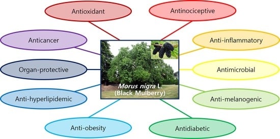
:Mulberry plants belonging to the Moraceae family have been grown for the purpose of being the nutrient source for silk worm and raw materials for the preparation of jams, marmalades, vinegars, juices, wines, and cosmetics. Morus nigra L. (black mulberry) is native to Southwestern Asia, and it has been used as a traditional herbal medicine for animals and humans. In this article, recent research progress on various biological and pharmacological properties of extracts, fractions, and isolated active constituents from different parts of M. nigra are reviewed. M. nigra exhibited a wide-spectrum of biological and pharmacological therapeutic effects including antinociceptive, anti-inflammatory, antimicrobial, anti-melanogenic, antidiabetic, anti-obesity, anti-hyperlipidemic, and anticancer activities. M. nigra also showed protective effects against various human organs and systems, mainly based on its antioxidant capacity. These findings strongly suggest that M. nigra can be used as a promising nutraceutical resource to control and prevent various chronic diseases.
1. Introduction
Morus, commonly known as mulberry, is the genus of a flowering plant belonging to the Moraceae family. They are widely distributed into subtropic regions of Asia (including Korea, Japan, China, and India), North America, and Africa [1]. In Asian countries, mulberry plants have been grown for the production of silk worms (Bombyx mori L.), because their leaves are a major and important nutrient source for silk worms [2]. Meanwhile, most European countries have usually used mulberry fruits to prepare jams, marmalades, vinegars, juices, wine, and cosmetic products [3]. Various parts of mulberry plants have also been used as traditional herbal medicines [4]. Diels-Alder-type adducts, flavonoids, benzofurans, stilbenes, and polyhydroxylated alkaloids are the most representative bioactive compounds identified from Sang-Bai-Pi (Chinese name for root barks of Morus species) [5]. Some previous review articles on Morus alba L. (M. alba), one of the most valuable plants rich in natural ingredients, have demonstrated that extracts, fractions and major constituents from M. alba exhibit numerous pharmacological activities such as antioxidant, anti-inflammatory, anticancer, antimicrobial, antifungal, skin-whitening, antidiabetic, anti-hyperlipidemic, anti-atherosclerotic, anti-obesity, cardioprotective, cognitive enhancing, hepatoprotective, anti-platelet, anxiolytic, anti-asthmatic, anthelmintic, antidepressant, and immunomodulatory activities [6,7,8].
Morus nigra L. (M. nigra), also called black mulberry, is native to Southwestern Asia. It has been grown throughout Europe and around the Mediterranean for centuries. Although biological and/or pharmacological activities of M. nigra have been relatively less studied compared to those of M. alba, several bioactive compounds isolated from M. nigra have also been used as herbal medicines for animals and humans due to their analgesic and anti-inflammatory effects [1]. Budiman et al. [9] briefly summarized chemical compounds isolated from various parts of M. nigra and their pharmacological activities. In this review article, we extensively covered recent research progress on biological and pharmacological properties of M. nigra extracts, fractions, and active constituents, suggesting its potential and usefulness as a nutraceutical resource. Major biological and pharmacological therapeutic activities of M. nigra were summarized in Table 1.
2. Antinociceptive Activity
In 2000, de Souza et al. [10] firstly reported on the antinociceptive effect of morusin, the main prenylflavonoid of M. nigra isolated from acetonic extract of its root barks. Morusin showed a significant inhibitory effect on acetic acid-induced abdominal constriction responses and formalin-induced pain, and it also resulted in prolongation of the latency period in a hot plate test in mice. Because morusin is also purified from other mulberry plants, such as M. alba [11], M. australis [12] and M. lhou [13], this study result alone is insufficient to fully reflect the analgesic activity of M. nigra. Nine years later, Padilha et al. [14] investigated the antinociceptive effect of methylene chloride extract of M. nigra leaves in mice. Similar to the results of de Souza et al. [10], M. nigra leaves extract showed significantly and dose-dependently reduced acetic acid-induced writhing and formalin-induced pain and increased response latency period in a tail-immersion test and hot plate test without any acute toxicity when the dose of the extract was up to 300 mg/kg.
Two studies by Chen et al. [15,16] recently evaluated the antinociceptive properties of total flavonoid extracts and main active ingredients from fresh fruits of M. nigra. In the first study [15], total flavonoids from M. nigra showed dose-dependent decreases in the duration of formalin-induced pain-response behaviors. In the second study, three different mulberry fruits (M. alba, M. nigra and M. mongolia) were compared [16]. M. nigra fruits had more anthocyanin and flavonol contents than other species. The duration of the formalin-induced secondary pain phase (inflammatory phase) in the group treated with total flavonoid extract from M. nigra was significantly shorter than that in the control group. Reduced development of inflammatory cytokine interleukin-6 (IL-6) and an increased level of an anti-inflammatory cytokine IL-10 associated with the nuclear factor kappa-light-chain-enhancer of activated B cells (NF-κB) and nitric oxide (NO) pathways were observed after treatment with M. nigra extract, suggesting the possible mechanism of its antinociceptive effects. Interestingly, the three main flavonoid ingredients (cyanidin-3-O-glucoside, rutin and isoquercetin) from M. nigra did not reduce the duration of formalin-induced pain individually, although they significantly decreased such duration when they were used as a mixture.
3. Anti-Inflammatory Activity
Inflammation is defined as a set of physiological defense mechanisms taking place in the body. However, inflammation is also considered an initial event of major chronic diseases such as cardiovascular, autoimmune, eye, age-related, neurodegenerative diseases, and cancers [17]. In this respect, inhibiting and controlling inflammatory responses in the human body can be one of fundamental approaches for treating chronic diseases.
As a follow-up research of a previous study on antinociceptive activity, Padilha et al. [18] evaluated the anti-inflammatory effects of methylene chloride extract of M. nigra leaves in male rats. M. nigra leaves extract significantly inhibited the volume of paw edema induced by intraplantar injection of carrageenan at a half-maximal inhibitory concentration (IC50) value of 15.2 mg/kg. M. nigra leaves also significantly inhibited the formation of granulomatous tissues in the chronic inflammation status using a cotton pellet-induced granuloma rat model (IC50 of 71.1 mg/kg). In the same year, Wang et al. [19] isolated three new compounds (mornigrol D, G and H) with six other known compounds (norartocarpetin, dihydrokaempferol, albanin A, albanin E, moracin M, and albafuran C) from the stem bark of M. nigra and assessed their anti-inflammatory activities by calculating the inhibition of releasing β-glucuronidase from rat polymorphonuclear leukocytes induced by platelet-activating factor. At a concentration of 10−5 M, mornigrol D and norartocarpetin showed potent anti-inflammatory properties, showing inhibition rates of 65.9% and 67.7%, respectively. In 2014, Zelová et al. [20] investigated into the anti-inflammatory activities of two Diels-Alder adducts (soroceal and sanggenon E) isolated from the root bark of M. nigra, by determining the attenuation of secretion of pro-inflammatory cytokines, tumor necrosis factor-alpha (TNF-α) and IL-1β, in lipopolysaccharide (LPS)-stimulated macrophages. Although sanggenon E significantly reduced the production of TNF-α compared to the vehicle control, both compounds failed to significantly affect the level of IL-1β.
Chen et al. [15] reported that the total flavonoid extract of M. nigra fruits can dose-dependently inhibit xylene-induced ear edema (edema rate 60.1% at a concentration of 200 mg/20 mL/kg) and carrageenan-induced paw edema (edema rate 9.5% at a concentration of 100 mg/20 mL/kg; 8.6% at a concentration of 200 mg/20 mL/kg) in mice. Levels of pro-inflammatory cytokines including IL-1β, TNF-α, NO, and interferon-gamma (IFN-γ) were also significantly decreased after the treatment of M. nigra fruit extract in mice with xylene-induced inflammation. In addition, M. nigra fruits extract significantly reduced levels of NO in LPS-stimulated RAW 264.7 cells without showing the cytotoxicity effect at the concentration of 50 to 100 μg/mL.
A very recent study [21] has shown that extracts of M. nigra pulps and leaves can improve survival rate and decrease the number of total leukocytes in bronchoalveloar lavage fluid in LPS-induced septic mice, indicating the reduction of inflammatory infiltrate in the lung. Although most hepatic and serum cytokine levels were not changed by the administration of M. nigra extracts, serum levels of TNF, an important mediator of sepsis, were significantly lower in the M. nigra extract-treated group than those in the septic animal group.
4. Antimicrobial Activity
Antibacterial activities of M. nigra leaves have been investigated in various organic fractions. Tahir et al. [22] reported that the ethyl acetate fraction of M. nigra leaves is active against four dental caries-causing bacterial strains: Streptococcus mutans, Escherichia coli (E. coli), Staphylococcus aureus (S. aureus), and Bacillus subtilis (B. subtilis). Also, the chloroform fraction showed antibacterial properties against Pseudomonas aeruginosa (P. aeruginosa) and B. subtilis, while the methanol fraction was only active against B. subtilis. No activity was observed for n-hexane or aqueous fraction. The inhibition rate of streptococcal biofilm formation (anti-adherence effect) by M. nigra ethyl acetate fraction was 87%. In another study conducted by Souza et al. [23], crude ethanol extract of M. nigra leaves exhibited bactericidal activities against Bacillus cereus (B. cereus), Enterococcus faecalis (E. faecalis), and E. coli, with minimal inhibitory concentration (MIC) and minimum bactericidal concentration (MBC) less than 0.195 mg/mL for all. Potent antibacterial activities against B. cereus and E. faecalis were also observed for hexane, chloroform and ethyl acetate extracts (MIC values < 0.195 mg/mL for all). However, their measured MBCs were over 6 mg/mL. It was noted that chloroform extract exclusively showed a bactericidal effect against Salmonella choleraesuis (MIC and MBC value < 0.195 mg/mL, respectively). The antibacterial activities of the total flavonoid extract of M. nigra fruits were evaluated against three inflammatory pain-causing bacteria, E. coli, P. aeruginosa and S. aureus. Its fruit extract strongly inhibited all three strains, with MBC values of 2 mg/mL or less [16].
The antimicrobial activities of fresh juice of M. nigra fruits against five Gram-positive and three Gram-negative bacterial strains have been compared with conventional antibiotics [24]. Although 100 μL of M. nigra fruits juice produced generally smaller zones of inhibition (ranging from 9.98 to 19.87 mm) than other antibiotics treated at their standard doses, it showed a broad-spectrum antimicrobial effect against both Gram-positive and Gram-negative bacteria, having the highest inhibition against P. aeruginosa. Minhas et al. [25] investigated into the antimicrobial effect of five M. nigra fruits extracts classified by different solvents against 16 bacterial and 2 fungal strains in comparison with conventional antibiotics and antifungal agent nystatin. Ethanolic and acetone extracts of M. nigra fruits showed highly-sensitive inhibition (defined as 20 mm or more longer diameter of zone of inhibition) against E. coli, S. aureus, and Neisseria spp.; methanolic extract against Klebsiella pneumoniae and Neisseria spp.; and chloroform extract against Serratia marcesscens, Staphylococcus epidermidis (S. epidermidis), P. aeruginosa, and S. aureus. Similar to the results of Khalid et al. [24], M. nigra extracts had smaller zones of inhibition than those observed with conventional drugs.
In a recent study assessing antibacterial activities against two strains causing acne, S. epidermidis and Propionibacterium acnes (P. acnes), the ethanolic extract of M. nigra fruits had MIC values of 2.5% for both strains and MBC values of 2.5% and 5%, respectively [26]. As a follow-up approach, a comparative study was performed for extracts from three parts (stem barks, fruits and leaves) of M. nigra on their antibacterial effects against S. epidermidis and P. acnes [27]. M. nigra stem barks possessed the most potent antibacterial activities against both strains, with an MIC value of 4 mg/mL for S. epidermidis and 2 mg/mL for P. acnes. In addition, M. nigra stem barks extract induced nucleic acid, protein, and ion leakages and cellular membrane damages against P. acnes. These results suggest that the antibacterial effect of M. nigra stem bark is related to reduced cell membrane fluidity and bacterial cell wall destruction.
Mazzimba et al. [28] reported that six isolated constituents (oxyresveratrol, moracin M, cyclomorusin, morusin, kuwanon C, and a derivative of kuwanon C) from aerial parts of M. nigra show antibacterial activities against S. aureus, B. subtilis, Micrococcus flavus, S. faecalis, Salmonella abony, and P. aeruginosa, with morusin having the most potent activity against B. subtilis (MIC value 3.91 μg/mL).
Tuberculosis (TB), an infectious disease caused by Mycobacterium tuberculosis (M. tuberculosis), is one of the top 10 causes of death in the world. TB is a curable and preventive disease, but resistance against conventional antibiotic medications for M. tuberculosis has increased the number of cases of multidrug-resistant or extensively drug-resistant TB [29]. In this respect, demand for new medications with novel therapeutic targets such as protein tyrosinase phosphatases (PTPs) is growing [30,31]. Mascarello et al. [32] evaluated the anti-tuberculosis activity of Diel–Alder-type adducts from M. nigra root bark to determine their potential as candidates for M. tuberculosis PTP inhibitor. A total of eight compounds (Kuwanon L, G, and H; cudraflavanone A; morusin, oxyresveratrol; chalcomoracin; and norartocarpetin) were isolated from M. nigra. They all significantly inhibited M. tuberculosis PTP-B (Mtb PtpB) with IC50 values ranging from 0.36 to 8.42 μM. Further enzyme kinetic analyses for Kuwanon G and H, two of the most potent compounds, showed that both compounds competitively inhibited Mtb PtpB, with inhibitory constant (Ki) values of 0.39 ± 0.27 μM and 0.20 ± 0.01 μM, respectively. In addition, Kuwanon G inhibited the growth of M. tuberculosis inside macrophages by 61.3% at a non-cytotoxic concentration (10 μg/mL, corresponding to 14.4 μM of Kuwanon G), indicating that it is the most promising anti-tuberculosis constituent isolated from M. nigra.
Antimicrobial activity of M. nigra against Candida spp., the most common cause of fungal infections around the world [33], was assessed with aqueous and methanol extracts of its fruits, by using a disc-diffusion assay [34]. Of nine selected Candida spp., both extracts exhibited anticandidal effect against Candida (C.) albicans, C. parapsilosis, C. tropicalis, and Geotricum candidum, with lower MIC values observed for the methanol extract (0.625–2.5 mg/mL) than those for the aqueous extract (1.25–5 mg/mL).
5. Anti-Melanogenic (Skin-Whitening) Activity
Although melanin pigmentation in the skin is an important defense mechanism against ultraviolet radiation, abnormal melanin hyperpigmentation catalyzed by tyrosinase can cause several serious aesthetic problems [35,36,37]. As an anti-melanogenic strategy, tyrosinase inhibitors have become increasingly important for treating skin disorders associated with pigmentation and to improve skin-whitening.
Zhang et al. [38] investigated the inhibitory effect of 2,4,2’,4’-tetrahydroxy-3-(3-methyl-2-butenyl)-chalcone (TMBC) isolated from the stem of M. nigra on tyrosinase activity and melanin biosynthesis. TMBC dose-dependently and competitively inhibited mushroom tyrosinase-mediated L-dopa oxidation (IC50 value 0.95 ± 0.04 μM), which was more potent than kojic acid (IC50 value 24.88 ± 1.13 μM), a well-known skin depigmenting agent. Furthermore, TMBC significantly reduced the melanin content and cellular tyrosinase activity in B16 melanoma cells, although it increased mRNA levels of cellular tyrosinase. Zheng et al. [39] screened tyrosinase inhibitory properties of a total of 29 constituents isolated from roots of M. nigra. Among them, nine compounds (5’-geranyl-5,7,2’,4’-tetrahydroxyflavone, steppogenin-7-O-β-d-glucoside, 2,4,2’,4’-tetrahydroxychalcone, moracin N, kuwanon H, mulberrofuran G, morachalcone A, oxyresveratrol-3’-O-β-d-glucopyranoside and oxyresveratrol-2-O-β-d-glucopyranoside) showed better tyrosinase inhibitory activities than kojic acid (IC50 value 46.95 ± 1.72 μM, with 2,4,2’,4’-tetrahydroxychalcone having the highest activity (IC50 value 0.062 ± 0.002 μM, 757-fold lower IC50 than kojic acid). More recently, de Freitas et al. [40] reported that five different batches of standardized ethanolic extracts of M. nigra leaves all exhibited tyrosinase inhibitory activities, with IC50 ranging from 5.00 to 8.49 μg/mL.
Koyu et al. [41] tested the microwave-assisted extraction of fresh fruits of M. nigra in variable conditions for optimizing and maximizing tyrosinase inhibitory activity. Consequently, the highest tyrosinase inhibitory activity (IC50 value 1.44 mg/mL) was observed in the optimum microwave extraction system yielding the highest amount of anthocyanin content (13.28 mg/g cyanidin-3-glucoside equivalent), suggesting the important potential of anthocyanins on tyrosinase inhibition.
6. Antidiabetic and Anti-Obesity Activity
Diabetes mellitus is a chronic endocrine disorder characterized by hyperglycemia related to metabolic impairment of insulin production, secretion, and/or utilization. It is closely associated with the development of several important complications in cardiovascular, neurological and renal systems that can lead to increased morbidity and mortality in diabetic patients [42]. Various classes of antihyperglycemic agents are now available. However, some undesirable adverse effects such as hypoglycemia, gastrointestinal symptoms, weight gain and hepato-renal toxicity caused by the administration of these medications have been arousing interests on the discovery of new effective and safer naturally-occurring antidiabetic agents with different therapeutic pathophysiological mechanisms and targets [43,44,45].
M. nigra has also shown good antidiabetic effects on extracts and active constituents from some parts of this plant. Abd El-Mawla et al. [46] investigated the hypoglycemic efficacy of M. nigra leaf extracts and its cell suspension cultures treated with methyl jasmonate to induce accumulation of flavonoid contents in cell cultures. Extracts from M. nigra leaves dose-dependently decreased plasma glucose concentrations and increased insulin levels up to 500 mg/kg/day in streptozotocin (STZ)-treated diabetic rats. In addition, a slightly higher hypoglycemic effect was observed when rats were treated with extracts from cultured cells, indicating the additive action of flavonoids induced by methyl jasmonate. Hydroethanolic extracts of M. nigra leaves also significantly decreased serum fasting and 2-h glucose concentrations (at dose of 50 mg/kg) and increased serum insulin level (at dose of 10 mg/kg) in nicotinamide-STZ-induced type 2 diabetic rats [47]. Diabetes-induced changes in blood vessels may enhance the pathophysiological activity of metalloproteinases (MMPs). It is known that the inhibition of MMPs can improve insulin resistance and oxidative stress [48,49]. Araujo et al. [49] demonstrated the hypoglycemic potential of M. nigra leaves via reduction of expression and activity of MMP-2 in livers of diabetic rats. In addition, several phenolic compounds and isoprenylated flavonoids isolated from extracts of M. nigra twigs showed good antidiabetic activities, involving mechanisms of peroxisome proliferators-activated receptor gamma (PPARγ) activation [50] and α-glucosidase inhibition [51]. On the other hand, 3-week treatment of aqueous extract of M. nigra leaves failed to affect serum glucose levels in non-diabetic or diabetic pregnant rats [52].
Although there is no published report on the antidiabetic activity of black mulberry fruit yet, its effects on obesity, associated with increased risk of many chronic adverse health effects including cardiovascular diseases, dyslipidemia, non-alcoholic hepatic disease, cancer, and type 2 diabetes [53,54] have been evaluated by Fabroni et al. [55]. They demonstrated that 80% hydroethanolic freeze-dried extract of fruits of M. nigra had moderate total anthocyanin and total phenolic contents, with an IC50 value for pancreatic lipase inhibition at 6.32 ± 0.01 mg/mL.
7. Anti-Hyperlipidemic and Anti-Atherosclerotic Activity
Cholesterol is a lipid molecule that acts as a structural component of cell membrane modulating fluidity and permeability, and as a precursor for steroid hormone and bile acid synthesis [56]. At the same time, hypercholesterolemia, a typical type of hyperlipidemia characterized by excessive accumulation of cholesterol in serum, is one of the crucial risk factors for coronary heart disease and atherosclerotic progression [57]. It has also been reported that reduction of low-density lipoprotein cholesterol (LDL-C) and improvement in levels of high-density lipoprotein cholesterol (HDL-C) can contribute to the anti-atherogenic condition [58,59].
Results from biochemical profile studies conducted by Volpato et al. [52] and Mahmoud [60] demonstrated that M. nigra extracts can decrease total cholesterol, triglyceride, LDL-C, and very low-density lipoprotein cholesterol (VLDL-C) levels and increase HDL-C in diabetic pregnant rats [52] and rats fed a high-fat diet [60]. Zeni et al. [61] evaluated the lipid-lowering effect of M. nigra leaf extract using Triton WR-1339-induced hyperlipidemic rats. The LDL-C level had significantly decreased after treatment with 100 mg/kg M. nigra infusion extract and HDL-C levels were restored in all groups treated with M. nigra extract at three different concentrations (100, 200 and 400 mg/kg), compared to those in the group only treated with Triton WR-1339. Atherogenic index and cardiac risk factor, indicators of likelihood of cardiovascular diseases associated with hyperlipidemia, were also decreased by M. nigra leaf extract. In another study by Jiang et al. [62], a high dose (210 mg/kg) of ethanolic extract of M. nigra fruit (EEBM) resulted in lowering mean body weight in rats fed a 6-week high-fat diet, which is comparable to the effect observed in the group treated with 5 mg/kg simvastatin. EEBM also dose-dependently improved serum lipid profiles, atherosclerosis indexes and lipid peroxidation compared to the control (high-fat diet-induced hyperlipidemic model) group. Histopathological changes in rat liver and thoracic aorta with reduction in the intima-media thickness of rat aortic arch after treatment with EEBM suggest that M. nigra fruit can effectively suppress the development and deterioration of atherosclerosis.
8. Organ-Protective Activity
8.1. Neuroprotective Effect
Turgut et al. [63] investigated the effect of M. nigra leaves extract on D-galactose-induced cognitive impairment and oxidative stress in mice. The results from the Morris water maze test showed significant and dose-dependent decreases in mean escape latency and time required to reach the target quadrant. Time spent in the target quadrant and number of times crossed the platform location were increased after the administration of lyophilized M. nigra extract, suggesting its potential neuroprotective role by preventing D-galactose-induced learning dysfunction and memory loss. M. nigra extract also showed DNA damage protection, reduced malondialdehyde (MDA) levels and augmented activities of three anti-oxidant enzymes, superoxide dismutase (SOD), glutathione peroxidase (GPx), and catalase (CAT) in the serum, brain and liver of D-galactose-treated mice. These antioxidant and anti-aging properties are considered as one of key mechanisms of M. nigra in delaying neurodegenerative processes.
Dalmagro et al. [64] performed a forced swimming test (FST) and tail suspension test (TST) to evaluate antidepressant-like activities of M. nigra and its major phenolic compounds syringic acid in mice. Acute and subchronic oral administration of aqueous extract of M. nigra leaves significantly decreased the immobility time in FST and TST except for acute administration at a dose of 100 mg/kg extract in TST. Acute treatment with 1 mg/kg and 10 mg/kg and subchronic treatment with 1 mg/kg of syringic acid also significantly decreased immobility time in TST. Nitro-oxidative stress in the serum and brain was assessed by measuring thiobarbituric acid reactive substances (TBARS), nitrite, protein carbonyl content (PC) and non-protein thiol groups (NPSH) levels, with some inconsistent and controversial study results. A significant decrease of TBARS level was observed at acute doses of 3 mg/kg M. nigra extract. However, TBARS levels were oppositely increased at subchronic doses of 3, 10, and 100 mg/kg extract in the serum and at a subchronic dose of 3 mg/kg extract in the brain. Levels of nitrites in the serum were significantly decreased after subchronic administration of 10, 30 and 100 mg/kg extracts of M. nigra leaves, and nitrites in the brain were also decreased after subchronic treatment with the extract at doses of 30 and 100 mg/kg. In addition, subchronic treatment with 1 mg/kg syringic acid resulted in significant changes in TBARS and nitrite levels in the serum and brain (all decreased, except TBARS level was increased in the brain). PC level was decreased after treatment with 30 mg/kg M. nigra extract and syringic acid. There was no significant change in NPSH level at all treatment conditions. Nevertheless, M. nigra leaf extract and syringic acid both exhibited good cell viabilities in hippocampal and cerebral cortex slices incubated with 100 mM glutamate, suggesting their proper neuroprotective effect against glutamate-induced toxicity.
8.2. Hepatoprotective Effect
Tag et al. [65] evaluated the hepatoprotective effect of the ethanolic extract of M. nigra leaves. With an IC50 value at 14.5 μg/mL in in vitro cytotoxicity to HepG2 (a well-differentiated human hepatocellular carcinoma) cell line, M. nigra leaf extract also significantly decreased levels of liver enzymes alanine aminotransaminase (ALT), aspartate aminotransaminase (AST), alkaline phosphatase (ALP), and lactate dehydrogenase (LDH) in male albino rats with methotrexate-induced hepatotoxicity. Hematosomatic index, defined as the ratio between liver- and body-weight and considered as an indicator for hepatic damage and liver inflammation, in the group co-treated with M. nigra extract and methotrexate, was also apparently decreased compared to that in methotrexate-only treated group. In histopathological studies, M. nigra treatment resulted in moderate enhancement in the hepatoprotection from methotrexate-related injury. Microscopic damage scores (hepatocyte degeneration, congestion, leukocyte infiltration, fibrosis, and total histopathology score) were significantly decreased when M. nigra extract was simultaneously administered compared to those in the group treated with methotrexate alone. In addition, methotrexate-induced progressive increases in collagen deposition of liver tissue were normalized by treatment with M. nigra leaf extract. Another study performed by Hassanalilou et al. [66] also showed that M. nigra leaf extract can lead to less fatty degeneration in liver tissue and smaller distension of hepatic cytoplasm due to fatty droplets in STZ-induced diabetic rats along with reduced fasting blood glucose, compared to glibenclamide, a well-known sulfonylurea antihyperglycemic agent.
Hepatoprotective activity of M. nigra fruits in carbon tetrachloride (CCl4, a well-known potent hepatotoxin)-treated HepG2 cells [67] and adult male Sprague-Dawley rats [68] have been reported. Extracts of M. nigra fruits dose-dependently and significantly reduced levels of hepatic enzymes AST, ALT and gamma-glutamyl transferase (GGT) compared to control (CCl4-treated group). At the same time, they significantly increased SOD and gluatathione peroxidase (GPx) enzymatic capacities and decreased expression levels and activities hepatic capsase-3 (a biomarker for cell apoptosis) and 8-oxo-2’-deoxyguanosine (a biomarker for oxidative stress) in rat liver tissues, indicating that the hepatoprotective effect of M. nigra fruits might be closely associated with its antioxidant activity [67,68].
8.3. Renal-Protective Effect
The effects of hydroalcoholic extract of M. nigra fruits on biochemical and histopathological changes in serum and kidney tissues have been evaluated in alloxan-induced diabetic rats [69]. Milder glomerular damage and no mesenchymal tissue expansion into renal glomerular vessels were observed in the group after 8 weeks of treatment with 800 mg/kg M. nigra fruit extract compared to those in diabetic and positive control (150 mg/kg metformin) groups. Although an increase in serum creatinine level was observed in the group treated with 800 mg/kg M. nigra extract, this group had lower serum glucose and urea levels compared to diabetic and positive control groups. These results suggest that M. nigra fruits have a protective effect on diabetic nephropathy and related kidney tissue injury. The extract of M. nigra leaves also significantly improved biochemical parameters reflecting kidney functions (serum creatinine, urea, and uric acid) and exhibited milder histopathological glycogen accumulation, fatty degeneration, and lymphocyte infiltration of renal convoluted tubules in STZ-induced diabetic rats compared to non-treated and glibenclamide-treated groups [66].
8.4. Gastroprotective Effect
Nesello et al. [70] reported that oral administration of methanolic extract from M. nigra fruits at a high dose (300 mg/kg) can protect gastric mucosa against acidified ethanol-induced acute gastric ulcer in female mice. This study result was confirmed by macroscopic and microscopic representative images, showing that the degree of epithelial damage in gastric tissue was decreased. To further investigate the underlying mechanisms for the gastroprotective effect, levels of lipid hydroperoxide (LOOH) and glutathione (GSH) in ulcerated gastric mucosa were quantified. M. nigra fruits extract prevented GSH depletion and promoted partial reduction of LOOH, suggesting its ability to ameliorate oxidative stress involved in the development of gastric injury by acidified ethanol. Because M. nigra fruits did not affect the activity of H+/K+-ATPase in their study, they have pharmacological advantages of being free from the risk of several side effects such as rebound acid hypersecretion, hypergastrinemia, gastric polyps, or atrophic gastritis [71] known to be associated with suppressed gastric acid secretion.
9. Activity on Female Reproductive System
De Queiroz et al. [72] investigated the estrogenic effect of M. nigra on the female reproductive system and embryonic development. Five different concentrations (25, 50, 75, 350, and 700 mg/kg) of hydroalcoholic extract of dried M. nigra leaves were administered in female Wistar rats for 15 days and their biological and clinical features were compared with the control group, in which distilled water instead of M. nigra extract was used as treatment. There were no significant differences in the number of deaths, clinical signs of toxicity, changes in food consumption, or body weight between groups, suggesting that M. nigra leaves did not cause maternal reproductive toxicity. Histological changes in ovarian structures, signs of edema, cystic follicles, retained oocytes, or thickened uterine epithelium were not observed. The number of corpora lutea, live fetuses, implants, resorptions, implantation, and pre- or post-implantation loss were not affected by the administration of M. nigra leaf extract either. Consequently, M. nigra exhibited no estrogenic effect or toxicity on the female reproductive system.
Another study conducted by Cavalcante et al. [73] showed that ethanolic extract of M. nigra fresh leaves at 0.1 mg/mL can improve percentages of follicular morphology, antrum formation, and fully grown oocytes, as well as the diameter of follicles compared to control group at 12 days after treatment. Furthermore, additive effects on follicular growth (described as follicular diameter increase and higher daily growth rate) were observed when M. nigra extract with supplemented medium and follicle-stimulating hormone (FSH) were used as co-treatment, indicating its capacity on ovine secondary follicle development.
10. Anticancer Activity
Cancer is a life-threatening disease state characterized by unregulated and permanent cell growth and proliferation [74]. Because of its ability to avoid programmed cell death (apoptosis) as one of the main driving forces for maintaining cancer cell proliferation, induction of apoptosis in cancer has been considered a reasonable strategy to treat cancer [75,76].
Morniga M, a mannose-specific jacalin-related lectin from the bark of M. nigra, can preferentially trigger the proliferation and activation of human T- and natural killer- (NK-)lymphocytes and dose-dependently induce cell death of α-CD3 activated T lymphocytes when compared with concanavalin A (Con A), a well-known mannose-specific legume leptin from Canavalia ensiformis [77]. Results from flow cytometry analysis have demonstrated that morniga M-induced cell death is probably associated with the apoptotic mechanism, suggesting the anticancer potential of morniga M via cell-death induction and immunomodulation as reported in previous studies with Con A [78,79]. Anticancer activities of morniga M were further investigated by Çakıroğlu et al. [80], in which they demonstrated that both M. nigra fruit extract and morniga M significantly and dose-dependently decreased cell viability against HT-29 cell line (human colorectal cancer). Another brief research by Qadir et al. [81] the demonstrated dose-dependent anticancer activity of n-hexane and aqueous methanol extract of M. nigra leaves against HeLa cell line (human cervical cancer), with IC50 values of 185.9 ± 8.3 µg/mL and 56.0 ± 1.7 µg/mL, respectively.
Anti-proliferative and apoptotic effects of M. nigra fruits against several human adenocarcinoma cell lines have been reported [80,82,83]. Ahmed et al. [82] compared the anticancer effects between fresh and dried fruit extracts of M. nigra on MCF-7 cell line (human breast cancer). Study results have shown that both ethanolic extracts dose- and time-dependently inhibit cellular growth of MCF-7 cells; exhibit apoptotic morphological changes in their cytoplasmic membranes, cell bodies, and nuclei; induce DNA fragmentations and single strand breaks; and decrease mitotic indexes, with better pharmacological properties in fresh fruit of M. nigra. Turan et al. [83] evaluated the anticancer activities of M. nigra fruit extract on PC-3 cells (human prostate cancer). Dimethyl sulfoxide (DMSO) extract of M. nigra exhibited moderate cytotoxicity against PC-3 cells with an IC50 value of 370.1 ± 5.8 μg/mL. It significantly increased the cell number at G0/G1 phase and decreased the cell number at S phase, indicating that M. nigra fruits inhibited the progression of the cell cycle at the G0/G1 phase. M. nigra fruit extract at a high dose (666 μg/mL) significantly increased the number of necrotic, early apoptotic and late apoptotic cells compared to the untreated control group. It also dose-dependently decreased mitochondrial membrane potential and increased activities of caspase 3 and 7 (key mediators of apoptosis) in PC-3 cells [83].
11. Antioxidant Activity
Oxidative stress is characterized by an excessive increase in intracellular oxidizing species such as reactive oxygen species (ROS) involved in the loss of antioxidant defense capacity. It plays a critical role in various clinical conditions including aging, cancer, diabetes, atherosclerosis, chronic inflammation, neurodegenerative diseases, rheumatoid arthritis, human immunodeficiency virus (HIV) infection, ischemia and reperfusion injury, and obstructive sleep apnea [84,85]. Many researchers are interested in the antioxidant activity of naturally-occurring ingredients because phenolic compounds and flavonoids, the largest phytochemical molecules from natural resources, possess a variety of biological properties including antioxidant activity [86,87,88,89]. It has also been widely reported that mulberries are rich in anthocyanin constituents having remarkable antioxidant activities and other health benefits such as anti-inflammatory, antimicrobial, anti-obesity, antidiabetic, anti-hyperlipidemic, antihypertensive, cardioprotective (reduced risk of coronary heart disease and stroke), and anticancer effects [90,91,92].
Numerous researches have proven antioxidant properties of M. nigra with different in vitro methods, including DPPH (2,2-diphenyl-1-picrylhydrazyl) radical scavenging assay [15,23,24,25,26,28,61,70,93,94,95,96,97,98,99,100,101,102,103,104,105,106,107,108,109,110,111,112,113], ABTS (2,2’-azino-bis(3-ethylbenzothiazoline)-6-sulfonic acid) radical scavenging assay [15,91,94,99,100,101,102,103,107,110,112,113,114,115,116,117], reducing power assay [15,99,113,118,119], superoxide anion radical (O2−) scavenging assay [15,118,120], hydroxyl radical (OH-) scavenging assay [15,113,120], lipid peroxidation assay [19,52,60,62,63,64,70,121,122], antioxidant enzyme activity assay [21,49,52,62,63,67], β-carotene bleaching assay [23,119,123], ferric-reducing antioxidant power (FRAP) assay [24,85,91,95,100,102,104,107,110,111], protein carbonyl assay [49,64,96], GSH measurement [67,70,112], hydrogen peroxide (H2O2)-induced injury assay [113,121], NO radical scavenging assay [111,118], SOD-like activity [96], cupric-ion reducing antioxidant capacity (CUPRAC) assay [102,107,108,110], H2O2 scavenging assay [108,119], phosphomolybdenum assay [108,119], and ROS measurement [112].
12. Other Pharmacological Activities
Malik et al. [124] investigated the cardiovascular activity of aqueous methanolic extract of M. nigra fruit in frogs. Treatment of M. nigra fruit extract showed significant and dose-dependent decreases in heart rate without direct effects on the contractility of frog’s heart. Results of phytochemical analysis revealed the presence of cardiac glycosides in M. nigra fruit, along with other active constituents including saponins, alkaloids, phenolic compounds, and flavonoids.
Crude extract and fractions of M. nigra fruits exhibit both in vitro and in vivo prokinetic, laxative, and antidiarrheal effects [125]. M. nigra extract significantly promoted the transit of charcoal meal through the small intestine, increased gastric emptying rate and the mean number of wet feces, and decreased castor oil-induced diarrhea in mice. In in vitro studies, chloroform and petroleum ether fractions of M. nigra fruits dose-dependently inhibited carbachol- and potassium ion-induced contractions of rabbit jejunum while aqueous and ethyl acetate fractions showed stimulatory effects on guinea-pig ileum. Suppression of maximum responses of acetylcholine and calcium ion (Ca2+) by M. nigra fruits was also observed, and most gastrointestinal effects were conversely affected by concomitant administration of atropine, suggesting that the underlying mechanisms of these prokinetic, laxative, and antidiarrheal activities might be associated with cholinergic control and Ca2+ channel antagonism [125].
Fahimi and Jahromy [126] described the effects of M. nigra fruit juice on levodopa-induced dyskinesia in mice with 1-methyl-4-phenyl-1,2,3,6-tetrahydropyridine (MPTP)-induced Parkinson’s disease. After 14 days of levodopa treatment, administration of 10 or 15 mL/kg of M. nigra fruit juice significantly decreased abnormal involuntary movement scale (AIMS) scores compared to levodopa treatment only.
13. Drug-Food Interaction and Toxicity
Food ingredients can cause drug-food interactions, most of which are pharmacokinetic interactions associated with the alteration in activities of drug-metabolizing enzymes or drug transporters [127]. A brief experimental report by Kim et al. [128] demonstrated that the fruit juice of M. nigra has a potent inhibitory effect of human liver microsomal cytochrome P450 3A (CYP3A) activity, with IC50 values for midazolam (a probe drug for CYP3A) 1‘-hydroxylation of 2.96 ± 0.33% (v/v, with 20-min preincubation) and 6.22 ± 0.47% (no preincubation). Because approximately 30% of clinically used drugs including macrolide antibiotics, antiarrhythmics, benzodiazepines, immune modulators, human immunodeficiency virus (HIV) antivirals, antihistamines, calcium channel blockers, and statins are metabolized by CYP3A [129,130], concomitant intake of CYP3A substrates with M. nigra fruit can lead to an increase in plasma drug exposure.
Figueredo et al. [131] assessed the acute and subacute toxicities of M. nigra leaves extract in Wistar rats. A single or 28-day oral dose of ethanolic extract of M. nigra leaves did not cause any adverse effects. It did not induce abnormal behaviors or mortality. M. nigra extract resulted in some significant but non-toxic changes in biochemical profiles (decreased urea and AST in males; decreased total cholesterol and AST in females) and leukocyte parameters (increased neutrophils in males; decreased white blood cell in females). M. nigra leaves did not affect lipid peroxidation and changes in renal and hepatic CAT enzymatic activities.

Table 1.
Summary of major biological and pharmacological therapeutic activities of M. nigra.
| Pharmacological Activity | Study Model | Used Part | SampleType a | Ref. |
|---|---|---|---|---|
| Antinociceptive | Swiss mice | Root bark | C | [10] |
| Male Swiss mice | Leaf | E | [14] | |
| Male Kunming mice | Fruit | E | [15] | |
| Male Kunming mice | Fruit | E,C | [16] | |
| Anti-inflammatory | Kunming male mice; RAW 264.7 cell | Fruit | E | [15] |
| Adult male rats | Leaf | E | [18] | |
| Rat polymorphonuclear leukocytes | Bark | C | [19] | |
| THP-1 human monocytic leukemia cell line | Root | C | [20] | |
| Male C57BL/6 mice | Pulp; leaf | E | [21] | |
| Antimicrobial | In vitro assay | Fruit | E | [16] |
| In vitro assay | Leaf | E,F | [22] | |
| In vitro assay | Leaf | E,F | [23] | |
| In vitro assay | Fruit | J | [24] | |
| In vitro assay | Fruit | E | [25] | |
| In vitro assay | Fruit | E | [26] | |
| In vitro assay | Stem bark; fruit; leaf | E | [27] | |
| In vitro assay | Stem bark; stem wood | E,C | [28] | |
| In vitro assay; THP-1 cell line | Root | E,C | [32] | |
| In vitro assay | Fruit | E | [34] | |
| Anti-melanogenic (Skin-whitening) | In vitro assay; B16 melanoma cells | Stem | C | [38] |
| In vitro assay | Root; twig | C | [39] | |
| In vitro assay | Leaf | E | [40] | |
| In vitro assay | Fruit | E | [41] | |
| Antidiabetic | Male Wistar rats | Leaf | E b | [46] |
| Male albino mice | Leaf | E | [47] | |
| Female albino Fischer rats | Pulp; leaf | E | [49] | |
| PPARγ-transfected HEK293 cells | Twig | C | [50] | |
| In vitro assay | Twig | C | [51] | |
| Anti-obesity | In vitro assay | Fruit | E | [55] |
| Anti-hyperlipidemic | Wistar rats | Leaf | E | [52] |
| Adult male albino Sprague-Dawley rats | Fruit | E | [60] | |
| Male Wistar rats | Leaf | E | [61] | |
| Male Spraque-Dawley rats | Fruit | E | [62] | |
| Organ-protective | Male BALB/c mice | Leaf | E | [63] |
| Male Swiss mice | Leaf | E,C | [64] | |
| HepG2 human hepatocellular carcinoma cell line; male albino rats | Leaf | E | [65] | |
| Male Wistar rats | Leaf | E | [66] | |
| HepG2 cells | Fruit | E | [67] | |
| Adult male Sprague-Dawley rats | Fruit | E | [68] | |
| Male Wistar rats | Fruit | E | [69] | |
| Female Swiss mice | Fruit | E | [70] | |
| Anticancer | Peripheral blood mononuclear cells (PBMCs); peripheral blood T lymphocytes; Jurkat T leukemia cells | Bark | C | [77] |
| HT-29 human colorectal adenocarcinoma cell line | Fruit | E,C | [80] | |
| HeLa human cervical cancer cell line | Leaf | E | [81] | |
| MCF-7 human breast cancer cell line | Fruit | E | [82] | |
| PC-3 human prostate adenocarcinoma cells | Fruit | E | [83] |
a, E, extract; F, fraction; C, isolated compound; J, juice. b, Cell suspension cultures of M. nigra extract were used.
14. Conclusions
M. nigra, especially its leaf and fruit parts, exhibited various pharmacological properties including antinociceptive, anti-inflammatory, antimicrobial, anti-melanogenic, antidiabetic, anti-obesity, anti-hyperlipidemic, and anticancer activities. M. nigra also showed protective and therapeutic effects on the central nervous system, liver, kidney, gastrointestinal tract, and female reproductive system. Most of these features were attributable to its antioxidant capacity due to abundant phytochemical constituents such as polyphenols, flavonoids and anthocyanins. These findings suggest that M. nigra can be used as a promising nutraceutical resource to control and prevent various chronic diseases. Given that most researches are performed in vitro and in animal models, further studies at the clinical level are required to establish the efficacy and safety of M. nigra in the human body.
Author Contributions
S.H.L. was responsible for collecting and summarizing literature data. C.-I.C. wrote and edited the manuscript.
Funding
This research was supported by Basic Science Research Program through the National Research Foundation of Korea (NRF) funded by the Ministry of Education (No. 2016R1D1A1B03933963).
Conflicts of Interest
The authors declare no conflict of interest.
References
- Hussain, F.; Rana, Z.; Shafique, H.; Malik, A.; Hussain, Z. Phytopharmacological potential of different species of Morus alba and their bioactive phytochemicals: A review. Asian Pac. J. Trop. Biomed. 2017, 7, 950–956. [Google Scholar] [CrossRef]
- Vijayan, K.; Chauhan, S.; Das, N.K.; Chakraborti, S.P.; Roy, B.N. Leaf yield component combining abilities in mulberry (Morus spp.). Euphytica 1997, 98, 47–52. [Google Scholar] [CrossRef]
- Natić, M.M.; Dabić, D.Č.; Papetti, A.; Fotirić Akšić, M.M.; Ognjanov, V.; Ljubojević, M.; Tešić, Ž. Analysis and characterisation of phytochemicals in mulberry (Morus alba L.) fruits grown in Vojvodina, North Serbia. Food Chem. 2014, 171, 128–136. [Google Scholar] [CrossRef] [PubMed]
- Sánchez-Salcedo, E.M.; Sendra, E.; Carbonell-Barrachina, Á.A.; Martínez, J.J.; Hernández, F. Fatty acids composition of Spanish black (Morus nigra L.) and white (Morus alba L.) mulberries. Food Chem. 2016, 190, 566–571. [Google Scholar] [CrossRef] [PubMed]
- Wei, H.; Zhu, J.J.; Liu, X.Q.; Feng, W.H.; Wang, Z.M.; Yan, L.H. Review of bioactive compounds from root barks of Morus plants (Sang-Bai-Pi) and their pharmacological effects. Cogent. Chem. 2016, 2, 1212320. [Google Scholar] [CrossRef]
- Yang, Y.; Tan, Y.X.; Chen, R.Y.; Kang, J. The latest review on the polyphenols and their bioactivities of Chinese Morus plants. J. Asian Nat. Prod. Res. 2014, 16, 690–702. [Google Scholar] [CrossRef] [PubMed]
- Chan, E.W.; Lye, P.Y.; Wong, S.K. Phytochemistry, pharmacology, and clinical trials of Morus alba. Chin. J. Nat. Med. 2016, 14, 17–30. [Google Scholar]
- Gryn-Rynko, A.; Bazylak, G.; Olszewska-Slonina, D. New potential phytotherapeutics obtained from white mulberry (Morus alba L.) leaves. Biomed. Pharmacother. 2016, 84, 628–636. [Google Scholar] [CrossRef]
- Budiman, A.; Sulastri, A.; Alfauziah, T.Q. Chemical compounds and pharmacological activity of Morus nigra as a potential product of drug: A review. Int. Res. J. Pharm. 2018, 9, 76–81. [Google Scholar] [CrossRef]
- De Souza, M.M.; Bittar, M.; Cechinel-Filho, V.; Yunes, R.A.; Messana, I.; Delle Monache, F.; Ferrari, F. Antinociceptive properties of morusin, a prenylflavonoid isolated from Morus nigra root bark. Z. Naturforsch. C. 2000, 55, 256–260. [Google Scholar] [CrossRef]
- Nomura, T.; Fukai, T.; Yamada, S.; Katayanagi, M. Studies on the constituents of the cultivated mulberry tree. I. Three new prenylflavones from the root bark of Morus alba L. Chem. Pharm. Bull. 1978, 26, 1394–1402. [Google Scholar] [CrossRef]
- Ko, H.H.; Yu, S.M.; Ko, F.N.; Teng, C.M.; Lin, C.N. Bioactive constituents of Morus australis and Broussonetia papyrifera. J. Nat. Prod. 1997, 60, 1008–1011. [Google Scholar] [CrossRef] [PubMed]
- Ryu, Y.B.; Ha, T.J.; Curtis-Long, M.J.; Ryu, H.W.; Gal, S.W.; Park, K.H. Inhibitory effects on mushroom tyrosinase by flavones from the stem barks of Morus lhou (S.) Koidz. J. Enzyme Inhib. Med. Chem. 2008, 23, 922–930. [Google Scholar] [CrossRef] [PubMed]
- Padilha, M.M.; Vilela, F.C.; da Silva, M.J.; dos Santos, M.H.; Alves-da-Silva, G.; Giusti-Paiva, A. Antinociceptive Effect of the extract of Morus nigra leaves in mice. J. Med. Food 2009, 12, 1381–1385. [Google Scholar] [CrossRef] [PubMed]
- Chen, H.; Pu, J.; Liu, D.; Yu, W.; Shao, Y.; Yang, G.; Xiang, Z.; He, N. Anti-Inflammatory and Antinociceptive Properties of Flavonoids from the Fruits of Black Mulberry (Morus nigra L.). PLoS ONE 2016, 11, e0153080. [Google Scholar] [CrossRef] [PubMed]
- Chen, H.; Yu, W.; Chen, G.; Meng, S.; Xiang, Z.; He, N. Antinociceptive and Antibacterial Properties of Anthocyanins and Flavonols from Fruits of Black and Non-Black Mulberries. Molecules 2018, 23, 4. [Google Scholar] [CrossRef] [PubMed]
- Chalons, P.; Amor, S.; Courtaut, F.; Cantos-Villar, E.; Richard, T.; Auger, C.; Chabert, P.; Schni-Kerth, V.; Aires, V.; Delmas, D. Study of Potential Anti-Inflammatory Effects of Red Wine Extract and Resveratrol through a Modulation of Interleukin-1-Beta in Macrophages. Nutrients 2018, 10, 1856. [Google Scholar] [CrossRef]
- Padilha, M.M.; Vilela, F.C.; Rocha, C.Q.; Dias, M.J.; Soncini, R.; dos Santos, M.H.; Alves-da-Silva, G.; Giusti-Paiva, A. Antiinflammatory properties of Morus nigra leaves. Phytother. Res. 2010, 24, 1496. [Google Scholar] [CrossRef]
- Wang, L.; Yang, Y.; Liu, C.; Chen, R.Y. Three new compounds from Morus nigra L. J. Asian Nat. Prod. Res. 2010, 12, 431–437. [Google Scholar] [CrossRef]
- Zelová, H.; Hanáková, Z.; Čermáková, Z.; Šmejkal, K.; Dalĺ Acqua, S.; Babula, P.; Cvačka, J.; Hošek, J. Evaluation of anti-inflammatory activity of prenylated substances isolated from Morus alba and Morus nigra. J. Nat. Prod. 2014, 77, 1297–1303. [Google Scholar] [CrossRef]
- De Pádua Lúcio, K.; Rabelo, A.C.S.; Araújo, C.M.; Brandão, G.C.; de Souza, G.H.B.; da Silva, R.G.; de Souza, D.M.S.; Talvani, A.; Bezerra, F.S.; Cruz Calsavara, A.J.; et al. Anti-Inflammatory and Antioxidant Properties of Black Mulberry (Morus nigra L.) in a Model of LPS-Induced Sepsis. Oxid. Med. Cell Longev. 2018, 2018, 5048031. [Google Scholar]
- Tahir, L.; Aslam, A.; Ahmed, S. Antibacterial activities of Diospyros blancoi, Phoenix dactylifera and Morus nigra against dental caries causing pathogens: An in vitro study. Pak. J. Pharm. Sci. 2017, 30, 163–169. [Google Scholar] [PubMed]
- Souza, G.R.; Oliveira-Junior, R.G.; Diniz, T.C.; Branco, A.; Lima-Saraiva, S.R.G.; Guimarães, A.L.; Oliveira, A.P.; Pacheco, A.G.M.; Silva, M.G.; Moraes-Filho, M.O.; et al. Assessment of the antibacterial, cytotoxic and antioxidant activities of Morus nigra L. (Moraceae). Braz. J. Biol. 2018, 78, 248–254. [Google Scholar] [CrossRef] [PubMed]
- Khalid, N.; Fawad, S.A.; Ahmed, I. Antimicrobial activity, phytochemical profile and trace minerals of black mulberry (Morus nigra L.) fresh juice. Pak. J. Bot. 2011, 43, 91–96. [Google Scholar]
- Minhas, M.A.; Begum, A.; Hamid, S.; Babar, M.; Ilyas, R.; Ali, S.; Latif, F.; Andleeb, S. Evaluation of Antibiotic and Antioxidant Activity of Morus nigra (Black Mulberry) Extracts Against Soil Borne, Food Borne and Clinical Human Pathogens. Pak. J. Zool. 2016, 48, 1381–1388. [Google Scholar]
- Budiman, A.; Aulifa, D.L.; Kusuma, A.S.W.; Sulastri, A. Antibacterial and Antioxidant Activity of Black Mulberry (Morus nigra L.) Extract for Acne Treatment. Pharmacogn. J. 2017, 9, 611–614. [Google Scholar] [CrossRef]
- Aulifa, D.L.; Fitriansyah, S.N.; Ardiansyah, S.A.; Wibowo, D.P.; Julata, Y.A.; Christy, D.S. Phytochemical Screening, Antibacterial Activity, and Mode of Action on Morus nigra. Pharmacogn. J. 2018, 10, 167–171. [Google Scholar] [CrossRef]
- Mazimba, O.; Majinda, R.R.T.; Motlhanka, D. Antioxidant and antibacterial constituents from Morus nigra. Afr. J. Pharm. Pharmacol. 2011, 5, 751–754. [Google Scholar] [CrossRef]
- Tuberculosis: WHO Fact Sheet No. 104. Available online: https://www.who.int/en/news-room/fact-sheets/detail/tuberculosis (accessed on 2 January 2019).
- Sajid, A.; Arora, G.; Singhal, A.; Kalia, V.C.; Singh, Y. Protein Phosphatases of Pathogenic Bacteria: Role in Physiology and Virulence. Annu. Rev. Microbiol. 2015, 69, 527–547. [Google Scholar] [CrossRef]
- Mascarello, A.; Chiaradia-Delatorre, L.D.; Mori, M.; Terenzi, H.; Botta, B. Mycobacterium tuberculosis-Secreted Tyrosine Phosphatases as Targets against Tuberculosis: Exploring Natural Sources in Searching for New Drugs. Curr. Pharm. Des. 2016, 22, 1561–1569. [Google Scholar] [CrossRef]
- Mascarello, A.; Orbem Menegatti, A.C.; Calcaterra, A.; Martins, P.G.A.; Chiaradia-Delatorre, L.D.; D’Acquarica, I.; Ferrari, F.; Pau, V.; Sanna, A.; De Logu, A.; et al. Naturally occurring Diels-Alder-type adducts from Morus nigra as potent inhibitors of Mycobacterium tuberculosis protein tyrosinase phosphatase B. Eur. J. Med. Chem. 2018, 144, 277–288. [Google Scholar] [CrossRef] [PubMed]
- Manolakaki, D.; Velmahos, G.; Kourkoumpetis, T.; Chang, Y.; Alam, H.B.; De Moya, M.M.; Mylonakis, E. Candida infection and colonization among trauma patients. Virulence 2010, 1, 367–375. [Google Scholar] [CrossRef] [PubMed]
- Yiğit, N.; Yiğit, D.; Özgen, U.; Aktaş, A.E. Anticandidal activity of black mulberry (Morus nigra L.). Türk. Mikrobiyol. Cem. Derg. 2007, 37, 169–173. [Google Scholar]
- Briganti, S.; Camera, E.; Picardo, M. Chemical and instrumental approaches to treat hyperpigmentation. Pigment Cell Res. 2003, 16, 101–110. [Google Scholar] [CrossRef] [PubMed]
- Slominski, A.; Tobin, D.J.; Shibahara, S.; Wortsman, J. Melanin pigmentation in mammalian skin and its hormonal regulation. Physiol. Rev. 2004, 84, 1155–1228. [Google Scholar] [CrossRef] [PubMed]
- Solano, F.; Briganti, S.; Picardo, M.; Ghanem, G. Hypopigmenting agents: An updated review on biological, chemical and clinical aspects. Pigment Cell Res. 2006, 19, 550–571. [Google Scholar] [CrossRef]
- Zhang, X.; Hu, X.; Hou, A.; Wang, H. Inhibitory effect of 2,4,2’,4’-tetrahydroxy-3-(3-methyl-2-butenyl)-chalcone on tyrosinase activity and melanin biosynthesis. Biol. Pharm. Bull. 2009, 32, 86–90. [Google Scholar] [CrossRef]
- Zheng, Z.P.; Cheng, K.W.; Zhu, Q.; Wang, X.C.; Lin, Z.X.; Wang, M. Tyrosinase inhibitory constituents from the roots of Morus nigra: A structure-activity relationship study. J. Agric. Food Chem. 2010, 58, 5368–5373. [Google Scholar] [CrossRef]
- De Freitas, M.M.; Fontes, P.R.; Souza, P.M.; William Fagg, C.; Neves Silva Guerra, E.; de Medeiros Nóbrega, Y.K.; Silveira, D.; Fonseca-Bazzo, Y.; Simeoni, L.A.; Homem-de-Mello, M.; et al. Extracts of Morus nigra L. Leaves Standardized in Chlorogenic Acid, Rutin and Isoquercitrin: Tyrosinase Inhibition and Cytotoxicity. PLoS ONE 2016, 11, e0163130. [Google Scholar] [CrossRef]
- Koyu, H.; Kazan, A.; Demir, S.; Haznedaroglu, M.Z.; Yesil-Celiktas, O. Optimization of microwave assisted extraction of Morus nigra L. fruits maximizing tyrosinase inhibitory activity with isolation of bioactive constituents. Food Chem. 2018, 248, 183–191. [Google Scholar] [CrossRef]
- Baynes, J.W. Role of oxidative stress in development of complications in diabetes. Diabetes 1991, 40, 405–412. [Google Scholar] [CrossRef] [PubMed]
- Hung, H.Y.; Qian, K.; Norris-Natschke, S.L.; Hsu, C.S.; Lee, K.H. Recent discovery of plant-derived anti-diabetic natural products. Nat. Prod. Rep. 2012, 29, 580–606. [Google Scholar] [CrossRef] [PubMed]
- Alkhalidy, H.; Wang, Y.; Liu, D. Dietary Flavonoids in the Prevention of T2D: An Overview. Nutrients 2018, 10, 438. [Google Scholar] [CrossRef] [PubMed]
- Xu, L.; Li, Y.; Dai, Y.; Peng, J. Natural products for the treatment of type 2 diabetes mellitus: Pharmacology and mechanisms. Pharmacol. Res. 2018, 130, 451–465. [Google Scholar] [CrossRef] [PubMed]
- Abd El-Mawla, A.M.; Mohamed, K.M.; Mostafa, A.M. Induction of Biologically Active Flavonoids in Cell Cultures of Morus nigra and Testing their Hypoglycemic Efficacy. Sci. Pharm. 2011, 79, 951–961. [Google Scholar] [CrossRef] [PubMed]
- AbouZid, S.F.; Ahmed, O.M.; Ahmed, R.R.; Mahmoud, A.; Abdella, E.; Ashour, M.B. Antihyperglycemic effect of crude extracts of some Egyptian plants and algae. J. Med. Food 2014, 17, 400–406. [Google Scholar] [CrossRef] [PubMed]
- Kłysik, A.B.; Naduk-Kik, J.; Hrabec, Z.; Goś, R.; Hrabec, E. Intraocular matrix metalloproteinase 2 and 9 in patients with diabetes mellitus with and without diabetic retinopathy. Arch. Med. Sci. 2010, 6, 375–381. [Google Scholar] [CrossRef] [PubMed]
- Araujo, C.M.; Lúcio Kde, P.; Silva, M.E.; Isoldi, M.C.; de Souza, G.H.; Brandão, G.C.; Schulz, R.; Costa, D.C. Morus nigra leaf extract improves glycemic response and redox profile in the liver of diabetic rats. Food Funct. 2015, 6, 3490–3499. [Google Scholar] [CrossRef]
- Xu, L.J.; Yu, M.H.; Huang, C.Y.; Niu, L.X.; Wang, Y.F.; Wu, C.Z.; Yang, P.M.; Hu, X. Isoprenylated flavonoids from Morus nigra and their PPAR γ agonistic activities. Fitoterapia 2018, 127, 109. [Google Scholar] [CrossRef]
- Xu, L.; Yu, M.; Niu, L.; Huang, C.; Wang, Y.; Wu, C.; Yang, P.; Hu, X. Phenolic compounds isolated from Morus nigra and their α-glucosidase inhibitory activities. Nat. Prod. Res. 2018. [Google Scholar] [CrossRef]
- Volpato, G.T.; Calderson, I.M.P.; Sinzato, S.; Campos, K.E.; Rudge, M.V.C.; Damasceno, D.C. Effect of Morus nigra aqueous extract treatment on the maternal-fetal outcome, oxidative stress status and lipid profile of streptozotocin-induced diabetic rats. J. Ethnopharmacol. 2011, 138, 691–696. [Google Scholar] [CrossRef] [PubMed]
- Jung, U.J.; Choi, M.S. Obesity and its metabolic complications: The role of adipokines and the relationship between obesity, inflammation, insulin resistance, dyslipidemia and nonalcoholic fatty liver disease. Int. J. Mol. Sci. 2014, 15, 6184–6223. [Google Scholar] [CrossRef] [PubMed]
- Nakayama, H.; Shimada, Y.; Zang, L.; Terasawa, M.; Nishiura, K.; Matsuda, K.; Toombs, C.; Langdon, C.; Nishimura, N. Novel Anti-Obesity Properties of Palmaria mollis in Zebrafish and Mouse Models. Nutrients 2018, 10, 1401. [Google Scholar] [CrossRef] [PubMed]
- Fabroni, S.; Ballistreri, G.; Amenta, M.; Romeo, F.V.; Rapisarda, P. Screening of the anthocyanin profile and in vitro pancreatic lipase inhibition by anthocyanin-containing extracts of fruits, vegetables, legumes and cereals. J. Sci. Food Agric. 2016, 96, 4713–4723. [Google Scholar] [CrossRef] [PubMed]
- Bhagavan, N.V.; Ha, C.E. Essentials of Medical Biochemistry, 1st ed.; Academic Press: Cambridge, MA, USA, 2011; pp. 209–223. [Google Scholar]
- Kannel, W.B.; Castelli, W.P.; Gordon, T. Choelsterol in the prediction of atherosclerotic disease: New perspectives based on the Framingham study. Ann. Intern. Med. 1979, 90, 85–91. [Google Scholar] [CrossRef] [PubMed]
- Lusis, A.J. Atherosclerosis. Nature 2000, 407, 233–241. [Google Scholar] [CrossRef] [PubMed]
- Assmann, G.; Nofer, J.R. Atheroprotective effects of high-density lipoproteins. Annu. Rev. Med. 2003, 54, 321–341. [Google Scholar] [CrossRef]
- Mahmoud, M.Y. Natural antioxidants effect of mulberry fruits (Morus nigra and Morus alba L.) on lipids profile and oxidative stress in hypercholestrolemic rats. Pak. J. Nutr. 2013, 12, 665–672. [Google Scholar] [CrossRef]
- Zeni, A.L.B.; Moreira, T.D.; Dalmagro, A.P.; Camargo, A.; Bini, L.A.; Simionatto, E.L.; Scharf, D.R. Evaluation of phenolic compounds and lipid-lowering effect of Morus nigra leaves extract. An. Acad. Bras. Cienc. 2017, 89, 2805–2815. [Google Scholar] [CrossRef]
- Jiang, Y.; Dai, M.; Nie, W.J.; Yang, X.R.; Zeng, X.C. Effects of the ethanol extract of black mulberry (Morus nigra L.) fruit on experimental atherosclerosis in rats. J. Ethnopharmacol. 2017, 200, 228–235. [Google Scholar] [CrossRef]
- Turgut, N.H.; Mert, D.G.; Kara, H.; Egilmez, H.R.; Arslanbas, E.; Tepe, B.; Gungor, H.; Yilmaz, N.; Tuncel, N.B. Effect of black mulberry (Morus nigra) extract treatment on cognitive impairment and oxidative stress status of D-galactose-induced aging mice. Pharm. Biol. 2016, 54, 1052–1064. [Google Scholar] [CrossRef] [PubMed]
- Dalmagro, A.P.; Camargo, A.; Zeni, A.L.B. Morus nigra and its major phenolic, syringic acid, have antidepressant-like and neuroprotective effects in mice. Metab. Brain Dis. 2017, 32, 1963–1973. [Google Scholar] [CrossRef]
- Tag, H.M. Hepatoprotective effect of mulberry (Morus nigra) leaves extract against methotrexate induced hepatotoxicity in male albino rat. BMC Complement. Altern. Med. 2015, 15, 252. [Google Scholar] [CrossRef] [PubMed]
- Hassanalilou, T.; Payahoo, L.; Shahabi, P.; Abbasi, M.M.; Jafar-abadi, M.A.; Bishak, Y.K.; Khordadmehr, M.; Esnaashari, S.; Barzegar, A. The protective effects of Morus nigra L. leaves on the kidney function tests and kidney and liver histological structures in streptozotocin-induced diabetic rats. Biomed. Res. 2017, 28, 6113–6118. [Google Scholar]
- Youssef, F.S.; Labib, R.M.; Eldahshan, O.A.; Singab, A.N.B. Synergistic Hepatoprotective and Antioxidant Effect of Artichoke, Fig, Blackberry Herbal Mixture on HepG2 Cells and Their Metabolic Profiling Using NMR Coupled with Chemometrics. Chem. Biodivers. 2017, 14, e1700206. [Google Scholar] [CrossRef] [PubMed]
- Deniz, G.Y.; Laloglu, E.; Koc, K.; Nadaroglu, H.; Geyikoglu, F. The effect of black mulberry (Morus nigra) extract on carbon tetrachloride-induced liver damage. Arch. Biol. Sci. 2018, 70, 371–378. [Google Scholar] [CrossRef]
- Rahimi-Madiseh, M.; Naimi, A.; Heydarian, E.; Rafieian-Kopaei, M. Renal biochemical and histopathological alterations of diabetic rats under treatment with hydro alcoholic Morus nigra extract. J. Renal Inj. Prev. 2016, 6, 56–60. [Google Scholar] [CrossRef] [PubMed]
- Nesello, L.A.N.; Beleza, M.L.M.L.; Mariot, M.; Mariano, L.N.B.; de Souza, P.; Campos, A.; Cechinel-Filho, V.; Andrade, S.F.; da Silva, L.M. Gastroprotective Value of Berries: Evidences from Methanolic Extracts of Morug nigra and Rubus niveus Fruits. Gastroenterol. Res. Pract. 2017, 7089697. [Google Scholar] [CrossRef] [PubMed]
- Park, H.W.; Jung, H.Y. Safety profile of acid suppression with proton pump inhibitors. Korean J. Helicobacter Up. Gastrointest. Res. 2009, 9, 11–17. [Google Scholar]
- De Queiroz, G.T.; Santos, T.R.; Macedo, R.; Peters, V.M.; Leite, M.N.; de Cássia da Silveira e Sá, R.; de Oliveira Guerra, M. Efficacy of Morus nigra L. on reproduction in female Wistar rats. Food Chem. Toxicol. 2012, 50, 816–822. [Google Scholar] [CrossRef] [PubMed]
- Cavalcante, A.; Lins, T.; Santos, J.; Barros, V.; Monte, A.; Barberino, R.S.; Almeida, J.; Matos, M. Supplemented Morus nigra extract-based medium associated with FSH enables the survival and growth of isolated ovine secondary ovarian follicles. Reprod. Domest. Anim. 2018, 53, 423–432. [Google Scholar] [CrossRef] [PubMed]
- Hsu, P.P.; Sabatini, D.M. Cancer cell metabolism: Warburg and beyond. Cell 2008, 134, 703–707. [Google Scholar] [CrossRef] [PubMed]
- Fisher, D.E. Apoptosis in cancer therapy: Crossing the threshold. Cell 1994, 78, 539–542. [Google Scholar] [CrossRef]
- Brown, J.M.; Attardi, L.D. The role of apoptosis in cancer development and treatment response. Nat. Rev. Cancer 2005, 5, 231–237. [Google Scholar] [CrossRef] [PubMed]
- Benoist, H.; Culerrier, R.; Poiroux, G.; Ségui, B.; Jauneau, A.; Van Damme, E.J.; Peumans, W.J.; Barre, A.; Rougé, P. Two structurally identical mannose-specific jacalin-related lectins display different effects on human T lymphocyte activation and cell death. J. Leukoc. Biol. 2009, 86, 103–114. [Google Scholar] [CrossRef] [PubMed]
- Miyagi, T.; Takehara, T.; Tatsumi, T.; Suzuki, T.; Jinushi, M.; Kanazawa, Y.; Hiramatsu, N.; Kanto, T.; Tsuji, S.; Hori, M.; et al. Concanavalin a injection activates intrahepatic innate immune cells to provoke an antitumor effect in murine liver. Hepatology 2004, 40, 1190–1196. [Google Scholar] [CrossRef] [PubMed] [Green Version]
- Amin, A.R.; Paul, R.K.; Thakur, V.S.; Aqarwal, M.L. A novel role for p73 in the regulation of Akt-Foxo1a-Bim signaling and apoptosis induced by the plant lectin, Concanavalin A. Cancer Res. 2007, 67, 5617–5621. [Google Scholar] [CrossRef]
- Çakıroğlu, E.; Uysal, T.; Çalıbaşı Koçal, G.; Aygenli, F.; Baran, G.; Baskın, Y. The role of Morus nigra extract and its active compounds as drug candidate on human colorectal adenocarcinoma cell line HT-29. Int. J. Clin. Oncol. Cancer Res. 2017, 2, 10–14. [Google Scholar]
- Qadir, M.I.; Ali, M.; Ibrahim, Z. Anticancer activity of Morus nigra leaves extract. Bangladesh J. Pharmacol. 2014, 9, 496–497. [Google Scholar] [CrossRef]
- Ahmed, A.; Ali, M.; El-Kholie, E.; El-Garawani, I.; Sherif, N. Anticancer activity of Morus nigra on human breast cancer cell line (MCF-7): The role of fresh and dry fruit extracts. J. Biosci. Appl. Res. 2016, 2, 352–361. [Google Scholar]
- Turan, I.; Demir, S.; Kilinc, K.; Burnaz, N.A.; Yaman, S.O.; Akbulult, K.; Mentese, A.; Aliyazicioglu, Y.; Deger, O. Antiproliferative and apoptotic effect of Morus nigra extract on human prostate cancer cells. Saudi Pharm. J. 2017, 25, 241–248. [Google Scholar] [CrossRef] [PubMed]
- Dröge, W. Free radicals in the physiological control of cell function. Physiol. Rev. 2002, 82, 47–95. [Google Scholar] [CrossRef] [PubMed]
- Collins, C.A.; Fry, F.H.; Holme, A.L.; Yiakouvaki, A.; Al-Qenaei, A.; Pourzand, C.; Jacob, C. Towards multifunctional antioxidants: Synthesis, electrochemistry, in vitro and cell culture evaluation of compounds with ligand/catalytic properties. Org. Biomol. Chem. 2005, 3, 1541–1546. [Google Scholar] [CrossRef] [PubMed]
- Li, A.N.; Li, S.; Zhang, Y.J.; Xu, X.R.; Chen, Y.M.; Li, H.B. Resources and Biological Activities of Natural Polyphenols. Nutrients 2014, 6, 6020–6047. [Google Scholar] [CrossRef] [PubMed] [Green Version]
- Kumar, S.; Pandey, A.K. Chemistry and Biological Activities of Flavonoids: An Overview. Sci. World J. 2013, 162750. [Google Scholar] [CrossRef] [PubMed]
- Saxena, M.; Saxena, J.; Pradhan, A. Flavonoids and phenolic acids as antioxidants in plants and human health. Int. J. Pharm. Sci. Rev. Res. 2012, 16, 130–134. [Google Scholar]
- Huyut, Z.; Beydemir, S.; Gülçin, I. Antioxidant and Antiradical Properties of Selected Flavonoids and Phenolic Compounds. Biochem. Res. Int. 2017, 7616791. [Google Scholar] [CrossRef]
- Zafra-Stone, S.; Yasmin, T.; Bagchi, M.; Chatterjee, A.; Vinson, J.A.; Bagchi, D. Berry anthocyanins as novel antioxidants in human health and disease prevention. Mol. Nutr. Food Res. 2007, 51, 675–683. [Google Scholar] [CrossRef]
- Özgen, M.; Serçe, S.; Kaya, C. Phytochemical and antioxidant properties of anthocyanin-rich Morus nigra and Morus alba fruits. Sci. Hortic. (Amsterdam) 2009, 119, 275–279. [Google Scholar] [CrossRef]
- Lee, Y.M.; Yoon, Y.; Yoon, H.; Park, H.M.; Song, S.; Yeum, K.J. Dietary Anthocyanins against Obesity and Inflammation. Nutrients 2017, 9, 1089. [Google Scholar] [CrossRef]
- Kalkan Yildirim, H. Evaluation of colour parameters and antioxidant activities of fruit wines. Int. J. Food Sci. Nutr. 2006, 57, 47–63. [Google Scholar] [CrossRef] [PubMed]
- Al-Mustafa, A.H.; Al-Thunibat, O.Y. Antioxidant activity of some Jordanian medicinal plants used traditionally for treatment of diabetes. Pak. J. Biol. Sci. 2008, 11, 351–358. [Google Scholar] [CrossRef] [PubMed]
- Ercisli, S.; Tosun, M.; Duralija, B.; Voća, S.; Sengul, M.; Turan, M. Phytochemical content of some black (Morus nigra L.) and purple (Morus rubra L.) mulberry genotypes. Food Technol. Biotechnol. 2010, 48, 102–106. [Google Scholar]
- El-Khawaga, O.Y.; Abou-Seif, M.A. Biochemical studies on antioxidant and oxidant activities of some plant extracts. Eur. Rev. Med. Pharmacol. Sci. 2010, 14, 731–738. [Google Scholar] [PubMed]
- Imran, M.; Khan, H.; Shah, M.; Khan, R.; Khan, F. Chemical composition and antioxidant activity of certain Morus species. J. Zhejiang Univ. Sci. B 2010, 11, 973–980. [Google Scholar] [CrossRef] [PubMed]
- Thabti, I.; Marzougui, N.; Elfalleh, W.; Ferchichi, A. Antioxidant composition and antioxidant activity of white (Morus alba L.), black (Morus nigra L.) and red (Morus rubra L.) mulberry leaves. Acta Bot. Gallica 2011, 158, 205–214. [Google Scholar] [CrossRef]
- Arfan, M.; Khan, R.; Rybarczyk, A.; Amarowicz, R. Antioxidant activity of mulberry fruit extracts. Int. J. Mol. Sci. 2012, 13, 2472–2480. [Google Scholar] [CrossRef]
- Iqbal, S.; Younas, U.; Sirajuddin; Chan, K.W.; Sarfraz, R.A.; Uddin, K. Proximate composition and antioxidant potential of leaves from three varieties of Mulberry (Morus sp.): A comparative study. Int. J. Mol. Sci. 2012, 13, 6651–6664. [Google Scholar] [CrossRef]
- Kostić, D.A.; Dimitrijević, D.S.; Mitić, S.S.; Mitić, M.N.; Stojanović, G.S.; Živanović, A.V. Phenolic content and antioxidant activities of fruit extracts of Morus nigra L (Moraceae) from Southeast Serbia. Trop. J. Pharm. Res. 2013, 12, 105–110. [Google Scholar] [CrossRef]
- Kamiloglu, S.; Serali, O.; Unal, N.; Capanoglu, E. Antioxidant activity and polyphenol composition of black mulberry (Morus nigra L.) products. J. Berry Res. 2013, 3, 41–51. [Google Scholar]
- Sánchez-Salcedo, E.M.; Mena, P.; García-Viguera, C.; Hernández, F.; Martínez, J.J. (Poly)phenolic compounds and antioxidant activity of white (Morus alba) and black (Morus nigra) mulberry leaves: Their potential for new products rich in phytochemicals. J. Funct. Foods 2015, 18, 1039–1046. [Google Scholar] [CrossRef]
- Dimitrova, M.P.; Petkova, N.T.; Denev, P.P.; Aleksieva, I.N. Carbohydrate composition and antioxidant activity of certain Morus species. Int. J. Pharmacogn. Phytochem. Res. 2015, 7, 621–627. [Google Scholar]
- Jiang, B.; Mantri, N.; Hu, Y.; Lu, J.; Jiang, W.; Lu, H. Evaluation of bioactive compounds of black mulberry juice after thermal, microwave, ultrasonic processing, and storage at different temperatures. Food Sci. Technol. Int. 2015, 21, 392–399. [Google Scholar] [CrossRef] [PubMed]
- Khattak, K.F.; Rahman, T.R. Effect of geographical distributions on the nutrient composition, phytochemical profile and antioxidant activity of Morus nigra. Pak. J. Pharm. Sci. 2015, 28, 1671–1678. [Google Scholar] [PubMed]
- Tomas, M.; Toydemir, G.; Boyacioglu, D.; Hall, R.; Beekwilder, J.; Capanoglu, E. The effects of juice processing on black mulberry antioxidants. Food Chem. 2015, 186, 277–284. [Google Scholar] [CrossRef] [PubMed]
- Celep, E.; Charehsaz, M.; Akyüz, S.; Acar, E.T.; Yesilada, E. Effect of in vitro gastrointestinal digestion on the bioavailability of phenolic components and the antioxidant potentials of some Turkish fruit wines. Food Res. Int. 2015, 78, 209–215. [Google Scholar] [CrossRef] [PubMed]
- Ionica, M.E.; Nour, V.; Trandafir, I. Bioactive compounds and antioxidant capacity of some Morus species. South.-west. J. Hortic. Biol. Environ. 2017, 8, 79–88. [Google Scholar]
- Tomas, M.; Toydemir, G.; Boyacioglu, D.; Hall, R.D.; Beekwilder, J.; Capanoglu, E. Processing black mulberry into jam: Effects on antioxidant potential and in vitro bioaccessibility. J. Sci. Food Agric. 2017, 97, 3106–3113. [Google Scholar] [CrossRef] [PubMed]
- Montenote, M.C.; Wajsman, V.Z.; Konno, Y.T.; Ferreira, P.C.; Silva, R.M.G.; Therezo, A.L.S.; Silva, L.P.; Martins, L.P.A. Antioxidant effect of Morus nigra on Chagas disease progression. Rev. Inst. Med. Trop. Sao. Paulo 2017, 59, e73. [Google Scholar] [CrossRef] [PubMed]
- Li, Y.; Bao, T.; Chen, W. Comparison of the protective effect of black and white mulberry against ethyl carbanate-induced cytotoxicity and oxidative damage. Food Chem. 2018, 243, 65–73. [Google Scholar] [CrossRef]
- Wang, W.; Li, X.; Bao, X.; Gao, L.; Tao, Y. Extraction of polysaccharides from black mulberry fruit and their effect on enhancing antioxidant activity. Int. J. Biol. Macromol. 2018, 120, 1420–1429. [Google Scholar] [CrossRef] [PubMed]
- Calín-Sánchez, A.; Martínez-Nicolás, J.J.; Munera-Picazo, S.; Carbonell-Barrachina, A.A.; Legua, P.; Hernández, F. Bioactive compounds and sensory quality of black and white mulberries grown in Spain. Plant. Foods Hum. Nutr. 2013, 68, 370–377. [Google Scholar] [CrossRef] [PubMed]
- Abbas, G.M.; Abdel Bar, F.M.; Baraka, H.N.; Gohar, A.A.; Lahloub, M.F. A new antioxidant stilbene and other constituents from the stem bark of Morus nigra L. Nat. Prod. Res. 2014, 28, 952–959. [Google Scholar] [CrossRef]
- Tarko, T.; Duda-Chodak, A.; Satora, P.; Sroka, P.; Pogoń, P.; Machalica, J. Chaenomeles japonica, Cornus mas, Morus nigra fruits characteristics and their processing potential. J. Food Sci. Technol. 2014, 51, 3934–3941. [Google Scholar] [CrossRef] [PubMed]
- Gundogdu, M.; Tunçtürk, M.; Berk, S.; Şekeroğlu, N.; Gezici, S. Antioxidant capacity and bioactive contents of mulberry species from Eastern Anatolia region of Turkey. Indian J. Pharm. Educ. Res. 2018, 52, S96–S101. [Google Scholar] [CrossRef]
- Nikkhah, E.; Khayami, M.; Heidari, R. In vitro screening for antioxidant activity and cancer suppressive effect of blackberry (Morus nigra). Int. J. Cancer Manag. 2008, 1, e80627. [Google Scholar]
- Issa, N.K.; Abd-Aljabar, R.S. Evaluation of antioxidant properties of Morus nigra L. fruit extracts [II]. Jordan J. Biol. Sci. 2013, 6, 258–265. [Google Scholar] [CrossRef]
- Feng, R.Z.; Wang, Q.; Tong, W.Z.; Xiong, J.; Wei, Q.; Zhou, W.H.; Yin, Z.Q.; Yin, X.Y.; Wang, L.Y.; Chen, Y.Q.; et al. Extraction and antioxidant activity of flavonoids of Morus nigra. Int. J. Clin. Exp. Med. 2015, 8, 22328–22336. [Google Scholar]
- Naderi, G.A.; Asgary, S.; Sarraf-Zadegan, N.; Oroojy, H.; Afshin-Nia, F. Antioxidant activity of three extracts of Morus nigra. Phytother. Res. 2004, 18, 365–369. [Google Scholar] [CrossRef]
- Mnaa, S.; Aniess, W.; Olwy, Y.; Shaker, E. Antioxidant activity of white (Morus alba L.) and black (Morus nigra L.) berries against CCl4 hepatotoxic agent. Adv. Tech. Biol. Med. 2014. [Google Scholar] [CrossRef]
- Hassimotto, N.M.; Genovese, M.I.; Lajolo, F.M. Antioxidant activity of dietary fruits, vegetables, and commercial frozen fruit pulps. J. Agric. Food Chem. 2005, 53, 2928–2935. [Google Scholar] [CrossRef] [PubMed]
- Malik, M.N.H.; Alamgeer; Salma, U.; Qayyum, A.; Samreen, S. Phytochemical analysis and cardiac depressant activity of aqueous methanolic extract of Morus nigra L. fruit. J. Appl. Pharm. Sci. 2012, 2, 39–41. [Google Scholar]
- Akhlaq, A.; Mehmood, M.H.; Rehman, A.; Ashraf, Z.; Syed, S.; Bawany, S.A.; Gilani, A.H.; Ilyas, M.; Siddiqui, B.S. The prokinetic, laxative, and antidiarrheal effects of Morus nigra: Possible muscarinic, Ca2+ channel blocking, and antimuscarinic mechanisms. Phytother. Res. 2016, 30, 1362–1376. [Google Scholar] [CrossRef] [PubMed]
- Fahimi, Z.; Jahromy, M.H. Effects of blackberry (Morus nigra) fruit juice on levodopa-induced dyskinesia in a mice model of Parkinson’s disease. J. Exp. Pharmacol. 2018, 10, 29–35. [Google Scholar] [CrossRef] [PubMed]
- Briguglio, M.; Hrelia, S.; Malaguti, M.; Serpe, L.; Canaparo, R.; Dell’Osso, B.; Galentino, R.; De Michele, S.; Dina, C.Z.; Porta, M.; et al. Food Bioactive Compounds and Their Interference in Drug Pharmacokinetic/Pharmacodynamic Profiles. Pharmaceutics 2018, 10, 277. [Google Scholar] [CrossRef] [PubMed]
- Kim, H.; Yoon, Y.J.; Shon, J.H.; Cha, I.J.; Shin, J.G.; Liu, K.H. Inhibitory effects of fruit juices on CYP3A activity. Drug Metab. Dispos. 2006, 34, 521–523. [Google Scholar] [CrossRef] [PubMed]
- Zanger, U.M.; Schwab, M. Cytochrome P450 enzymes in drug metabolism: Regulation of gene expression, enzyme activities, and impact of genetic variation. Pharmacol. Ther. 2013, 138, 103–141. [Google Scholar] [CrossRef] [Green Version]
- Drug Interactions Flockhart Table. Available online: https://drug-interactions.medicine.iu.edu/main-table.aspx (assessed on 11 February 2019).
- Figueredo, K.C.; Guex, C.G.; Reginato, F.Z.; Haas da Silva, A.R.; Cassanego, G.B.; Lhamas, C.L.; Boligon, A.A.; Lopes, G.H.H.; de Freitas Bauermann, L. Safety assessment of Morus nigra L. leaves: Acute and subacute oral toxicity studies in Wistar rats. J. Ethnopharmacol. 2018, 224, 290–296. [Google Scholar] [CrossRef]
© 2019 by the authors. Licensee MDPI, Basel, Switzerland. This article is an open access article distributed under the terms and conditions of the Creative Commons Attribution (CC BY) license (http://creativecommons.org/licenses/by/4.0/).
Share and Cite
MDPI and ACS Style
Lim, S.H.; Choi, C.-I. Pharmacological Properties of Morus nigra L. (Black Mulberry) as A Promising Nutraceutical Resource. Nutrients 2019, 11, 437. https://doi.org/10.3390/nu11020437
AMA Style
Lim SH, Choi C-I. Pharmacological Properties of Morus nigra L. (Black Mulberry) as A Promising Nutraceutical Resource. Nutrients. 2019; 11(2):437. https://doi.org/10.3390/nu11020437
Chicago/Turabian StyleLim, Sung Ho, and Chang-Ik Choi. 2019. "Pharmacological Properties of Morus nigra L. (Black Mulberry) as A Promising Nutraceutical Resource" Nutrients 11, no. 2: 437. https://doi.org/10.3390/nu11020437
Note that from the first issue of 2016, this journal uses article numbers instead of page numbers. See further details here.