Article

Exposure to West Nile Virus Increases Bacterial Diversity and Immune Gene Expression in Culex pipiens

by
Steven D. Zink
1,
Greta A. Van Slyke
1,
Michael J. Palumbo
2,
Laura D. Kramer
1 and
Alexander T. Ciota
1,*
1
Griffin Laboratory, Wadsworth Center, New York State Department of Health, Slingerlands, NY 12159, USA
2
Wadsworth Center Bioinformatics Core, Wadsworth Center, New York State Department of Health, Albany, NY 12222, USA
*
Author to whom correspondence should be addressed.
Viruses 2015, 7(10), 5619-5631; https://doi.org/10.3390/v7102886
Submission received: 24 June 2015 / Revised: 23 September 2015 / Accepted: 15 October 2015 / Published: 27 October 2015
(This article belongs to the Special Issue Impact of the Insect Microbiome on Arbovirus Transmission)

Abstract

:
Complex interactions between microbial residents of mosquitoes and arboviruses are likely to influence many aspects of vectorial capacity and could potentially have profound effects on patterns of arbovirus transmission. Such interactions have not been well studied for West Nile virus (WNV; Flaviviridae, Flavivirus) and Culex spp. mosquitoes. We utilized next-generation sequencing of 16S ribosomal RNA bacterial genes derived from Culex pipiens Linnaeus following WNV exposure and/or infection and compared bacterial populations and broad immune responses to unexposed mosquitoes. Our results demonstrate that WNV infection increases the diversity of bacterial populations and is associated with up-regulation of classical invertebrate immune pathways including RNA interference (RNAi), Toll, and Jak-STAT (Janus kinase-Signal Transducer and Activator of Transcription). In addition, WNV exposure alone, without the establishment of infection, results in similar alterations to microbial and immune signatures, although to a lesser extent. Multiple bacterial genera were found in greater abundance in WNV-exposed and/or infected mosquitoes, yet the most consistent and notable was the genus Serratia.

1. Introduction

West Nile virus (WNV; Flaviviridae, Flavivirus) is the most prevalent arthropod-borne virus (arbovirus) in the U.S. and the most geographically widespread arbovirus in the world. Although the majority of WNV infections go undiagnosed, over 40,000 cases of disease and 1400 deaths have been attributed to WNV over the last 15 years in the U.S. WNV is maintained in nature in an enzootic cycle between Culex spp. mosquitoes and birds. Culex pipiens Linnaeus is the primary enzootic vector of WNV in the northeastern U.S. and is likely to also play a principal role in both human spillover and seasonal maintenance (reviewed in [1,2]).
Mosquitoes are constantly exposed to a diverse range of microbes which directly or indirectly interact and can significantly alter innate immunity and fitness [3,4,5,6]. Larval habitats are home to diverse microbial communities and although many bacterial species may be found exclusively in mosquito larvae, numerous species are also maintained transstadially [7,8,9,10,11]. Adult mosquitoes may be exposed to additional bacteria through sugar feeding and breaks in their cuticle. While the essential function of the nutrient rich blood meal is to facilitate egg development, it also results in substantial alterations to microbial diversity and load [12]. These microbial alterations are, therefore, inherently bound to arbovirus exposure and infection.
Pioneered largely by studies of Drosophila, our understanding of the complexity and redundancy of the multifunctional invertebrate immune system has greatly increased in recent years [13]. Although RNA interference (RNAi) may be the primary immune response to arboviruses in mosquitoes [14], classic innate immune responses, including the Toll, Imd (Immune Deficiency), and Jak-STAT(Janus kinase-Signal Transducer and Activator of Transcription) signaling pathways, have also been implicated in control of viral infections of invertebrates, including mosquitoes [15]. In addition, the historic assumption of a benign relationship between arboviruses and their vectors has been challenged, with documented associations between arbovirus exposure and alterations to mosquito life-history traits, including effects of WNV exposure on longevity, blood feeding, and fecundity [16,17,18]. Understanding the complex interactions between microbial communities, arboviruses, and mosquito immunity has direct implications in our understanding of the factors contributing to vectorial capacity, including arbovirus competence, mosquito survival, and blood feeding behavior. In addition, as demonstrated with the introduction of Wolbachia-infected Aedes aegypti to limit Dengue virus (DENV) transmission [17,19,20,21], characterizing these relationships can ultimately result in novel control strategies.
Although the microbiome of the midgut has been shown to influence the fitness of arboviruses within the mosquito [3,22], the specifics of these interactions remain largely uncharacterized, particularly in the Culex-WNV system. In addition, past studies have focused primarily on assessing the influence of the bacterial community on arbovirus competence, yet have largely ignored the effect of the virus on microbial composition. In this study we utilized next-generation sequencing of 16S bacterial genes derived from Cx. pipiens following WNV exposure and/or infection and compared bacterial populations and broad immune responses to unexposed mosquitoes. These data demonstrate that unique microbial signatures are associated with WNV exposure and infection, providing insight into the multifaceted interactions between arboviruses, microbial communities, and mosquito immunity which have broad implications for our understanding of the complexity of transmission of WNV and other arboviruses.

2. Materials and Methods

2.1. Mosquito Blood Feeding and Testing

Cx. pipiens egg rafts were originally collected in Pennsylvania in 2004 (courtesy of M. Hutchinson) and colonized at the arbovirus laboratory, Wadsworth Center (Albany, NY, USA). Mosquitoes were reared and maintained in 30.5 cm3 cages at 26 °C, 45%–65% relative humidity with a photoperiod of 16:8 (light:dark) hours and provided cotton pads with 10% sucrose ad libitum. A total of 300 5–7 day old female mosquitoes from the same generation were collected for each experimental group upon emergence, held in mesh top 3.8 L paper cartons, and deprived of sucrose for 24 h prior to blood feeding. All mosquitoes were fed on the same day on defibrinated chicken blood (Hema Resources, Aurora, Oregon, USA) with 2.5% sucrose together with either 1 mL EMEM (Earle’s Balanced Salt Solution, nonessential amino acids, 2 mM l-glutamine, 1 mM sodium pyruvate, and 1500 mg/L sodium bicarbonate [ATCC]) for the unexposed group, or 1 mL WNV strain NY1986 [23], amplified once on mosquito cell culture, and diluted in EMEM for the WNV exposed groups. Feeding of control and virus-challenged groups was carried out for 1 h using Hemotek membrane feeders (Discovery Workshops, Accrington, UK) with sausage casing heated to 37 °C. Mosquitoes were anesthetized using CO2 and only engorged individuals were maintained in pint cups with standard rearing conditions.
At 7 days post blood feeding mosquitoes were anesthetized with triethyamine (Sigma-Aldrich, St. Louis, MO, USA), separated, and individually surface sterilized in 1.8 mL tubes by using a single wash with 1 mL 70% EtOH followed by two washes with 1 mL phosphate buffer solution under sterile conditions. Following washes, individual mosquitoes were homogenized in 1 mL lysis buffer and DNA and RNA were extracted using an AllPrep DNA/RNA minikit (Qiagen, Valencia, CA, USA) according to the manufacturer’s protocol. The presence of WNV in the exposed mosquitoes was confirmed by reverse-transcription (RT)-PCR as previously described [24]. Mosquitoes were then separated into unexposed (UNEXP), WNV positive (WNV+) or WNV negative (WNV−) groups and DNA was used for bacterial 16S sequencing.

2.2. Bacterial Sequencing and Analysis

In order to assess the relationship between bacterial populations and WNV exposure and/or infection, deep-sequencing of bacterial 16S v3 and v4 hypervariable regions were completed for Cx. pipiens seven days post-feeding on blood meals, with or without virus, using the established MiSeq 16S pipeline (Illumina, San Diego, CA, USA). In short, 16S amplicons covering V3 and V4 hypervariable regions with overhang adapters were created via Taq PCR (New England Biolabs, Ipswich, MA, USA) and verified for size (459 bp), indices and Illumina sequencing adapters were attached using the Nextera XT Index kit (Illumina), and samples were normalized and pooled prior to sequencing at the Wadsworth Center Applied Genomics Core. Automated cluster generation and paired-end sequencing (250-bp reads) was performed on the Illumina MiSeq system. Analysis of 16S reads was completed using MiSeq Reporter, which utilizes the Greengenes 16S ribosomal RNA (rRNA) database for taxonomic assignments [25]. Additional microbiome analyses were completed at the Wadsworth Center Bioinfomatics Core using the open source pipeline Qiime v 1.9.0 [26]. Forward and reverse paired-end Illumina sequence data was combined using the join_paired_ends.py script. The command split_libraries_fastq.py was called to modify the fasta header identifiers to be Qiime compatible. Open reference OTU picking was done with pick_open_reference_otus.py using the default uclust method and the Greengenes OTU database. A mapping file was created to group the samples by experimental condition and used with subsequent analysis to process data. The command filter_samples_from_otu_table.py was used to create OTU tables (i.e., biom files) with a minimum number of observations in each sample (20,000). Taxonomy plots were created with successive calls to summarize_otu_by_category.py, summarize_taxa.py and plot_taxa_summary.py. Alpha and beta diversity analysis and plots were created with core_diversity_analyses.py and specifying a sampling depth of 20,000.
GraphPad Prism v.5 was used for additional statistical analyses including chi-square tests used to compare proportion of OTUS among samples and groups, and Pearson’s correlation tests used to compare the relationships between proportions of individual genera.

2.3. Immune Transcript Quantification

Assays were performed according to standard protocol [27]. For each population (WNV+, WNV−, UNEXP), pools of 10 females from the original blood meal feeding groups were homogenized in 1 mL mosquito diluent (20% heat-inactivated fetal bovine serum (FBS) in Dulbecco’s phosphate-buffered saline (PBS) plus 50 μg/mL penicillin/streptomycin, 50 μg/mL gentamicin, and 2.5 μg/mL Fungizone) and briefly centrifuged. The supernatant was transferred to new tubes and total RNA was extracted with Trizol, quantified using a Nanodrop 2000 spectrophotometer (Thermo Scientific, Waltham, MA, USA) and subjected to reverse transcription using Superscript III (Invitrogen, Carlsbad, CA, USA) with random hexamers. Five microliters of complementary DNA (cDNA) was used for quantitative RT-PCR (qPCR). Real-time quantification was performed using the QuantiTect SYBR Green PCR Kit (Qiagen) and ABI Detection System ABI Prism 7500 (Life Technologies, Grand Island, NY, USA). Primer sequences have been previously published [28,29,30,31]. All qPCR reactions were performed in triplicate; to check for the specificity of the PCR reactions, melting curves were analyzed. The levels of expression in test samples were determined by normalizing results using the ribosomal rpl8 gene levels. The differential expression of REL1, HOP, DCR2, and TEP1 was calculated using the ΔΔCt method [32] following comparison to rpl8 expression. Results show the fold-difference of transcripts relative to the UNEXP group.

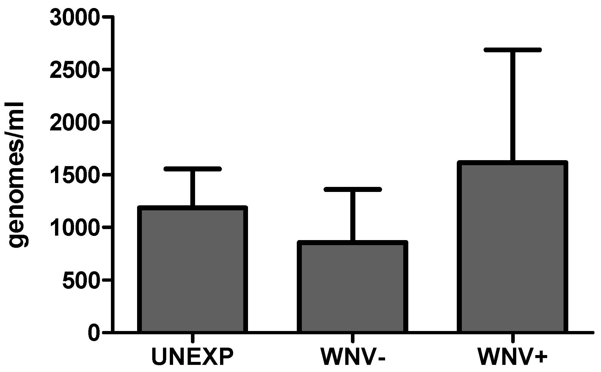
2.4. Total Microbial Load Determination

The amount of total bacterial DNA was determined in triplicate for individual mosquitoes using QuantiTect SYBR Green qPCR (Qiagen) with the same primers utilized for 16S sequencing (V3V4 hypervariable region). A standard curve was generated by extracting DH5-Alpha Escherichia coli, quantitating DNA utilizing a NanoDrop 2000 and calculating genome copies. A 10-fold serial dilution was created from 102 to 108 genome copies.

3. Results

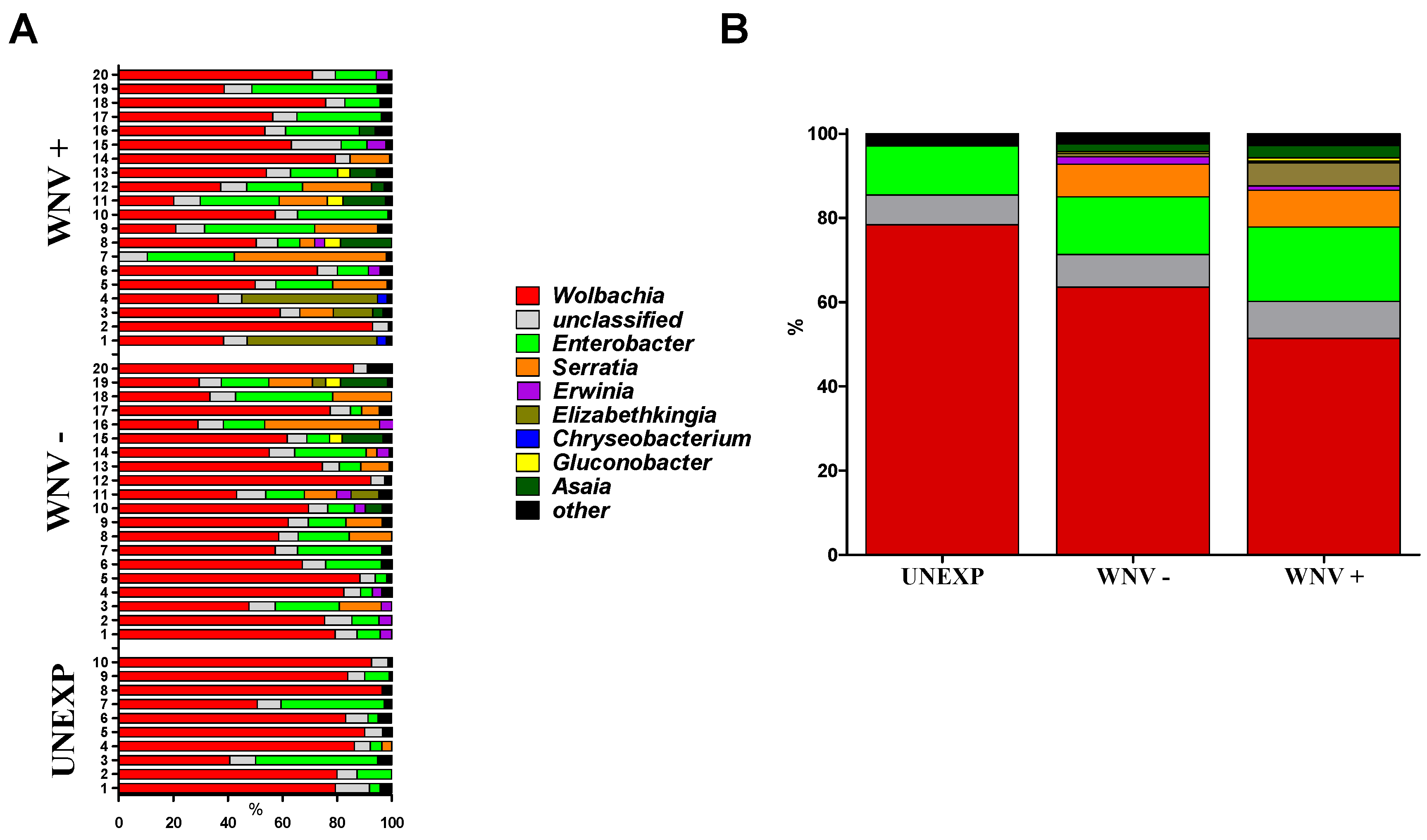
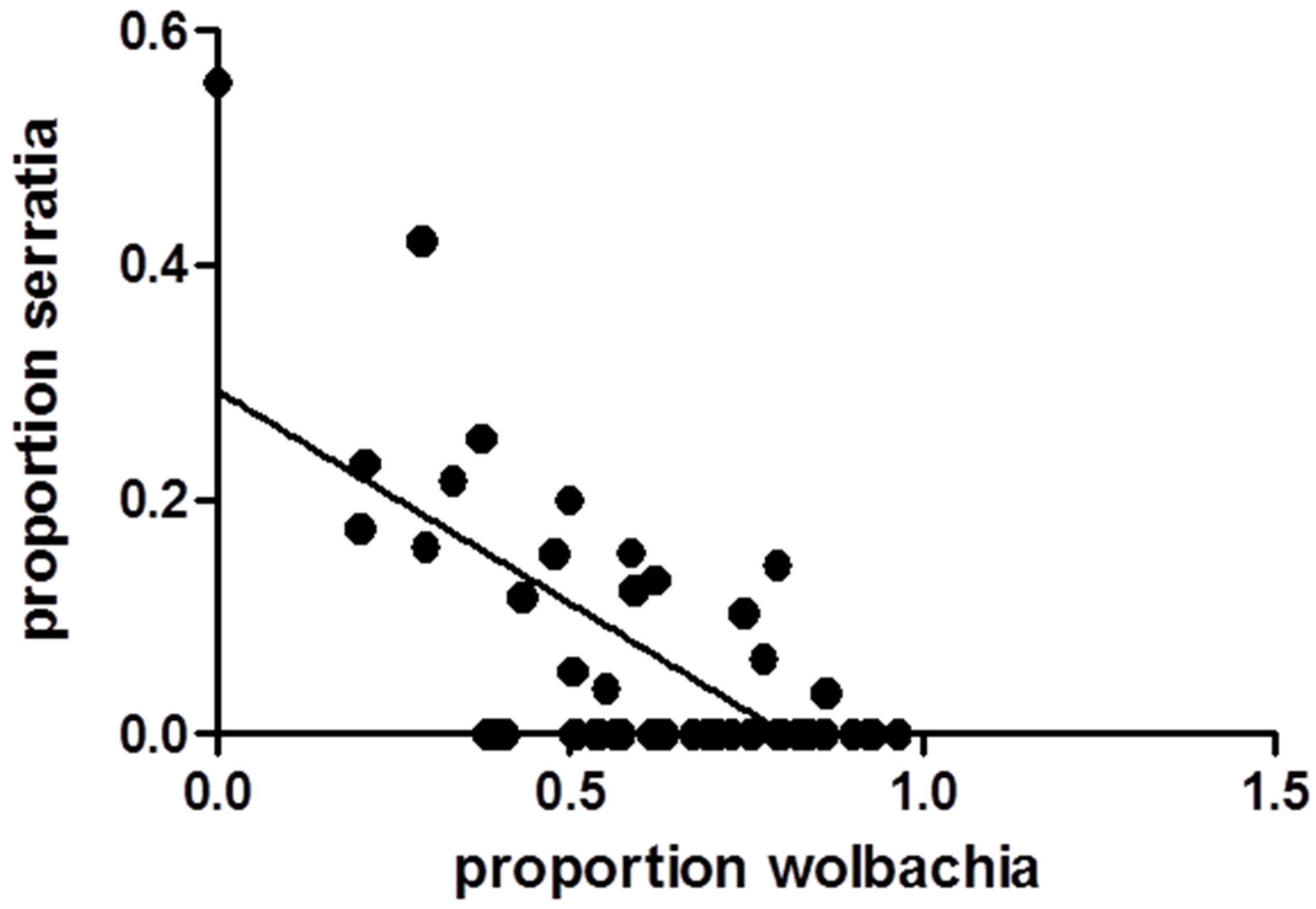
3.1. Relationships between WNV Exposure and Microbial Signatures in Cx. pipiens

WNV titer of infectious bloodmeals was 7.3 log10 pfu/mL, a realistic natural dose [33]. 120 fully engorged Cx. pipiens were tested at day seven post-feeding for WNV RNA, of which 39 (32.5%) were WNV positive. Bacterial microbiome characterization was completed for a subset of these including 20 WNV+, 20 WNV− and 10 unexposed control (UNEXP) mosquitoes. A mean of 193,542 reads/sample were identified by Illumina metagenomics analyses as bacterial in origin and members of the Proteobacteria phylum represented over 96% all sequences (sequences available upon request). Substantial diversity was found within this phylum, with representatives from means of 61, 100, and 106 families, genera and species, respectively. Since the classification rate for the species level was less than 25% for all samples, genera were used for taxonomic assignments and diversity analyses. Results demonstrate substantial variability within and among groups in microbial compositions (Figure 1). Means of 85, 105, and 98 genera were identified in UNEXP, WNV− and WNV+ groups, respectively. Although experimental treatment was not associated with distinct phylogenetic signatures (Supplementary Figure S1), increased bacterial diversity was associated with WNV exposure and further elevated with WNV infection (Table 1; Supplementary Figure S2). When diversity is corrected for sampling (read depth) both Shannon entropy and Simpson’s diversity indices demonstrate a clear trend of increasing genus level diversity with UNEXP relative to WNV—and additionally with WNV—relative to WNV+ (Table 1). The most dominant genus was Wolbachia for all groups, comprising 61.7% of all reads, yet the mean percentage of Wolbachia in the UNEXP group (78.4) was significantly higher than the WNV− group (63.6; Chi-square, p < 0.001), which was further decreased with WNV infection (51.4; Figure 1). Enterobacter, the second most prevalent genus, was modestly higher with WNV exposure and infection, with percentages of 11.6, 13.6, and 17.6 in UNEXP, WNV−, and WNV+ groups, respectively. While none of the UNEXP samples had classifiable representatives outside of these two genera at proportions greater than 3.5%, 75% (30/40) of all WNV exposed (WNV+ and WNV−) mosquitoes had at least one other genus represented at this level. The most striking difference between the UNEXP and WNV exposed groups was in the genus Serratia. While just 0.36% of UNEXP sequences were identified as Serratia, 7.8% and 8.7% of the reads from WNV− and WNV+ were identified as Serratia, respectively, representing a greater than 20-fold increase in proportion in WNV exposed groups relative to the UNEXP group. There was a significant negative correlation between the proportions of Wolbachia and Serratia in the samples sequenced (Pearson’s correlation, r = −0.687, p < 0.0001; Figure 2). All reads belonging to these genera which were resolved on the species level were identified as Wolbachia pipientis and Serratia entomophilia.
Quantitative PCR was used to assess if differences in diversity among groups correlate to generic differences in bacterial load. Results demonstrate significant variability in bacterial load among samples that did not consistently correlate to difference in bacterial diversity. Relative to the UNEXP group, mean bacterial load was modestly higher for the WNV+ group and modestly lower for the WNV− group, yet these differences were not significant (t-test, p > 0.05; Figure 3). Although it should be noted that the 16s copy number can be species-dependent to some degree, these results support the idea that neither WNV exposure nor infection status is associated with consistent variations in total bacteria. Interestingly, the individual mosquito with the highest bacterial load was the only sample in which no Wolbachia was identified and Serratia was the dominant genus.
Figure 1. Proportions of bacterial genera identified in individual (A) or combined (B) Cx. pipiens at seven days post blood feeding. Mosquitoes are classified as unexposed (UNEXP, non-infectious blood meal), WNV negative (WNV−, WNV exposed but uninfected) or WNV positive (WNV+). Proportions were generated from 16S rRNA metagenomics workflow on MiSeq Reporter. “Other” refers to genera found at proportion below 3.5%.
Viruses 07 02886 g001
Table 1. Bacterial diversity identified in Cx. pipiens seven days post blood feeding.
GroupFamilies/SampleGenera/SampleSpecies/Sample1-D 1Sn 2
UNEXP5485830.370.052
WNV−661091180.550.080
WNV+60981050.680.102
1 Simpson’s diversity index (1-D), D = ∑ni(ni-1)/N(N-1), where ni=reads from individual genera and N = total number of reads. 2 normalized Shannon entropy (Sn) = ∑-i Pi lnPi/ln N, where pi = frequency of individual genera and N = total number of reads.
Figure 2. Relationship between the proportions of Wolbachia and Serratia in Cx. pipiens at seven days post blood feeding. Data points represent individual mosquitoes and the line represents the best-fit relationship resulting from linear regression analysis. A significant negative correlation was found (Pearson’s correlation, r = −0.687, p < 0.0001).
Viruses 07 02886 g002
Figure 3. Total bacterial load in Cx. pipiens at seven days post blood feeding for unexposed (UNEXP), WNV−, and WNV+ mosquitoes. Bars represent means +/− SEM.
Viruses 07 02886 g003

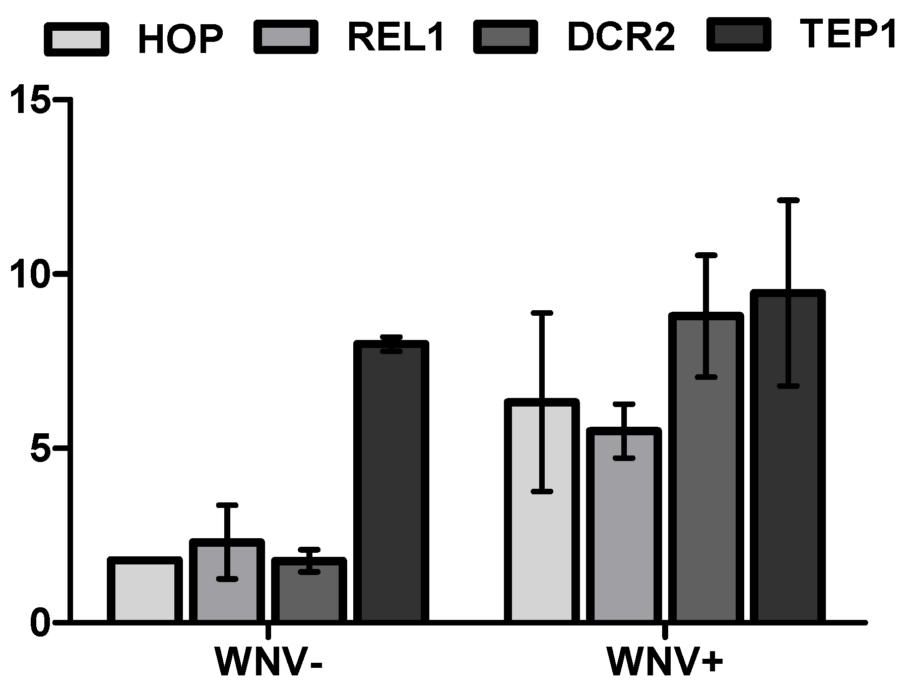
3.2. Mosquito Immune Gene Expression Following WNV Exposure and Infection

In order to assess if WNV exposure and infection, as well as unique microbial signatures, were associated with variability in the regulation of innate immune pathways, transcript levels of markers of individual pathways including Jak-STAT (HOP), Toll (REL1), and RNAi (DCR2), as well as TEP1, were quantified from Cx. pipiens pools after experimental treatments identical to those used to attain individual microbial signatures. Values for ΔΔCT, which control for small differences in the housekeeping gene and are expressed as relative differences as compared to the UNEXP group, demonstrate that all four markers are up-regulated at seven days post WNV exposure (Figure 4, t-test, p < 0.01). An approximately two-fold increase was measured for HOP, REL1, and DCR2, while a much larger eight-fold difference in TEP1 expression was identified. More substantial increases in levels of expression of all transcripts were measured in the WNV+ group, with approximately 5–10-fold increases measured relative to the UNEXP group. With the exception of TEP1, all values were also higher in the WNV+ group relative to the WNV− group, and the largest difference measured between these groups was for DCR2 expression. Together these data demonstrate that there is a generic up-regulation of the mosquito immune response which coincides with increases in bacterial diversity and is sustained following WNV exposure, even in the absence of established infection.
Figure 4. Cx. pipiens immune gene transcript levels at seven days post blood feeding for WNV+ and WNV− mosquitoes. Differences are expressed as mean fold change in qPCR CT (cycle threshold) +/− SD relative to unexposed mosquitoes and normalized by rpl8 transcript levels (ΔΔCT). Statistically significant differences (t-test, p < 0.001) were measured for all transcript levels for WNV exposed (− or +) relative to unexposed mosquitoes and WNV− relative to WNV+ mosquitoes, with the exception of TEP1, for which WNV− and WNV+ groups were statistically equivalent (p = 0.099).
Viruses 07 02886 g004

4. Discussion

Interactions between resident microbial communities of mosquitoes and arboviruses can significantly alter mosquito immune and metabolic pathways and may influence many aspects of vectorial capacity including vector competence, blood feeding behavior, and longevity [3,22,34,35,36,37]. Although previous studies have largely focused on the capacity of Wolbachia pipientis to decrease fitness of DENV [38,39,40], WNV [41,42] and other arboviruses [43,44,45], there are likely numerous direct and indirect microbial interactions that could influence virus transmissibility [3]. Here, we add to the growing body of literature demonstrating such interactions, focusing on the effect of WNV exposure and infection on bacterial signatures in Cx. pipiens. Our results demonstrate that WNV infection increases the diversity of bacterial populations and is associated with up-regulation of classical invertebrate immune pathways. In addition, we offer the novel observation that WNV exposure without the establishment of infection results in lasting alterations to microbial and immune signatures.
While the effect of WNV on bacterial populations has not been previously assessed, a limited number of studies have characterized bacterial communities of Culex spp., focusing primarily on culturable bacteria [46,47,48]. Although colonized mosquitoes were used in the current study, as has been found in most studies with field mosquitoes, the dominant bacteria we identified are gram-negative Proteobacteria [37,47,48,49,50,51]. It is somewhat surprising that the levels of bacterial diversity in colonized mosquitoes are generally higher than those found in previous studies with field populations, yet this may be a result of methodology, as here we utilized next-generation sequencing technologies with significant depth (~200,000 reads/sample) and did not limit testing to the midgut or other individual tissues. Although Wolbachia was the dominant genus in the majority of mosquitoes, we also found substantial variability among individuals despite the homogeneity of rearing conditions. While competence is likely to always be stochastic to some degree, and mosquito genetics may also contribute, these unique microbial signatures could partially account for individual variability in WNV vector competence [22]. Although we did not identify any specific genera that were uniquely or consistently associated with WNV resistance in Cx. pipiens, results indicate that WNV exposure together with the previously documented effect of blood feeding [12] are likely to significantly disrupt populations, perhaps hindering the capacity to identify signatures of resistance at seven days post-feeding. The fact that the proportion of Wolbachia was significantly higher in WNV− mosquitoes relative to WNV+ mosquitoes stands in contrast to the idea that Wolbachia has the capacity to limit virus infection and spread, yet it has been demonstrated that this effect is strain-dependent [52,53]. In addition, it has been shown that Wolbachia conversely has the ability to enhance WNV infection in Cx. tarsalis, yet it should be noted that in these experiments Wolbachia was injected rather than stably-inherited and, therefore, likely resulted in altered tropism and biological consequences [54]. A comparison to unexposed mosquitoes suggests that lower Wolbachia levels are a result of WNV exposure, and more so infection, rather than a determinant of WNV competence. WNV could benefit from the capacity to directly inhibit Wolbachia [41], yet data suggest that differences in proportions of Wolbachia and other taxa more likely result from the indirect consequences of immune modulation [55,56,57] and/or resource competition associated with viral infection [58].
The association between WNV infection and increased bacterial diversity is consistent with a recent study with Chikungunya virus (CHIKV) and Ae. albopictus, which demonstrates an increase in Enterobacter together with a decrease in Wolbachia [51]. Similar to WNV results, this study additionally shows that the increased diversity is not necessarily associated with an overall increase in bacterial load but instead a shift in composition.
WNV infection was also associated with up-regulation of all major immune pathways evaluated here. RNAi is considered the primary invertebrate immune response associated with arbovirus infections [14]. Consistent with this is the increased expression of dicer2 (DCR2) in WNV+ mosquitoes relative to both UNEXP and WNV− mosquitoes. More surprising was the up-regulation of markers of other pathways historically associated with responding to bacteria and other microbes. These results are consistent with more recent studies demonstrating a broader role for these pathways, including in responding to viral infections [59]. Jak-STAT has been shown to restrict WNV as a result of secreted Vago in mosquito cells [60] and the Toll pathway has been implicated in control of DENV Ae. aegypti [61,62]. The Imd pathway has also been shown to play a role in defense against RNA viruses in Drosophila [63,64], yet is most often associated with activation by Gram-negative bacteria [65,66]. The broad reactivity of these pathways provides a plausible mechanism by which bacterial and viral populations might indirectly interact and result in reciprocal modifications. Indeed, the more mosquito microbiota are investigated the more it becomes clear that outcomes of individual infections likely need to be considered in the context of the holobiome, including not just arboviruses and bacterial populations, but also mosquito-specific viruses and fungi [67]. Further studies assessing microbial signatures and immune regulation together with WNV kinetics in Cx. pipiens will provide a clearer picture of the causal nature of such interactions.
Perhaps the most surprising result in the current study is the fact that WNV exposure alone is associated with lasting increases in bacterial diversity and immune gene expression in Cx. pipiens. Previous studies demonstrate that WNV resistance can be associated with decreased longevity of Cx. pipiens, suggesting initial infection may be established and quickly quelled, but at a fitness cost [16,68]. One plausible explanation for this cost offered by the current data is that the lasting immune activation may come at a metabolic price, as has been seen with studies of pathogen resistance in Drosophila [69]. Alternatively, costs of both resistance and infection could be “multiple fronts costs” in which the immune response to the invading virus decreases the capacity to keep particular bacteria below pathogenic levels [70].
Although there was substantial variability among individual mosquitoes, the most consistent and sizable increase identified in WNV exposed mosquitoes was with the proportion of Serratia. Similar to Wolbachia, Serratia spp. have been shown to be passed both vertically and transtadially by mosquitoes [71] and S. odorifera have been demonstrated to enhance susceptibility of Ae. aegypti to both CHIKV and DENV with co-feeding experiments [71,72]. S. marcescans has the capacity to modulate Plasmodium infection and be pathogenic to mosquitoes [73,74,75], as well as having other antimicrobial properties [76,77]. A significant negative correlation between Wolbachia and Serratia was observed. This is consistent with there being a competitive interaction between these microbes, as has been observed with Wolbachia and other co-infecting endosymbionts [78,79], as well as with other maternally inherited bacteria [80,81]. A recent study demonstrated inhibition of Wolbachia by Asaia bacteria in Anopheles mosquitoes [82], a genus which was also identified in a number of WNV exposed mosquitoes in the current study. Serratia sequences for which species-level classification was possible in the current study were identified as S. entomophilia. Although this species has not been previously associated with mosquitoes, it is documented to be pathogenic in coleopteran larvae [83] and has been commercially used as an insecticide [84]. While the relationship between bacterial composition and vector competence is frequently discussed, the possibility that interactions among bacteria and/or between bacteria and viruses could alter other aspects of vectorial capacity, including vector longevity, has generally been overlooked. Further investigation of these interactions using field populations will help elucidate the likelihood that such relationships could ultimately be exploited in novel control strategies.

Supplementary Files

Supplementary File 1

Acknowledgments

Sequencing and metagenomics was completed by the Wadsworth Center Applied Genomics Technology Core and additional bioinformatics were completed at the Wadsworth Center Bioinformatics Core. Cells and media were provided by the Wadsworth Center Media and Tissue Culture Facility. We thank Pamela Chin for assistance with mosquito rearing and maintenance. This work was supported by federal funds from the National Institute of Health (grant number RO1-AI-077669).

Author Contributions

S.D.Z. performed experiments and wrote the manuscript. G.A.V.S. performed experiments. L.D.K. coordinated experiments, provided laboratory resources and edited the manuscript. M.J.B. completed bioinformatics and sequence analyses. A.T.C. conceived, coordinated and performed experiments, analyzed data and wrote the manuscript.

Conflicts of Interest

The authors declare no conflict of interest.

References

  1. Ciota, A.T.; Chin, P.A.; Kramer, L.D. The effect of hybridization of Culex pipiens complex mosquitoes on transmission of West Nile virus. Parasit. Vectors 2013, 6. [Google Scholar] [CrossRef]
  2. Nelms, B.M.; Fechter-Leggett, E.; Carroll, B.D.; Macedo, P.; Kluh, S.; Reisen, W.K. Experimental and natural vertical transmission of West Nile virus by California Culex (Diptera: Culicidae) mosquitoes. J. Med. Entomol. 2013, 50, 371–378. [Google Scholar] [CrossRef] [PubMed]
  3. Dennison, N.J.; Jupatanakul, N.; Dimopoulos, G. The mosquito microbiota influences vector competence for human pathogens. Curr. Opin. Insect. Sci. 2014, 3, 6–13. [Google Scholar] [CrossRef] [PubMed]
  4. Minard, G.; Mavingui, P.; Moro, C.V. Diversity and function of bacterial microbiota in the mosquito holobiont. Parasit. Vectors 2013, 6, e146. [Google Scholar] [CrossRef] [PubMed]
  5. Ricci, I.; Mosca, M.; Valzano, M.; Damiani, C.; Scuppa, P.; Rossi, P.; Crotti, E.; Cappelli, A.; Ulissi, U.; Capone, A.; et al. Different mosquito species host Wickerhamomyces anomalus (Pichia anomala): Perspectives on vector-borne diseases symbiotic control. Antonie Leeuwenhoek 2011, 99, 43–50. [Google Scholar] [CrossRef] [PubMed] [Green Version]
  6. Ryu, J.H.; Kim, S.H.; Lee, H.Y.; Bai, J.Y.; Nam, Y.D.; Bae, J.W.; Lee, D.G.; Shin, S.C.; Ha, E.M.; Lee, W.J. Innate immune homeostasis by the homeobox gene caudal and commensal-gut mutualism in Drosophila. Science 2008, 319, 777–782. [Google Scholar] [CrossRef] [PubMed]
  7. Duguma, D.; Rugman-Jones, P.; Kaufman, M.G.; Hall, M.W.; Neufeld, J.D.; Stouthamer, R.; Walton, W.E. Bacterial communities associated with culex mosquito larvae and two emergent aquatic plants of bioremediation importance. PLoS ONE 2013, 8, e72522. [Google Scholar] [CrossRef] [PubMed]
  8. Rejmankova, E.; Harbin-Ireland, A.; Lege, M. Bacterial abundance in larval habitats of four species of Anopheles (Diptera: Culicidae) in Belize, Central America. J. Vector Ecol. 2000, 25, 229–239. [Google Scholar] [PubMed]
  9. Volf, P.; Kiewegova, A.; Nemec, A. Bacterial colonisation in the gut of Phlebotomus duboseqi (Diptera: Psychodidae): Transtadial passage and the role of female diet. Folia Parasit. (Praha) 2002, 49, 73–77. [Google Scholar] [CrossRef]
  10. Xu, Y.; Chen, S.; Kaufman, M.G.; Maknojia, S.; Bagdasarian, M.; Walker, E.D. Bacterial community structure in tree hole habitats of Ochlerotatus triseriatus: Influences of larval feeding. J. Am. Mosq. Control Assoc. 2008, 24, 219–227. [Google Scholar] [CrossRef] [PubMed]
  11. Yee, D.A.; Allgood, D.; Kneitel, J.M.; Kuehn, K.A. Constitutive differences between natural and artificial container mosquito habitats: Vector communities, resources, microorganisms, and habitat parameters. J. Med. Entomol. 2012, 49, 482–491. [Google Scholar] [CrossRef] [PubMed]
  12. Oliveira, J.H.; Goncalves, R.L.; Lara, F.A.; Dias, F.A.; Gandara, A.C.; Menna-Barreto, R.F.; Edwards, M.C.; Laurindo, F.R.; Silva-Neto, M.A.; Sorgine, M.H.; et al. Blood meal-derived heme decreases ROS levels in the midgut of Aedes aegypti and allows proliferation of intestinal microbiota. PLoS Pathog. 2011, 7, e1001320. [Google Scholar] [CrossRef] [PubMed]
  13. Broderick, N.A.; Buchon, N.; Lemaitre, B. Microbiota-induced changes in drosophila melanogaster host gene expression and gut morphology. mBio 2014, 5, e01117-14. [Google Scholar] [CrossRef] [PubMed]
  14. Blair, C.D. Mosquito RNAi is the major innate immune pathway controlling arbovirus infection and transmission. Future Microbiol. 2011, 6, 265–277. [Google Scholar] [CrossRef] [PubMed]
  15. Arjona, A.; Wang, P.; Montgomery, R.R.; Fikrig, E. Innate immune control of West Nile virus infection. Cell Microbiol. 2011, 13, 1648–1658. [Google Scholar] [CrossRef] [PubMed]
  16. Ciota, A.T.; Styer, L.M.; Meola, M.A.; Kramer, L.D. The costs of infection and resistance as determinants of West Nile virus susceptibility in Culex mosquitoes. BMC Ecol. 2011, 11, e23. [Google Scholar] [CrossRef] [PubMed]
  17. Hill, C.L.; Sharma, A.; Shouche, Y.; Severson, D.W. Dynamics of midgut microflora and dengue virus impact on life history traits in Aedes aegypti. Acta Trop. 2014, 140, 151–157. [Google Scholar] [CrossRef] [PubMed]
  18. Styer, L.M.; Meola, M.A.; Kramer, L.D. West Nile virus infection decreases fecundity of Culex tarsalis females. J. Med. Entomol. 2007, 44, 1074–1085. [Google Scholar] [CrossRef] [PubMed]
  19. McMeniman, C.J.; O’Neill, S.L. A virulent Wolbachia infection decreases the viability of the dengue vector Aedes aegypti during periods of embryonic quiescence. PLoS Negl. Trop. Dis. 2010, 4, e748. [Google Scholar] [CrossRef] [PubMed]
  20. Putnam, J.L.; Scott, T.W. Blood-feeding behavior of dengue-2 virus-infected Aedes aegypti. Am. J. Trop. Med. Hyg. 1995, 52, 225–227. [Google Scholar] [PubMed]
  21. Walker, T.; Johnson, P.H.; Moreira, L.A.; Iturbe-Ormaetxe, I.; Frentiu, F.D.; McMeniman, C.J.; Leong, Y.S.; Dong, Y.; Axford, J.; Kriesner, P.; et al. The wMel Wolbachia strain blocks dengue and invades caged Aedes aegypti populations. Nature 2011, 476, 450–453. [Google Scholar] [CrossRef] [PubMed]
  22. Weiss, B.; Aksoy, S. Microbiome influences on insect host vector competence. Trends Parasitol. 2011, 27, 514–522. [Google Scholar] [CrossRef] [PubMed]
  23. Ebel, G.D.; Carricaburu, J.; Young, D.; Bernard, K.A.; Kramer, L.D. Genetic and phenotypic variation of West Nile virus in New York, 2000–2003. Am. J. Trop. Med. Hyg. 2004, 71, 493–500. [Google Scholar] [PubMed]
  24. Shi, P.-Y.; Kauffman, E.B.; Ren, P.; Felton, A.; Tai, J.H.; Dupuis, A.P., II; Jones, S.A.; Ngo, K.A.; Nicholas, D.C.; Maffei, J.G.; et al. High throughput detection of West Nile virus RNA. J. Clin. Microbiol. 2001, 39, 1264–1271. [Google Scholar] [CrossRef] [PubMed]
  25. Caporaso, J.G.; Lauber, C.L.; Walters, W.A.; Berg-Lyons, D.; Lozupone, C.A.; Turnbaugh, P.J.; Fierer, N.; Knight, R. Global patterns of 16S rRNA diversity at a depth of millions of sequences per sample. Proc. Natl. Acad. Sci. USA 2011, 108 (Suppl. 1), 4516–4522. [Google Scholar] [CrossRef] [PubMed]
  26. Caporaso, J.G.; Kuczynski, J.; Stombaugh, J.; Bittinger, K.; Bushman, F.D.; Costello, E.K.; Fierer, N.; Pena, A.G.; Goodrich, J.K.; Gordon, J.I.; et al. QIIME allows analysis of high-throughput community sequencing data. Nat. Methods 2010, 7, 335–336. [Google Scholar] [CrossRef] [PubMed]
  27. Dong, Y.; Aguilar, R.; Xi, Z.; Warr, E.; Mongin, E.; Dimopoulos, G. Anopheles gambiae immune responses to human and rodent Plasmodium parasite species. PLoS Pathog. 2006, 2, e52. [Google Scholar] [CrossRef] [PubMed]
  28. Carvalho-Leandro, D.; Ayres, C.F.; Guedes, D.R.; Suesdek, L.; Melo-Santos, M.A.; Oliveira, C.F.; Cordeiro, M.T.; Regis, L.N.; Marques, E.T.; Gil, L.H.; et al. Immune transcript variations among Aedes aegypti populations with distinct susceptibility to dengue virus serotype 2. Acta Trop. 2012, 124, 113–119. [Google Scholar] [CrossRef] [PubMed]
  29. Garver, L.S.; Dong, Y.; Dimopoulos, G. Caspar controls resistance to Plasmodium falciparum in diverse anopheline species. PLoS Pathog. 2009, 5, e1000335. [Google Scholar] [CrossRef] [PubMed]
  30. Goic, B.; Vodovar, N.; Mondotte, J.A.; Monot, C.; Frangeul, L.; Blanc, H.; Gausson, V.; Vera-Otarola, J.; Cristofari, G.; Saleh, M.C. RNA-mediated interference and reverse transcription control the persistence of RNA viruses in the insect model Drosophila. Nat. Immunol. 2013, 14, 396–403. [Google Scholar] [CrossRef] [PubMed]
  31. Maeda, H.; Fujimoto, C.; Haruki, Y.; Maeda, T.; Kokeguchi, S.; Petelin, M.; Arai, H.; Tanimoto, I.; Nishimura, F.; Takashiba, S. Quantitative real-time PCR using TaqMan and SYBR Green for Actinobacillus actinomycetemcomitans, Porphyromonas gingivalis, Prevotella intermedia, tetQ gene and total bacteria. FEMS Immunol. Med. Microbiol. 2003, 39, 81–86. [Google Scholar] [CrossRef]
  32. Livak, K.J.; Schmittgen, T.D. Analysis of relative gene expression data using real-time quantitative PCR and the 2(-Delta Delta C(T)) Method. Methods 2001, 25, 402–408. [Google Scholar] [CrossRef] [PubMed]
  33. McLean, R.G.; Ubico, S.R.; Docherty, D.E.; Hansen, W.R.; Sileo, L.; McNamara, T.S. West Nile virus transmission and ecology in birds. Ann. N. Y. Acad. Sci. 2001, 951, 54–57. [Google Scholar] [CrossRef] [PubMed]
  34. Azambuja, P.; Garcia, E.S.; Ratcliffe, N.A. Gut microbiota and parasite transmission by insect vectors. Trends Parasitol. 2005, 21, 568–572. [Google Scholar] [CrossRef] [PubMed]
  35. Brownlie, J.C.; Johnson, K.N. Symbiont-mediated protection in insect hosts. Trends Microbiol. 2009, 17, 348–354. [Google Scholar] [CrossRef] [PubMed]
  36. Cirimotich, C.M.; Ramirez, J.L.; Dimopoulos, G. Native microbiota shape insect vector competence for human pathogens. Cell Host Microbe 2011, 10, 307–310. [Google Scholar] [CrossRef] [PubMed]
  37. Gaio Ade, O.; Gusmao, D.S.; Santos, A.V.; Berbert-Molina, M.A.; Pimenta, P.F.; Lemos, F.J. Contribution of midgut bacteria to blood digestion and egg production in Aedes aegypti (diptera: Culicidae) (L.). Parasit. Vectors 2011, 4, e105. [Google Scholar] [CrossRef] [PubMed]
  38. Bian, G.; Xu, Y.; Lu, P.; Xie, Y.; Xi, Z. The endosymbiotic bacterium Wolbachia induces resistance to dengue virus in Aedes aegypti. PLoS Pathog. 2010, 6, e1000833. [Google Scholar] [CrossRef] [PubMed]
  39. Hoffmann, A.A.; Montgomery, B.L.; Popovici, J.; Iturbe-Ormaetxe, I.; Johnson, P.H.; Muzzi, F.; Greenfield, M.; Durkan, M.; Leong, Y.S.; Dong, Y.; et al. Successful establishment of Wolbachia in Aedes populations to suppress dengue transmission. Nature 2011, 476, 454–457. [Google Scholar] [CrossRef] [PubMed]
  40. Moreira, L.A.; Iturbe-Ormaetxe, I.; Jeffery, J.A.; Lu, G.; Pyke, A.T.; Hedges, L.M.; Rocha, B.C.; Hall-Mendelin, S.; Day, A.; Riegler, M.; et al. A Wolbachia symbiont in Aedes aegypti limits infection with dengue, Chikungunya, and Plasmodium. Cell 2009, 139, 1268–1278. [Google Scholar] [CrossRef] [PubMed]
  41. Glaser, R.L.; Meola, M.A. The native Wolbachia endosymbionts of Drosophila melanogaster and Culex quinquefasciatus increase host resistance to West Nile virus infection. PLoS ONE 2010, 5, e11977. [Google Scholar] [CrossRef] [PubMed]
  42. Micieli, M.V.; Glaser, R.L. Somatic Wolbachia (Rickettsiales: Rickettsiaceae) levels in Culex quinquefasciatus and Culex pipiens (Diptera: Culicidae) and resistance to West Nile virus infection. J. Med. Entomol. 2014, 51, 189–199. [Google Scholar] [CrossRef] [PubMed]
  43. Hedges, L.M.; Brownlie, J.C.; O’Neill, S.L.; Johnson, K.N. Wolbachia and virus protection in insects. Science 2008, 322, 702. [Google Scholar] [CrossRef] [PubMed]
  44. Mousson, L.; Martin, E.; Zouache, K.; Madec, Y.; Mavingui, P.; Failloux, A.B. Wolbachia modulates Chikungunya replication in Aedes albopictus. Mol. Ecol. 2010, 19, 1953–1964. [Google Scholar] [CrossRef] [PubMed]
  45. Van den Hurk, A.F.; Hall-Mendelin, S.; Pyke, A.T.; Frentiu, F.D.; McElroy, K.; Day, A.; Higgs, S.; O’Neill, S.L. Impact of Wolbachia on infection with chikungunya and yellow fever viruses in the mosquito vector Aedes aegypti. PLoS Negl. Trop. Dis. 2012, 6, e1892. [Google Scholar] [CrossRef] [PubMed] [Green Version]
  46. Chandel, K.; Mendki, M.J.; Parikh, R.Y.; Kulkarni, G.; Tikar, S.N.; Sukumaran, D.; Prakash, S.; Parashar, B.D.; Shouche, Y.S.; Veer, V. Midgut microbial community of Culex quinquefasciatus mosquito populations from India. PLoS ONE 2013, 8, e80453. [Google Scholar] [CrossRef] [PubMed]
  47. Demaio, J.; Pumpuni, C.B.; Kent, M.; Beier, J.C. The midgut bacterial flora of wild Aedes triseriatus, Culex pipiens, and Psorophora columbiae mosquitoes. Am. J. Trop. Med. Hyg. 1996, 54, 219–223. [Google Scholar] [PubMed]
  48. Pidiyar, V.J.; Jangid, K.; Patole, M.S.; Shouche, Y.S. Studies on cultured and uncultured microbiota of wild culex quinquefasciatus mosquito midgut based on 16s ribosomal RNA gene analysis. Am. J. Trop. Med. Hyg. 2004, 70, 597–603. [Google Scholar] [PubMed]
  49. Lindh, J.M.; Terenius, O.; Faye, I. 16S rRNA gene-based identification of midgut bacteria from field-caught Anopheles gambiae sensu lato and A. funestus mosquitoes reveals new species related to known insect symbionts. Appl. Environ. Microbiol. 2005, 71, 7217–7223. [Google Scholar] [CrossRef] [PubMed]
  50. Rani, A.; Sharma, A.; Rajagopal, R.; Adak, T.; Bhatnagar, R.K. Bacterial diversity analysis of larvae and adult midgut microflora using culture-dependent and culture-independent methods in lab-reared and field-collected Anopheles stephensi-an Asian malarial vector. BMC Microbiol. 2009, 9, e96. [Google Scholar] [CrossRef] [PubMed]
  51. Zouache, K.; Michelland, R.J.; Failloux, A.B.; Grundmann, G.L.; Mavingui, P. Chikungunya virus impacts the diversity of symbiotic bacteria in mosquito vector. Mol. Ecol. 2012, 21, 2297–2309. [Google Scholar] [CrossRef] [PubMed]
  52. Hussain, M.; Lu, G.; Torres, S.; Edmonds, J.H.; Kay, B.H.; Khromykh, A.A.; Asgari, S. Effect of wolbachia on replication of west nile virus in a mosquito cell line and adult mosquitoes. J. Virol. 2013, 87, 851–858. [Google Scholar] [CrossRef] [PubMed]
  53. Martinez, J.; Longdon, B.; Bauer, S.; Chan, Y.S.; Miller, W.J.; Bourtzis, K.; Teixeira, L.; Jiggins, F.M. Symbionts commonly provide broad spectrum resistance to viruses in insects: A comparative analysis of Wolbachia strains. PLoS Pathog. 2014, 10, e1004369. [Google Scholar] [CrossRef] [PubMed]
  54. Dodson, B.L.; Hughes, G.L.; Paul, O.; Matacchiero, A.C.; Kramer, L.D.; Rasgon, J.L. Wolbachia enhances West Nile virus (WNV) infection in the mosquito Culex tarsalis. PLoS Negl. Trop. Dis. 2014, 8, e2965. [Google Scholar] [CrossRef] [PubMed]
  55. Kambris, Z.; Cook, P.E.; Phuc, H.K.; Sinkins, S.P. Immune activation by life-shortening Wolbachia and reduced filarial competence in mosquitoes. Science 2009, 326, 134–136. [Google Scholar] [CrossRef] [PubMed]
  56. Pan, X.; Zhou, G.; Wu, J.; Bian, G.; Lu, P.; Raikhel, A.S.; Xi, Z. Wolbachia induces reactive oxygen species (ROS)-dependent activation of the Toll pathway to control dengue virus in the mosquito Aedes aegypti. Proc. Natl. Acad. Sci. USA 2012, 109, E23–E31. [Google Scholar] [CrossRef] [PubMed]
  57. Rances, E.; Ye, Y.H.; Woolfit, M.; McGraw, E.A.; O’Neill, S.L. The relative importance of innate immune priming in Wolbachia-mediated dengue interference. PLoS Pathog. 2012, 8, e1002548. [Google Scholar] [CrossRef] [PubMed]
  58. Caragata, E.P.; Rances, E.; O’Neill, S.L.; McGraw, E.A. Competition for amino acids between Wolbachia and the mosquito host, Aedes aegypti. Microb. Ecol. 2014, 67, 205–218. [Google Scholar] [CrossRef] [PubMed]
  59. Fragkoudis, R.; ttarzadeh-Yazdi, G.; Nash, A.A.; Fazakerley, J.K.; Kohl, A. Advances in dissecting mosquito innate immune responses to arbovirus infection. J. Gen. Virol. 2009, 90, 2061–2072. [Google Scholar] [CrossRef] [PubMed]
  60. Paradkar, P.N.; Trinidad, L.; Voysey, R.; Duchemin, J.B.; Walker, P.J. Secreted Vago restricts West Nile virus infection in Culex mosquito cells by activating the Jak-STAT pathway. Proc. Natl. Acad. Sci. USA 2012, 109, 18915–18920. [Google Scholar] [CrossRef] [PubMed]
  61. Ramirez, J.L.; Souza-Neto, J.; Torres, C.R.; Rovira, J.; Ortiz, A.; Pascale, J.M.; Dimopoulos, G. Reciprocal tripartite interactions between the Aedes aegypti midgut microbiota, innate immune system and dengue virus influences vector competence. PLoS. Negl. Trop. Dis. 2012, 6, e1561. [Google Scholar] [CrossRef] [PubMed]
  62. Xi, Z.; Ramirez, J.L.; Dimopoulos, G. The Aedes aegypti toll pathway controls dengue virus infection. PLoS Pathog. 2008, 4, e1000098. [Google Scholar] [CrossRef] [PubMed]
  63. Avadhanula, V.; Weasner, B.P.; Hardy, G.G.; Kumar, J.P.; Hardy, R.W. A novel system for the launch of alphavirus RNA synthesis reveals a role for the Imd pathway in arthropod antiviral response. PLoS Pathog. 2009, 5, e1000582. [Google Scholar] [CrossRef] [PubMed]
  64. Costa, A.; Jan, E.; Sarnow, P.; Schneider, D. The Imd pathway is involved in antiviral immune responses in Drosophila. PLoS ONE 2009, 4, e7436. [Google Scholar] [CrossRef] [PubMed]
  65. Ferrandon, D.; Imler, J.L.; Hetru, C.; Hoffmann, J.A. The Drosophila systemic immune response: Sensing and signalling during bacterial and fungal infections. Nat. Rev. Immunol. 2007, 7, 862–874. [Google Scholar] [CrossRef] [PubMed]
  66. Nehme, N.T.; Quintin, J.; Cho, J.H.; Lee, J.; Lafarge, M.C.; Kocks, C.; Ferrandon, D. Relative roles of the cellular and humoral responses in the Drosophila host defense against three gram-positive bacterial infections. PLoS ONE 2011, 6, e14743. [Google Scholar] [CrossRef] [PubMed] [Green Version]
  67. Chandler, J.A.; Liu, R.M.; Bennett, S.N. RNA shotgun metagenomic sequencing of northern California (USA) mosquitoes uncovers viruses, bacteria, and fungi. Front. Microbiol. 2015, 6, e185. [Google Scholar] [CrossRef] [PubMed]
  68. Ciota, A.T.; Matacchiero, A.C.; Kilpatrick, A.M.; Kramer, L.D. The effect of temperature on life history traits of Culex mosquitoes. J. Med. Entomol. 2013, 51, 55–62. [Google Scholar] [CrossRef]
  69. McKean, K.A.; Nunney, L. Sexual selection and immune function in Drosophila melanogaster. Evolution 2008, 62, 386–400. [Google Scholar] [CrossRef] [PubMed]
  70. McKean, K.A.; Yourth, C.P.; Lazzaro, B.P.; Clark, A.G. The evolutionary costs of immunological maintenance and deployment. BMC. Evol. Biol. 2008, 8, e76. [Google Scholar] [CrossRef] [PubMed]
  71. Apte-Deshpande, A.; Paingankar, M.; Gokhale, M.D.; Deobagkar, D.N. Serratia odorifera a midgut inhabitant of Aedes aegypti mosquito enhances its susceptibility to dengue-2 virus. PLoS ONE 2012, 7, e40401. [Google Scholar] [CrossRef] [PubMed]
  72. Apte-Deshpande, A.D.; Paingankar, M.S.; Gokhale, M.D.; Deobagkar, D.N. Serratia odorifera mediated enhancement in susceptibility of Aedes aegypti for chikungunya virus. Indian J. Med. Res. 2014, 139, 762–768. [Google Scholar] [PubMed]
  73. Bando, H.; Okado, K.; Guelbeogo, W.M.; Badolo, A.; Aonuma, H.; Nelson, B.; Fukumoto, S.; Xuan, X.; Sagnon, N.; Kanuka, H. Intra-specific diversity of Serratia marcescens in Anopheles mosquito midgut defines Plasmodium transmission capacity. Sci. Rep. 2013, 3, 1641. [Google Scholar] [CrossRef] [PubMed]
  74. Gonzalez-Ceron, L.; Santillan, F.; Rodriguez, M.H.; Mendez, D.; Hernandez-Avila, J.E. Bacteria in midguts of field-collected Anopheles albimanus block Plasmodium vivax sporogonic development. J. Med. Entomol. 2003, 40, 371–374. [Google Scholar] [CrossRef] [PubMed]
  75. Seitz, H.M.; Maier, W.A.; Rottok, M.; Becker-Feldmann, H. Concomitant infections of Anopheles stephensi with Plasmodium berghei and Serratia marcescens: Additive detrimental effects. Zentralbl. Bakteriol. Mikrobiol. Hyg. A 1987, 266, 155–166. [Google Scholar] [CrossRef]
  76. Okamoto, N. Norrie disease. Ryoikibetsu Shokogun Shirizu 1998, 19 Pt 2, 627–629. [Google Scholar]
  77. Someya, Y. Caliciviruses. Uirusu 2000, 50, 173–184. [Google Scholar] [CrossRef] [PubMed]
  78. Goto, S.; Anbutsu, H.; Fukatsu, T. Asymmetrical interactions between Wolbachia and Spiroplasma endosymbionts coexisting in the same insect host. Appl. Environ. Microbiol. 2006, 72, 4805–4810. [Google Scholar] [CrossRef] [PubMed]
  79. Kondo, N.; Shimada, M.; Fukatsu, T. Infection density of Wolbachia endosymbiont affected by co-infection and host genotype. Biol. Lett. 2005, 1, 488–491. [Google Scholar] [CrossRef] [PubMed]
  80. de la Fuente, J.; Garcia-Garcia, J.C.; Blouin, E.F.; Kocan, K.M. Characterization of the functional domain of major surface protein 1a involved in adhesion of the rickettsia Anaplasma marginale to host cells. Vet. Microbiol. 2003, 91, 265–283. [Google Scholar] [CrossRef]
  81. Macaluso, K.R.; Sonenshine, D.E.; Ceraul, S.M.; Azad, A.F. Rickettsial infection in Dermacentor variabilis (Acari: Ixodidae) inhibits transovarial transmission of a second Rickettsia. J. Med. Entomol. 2002, 39, 809–813. [Google Scholar] [CrossRef] [PubMed]
  82. Hughes, G.L.; Dodson, B.L.; Johnson, R.M.; Murdock, C.C.; Tsujimoto, H.; Suzuki, Y.; Patt, A.A.; Cui, L.; Nossa, C.W.; Barry, R.M.; et al. Native microbiome impedes vertical transmission of Wolbachia in Anopheles mosquitoes. Proc. Natl. Acad. Sci. USA 2014, 111, 12498–12503. [Google Scholar] [CrossRef] [PubMed]
  83. Nunez-Valdez, M.E.; Mahanty, H.K. The amb2 locus from Serratia entomophila confers anti-feeding effect on larvae of Costelytra zealandica (Coleoptera: Scarabaeidae). Gene 1996, 172, 75–79. [Google Scholar] [CrossRef]
  84. Grimont, P.A.D.; Jackson, T.A.; Ageron, E.; Noonan, M.J. Serratia entomophila sp. nov. Associated with Amber Disease in the New Zealand Grass Grub Costelytra zealandica. Int. J.Syst. Bacteriol. 1988, 38, 1–6. [Google Scholar] [CrossRef]

