Article

The First Complete Genome Sequence of a Novel Tetrastichus brontispae RNA Virus-1 (TbRV-1)

1
State Key Laboratory of Ecological Pest Control of Fujian-Taiwan Crops, Fujian Agriculture and Forestry University, Fuzhou 350002, China
2
Qingzhou Yatai Agricultural Technology Company Limited, Qingzhou 262500, China
3
Fujian Provincial Key Laboratory of Insect Ecology, College of Plant Protection, Fujian Agriculture and Forestry University, Fuzhou 350002, China
4
Fuzhou Entry-Exit Inspection & Quarantine Bureau of P.R.C, Fuzhou 350002, China
*
Author to whom correspondence should be addressed.
Viruses 2019, 11(3), 257; https://doi.org/10.3390/v11030257
Submission received: 19 January 2019 / Revised: 9 March 2019 / Accepted: 10 March 2019 / Published: 13 March 2019
(This article belongs to the Section Insect Viruses)

Abstract

:
The complete sequence of a novel RNA virus isolated from Tetrastichus brontispae (TbRV-1) was determined to be 12,239 nucleotides in length with five non-overlapping, linearly arranged coding sequences (CDS), potentially encoding nucleoproteins, hypothetical proteins, matrix proteins, glycoproteins, and RNA-dependent RNA polymerases. Sequence analysis indicated that the RNA-dependent RNA polymerase of TbRV-1 shares a 65% nucleotide and 67% amino acid sequence identity with Hubei dimarhabdovirus 2, suggesting that TbRV-1 is a member of the dimarhabdovirus supergroup. This corresponded to the result of the phylogenetic analysis. The affiliation of TbRV-1 with members of the family Rhabdoviridae was further validated by similar transcription termination motifs (GGAACUUUUUUU) to the Drosophila sigmavirus. The prevalence of TbRV-1 in all tissues suggested that the virus was constitutive of, and not specific to, any wasp tissue. To our knowledge, this is the first report on the complete genome sequence of a dimarhabdovirus in parasitoids.

1. Introduction

Rhabdoviridae, a family of viruses in the Mononegavirales order, are negative-sense (–) single-stranded RNA viruses. Most of them infect plants and animals (mammals, birds, reptiles, fish and arthropods) and are categorized into arthropod-vectored viruses of plants or vertebrates, and vertebrate- and arthropod-specific viruses on the basis of the host range [1]. Based on the strong nexus of virus clustering and the host types, as well as the broad host range of some rhabdovirus, the insects are proposed to be the primary determinants of host range of the plant and animal rhabdovirus [2,3]. Currently, 20 officially recognized genera and one viruses are reported [4]. The main insect-associated rhabdoviridae are from the genera Vesiculovirus, Cytorhabdovirus, Nucleorhabdovirus, Sigmavirus, Ephemerovirus and the superfamily dimarhabdovirus [1], where the majority insect vectors are from Diptera, Hemiptera, and Lepidopteara [3].
Among the arthropod-specific viruses, one group of predominantly insect-associated viruses is a sister group to the large clade of plant viruses, while the other group is the sigma virus clade, and are derived from the vector-borne dimarhabdoviruses [3]. Dimarhabdovirus, the dominant type of vector-borne rhabdovirus, which mainly infects Diptera, is the causative agent of significant morbidity and mortality among humans and animals globally [5]. In contrast to the cytolytic sides of rhabdoviruses to the mammalian, little or no cell killing is observed when insects are infected [6]. The rhabdoviral infection of an insect is typically persistent, and includes an initial phase of rapid replication, an acute phase of viral production, and a persistent phase with lower viral replication levels [6]. Even so, most insects infected with rhabdoviruses become more sensitive to paralysis and even die on exposure to CO2, such as the effect of sigmavirus on Drosophila [7,8]. Up to now, most research on insect-associated rhabdovirus still focuses on the initial characterization, with fewer reports on their roles.
Parasitoid wasps as vital biocontrol agents against pests are widely used in agro-ecosystems. In most cases, the higher efficiency of the parasitism ratio of Icheneumonidae and Braconidae wasps results in the presentation of viruses or virus-like particles detected in parasitoid wasps, such as the polydnavirus [9,10,11,12]. In other cases, the viruses presented in parasitoid wasps affect reproductive efficiency. For example, the Pteromalus puparum negative-strand RNA virus 1 (PpNSRV-1) reduced female offspring numbers, while it increased the longevity of the wasp P. puparum [13]. For the rhabdovirus from Hymenoptera, the Diachasminorpha longicaudata rhabdovirus (DlRhV) was introduced into the host Anastrepha suspense upon parasitism, and no indispensable role of DlRhV in successful parasitism was reported [14,15]. In addition, the genome of DlRhV was sequenced in 2016, and still is the only known genome of rhabodovirus from Hymenoptera [15].
Tetrastichus brontispae, a type of endoparasitoid wasp, is an efficient bio-control agent against several invasive leaf beetle species, such as Octodonta nipae and Brontispa longissima [5,16,17,18,19]. Previous studies showed that a novel mixture of systemic active and local active regulation was used for T. brontispae to escape host encapsulation in O. nipae pupae [20]. The role of virus-like particles and calyx fluid in achieving local active regulation has been widely reported [21,22]. Hence, the types of virus present in T. brontispae were detected by next generation sequencing (NGS).
In this paper, the detection of virus types was performed by metatranscriptional analysis, and the complete genome of a novel dimarhabdovirus from T. brontispae, named T. brontispae RNA virus 1 (TbRV-1) was reported. The TbRV-1 was somewhat similar to Hubei dimarhabdovirus, a novel virus discovered in Odonata by transcriptome and assigned to the dimarhabdovirus superfamily by bioinformatics [23].This study broadens the current knowledge on the fundamental patterns and processes of the viral evolution of rhabdoviruses.

2. Materials and Methods

2.1. Insect Collection

The wasps, T. brontispae, were reared on Octodonta nipae or Brontispa longissima (Coleoptera: Chrysomelidae) on the campus of Fujian Agriculture and Forestry University for nearly four years, after being collected from the Chinese Academy of Tropical Agricultural Sciences in 2012.

2.2. Transcriptome of the Tetrastichus brontispae Abdomen

The transcriptome of T. brontispae was sequenced as described by Tang et al. (2014) [24]. The total RNA from the 575 abdomens of female wasps was extracted using the Trizol reagent (Invitrogen, New York, NY, USA) and treated with DNase I. The concentration and integrity of the total RNA were assessed by a 2100 Bioanalyzer (Agilent Technologies, Santa Clara, CA, USA) before library construction.
The complementary DNA (cDNA) library was constructed with NEBNext® Ultra RNA Library Prep Kit (New England Biolabs, Ipswich, MA, USA). Poly (A) mRNA was isolated using oligo-dT beads (Qiagen, Tokyo, Japan). All mRNA was broken into short fragments (200 nt) by adding a fragmentation buffer. First-strand cDNA was generated using random hexamer-primed reverse transcription, followed by the synthesis of the second-strand cDNA using RNase H and DNA polymerase I. The cDNA fragments were purified using a QIAquick PCR extraction kit. These purified fragments were then washed with EB buffer for the addition of end reparation poly (A), and then they were ligated to sequencing adapters. Following agarose gel electrophoresis and the extraction of cDNA from gels, the cDNA fragments (200 ± 25 bp) were purified and enriched by PCR to construct the final cDNA library. The cDNA library was qualified and quantified with an Agilent 2100 Bioanalyzer and an ABI StepOnePlus Real-time PCR system, respectively, and then sequenced for 125 bp on the Illumina HiSeq TM 2500 platform using the single-end/paired-end technology in a single run at Genedenovo (Guangzhou, China).

2.3. Viral Metagenomics Analysis

For the reads derived from RNA-Seq, the clean data was obtained by filtering out unknown nucleotides larger than 5% and low quality reads and used for assembly with Trinity [25]. Those assembled sequences were outputted as unigenes. Unigenes aligned to Rhabdoviridae by Blastx with an E-value cut-off of 1 × 10⁻⁵ against the NCBI non-redundant (NR) database were chosen for further analysis because of the scarce reports regarding the Rhabdoviridae in Hymenoptera.

2.4. Virus Genome Sequencing

The canonical genome structure of rhabdovirus is 3’-nucleoprotein (N)-phosphoprotein (P)-matrix protein (M)-glycoprotein (G)-RNA-dependent RNA polymerase (L) [26]. Based on the typical genome layout of rhabdovirus (3’-N-P-M-G-L-5’), the gaps among the unigenes of TbRV-1 were first amplified by a routine polymerase chain reaction. And the gaps between the genes and the terminus end of TbRV-1 were obtained by the rapid amplification of cDNA ends (RACE PCR) with a SMARTerTM RACE PCR kit (Clonetech, Shiga, Japan). The specific primers for amplifying the whole genome of the putative rhabdovirus were designed according to the obtained sequences (Supplementary Table S1).
The PCR products were sequenced by Invitrogen (Guangzhou, China) after ligation into the T1 vector (TransGen Biotech, Beijing, China) and transformation into Escherichia coli T1. The final sequences were assembled using the DNAMAN 7 program (Lynnon, Quebec, Canada). The full length of the coding sequence was further validated after sequencing the products of the routine PCR acquired with a fastfly pfu enzyme (TransGen Biotech).
The complete genome of TbRV-1 was aligned to the known viruses by Blastx and Blastn to identify its closest relatives. The putative open reading frames (ORFs) were predicted by the ORF finder (https://www.ncbi.nlm.nih.gov/orffinder/). ORFs were analyzed on InterPro Scan [27] and PROSITE databases to find matches to known protein families and the potential functions of predicted proteins [28]. A maximum likelihood phylogenetic tree constructed with RdRP, the most conservative sequence domain across all RNA viruses [23,26], and was performed by MEGA 5.2 using the rtREV substitution model with gamma distributed and invariant sites (G + I) and the bootstrap method with 1000 repetitions [29]. The information on the sequences used for the analysis is listed in Supplementary Table S2.

2.5. MicroRNA Library Construction and Sequencing

As suggested earlier, infection caused by arboviruses is typically persistent, and is characterized by an initial phase of rapid replication and substantial viral production [6]. In response to infection, invertebrates process replicating viral RNA genomes into short interfering RNAs (siRNAs) of discrete sizes to guide virus clearance by RNA interference [30]. Therefore, to detect if the TbRV-1 were replicating in the wasps, the profile of small RNA tags from TbRV-1 was searched.
The small RNA library was constructed as follows. Seventy individuals of T. brontispae were used for every library. The total RNA was extracted by Trizol reagent. The RNA molecules in a size of 18–30 nts (nucleotides) were enriched from the total RNA of T. brontispae by polyacrylamide gel electrophoresis. After the ligation of 3’ and 5’ adapters, the ligation products were reverse-transcribed by PCR amplification, where the PCR products were enriched to generate a cDNA library and sequenced using IlluminaHiseqTM 2500 by Gene Denovo (Guangzhou, China).
Before further analysis, the clean tags were retrieved by trimming the adaptors after removing low quality reads containing more than one low quality base (Q-value ≤ 20) or unknown nucleotides. The clean reads were de novo assembled by Velvet with a k-mer of 17 [31]. To check if there were small RNA tags from TbRV-1, the assembled reads were subjected to Blastn analysis against the genome of TbRV-1 with a threshold E value of 10−5.

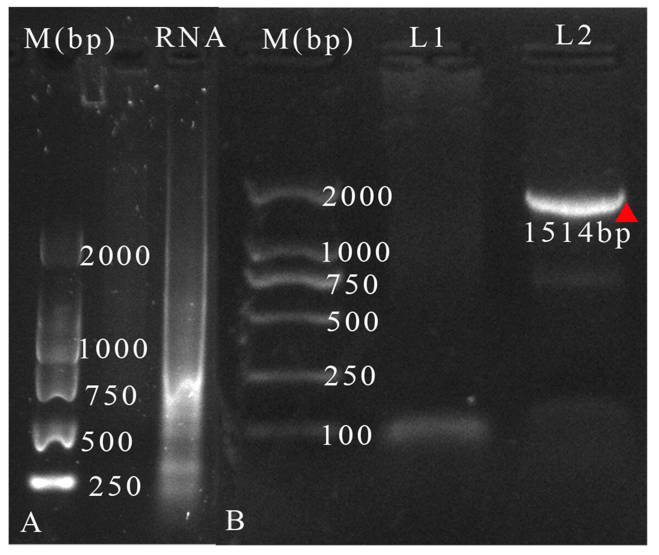
2.6. Isolation of Virus RNA

The virus pellet was isolated following the procedure described by Suzuki and Tanake (2005) [32], with some modification. Briefly, the wasps T. brontispae were homogenized with pipette. Then, the supernatants drawn from the homogenization after centrifuging at 600 g for 10 min were used for re-centrifuging at 15,000 g for 30 min at 4 °C. The virus pellets were resuspended in sterilized PBS, filtered with 0.45 μm Millipore filter, and used for RNA extraction with Qiagen virus RNA extraction kit (Qiagen, Tokyo, Japan). Then, the RNA was visualized by 1% agarose gel electrophoresis.
To detect if there was TbRV-1 in the mixture, the RNA extracted above was reverse-transcribed to cDNA using the SMARTerTM RACE cDNA amplification kit (Clontech laboratories Incorporation, Japan). A pair of primers (MS and MA), amplifying 1541 bp, was used for PCR amplification. The PCR was run with fastfly pfu enzyme with the following conditions: 30 s at 94 °C, 30 s at 49.8 °C, and 45 s at 72 °C for 35 cycles. The PCR reaction, with distilled water as the template, was used as a negative control. The PCR results were visualized by agarose gel electrophoresis.

2.7. Tissue Distribution and Developmental Expression Patterns of TbRV-1

The main parasitism factors of endoparasitoids are located in the ovaries and venom apparatus, while the gut appears to be the main barrier that must be circumvented for transmission [2,33]. Therefore, the tissue tropism of TbRV-1 was measured to obtain a better conjecture of the role of TbRV-1. The total RNA copy numbers of TbRV-1 in the head, thorax, venom apparatus, ovaries, and gut from the female wasp and the whole male body were measured. The gene used for the quantification was RdRP, which codes the protein required for transcription, plays a role in genome replication, and is the most conserved sequence domain among RNA viruses [23].
For quantification, the total RNA (about 800 ng RNA, 500 wasps) extracted from the above tissues was subjected to the Thermo Scientific Verso cDNA kit (Thermo Fisher Scientific Incorporation, Waltham, MA, USA) to synthesize the cDNA as a template, where the RT enhancers were used to remove the contaminating genomic DNA. The abundance of RdRP gene copies (copies/μL) among tissues was measured with real time quantitative PCR (qPCR) using ABI 7500 after constructing the absolute standard curve. Before qPCR, a recombinant plasmid named pEASY-Blunt-RdRP was constructed by inserting the purified PCR product of RdRP to the pEASY-Blunt cloning vector (TransGen Biotech, Beijing, China). Then, the recombinant plasmid pEASY-Blunt-RdRP was digested with the endonuclease Sal I (New England BioLabs, USA). The digested plasmid was diluted to 102, 104, 105, 106, and 107 with distilled water for the absolute standard curve construction. Three biological and two technical repeats were performed. The primers for this study are shown in Supplementary Table S1. The non-template control was included in every plate. The amount of TbRV-1 was compared using a one-way ANOVA analysis, followed by Tukey’s multiple comparison test (p < 0.05).

3. Results

3.1. Five Types of RNA Virus Present in the T. brontispae Transcriptome

The Tetrastichus brontispae RNA virus 1 (TbRV-1), Tetrastichus brontispae RNA virus 2 (TbRV-2), Tetrastichus brontispae RNA virus 3 (TbRV-3), Tetrastichus brontispae RNA virus 4 (TbRV-4), and Tetrastichus brontispae RNA virus 5 (TbRV-5) were identified through transcriptome mining (Table 1). The Blast results showed that TbRV-1, TbRV-2, TbRV-3, TbRV-4, and TbRV-5 were aligned to the viruses that belonged to Rhabdoviridae, Orthomyxoviridae, Iflaviridae, Narnaviridae, and Reoviridae families, respectively. Considering the rare reports of rhabdovirus in Hymenoptera, the genome and organization of TbRV-1 are further dicussed.

3.2. The Genome Organization of TbRV-1

A sequence of 12,239 nts (nucleotides) flanking 98 nt from the 5’ leader and 112 nt from the 3’ trailer was assembled for TbRV-1 (GenBank accession number: MH643740), where A+T pairs comprised 69.5% of the nucleotides. By comparing the 5’/3’untranslated regions (5’ UTR and 3’ UTR) and intergenic regions of the TbRV-1 genomes, the putative transcription termination motifs (TTP: GGAACUUUUUUU) similar to the TTP motif G(U)7 of the Sigmavirus from Drosophila [34,35] and the different putative conserved transcription initiations (Ti: GAUUKRU) of the Sigmavirus (Ti, GUUGUNG ) were observed. These conserved Ti and TTP typically flanked the structural genes and made the genes independently transcribed [34,36].
TbRV-1 has five putative ORFs (ORF I–V), located at nt positions 113–1393 (N), 1689–2600 (H), 2699–3355 (M), 3761–5410 (G), and 5781-12141 (L). These five ORFs, except for ORF II, have a conserved domain and are aligned to the corresponding proteins of Hubei dimarhabdovirus 2 or the Wuhan insect virus 7 by Blastp, rather than Blastn (Table 2). According to the bioinformatics analysis, the TbRV-1 putatively encoded N, H, M, G, and L (Figure 1), which is somewhat analogous to the general organization of the rhabdovirus genome structure “N-P-M-G-L” [25]. The P of the rhabdovirus is a multifunctional protein that plays multiple roles in viral transcription and replication processes [37], although it is also coupled with a high rate of evolution. The highly divergent protein sequence of phosphoprotein among rhabdoviruses, shown as Supplementary Figure S1, may be related to the highly selective pressures of its hosts, such as the RNAi machinery [38], which needs further validation.

3.3. Detection of TbRV-1 siRNAs

After a thorough Blastn analysis against the genome of TbRV-1, three corresponding small RNA sequences were detected. The corresponding coverages of these three sequences were 3122–3163, 7790–7831, and 11037–12078, with 100% identity. In detail, the first two sequences were aligned to ORF III (putative Matrix protein), and the last two sequences were aligned to ORF V (putative RdRP Protein). The sequence of the three unigenes and the blast results are shown below (Table 3).

3.4. Identification of Virus RNA from the Isolated Mixtures

After filtration, RNA extraction with the Qiagen kit was mainly a mixture of virus RNA. After agarose gel electrophoresis, a smear was observed in the respective lane when the extracted RNA was loaded into the gel (Figure 2A). For PCR validation, only the PCR with the cDNA as a template showed a visible band, while the products of negative PCR did not (Figure 2B). This further validated the presence of TbRV-1 in the mixture.

3.5. The Quantification of Virus RNA Copy Numbers

The standard curves of RdRp were calculated using the number of the copies and the corresponding cycle threshold (ct). The equation is y = −0.282x + 10.03, R2 = 0.9994, where y is the number after the log function of copies/ng of the genes, and x represents the corresponding ct. The gene copies of TbRV-1 were the two-folded reduction of the calculated values, as double-stranded plasmid was used for standard curves. The amplification efficiency in this range was 91.02%, the respective limitations of quantification of the RdRP gene copies were 18.9–1.89 × 10⁶, and their corresponding ct values were 13.43–31.00. The quantitative analysis did not detect any significant differences in the gene copies of RdRP among the tissues (F4,10 = 1.706, p = 0.225) (Table 4).

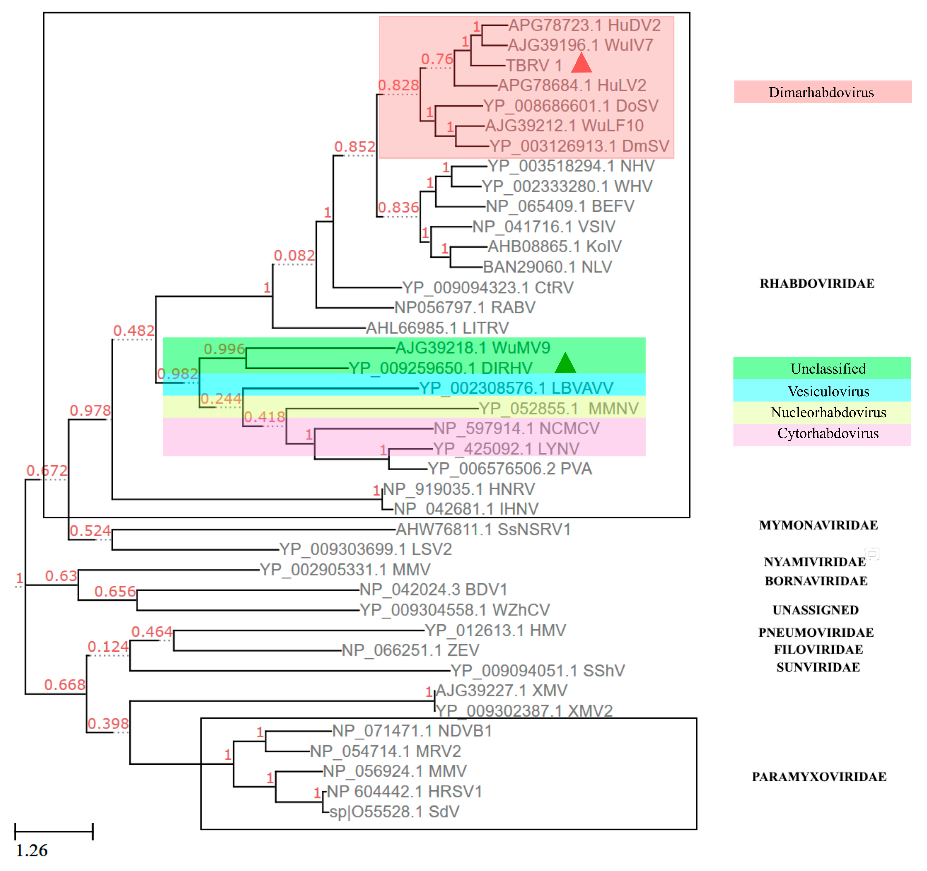
3.6. Phylogenetic Analysis Using the Protein Sequence of RdRP

The phylogenetic analysis-based RdRP showed that the TbRV-1 was clustered into a clade with two dimarhabdoviruses: Hubei dimarhabdovirus 2 (HuDV2, Accession number: KX884426) and Wuhan insect virus 7 (WuIV7, Accession number: KM817653.1) at high bootstrap values (BP = 100%, Figure 3), where both dimarhabdoviruses were newly discovered by RNA-seq sequencing [23,39]. The DlRhV was clustered with other unassigned viruses, and was more familiar with vesiculovirus. With a lower similarity (25% identity, e = 1 × 10−81) in the amino acid sequence of RdRp to DlRhV, another rhabdovirus from parasitoids TbRV-1 was observed. In all, TbRV-1 is a novel virus, different from the previously reported DlRhV.

4. Discussion

The genome of TbRV-1 exhibited a significant divergence from DlRhV by Blastn and Blastp, but a higher similarity to two dimarhabdovirus supergroups, HuDV2 and WuIV7 [23,39]. Other supports for this finding are the similarly conserved TTP domains and the clusters with the Sigmavirus from Drosophila, which is a member of the dimarhabdovirus group [40], while the DlRhV, another rhabdovirus isolated from parasitoid, is more closely related to other arthropod-infecting rhabdoviruses, but distantly related to the sigma viruses [15]. This is the first report of a dimarhabdovirus in an endoparasitoid, where the general insect host of dimarhabdoviruses are dipteran insects, such as Ochlerotatus sp., mosquitoes [26,41], Drosophila [41], and Odonata [23].
To fight against virus infection, the replicating viral RNA genomes were processed into small interfering RNA (siRNAs) by invertebrates [30]. In accordance with this, three siRNAs were retrieved from a small amount of RNA data. The low frequencies of siRNA in the small RNA library may also indicate a lower infection rate of this virus. Further, lower RNA copy numbers were quantified in the detected tissues, which imply that the virus might have a long latent period that is required for the rhabdovirus to exist in a vector insect before transmission [2,42]. The presence of TbRV-1 in all tissues of female and male wasps suggests that the expression of TbRV-1 is constitutive of, and not specific to, any wasp tissue.
The evolutionary challenge faced by RNA viruses is to maximize functional diversity with several constraints concerning the genome size. For dimarhabdoviruses, the evolution of genomic plasticity has been associated with that of alternative, overlapping and consecutive ORFs within the major structural protein genes as well as the insertion and loss of additional ORFs in each gene junction in a clade-specific manner [34]. In this study, the differences in the genome structure of TbRV-1 and the Sigmavirus clade are observed where the genome of TbRV-1 lacked a sigmaX gene, which is characteristic of Sigmavirus. Nonetheless, it cannot be inferred from this that TbRV-1 is classified as a novel species or genus because the sequence comparisons alone cannot be the only source in developing the classification schemes of viruses at this time, [43]. Meanwhile, the ability to accurately read the sequence information and robustly infer the phenotypic data is also fundamental for virus taxonomy based on the sequence information alone [44].
In general, viruses or virus-like particles (VLPs) in parasitoid wasps (Hymenoptera: Apocrita), such as the well-known polydnaviruses [9,10,45], VLPs [46,47,48,49], and some other viruses such as reoviruses, facilitate their parasitism success [50]. For example, the iflavirus, named Dinocampus coccinellae paralysis virus (DcPV), can induce changes in the lady beetle (Coleomegilla maculate), such as tremors, gait disturbance, and limitations in movement during parasitism [51]. The P. puparum negative-strand RNA virus 1 (PpNSRV-1), a novel member of Nyamiviridae, impaired several fitness parameters of the wasp, such as adult longevity and the offspring sex ratio but also affected parasitism [13]. For rhabdoviruses, only one virus, DlRhV, was observed in the venom apparatus of D. longicaudata parasitoids, and no role of DlRhV in parasitism was ascertained [14,15]. In this study, the TbRV-1 viruses were also transcribed in the venom apparatus, a major organ filled with parasitoid factors, which may be an indicator of the potential roles of TbRV-1 in parasitism. In addition, Longdon et al. (2010) discussed the sensitivity of the host, such as Drosophila or the mosquito, with rhabdovirus to CO2 and its negative effects, such as the reduced longevity [41]. The experiments on the role of TbRV-1 on anoxia, and the potential role on parasitism are underway. Meanwhile, for a more detailed study on the role of TbRV-1, we are establishing a virus cell culture system.

Supplementary Materials

The following are available online at https://www.mdpi.com/1999-4915/11/3/257/s1, Figure S1: Amino acid sequences alignment of the phosphoprotein of rhabdovirus. Similar residues are boxed and marked red, Table S1: Primers used in this study, Table S2: Sequences used for phylogenetic tree construction in this study.

Author Contributions

Methodology, E.M., B.T. F.J.S.-G. Y.W. and Y.-M.H.; software, E.M., Y.-M.H. Z.-M.C. Y.W. and J.-L.W.; validation, E.M., T.Q. and L.F.; resources, T.Q. and L.F.; writing—original draft preparation, E.M. and Y.-M.H.; writing—review and editing, E.M. B.T. F.J.S.-G. and Y.-M.H.; supervision, B.T.; project administration, B.T. and Y.-M.H.; funding acquisition, Y.-M.H.

Funding

This research was funded by grants from the National Science Foundation of China (31471829).

Conflicts of Interest

The authors declare no conflict of interest. The funders had no role in the design of the study; in the collection, analyses, or interpretation of data; in the writing of the manuscript, or in the decision to publish the results.

References

  1. Walker, P.; Blasdell, K.; Calisher, C.H.; Dietzgen, R.G.; Kondo, H.; Kurath, G.; Longdon, B.; Stone, D.M.; Tesh, R.B.; Tordo, N.; et al. ICTV Report Consortium. ICTV Virus Taxonomy Profile: Rhabdoviridae. J. Gen. Virol. 2018, 99, 447–448. [Google Scholar] [CrossRef] [PubMed]
  2. Hogenhout, S.A.; Redinbaugh, M.G.; Ammar, E.-D. Plant and animal rhabdovirus host range: A bug’s view. Trends Microbiol. 2003, 11, 264–271. [Google Scholar] [CrossRef]
  3. Longdon, B.; Murray, G.G.R.; Palmer, W.J.; Day, J.P.; Parker, D.J.; Welch, J.J.; Obbard, D.J.; Jiggins, F.M. The evolution, diversity, and host associations of rhabdoviruses. Virus Evol. 2015, 1, e014. [Google Scholar] [CrossRef]
  4. International Committee Taxonomy of Viruses. Available online: http://www.ictvonline.org/virus-Taxonomy.asp (accessed on 12 March 2019).
  5. Hosang, M.L.A.; Alouw, J.C.; Novarianto, H. Biological control of Brontispa longissima (Gestro) in Indonesia. In Report of the Expert Consultation on Coconut Beetle Outbreak in APPPC Member Countries; RAP Publication: Manado, Indonesia, 2004. [Google Scholar]
  6. Kuzmin, I.V.; Novella, I.S.; Dietzgen, R.G.; Padhi, A.; Rupprecht, C.E. The rhabdoviruses: Biodiversity, phylogenetics, and evolution. Infect. Genet. Evol. 2009, 9, 541–553. [Google Scholar] [CrossRef] [PubMed]
  7. Longdon, B.; Jiggins, F.M. Vertically transmitted viral endosymbionts of insects: Do sigma viruses walk alone? Proc. Biol. Sci. 2012, 279, 3889–3898. [Google Scholar] [CrossRef] [PubMed]
  8. L‘héritier, P.H. Sensitivity to CO2 in Drosophila a review. Heredity 1948, 2, 325–348. [Google Scholar] [CrossRef]
  9. Strand, M.R.; Burke, G.R. Polydnaviruses from discovery to current insights. Virology 2015, 479–480, 393–402. [Google Scholar] [CrossRef]
  10. Gundersen-Rindala, D.; Dupuy, C.; Huguet, E.; Drezen, J.-M. Parasitoid polydnaviruses: Evolution, pathology and applications. Biocontrol Sci. Technol. 2013, 23, 1–61. [Google Scholar] [CrossRef]
  11. Madanagopal, N.; Kim, Y. Parasitism by Cotesia glomerata induces immunosuppression of Pieris rapae: Effects of ovarian protein and polydnavirus. J. Asia-Pacif. Entomol. 2006, 9, 339–346. [Google Scholar] [CrossRef]
  12. Furihata, S.; Tanaka, K.; Ryuda, M.; Ochiai, M.; Matsumoto, H.; Csikos, G.; Hayakawa, Y. Immunoevasive protein (IEP)-containing surface layer covering polydnavirus particles is essential for viral infection. J. Invertebr. Pathol. 2014, 115, 26–32. [Google Scholar] [CrossRef] [PubMed]
  13. Wang, F.; Fang, Q.; Wang, B.; Yan, Z.; Hong, J.; Bao, Y.; Kuhn, J.H.; Werren, J.H.; Song, Q.; Ye, G. A novel negative-stranded RNA virus mediates sex ratio in its parasitoid host. PLoS Pathog. 2016, 13, e1006201. [Google Scholar] [CrossRef]
  14. Lawrence, P.O.; Matos, L.F. Transmission of the Diachasmimorpha longicaudata rhabdovirus (DlRhV) to wasp offspring: An ultrastructural analysis. J. Insect Physiol. 2005, 51, 235–241. [Google Scholar] [CrossRef] [PubMed]
  15. Simmondsa, T.J.; Carrillo, D.; Burke, G.R. Characterization of a venom gland-associated rhabdovirus in the parasitoid wasp Diachasmimorpha longicaudata. J. Insect Physiol. 2016, 91–92, 48–55. [Google Scholar] [CrossRef]
  16. Chen, Q.; Peng, Z.; Xu, C.; Tang, C.; Lu, B.; Jin, Q.; Wen, H.; Wan, F. Biological assessment of Tetrastichus brontispae, a pupal parasitoid of coconut leaf beetle Brontispa longissima. Biocontrol Sci. Technol. 2010, 20, 283–295. [Google Scholar] [CrossRef]
  17. Nguyen, H.T.; Oo, T.T.; Ichiki, R.T.; Takano, S.; Murata, M.; Takasu, K.; Konishi, K.; Tunkumthong, S.; Chomphookhiaw, N.; Nakamura, S. Parasitisation of Tetrastichus brontispae (Hymenoptera: Eulophidae), a biological control agent of the coconut hispine beetle Brontispa longissima (Coleoptera: Chrysomelidae). Biocontrol Sci. Technol. 2012, 22, 955–968. [Google Scholar] [CrossRef]
  18. Tang, B.Z.; Xu, L.; Hou, Y.M. Effects of rearing conditions on the parasitism of Tetrastichus brontispae on its pupal host Octodonta nipae. BioControl 2014, 59, 647–657. [Google Scholar] [CrossRef]
  19. Liu, K.; Fu, B.L.; Lin, J.R.; Fu, Y.G.; Peng, Z.Q.; Jin, Q.A.; Tang, L.D. Parasitism performance of Tetrastichus brontispae Ferriere over the coconut hispine beetle, Brontispa longissima (Gestro). Neotrop. Entomol. 2016, 45, 420–426. [Google Scholar] [CrossRef] [PubMed]
  20. Meng, E.; Tang, B.; Hou, Y.; Chen, X.; Chen, J.; Yu, X. Altered immune function of Octodonta nipae (Maulik) to its pupal endoparasitoid, Tetrastichus brontispae Ferrière. Comp. Biochem. Physiol. B Biochem. Mol. Biol. 2016, 198, 100–109. [Google Scholar] [CrossRef] [PubMed]
  21. Rotheram, S. Immune surface of eggs of a parasitic insect. Nature 1967, 214, 700. [Google Scholar] [CrossRef] [PubMed]
  22. Feddersen, I.; Sander, K.; Schmidt, O. Virus-like particles with host protein-like antigenic determinants protect an insect parasitoid from encapsulation. Experientia 1986, 42, 1278–1281. [Google Scholar] [CrossRef]
  23. Shi, M.; Lin, X.; Tian, J.; Chen, L.; Chen, X.; Li, C.; Qin, X.; Li, J.; Cao, J.; Eden, J.S.; et al. Redefining the invertebrate RNA virosphere. Nature 2016, 540, 539–543. [Google Scholar] [CrossRef] [PubMed]
  24. Tang, B.Z.; Chen, J.; Hou, Y.M.; Meng, E. Transcriptome immune analysis of the invasive beetle Octodonta nipae (Maulik) (Coleoptera: Chrysomelidae) parasitized by Tetrastichus brontispae Ferrière (Hymenoptera: Eulophidae). PLoS ONE 2014, 9, 1–12. [Google Scholar] [CrossRef] [PubMed]
  25. Grabherr, M.G.; Haas, B.J.; Yassour, M.; Levin, J.Z.; Thompson, D.A.; Amit, I.; Adiconis, X.; Fan, L.; Raychowdhury, R.; Zeng, Q.; et al. Full-length transcriptome assembly from RNA-Seq data without a reference genome. Nat. Biotechnol. 2011, 29, 644–652. [Google Scholar] [CrossRef] [PubMed] [Green Version]
  26. Reuter, G.; Boros, A.; Pal, J.; Kapusinszky, B.; Delwart, E.; Pankovics, P. Detection and genome analysis of a novel (dima)rhabdovirus (Riverside virus) from Ochlerotatus sp. mosquitoes in Central Europe. Infect. Genet. Evol. 2016, 39, 336–341. [Google Scholar] [CrossRef] [PubMed]
  27. Jones, P.; Binns, D.; Chang, H.-Y.; Fraser, M.; Li, W.; McAnulla, C.; McWilliam, H.; Maslen, J.; Mitchell, A.; Nuka, G.; et al. InterProScan 5: Genome-scale protein function classification. Bioinformatics 2014, 30, 1236–1240. [Google Scholar] [CrossRef]
  28. Kasuya, A.; Thornton, J.M. Three-dimensional structure analysis of PROSITE patterns. J. Mol. Biol. 1999, 286, 1673–1691. [Google Scholar] [CrossRef]
  29. Tamura, K.; Peterson, D.; Peterson, N.; Stecher, G.; Nei, M.; Kumar, S. MEGA5: Molecular evolutionary genetics analysis using maximum likelihood, evolutionary distance, and maximum parsimony methods. Mol. Biol. Evol. 2011, 28, 2731–2739. [Google Scholar] [CrossRef]
  30. Wu, Q.; Luo, Y.; Lu, R.; Lau, N.; Lai, E.C.; Li, W.X.; Ding, S.W. Virus discovery by deep sequencing and assembly of virus-derived small silencing RNAs. Proc. Natl. Acad. Sci. USA 2010, 107, 1606–1611. [Google Scholar] [CrossRef] [Green Version]
  31. Zerbino, D.R.; Birney, E. Velvet: Algorithms for de novo short read assembly using de Bruijn graphs. Genome Res. 2008, 18, 821–829. [Google Scholar] [CrossRef]
  32. Suzuki, M.; Tanaka, T. Virus-like particles in venom of Meteorus pulchricornis induce host hemocyte apoptosis. J. Insect Physiol. 2006, 52, 602–613. [Google Scholar] [CrossRef]
  33. Sylvester, E.S.; Richardson, J. Aphid-borne Rhabdoviruses—Relationships with Their Vectors. In Advances in Disease Vector Research; Harris, K.F., Ed.; Springer: New York, NY, USA, 1992; Volume 9. [Google Scholar]
  34. Walker, P.J.; Firth, C.; Widen, S.G.; Blasdell, K.R.; Guzman, H.; Wood, T.G.; Paradkar, P.N.; Holmes, E.C.; Tesh, R.B.; Vasilakis, N. Evolution of genome size and complexity in the rhabdoviridae. PLoS Pathog. 2015, 11, e1004664. [Google Scholar] [CrossRef]
  35. Gao, F.; Shen, J.; Liao, F.; Cai, W.; Lin, S.; Yang, H.; Chen, S. The first complete genome sequence of narcissus latent virus from Narcissus. Arch. Virol. 2018, 163, 1383–1386. [Google Scholar] [CrossRef] [PubMed]
  36. Barr, J.N.; Whelan, S.P.J.; Wertz, G.W. Transcriptional control of the RNA-dependent RNA polymerase of vesicular stomatitis virus. Biochim. Biophys. Acta 2002, 1577, 337–353. [Google Scholar] [CrossRef]
  37. Leyrat, C.; Ribeiro, E.A.; Gérard, F.C.; Ivanov, I.; Ruigrok, R.W.; Jamin, M. Structure, interactions with host cell and functions of rhabdovirus phosphoprotein. Future Virol. 2011, 6, 465–481. [Google Scholar] [CrossRef]
  38. Cook, S.; Chung, B.Y.; Bass, D.; Moureau, G.; Tang, S.; McAlister, E.; Culverwell, C.L.; Glucksman, E.; Wang, H.; Brown, T.D.; et al. Novel virus discovery and genome reconstruction from field RNA samples reveals highly divergent viruses in dipteran hosts. PLoS ONE 2013, 8, e80720. [Google Scholar] [CrossRef] [PubMed]
  39. Li, C.X.; Shi, M.; Tian, J.H.; Lin, X.D.; Kang, Y.J.; Chen, L.J.; Qin, X.C.; Xu, J.; Holmes, E.C.; Zhang, Y.Z. Unprecedented genomic diversity of RNA viruses in arthropods reveals the ancestry of negative-sense RNA viruses. eLife 2015, 4, e05378. [Google Scholar] [CrossRef]
  40. Kang, Y.-J.; Zhang, Y.-Z. Taxonomy and evolution of Rhabdoviruses. Chin. J. Vector Biol. Control 2014, 25, 374–377. [Google Scholar]
  41. Longdon, B.; Obbard, D.J.; Jiggins, F.M. Sigma viruses from three species of Drosophila form a major new clade in the rhabdovirus phylogeny. Proc. Biol. Sci. 2010, 277, 35–44. [Google Scholar] [CrossRef]
  42. Liu, S.; Vijayendran, D.; Bonning, B.C. Next generation sequencing technologies for insect virus discovery. Viruses 2011, 3, 1849–1869. [Google Scholar] [CrossRef]
  43. Simmonds, P. Methods for virus classification and the challenge of incorporating metagenomic sequence data. J. Gen. Virol. 2015, 96, 1193–1206. [Google Scholar] [CrossRef]
  44. Simmonds, P.; Adams, M.J.; Benko, M.; Breitbart, M.; Brister, J.R.; Carstens, E.B.; Davison, A.J.; Delwart, E.; Gorbalenya, A.E.; Harrach, B.; et al. Consensus statement: Virus taxonomy in the age of metagenomics. Nat. Rev. Microbiol. 2017, 15, 161–168. [Google Scholar] [CrossRef]
  45. Burke, G.R.; Strand, M.R. Polydnaviruses of parasitic wasps domestication of viruses to act as gene delivery vectors. Insects 2012, 3, 91–119. [Google Scholar] [CrossRef]
  46. Lawrence, P.O. Morphogenesis and cytopathic effects of the Diachasmimorpha longicaudata entomopoxvirus in host haemocytes. J. Insect Physiol. 2005, 51, 221–233. [Google Scholar] [CrossRef]
  47. Barratt, B.I.; Murney, R.; Easingwood, R.; Ward, V.K. Virus-like particles in the ovaries of Microctonus aethiopoides Loan (Hymenoptera: Braconidae): Comparison of biotypes from Morocco and Europe. J. Invertebr. Pathol. 2006, 91, 13–18. [Google Scholar] [CrossRef]
  48. Varaldi, J.; Ravallec, M.; Labrosse, C.; Lopez-Ferber, M.; Bouletreau, M.; Fleury, F. Artifical transfer and morphological description of virus particles associated with superparasitism behaviour in a parasitoid wasp. J. Insect Physiol. 2006, 52, 1202–1212. [Google Scholar] [CrossRef]
  49. Suzuki, M.; Miura, K.; Tanaka, T. Effects of the virus-like particles of a braconid endoparasitoid, Meteorus pulchricornis on hemocytes and hematopoietic organs of its noctuid host Pseudaletia separata. Appl. Entomol. Zool. 2009, 44, 115–125. [Google Scholar] [CrossRef]
  50. Renaulta, S.; Stasiaka, K.; Federicib, B.; Bigota, Y. Commensal and mutualistic relationships of reoviruses with their parasitoid wasp hosts. J. Insect Physiol. 2005, 51, 137–148. [Google Scholar] [CrossRef]
  51. Dheilly, N.M.; Maure, F.; Ravallec, M.; Galinier, R.; Doyon, J.; Duval, D.; Leger, L.; Volkoff, A.N.; Misse, D.; Nidelet, S.; et al. Who is the puppet master? Replication of a parasitic wasp-associated virus correlates with host behaviour manipulation. Proc. Biol. Sci. 2015, 282, 20142773. [Google Scholar] [CrossRef]
Figure 1. Schematic representation of the genome organization and location of each putative open reading frame (ORF). N: nucleoprotein; H: hypothetical protein; M: matrix protein; G: glycoprotein; and L: RNA-dependent RNA polymerase.
Viruses 11 00257 g001
Figure 2. (A) Gel electrophoresis of viral RNA, and (B) the PCR products obtained from the viral cDNA and distilled water. M: Marker DL 2000 (TransGen Biotech); L1: PCR products with distilled water as template; L2: PCR products with cDNA template. The band was marked with a red triangle.
Viruses 11 00257 g002
Figure 3. Maximum likelihood phylogenetic tree of TbRV-1 based on the RdRP protein and alignment. The numbers near the branches represent the percentage of the 1000 bootstrap iterations supporting the branches. The phylogeny was drawn with ETE Toolkit v3 software. The virus TbRV-1 and DlRhV were respectively marked with red and green triangles.
Viruses 11 00257 g003
Table 1. Virus species identified through transcriptome mining.
Query VirusGene IDLength (bp)Subject ProteinSubject VirusE ValueTaxonomy of Subject VirusCoverage Range
Tetrastichus brontispae RNA virus-1 (TbRV-1)Unigene00175631431NucleoproteinOita rhabdovirus 296/19728.2 × 10−100Rhabdoviridae (negative sense single strand RNA viruses)1–1431
Unigene00195681954Matrix proteinTibtogaran virus9 × 10−61442–3395
Unigene00230515167RdRpScophthalmus maximus Rhabdovirus03411–8578
Unigene00144531037RdRp2 × 10−2111,075–12,171
Unigene0016900339RdRp4 × 10−359036–9375
Unigene0016901404RdRp4 × 10−338656–9058
Unigene0008666895RdRp1 × 10−359744–10,603
Unigene0005445216RdRpGrass carp rhabdovirus V769 × 10−1310,581–10,796
Tetrastichus brontispae RNA virus -2 (TbRV-2)Unigene00001182396Polymerase PB1Quaranfil virus0Orthomyxoviridae (negative sense single strand RNA viruses, 6–8 segments)/
Unigene00227622405Polymerase PB17 × 10−171
Unigeno00221601447Polymerase PA1 × 10−36
Unigene00184602416Polymerase PA6 × 10−39
Unigene00207692500Polymerase PB22 × 10−21
Unigene0000506427Polymerase PB22 × 10−7
Unigene00213531762Hypothetical protein1 × 10−36
Unigene00214821772Hypothetical protein4 × 10−20
Unigene00161371656Hemagglutinin2 × 10−32
Unigene00207781614Hemagglutinin3 × 10−42
Tetrastichus brontispae RNA virus 3 (TbRV-3)Unigene0009037027PolyproteinDeformed wing virus1 × 10−69Iflaviridae/
Unigene0009052933Polyprotein4 × 10−101
Tetrastichus brontispae RNA virus -4 (TbRV-4)Unigene00222282784RdRpSaccharomyces 23S RNA Narnavirus5 × 10−18Narnaviridae (positive sense single strand RNA viruses)/
Tetrastichus brontispae RNA virus -5 (TbRV-5)Unigene0008109260VP3Kadipiro virus5 × 10−8Reoviridae (double strand RNA viruses)/
Unigene00021262047VP43 × 10−109
Unigene00114791396VP54 × 10−46
Unigene00049821092VP72 × 10−27
Unigene0005372850VP123 × 10−22
Unigene0012923483RdRpLiaoning virus1 × 10−26
Unigene0012924204RdRp1 × 10−12
Unigene0013487253VP14 × 10−18
Unigene0002892281VP39 × 10−21
Unigene0010175228VP21 × 10−17
Unigene00166081775VP22 × 10−88
Unigene0016692426VP21 × 10−27
Unigene0016693270VP21 × 10−16
Unigene0006368206VP32 × 10−13
Unigene0005945251VP33 × 10−17
Unigene0011375856VP61 × 10−26
Unigene00085231035VP88 × 10−48
Unigene0014543278RdRp3 × 10−14
Unigene0006593250RdRp4 × 10−16
Unigene0007896425Capping enzyme1 × 10–13
Unigene0001578236VP1Banna virus2 × 10–12
Table 2. Calculated and predicted properties of TbRV-1 (Tetrastichus brontispae RNA virus 1) ORFs.
Virus ORFPosition (bp)ORF Length (nt)Protein LengthProtein Mass (kDa)pISignal PeptideNo. of O-linked Glycosylation SitesNo. of Phosphorylation SitesTop Blastx MatchE ValueDomainE Value of Domain
TYS
ORF I113–1393128742649.276.92NO916926Putative nucleoprotein (Hubei dimarhabdovirus 2)1 × 10−100Rhabdo-ncap2.78 × 10−64
ORF II1689–260091230334.795.50NO1311315ND-ND-
ORF III2699–335565721825.976.91NO01067Matrix protein (Hubei dimarhabdovirus 2)2 × 10⁻²²Vesiculo_matrix super family2.49 × 10−4
ORF IV3761–5410165054963.287.45NO219927Glycoprotein (Wuhan insect virus 7)2 × 10−61Rhabdo_glycop super family1.78 × 10−23
ORF V5781–12,14163602119245.178.27NO106738113RdRp (Hubei dimarhabdovirus 2)0Mononeg_RNA-pol super family; Methyltrans_Mon super family; Mononeg_mRNAcap super family;01.15 × 10−132; 4.61 × 10−50
ND means no significant homology was detected.
Table 3. The Blastn results of small interfering RNA against the genome of TbRV-1.
LibrarySmall Interfering RNA SequenceIdentityCoverage Range (nt)Domain
TbBL1CAGGTTTTTTATTATTTTATCCTCTTGATTTGTGTCTCTAAC100%11,037–11,078RdRP
TbBL2TTGGAGTGTTGTTAGTGCATTAGCCATAGAAAGGGAATCCAA100%7790–7831RdRP
TbBL3TTTAAGTTTAAATAAAATATTAACCTGATGATCTGGAGCTCC100%3122–3163Matrix
Note: TbBL1, TbBL2, and TbBL3 represent three small RNA libraries of the Tetrastichus brontispae reared on Brontispa longissima. The column of domain shows in which domains the three separate siRNA were blasted. RdRP: RNA-dependent RNA polymerase; Matrix: putative matrix protein.
Table 4. Viral loads of TbRV-1 in different tissues of T. brontispae wasps.
SexTissuesCopies/ng Total RNA
Female waspsHead and thorax690 ± 189 a
Ovary706 ± 120 a
Venom apparatus446 ± 130 a
Remaining abdomen376 ± 6 a
Male waspsWhole body376 ± 119 a
Data represent means ± standard error. The columns annotated with the same letters are not significantly different (Tukey’s multiple comparison tests). The copies were obtained by a two-fold reduction of the calculated value.

