Hemostatic Efficiency and Wound Healing Properties of Natural Zeolite Granules in a Lethal Rabbit Model of Complex Groin Injury


Abstract
:1. Introduction


2. Experimental Section
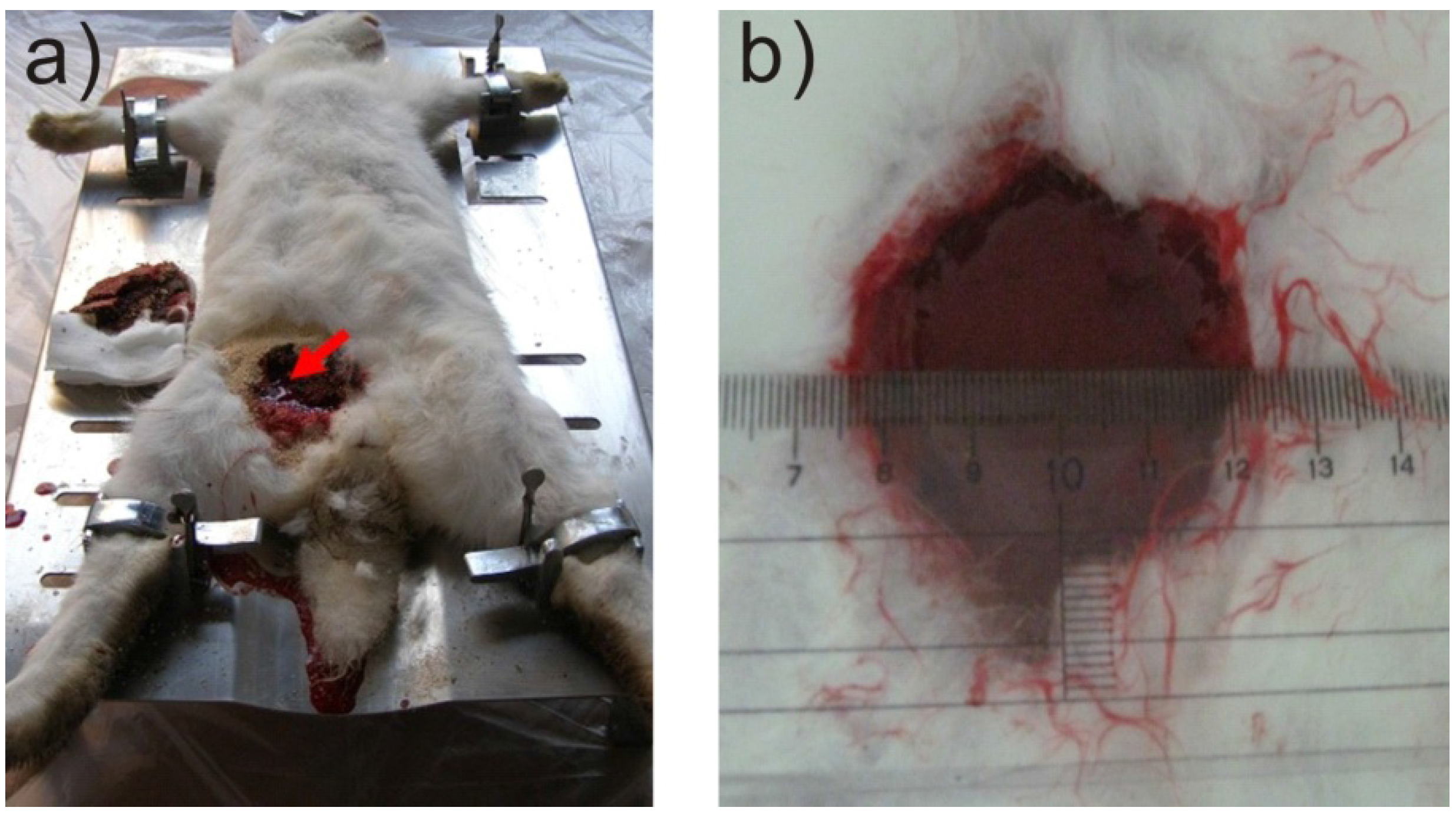
2.1. Model and Animal Preparation
2.2. Measurement of Exothermic Reaction
2.3. Statistical Analysis
3. Results and Discussion
3.1. Mortality

| Clotting percent | Surviving animals | Mortality | Wound infection | Infection percent | |
|---|---|---|---|---|---|
| Quikclot | 100% | 9/19 | 52.6% | 3/4 | 75% |
| NZG-JY | 100% | 15/19 | 21.0% | 0/4 | 0 |
| Medical Gauze | 5.6% | 6/18 | 66.7% | – | – |
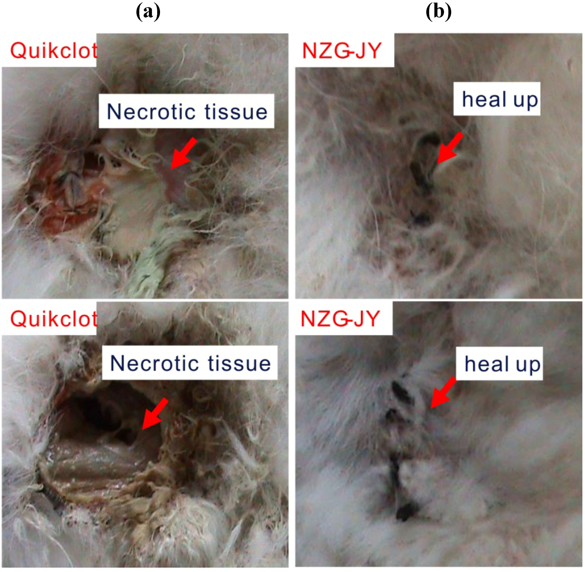
3.2. Wound Healing

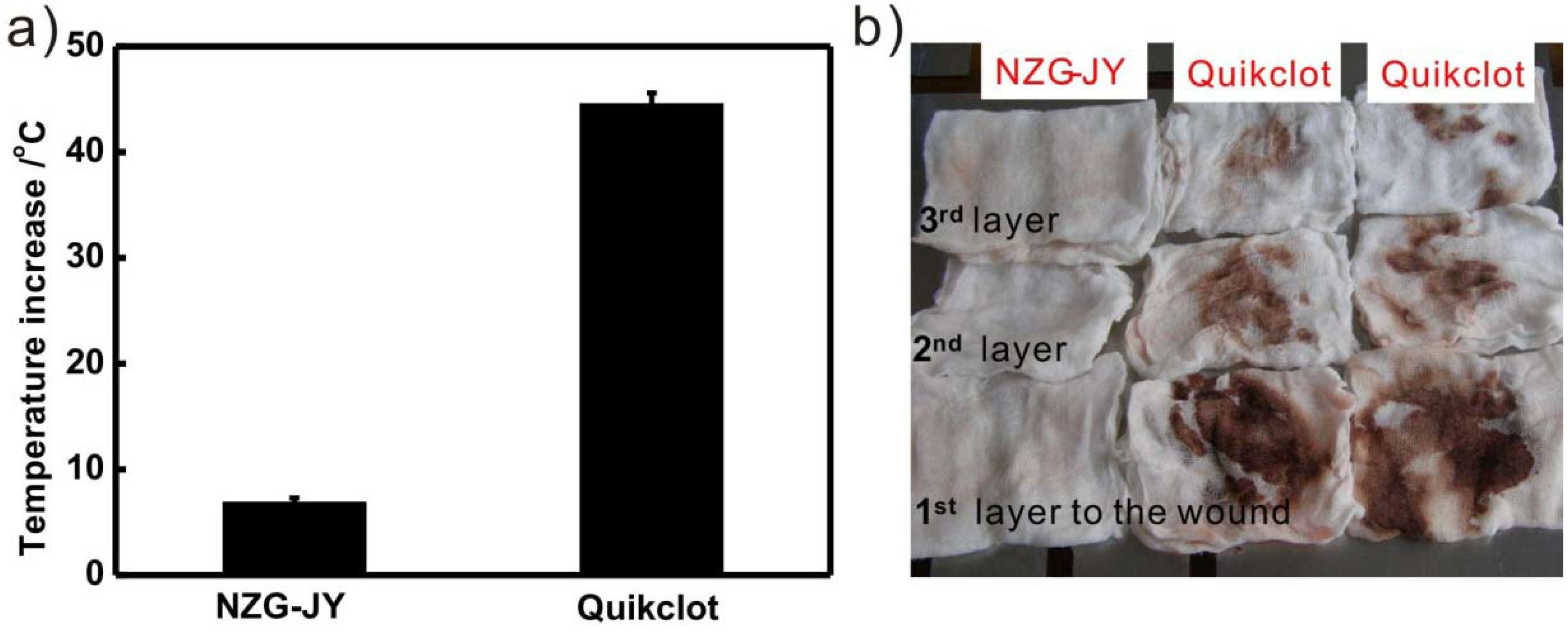
3.3. Exothermic Reaction

| Elements | Al | Ca | Fe | K | Mg | Na | Si/Al |
|---|---|---|---|---|---|---|---|
| NZG-JY | 6.10 | 1.93 | 0.68 | 2.27 | 0.11 | 1.41 | 5.16 |
| Quikclot | 16.11 | 11.43 | 0 | 0 | 0 | 0.57 | 1 |
4. Conclusions
Acknowledgments
References
- Achneck, H.E.; Sileshi, B.; Jamiolkowski, R.M.; Albala, D.M.; Shapiro, M.L.; Lawson, J.H. A comprehensive review of topical hemostatic agents efficacy and recommendations for use. Ann. Surg. 2010, 251, 217–228. [Google Scholar] [CrossRef] [PubMed]
- Galownia, J.; Martin, J.; Davis, M.E. Aluminophosphate-based, microporous materials for blood clotting. Microporous Mesoporous Mater. 2006, 92, 61–63. [Google Scholar] [CrossRef]
- Ward, K.R.; Tiba, M.H.; Holbert, W.H.; Blocher, C.R.; Draucker, G.T.; Proffitt, E.K.; Bowlin, G.L.; Ivatury, R.R.; Diegelmann, R.F. Comparison of a new hemostatic agent to current combat hemostatic agents in a swine model of lethal extremity arterial hemorrhage. J. Trauma Inj. Infect. Crit. Care 2007, 63, 276–283. [Google Scholar] [CrossRef]
- Johansson, P.I.; Stensballe, J.; Ostrowski, S.R. Current management of massive hemorrhage in trauma. Scand. J. Trauma Resusc. Emerg. Med. 2012. [Google Scholar] [CrossRef]
- Dai, C.L.; Yuan, Y.; Liu, C.S.; Wei, J.; Hong, H.; Li, X.S.; Pan, X.H. Degradable, antibacterial silver exchanged mesoporous silica spheres for hemorrhage control. Biomaterials 2009, 30, 5364–5375. [Google Scholar] [CrossRef] [PubMed]
- Kheirabadi, B.S.; Mace, J.E.; Terrazas, I.B.; Fedyk, C.G.; Estep, J.S.; Dubick, M.A.; Blackbourne, L.H. Safety evaluation of new hemostatic agents, smectite granules, and kaolin-coated gauze in a vascular injury wound model in swine. J. Trauma Inj. Infect. Crit. Care 2010, 68, 269–277. [Google Scholar] [CrossRef]
- Cancio, L.C. Comparison of a new hemostatic agent to current combat hemostatic agents in a swine model of lethal extremity arterial hemorrhage—Editorial comment. J. Trauma Inj. Infect. Crit. Care 2007, 63, 283–284. [Google Scholar]
- Alam, H.B.; Uy, G.B.; Miller, D.; Koustova, E.; Hancock, T.; Inocencio, R.; Anderson, D.; Llorente, O.; Rhee, P. Comparative analysis of hemostatic agents in a swine model of lethal groin injury. J. Trauma Inj. Infect. Crit. Care 2003, 54, 1077–1082. [Google Scholar] [CrossRef]
- Alam, H.B.; Burris, D.; DaCorta, J.A.; Rhee, P. Hemorrhage control in the battlefield: Role of new hemostatic agents. Mil. Med. 2005, 170, 63–69. [Google Scholar] [PubMed]
- Alam, H.B.; Chen, Z.; Jaskille, A.; Querol, R.; Koustova, E.; Inocencio, R.; Conran, R.; Seufert, A.; Ariaban, N.; Toruno, K.; Rhee, P. Application of a zeolite hemostatic agent achieves 100% survival in a lethal model of complex groin injury in swine. J. Trauma Inj. Infect. Crit. Care 2004, 56, 974–983. [Google Scholar] [CrossRef]
- Carraway, J.W.; Kent, D.N.; Young, K.; Cole, A.; Friedman, R.; Ward, K.R. Comparison of a new mineral based hemostatic agent to a commercially available granular zeolite agent for hemostasis in a swine model of lethal extremity arterial hemorrhage. Resuscitation 2008, 78, 230–235. [Google Scholar] [CrossRef] [PubMed]
- Kheirabadi, B.S.; Mace, J.E.; Terrazas, I.B.; Fedyk, C.G.; Estep, J.S.; Dubick, M.A.; Blackbourne, L.H. Safety evaluation of new hemostatic agents, smectite granules, and kaolin-coated gauze in a vascular injury wound model in swine. J. Trauma Inj. Infect. Crit. Care 2010, 68, 1263–1263. [Google Scholar] [CrossRef]
- Ostomel, T.A.; Stoimenov, P.K.; Holden, P.A.; Alam, H.B.; Stucky, G.D. Host–guest composites for induced hemostasis and therapeutic healing in traumatic injuries. J. Thromb. Thrombolysis 2006, 22, 55–67. [Google Scholar] [CrossRef] [PubMed]
- Pavelic, K.; Hadzija, M.; Bedrica, L.; Pavelic, J.; Dikic, I.; Katic, M.; Kralj, M.; Bosnar, M.H.; Kapitanovic, S.; Poljak-Blazi, M.; et al. Natural zeolite clinoptilolite: New adjuvant in anticancer therapy. J. Mol. Med. 2001, 78, 708–720. [Google Scholar] [CrossRef]
- Katic, M.; Bosnjak, B.; Gall-Troselj, K.; Dikic, I.; Pavelic, K. A clinoptilolite effect on cell media and the consequent effects on tumor cells in vitro. Front. Biosci. 2006, 11, 1722–1732. [Google Scholar] [CrossRef]
- Rodriguez-Fuentes, G.; Barrios, M.A.; Iraizoz, A.; Perdomo, I.; Cedre, B. Enterex: Anti-diarrheic drug based on purified natural clinoptilolite. Zeolites 1997, 19, 441–448. [Google Scholar] [CrossRef]
- Mumpton, F.A. La roca magica: Uses of natural zeolites in agriculture and industry. Proc. Natl. Acad. Sci. USA 1999, 96, 3463–3470. [Google Scholar] [CrossRef] [PubMed]
- Rimoli, M.G.; Rabaioli, M.R.; Melisi, D.; Curcio, A.; Mondello, S.; Mirabelli, R.; Abignente, E. Synthetic zeolites as a new tool for drug delivery. J. Biomed. Mater. Res. A 2008, 87, 156–164. [Google Scholar] [CrossRef] [PubMed]
- Acheson, E.M.; Kheirabadi, B.S.; Deguzman, R.; Dick, E.J.; Holcomb, J.B. Comparison of hemorrhage control agents applied to lethal extremity arterial hemorrhages in swine. J. Trauma Inj. Infect. Crit. Care 2005, 59, 65–874. [Google Scholar] [CrossRef]
- Kilbourne, M.; Keledjian, K.; Hess, J.R.; Scalea, T.; Bochicchio, G.V. Hemostatic efficacy of modified amylopectin powder in a lethal porcine model of extremity arterial injury. Ann. Emerg. Med. 2009, 53, 804–810. [Google Scholar] [CrossRef] [PubMed]
- Ziv-Polat, O.; Topaz, M.; Brosh, T.; Margel, S. Enhancement of incisional wound healing by thrombin conjugated iron oxide nanoparticles. Biomaterials 2010, 31, 741–747. [Google Scholar] [CrossRef] [PubMed]
- Vogler, E.A.; Siedlecki, C.A. Contact activation of blood-plasma coagulation. Biomaterials 2009, 30, 857–1869. [Google Scholar] [CrossRef]
- Schopka, S.; Schmid, T.; Schmid, C.; Lehle, K. Current strategies in cardiovascular biomaterial functionalization. Materials 2010, 3, 638–655. [Google Scholar] [CrossRef]
- Baker, S.E.; Sawvel, A.M.; Zheng, N.; Stucky, G.D. Controlling bioprocesses with inorganic surfaces: Layered clay hemostatic agents. Chem. Mater. 2007, 19, 4390–4392. [Google Scholar] [CrossRef]
© 2012 by the authors; licensee MDPI, Basel, Switzerland. This article is an open access article distributed under the terms and conditions of the Creative Commons Attribution license (http://creativecommons.org/licenses/by/3.0/).
Share and Cite
Li, Y.; Li, H.; Xiao, L.; Zhou, L.; Shentu, J.; Zhang, X.; Fan, J. Hemostatic Efficiency and Wound Healing Properties of Natural Zeolite Granules in a Lethal Rabbit Model of Complex Groin Injury. Materials 2012, 5, 2586-2596. https://doi.org/10.3390/ma5122586
Li Y, Li H, Xiao L, Zhou L, Shentu J, Zhang X, Fan J. Hemostatic Efficiency and Wound Healing Properties of Natural Zeolite Granules in a Lethal Rabbit Model of Complex Groin Injury. Materials. 2012; 5(12):2586-2596. https://doi.org/10.3390/ma5122586
Chicago/Turabian StyleLi, Yunlong, Hui Li, Liping Xiao, Lin Zhou, Jianzhong Shentu, Xumin Zhang, and Jie Fan. 2012. "Hemostatic Efficiency and Wound Healing Properties of Natural Zeolite Granules in a Lethal Rabbit Model of Complex Groin Injury" Materials 5, no. 12: 2586-2596. https://doi.org/10.3390/ma5122586
APA StyleLi, Y., Li, H., Xiao, L., Zhou, L., Shentu, J., Zhang, X., & Fan, J. (2012). Hemostatic Efficiency and Wound Healing Properties of Natural Zeolite Granules in a Lethal Rabbit Model of Complex Groin Injury. Materials, 5(12), 2586-2596. https://doi.org/10.3390/ma5122586