Responses of Antioxidant Enzymes in Catfish Exposed to Liquid Crystals from E-Waste

1
Department of Chemistry, Jackson State University, Jackson, MS 39217, USA
2
Department of Civil & Environmental Engineering, Jackson State University, Jackson, MS 39217, USA
3
School of Environmental Science and Engineering, South China University of Technology, Guangzhou 510641, P. R. China
*
Author to whom correspondence should be addressed.
Int. J. Environ. Res. Public Health 2008, 5(2), 99-103; https://doi.org/10.3390/ijerph5020099
Submission received: 30 October 2007
/
Accepted: 30 April 2008
/
Published: 30 June 2008
Abstract
:Liquid crystals (LCs) are typically elongated organic molecules with a non-uniform distribution of electrical charges leading to a dipole. LCs are widely used in displays of computers and other electronic devices. The rapid obsolescence rate of electronics results in large amounts of liquid crystal displays (LCDs) entering the environment. Data on health effects of LCs on living creatures are currently limited to some acute toxicity tests by a few major LC manufacturers. These tests concluded that the vast majority of LCs are not acutely toxic. Since the amount of LCs in electronic devices is very small, the health effects of LCs at low concentrations or doses become important. Catfish were used as the test animals in this study. Four major enzymes of the fish’s antioxidant defense system catalase (CAT), superoxide dismutase (SOD), selenium-dependent glutathione peroxidase (Se-GPx), and glutathione-S-transferase (GST) were chosen as biomarkers to examine effects of LCs, which were taken from obsolete laptop personal computers made in the early 1990s. The catfish were fed with food containing different contents of LCs for 40 days. Activities of the four chosen enzymes in fish livers were assayed. The results showed that there were significant inductions of CAT, SOD, and Se-GPx activities in response to the LC doses. The plots of the enzyme activities versus LC doses suggested an occurrence of oxidative stress when the dose reached about 20 μg LC/g fish·d. It was concluded that LCs can cause pollutant-induced stress to catfish at low doses. CAT, SOD and Se-GPx are effective biomarkers to give early warning on potential health effects of LCs on some aquatic lives including catfish.
Introduction
As a new generation of display technology, the liquid crystal displays (LCDs) have increased dramatically on the market. The key materials in a LCD are liquid crystal (LC) compounds or liquid crystals (LCs). There are more than ten thousand LCs used in different LCDs, but information on the structure of the LCs is kept confidential between LC and LCD manufacturers. Therefore, studies on the effect of LCs on the environment have been mainly conducted by LC manufacturers. MERCK, the world’s largest LC producer based in Germany, studied the acute oral toxicity on rats and aquatic living organisms, skin and eye irritation on rabbits, and mutagenic potential on bacteria of LC products of three world’s leading LC manufacturers [1]. These tests were all short-term and high-dose (up to 2000 mg/kg body weight). Based on the study, MERCK concluded that the LCs the companies put on the market are not harmful to living organisms. However, some studies have indicated that the absence of acute effect does not necessarily mean there is no chronic effect for the same contaminant [2–4]. Since the amounts of LCs in electronic devices are very small—approximately 0.6 mg/cm2 in a typical LCD, data on the effects of LCs at low concentrations or doses become important.
Living creatures have an antioxidant defense (AD) system which can neutralize the harmful effects of reactive oxygen species (ROS) including hydroxyl radicals (•OH), superoxide radical anion (
), and hydrogen peroxide (H2O2). The AD system includes antioxidant enzymes catalase (CAT), superoxide dismutase (SOD), glutathione peroxidase (GPx), glutathione-S-transferase (GST), and other lower molecular weight substances such as glutathione (GSH), vitamins and proteins located in different tissues [5]. The antioxidant enzymes are found in almost all tissues of vertebrates, and their activities are especially high in the liver, a major organ responsible for the transformation of ROS. The ROS are generated during normal metabolism and the amounts are well-controlled under normal physiological conditions. When pollutants such as xenobiotic molecules enter the body, they undergo redox cycling and generate ROS. The body will produce more antioxidant enzymes to get rid of the undesired ROS. This response is also called induction of antioxidant enzymes. When the generation of ROS overwhelms the AD system, damages to lipids, proteins and DNA occur and this condition is referred to as oxidative stress [6, 7]. The induction of the antioxidant enzymes gives an early warning on adverse health effects of certain pollutants on living organisms.
In this study, responses of the four major hepatic enzymes CAT, SOD, selenium-dependent GPx (Se-GPx), and GST of catfish’s AD system exposed to LCs were tested to examine early effects of LCs at low doses.
Materials and Methods
Test Animals
Freshwater catfish of 80 days old were used as test animals. The average length and weight of the catfish were 8.0±0.3 cm and 22.0±0.2 g, respectively. Catfish grow very slowly. It normally takes 2 to 4 years for a catfish to reach a weight of 500 g. Catfish usually get food from the bottom of the water body. This is very suitable for this study because LCs are not soluble in water and normally adhere to detritus and sink to the bottom of the water body.
Liquid Crystal Stock Solution
LCs were collected from LCDs of laptop personal computers (IBM Thinkpad 390) made in the late 1990s. The LCDs used for this study were produced by SHARP, the main supplier of LCDs over that period of time. Laptops manufactured by Macintosh, Compaq, and Toshiba all used LCDs made by SHARP. The effective size of each LCD was 390 cm2. The amount of LCs sandwiched between the two thin glass layers was measured to be 0.23 g and the areal density of LCs in a LCD was calculated to be 0.60 mg/cm2. During the collection, each LCD was carefully separated into two glass layers. A total of 4 LCDs were used for LC collection. Each layer was wiped 10 times with a cotton ball wetted with dimethyl sulfoxide (DMSO). After each wipe, the liquid (LC-containing DMSO) in the cotton ball was squeezed into a container. About 30 mL of DMSO were in the beaker used for the cotton ball to be dipped after each wipe. After LC collection, the remaining DMSO in the beaker and the liquid in the other container were transferred into a volumetric flask and the total volume was adjusted to 50 mL with fresh DMSO. The final concentration of LCs in DMSO was calculated to be 18 g/L. This was the LC stock solution for all the fish exposure tests.
Fish Food
Three different types of fish food were prepared for the test. One was the pure fish food purchased from a pet shop. It was in the form of dry pellets made from shrimps and other ingredients. The pure fish food was used for negative control. The second was DMSO-soaked fish food made by adding 32 mL of DMSO to 165 g pure fish food. It was used for blank control. The third was made by adding 32 mL of LC stock solution to 165 g of pure fish food. The content of LCs in this food was 3.5 mg LC/g food. This was called LC-containing food.
Exposure Test
Glass fish tanks of 37.8 L were used to accommodate the fish. The pH value of the water was 6.8±0.2 and temperature 20±2ºC. The water in the tanks was constantly aerated. Each tank held 8 fish. The fish were first acclimated for 7 days. During this period, only the pure fish food was fed at 2.0 g per day per tank. To keep the water fresh, one-third of the water in each tank was replaced by newly prepared water every day after the fish had eaten up all the food. The total mortality of fish was zero during the 7 day acclimatization period.
After acclimatization, the fish were divided into 6 groups. Each group consisted of 8 fish in one fish tank. Group 1 was the negative control, fed with the pure fish food at 2.0 g/d. Group 2 was the LC-free blank control, fed with the DMSO-soaked fish food at 2.0 g/d. Groups 3, 4, 5 and 6 were fed with LC-containing food at 0.2, 0.5, 1.0, and 2.0 g/d respectively. The total food amount was made up to 2.0 g/d by pure fish food for Groups 3, 4, and 5. For instance, Group 4 was fed with 0.5 g/d of LC-containing food and 1.5 g/d of pure fish food. This exposure test lasted for 40 d. Other similar studies used exposure periods of 2, 4, 7, 14, 21, 28, and 40 d [3–4, 8–10].
Sample Preparation
After the 40 day exposure, all fish were weighed and dissected. The livers were taken out and rinsed by physiological salt water. About 0.30 g of liver tissue from each fish was homogenized in 3 mL of 10 mM Tris buffer (pH 7.5) which contained 0.01 M Tris, 0.25 M sucrose, and 0.01 M EDTA. The homogenized solution was centrifuged at 10,000 rpm at 4ºC for 10 min. The supernatant solution was called enzyme solution. It was stored at −80ºC for further analysis of protein content and activities of four antioxidant enzymes.
Sample Analysis
The total protein content of the enzyme solution of each fish liver was determined according to the method by Bradford [11]. A G-250 solution was prepared by adding 100 mg Coomassie Brilliant Blue into 50 mL of 95% ethanol and 100 mL of 85% phosphoric acid, and diluted with distilled water to 1,000 mL. A volume of 10 μL of the enzyme solution was added into 5 mL of the G-250 solution. The absorbance at 595 nm was measured by a spectrophotometer to determine the protein content. The calibration curve was established by standard bovine serum albumen (BSA) solutions of 0, 50, 100, 200, 300, 400, and 500 μg/mL.
CAT activity was determined by the method of Xu et al. [12]. This method is based on the first-order reaction of CAT with H2O2. A 10 μL enzyme solution was added to 3.0 mL H2O2-phosphate buffer solution and the absorbance of H2O2 at 250 nm was measured every 5 s for 60 s by a spectrophotometer. The H2O2-phosphate buffer solution was made by diluting 0.16 mL H2O2 (30% w/v) to 100 mL with a 67 mM phosphate buffer (pH=7.0). The phosphate buffer was made by dissolving 3.522 g KH2PO4 and 7.268 g Na2HPO4·H2O into 1000 mL pure water. The reference cell was filled with this phosphate buffer alone. One unit of CAT activity (U) was defined as 1 U = 100K/ln2, where K is the slope of the curve lnA versus time and A is the absorbance at 250 nm.
SOD activity was determined according to the method by Zhu et al. [13]. This method was based on inhibition of SOD by auto-oxidation of 1,2,3-benzenetriol. First, the auto-oxidation rate (Ko) of 1,2,3-benzenetriol in 0.05 M Tris-HCl buffer solution (pH=8.2) was determined by spectrophotometry at 325 nm. Then the SOD-inhibited auto-oxidation rate (KSOD) was measured by adding 10 μL enzyme solution to the same buffer solution. One unit of SOD activity (U) is defined as the quantity of enzyme protein used to inhibit 50% of the auto-oxidation rate of 1,2,3-benzenetriol at 25oC. It was calculated as 1 U = (Ko-KSOD)/(3.1x Ko/2).
Se-GPx was assayed according to the method by Hafeman [14] with slight modification by Zhang et al. [8]. This method is based on the theory that Se-GPx can catalyze the reaction between GSH and peroxides. The remaining amount of GSH in the reaction can indicate the activity of Se-GPx. The remaining GSH was determined by adding 5,5′-dithio-bis-2-nitrobenzoic acid (DTNB) at 37oC, which reacts with GSH to produce 5-thio-2-nitrobenzoic acid anion. This product has a maximum absorbance at 412 nm. The absorbance is proportional to the amount of remaining GSH and inversely proportional to the activity of Se-GPx. Since GSH can react with H2O2 without the existence of Se-GPx, the reduction of GSH caused by non-enzyme reaction was deducted in the calculation. One unit of Se-GPx activity (U) was defined as the amount of enzyme that oxidizes 1 μmol/L of GSH per min.
GST activity was determined by the method of Habig et al. [15]. A reaction mixture containing 2.8 mL phosphate buffer (pH=6.5), 0.5 mL of 20 mM GSH water solution and 50 μL enzyme solution was prepared at 25oC. Then 100 μL of 30 mM 1-chloro-2,4-dinitrobenzene (CDNB) ethanol solution was added for conjugation of GST with CDNB. After 3 min reaction, 0.3 mL of 33% trichloroacetic acid (TCA) was added to terminate the reaction. The mixture was centrifuged at 3,000 rpm for 5 min. The supernatant was collected and the GST activity was determined by the change of optic density (OD) or absorbance at 340 nm as compared to that of the same mixture into which CDNB and TCA were added at the same time (no conjugation). One unit of GST activity (U) was expressed as the amount of enzyme necessary to conjugate 1 μmol of CDNB per min.
Finally, all enzyme activity data were normalized by the total protein content in the livers and were expressed as units of activity per mg of protein (U/mg Pr).
Results and Discussion
Doses of Test Groups
During the 40 d test period, there was no appreciable amount of growth in the body weight of the catfish. The LC doses for Groups 1 through 6 were calculated to be 0, 0, 4.0, 10, 20, and 40 μg LC/g fish·d, respectively. Following is a sample calculation of the LC dose for Group 3 to which 0.2 g/d of LC-containing food was fed.
The responses of the activities of the four antioxidant enzymes in catfish livers of the six test groups to LC doses are shown in Figures 1, 2, 3, and 4. The error bars in all the graphs represent the confidence limits for 95% confidence level from the data for each group. Statistical differences among group means were determined using the one-way ANOVA. The differences between any dose group and the negative control group or between any two dose groups were assessed using Student’s t-test. The differences were considered statistically significant when p<0.05.
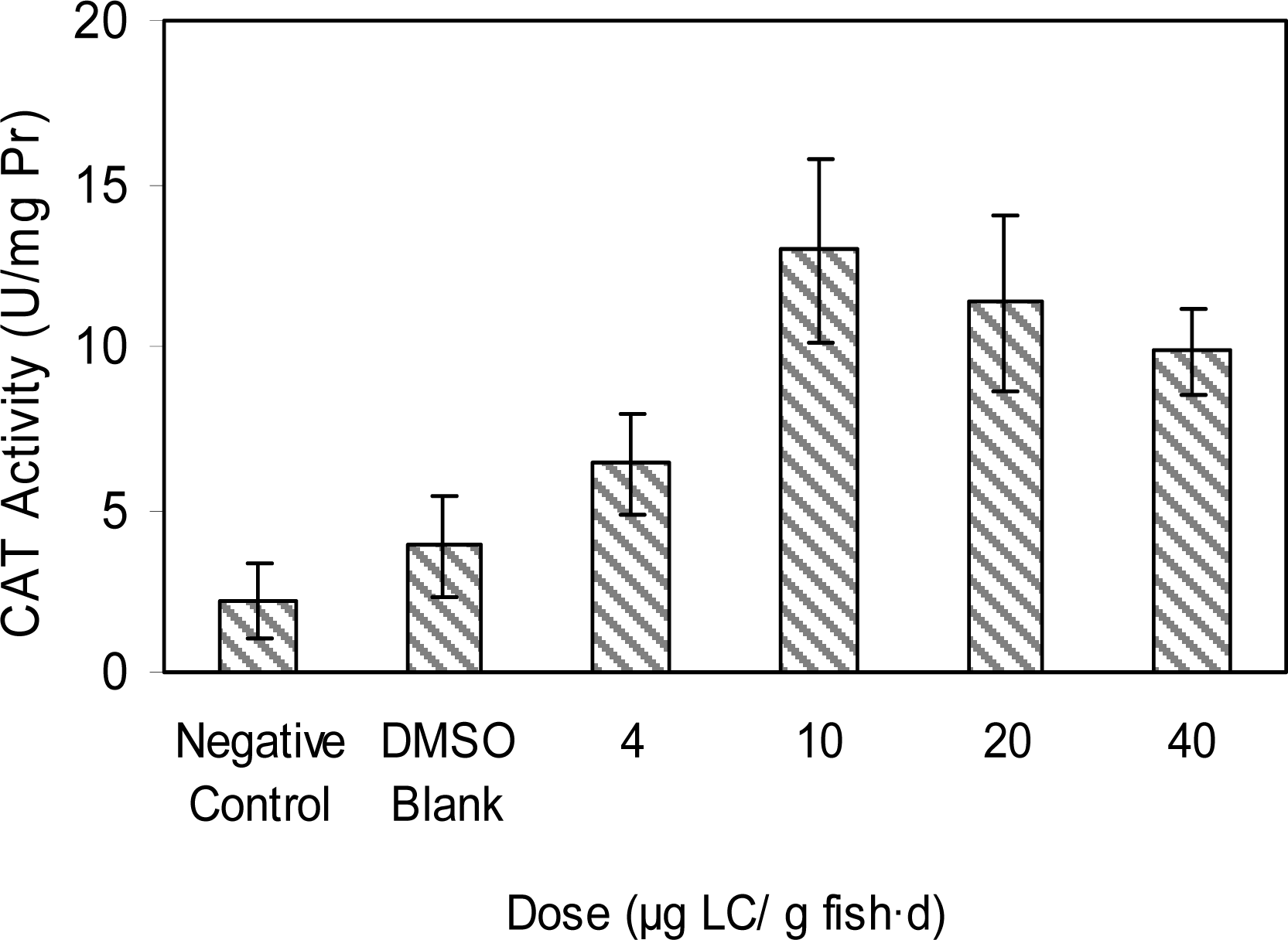
Clear induction of CAT activity was observed with the increase of LC dose (Figure 1). Statistical differences among the data for all groups were significant (p<0.01, ANOVA). The differences between any one of the four dose groups and the negative or the DSMO blank control were also significant (p<0.05, t-test). Difference was not significant between the negative control and DMSO blank control (p>0.1, t-test). CAT activity reached a peak between 10 and 20 μg LC/g fish·d, then started to decrease with LC dose increasing to 40 μg LC/g fish·d. While the difference between the 10 and 20 μg LC/g fish·d dose groups was not significant (p>0.4), the 40 μg LC/g fish·d dose group differed significantly from the 10 LC/g fish·d dose group (p<0.05, t-test). Compared to the negative control, SOD activities for all the dose groups were induced significantly (p<0.05, t-test). A significant response of SOD activity to the DMSO blank control (Figure 2) was observed (p<0.001), indicating that DMSO solvent could cause induction of this enzyme. The activity of SOD peaked around 20 μg LC/g fish·d and decreased slightly at 40 μg LC/g fish·d. However, the differences among the 10, 20, and 40 μg LC/g fish·d dose groups were insignificant (p>0.70, ANOVA).
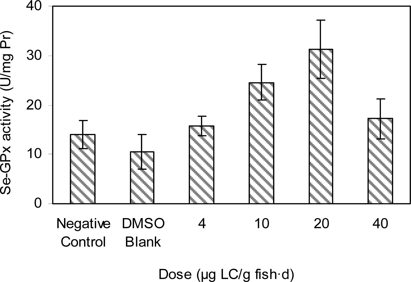
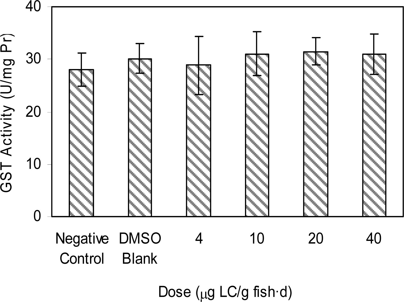
For Se-GPx, its activity was not significantly induced until the LC dose reached 10 μg LC/g fish·d. It peaked around 20 μg LC/g fish·d (Figure 3) and dropped significantly at 40 μg LC/g fish·d (p<0.05, t-test for 20 and 40 dose groups). The increase of antioxidant enzyme activities at lower LC doses suggests a compensatory response to a pollutant-induced stress. When the LC dose exceeded the compensatory threshold, oxidative stress may have occurred and the compensatory mechanism started to fail. The activity of GST did not show significant changes with all the LC doses (p=0.90, ANOVA), suggesting that GST may not be involved in the metabolism of these chemicals. Several other studies also reported that GST’s response to some pollutants was insignificant [8, 16, 17].
Conclusions
The results presented here show significant responses of the activities of three selected hepatic enzymes CAT, SOD, and Se-GPx to LC dose. These enzymes can be potential biomarkers for early warning of adverse health effects of LCs on some aquatic lives including catfish. The response patterns suggest that antioxidant enzymes are induced when catfish are exposed to low doses of LCs. When the LC dose reached about 20 μg LC/g fish·d, oxidative stress occurred. The LCs contained in LCDs of obsolete electronic devices such as laptop personal computers manufactured in the early 1990s may have adverse health effects on aquatic lives at fairly low concentrations. Proper handling and disposal of LCD-containing electronic waste is essential for the preservation of a healthy environment. Further comprehensive studies on the chronic toxicity of LCs on other living creatures and human beings are necessary.
Acknowledgments
This research was supported by a grant from the National Science Foundation (Grant No. BES-0238765).
References
- Werner, B; Simon-Hettich, B; Hoenicke, P. Toxicological and Ecotoxicological Investigations of Liquid Crystals. MERCK. http://www.MERCK.com. 2002.
- Geracitano, L; Monserrat, JM; Bianichini, A. Physiological and antioxidant enzyme responses to acute and chronic exposure of Laeonereis acuta (Polychaeta Nereididae) to copper. J Exp Marine Biol Ecol 2002, 277, 145–156. [Google Scholar]
- Webb, NA; Shaw, JR; Moran, J; Hogstrand, C; Wood, CM. Acute and Chronic physiological effects of silver exposure in three marine teleosts. Aquat Toxicol 2001, 54, 161–178. [Google Scholar]
- Ronisz, D; Finne, EF; Karlsson, H; Forlin, L. Effects of HBCDD and TBBPA on hepatic enzymes and other biomarkers in juvenile rainbow trout and feral eelpout. Aquat Toxicol 2004, 69, 229–245. [Google Scholar]
- Frei, B. Molecular and biological mechanisms of antioxidant action. FASEB J 1999, 13, 963–964. [Google Scholar]
- Di Guilio, RT; Washburn, PC; Wenning, RJ; Winston, GW; Jewell, CS. Biochemical responses in aquatic animals: a review of determinants of oxidative stress. Environ Toxicol Chem 1989, 8, 1103–1123. [Google Scholar]
- Storey, KB. Oxidative stress: animal adaptations in nature. Braz. J Med Biol Res 1996, 29, 1715–1733. [Google Scholar]
- Zhang, J; Shen, H; Wang, X; Wu, J; Xue, Y. Effects of chronic exposure of 2,4-dichlorophenol on the antioxidant system in liver of freshwater fish Carassius auratus. Chemoshpere 2004, 55, 167–174. [Google Scholar]
- Pedrajas, JR; Lopez-Barea, J; Peinado, J. Dieldrin induces peroxisomal enzymes in fish (sparus aurata) liver. Comp Biochem Physiol 1996, 115C, 125–131. [Google Scholar]
- Wijeyaratne, WMDN; Pathiratne, A. Acetylcholinesterase inhibition and gill lesions in Rasbora caverii, and indigenous fish inhabiting ric field associated waterbodies in Sri Lanka. Ecotoxicology 2006, 15, 609–619. [Google Scholar]
- Bradford, MM. A rapid and sensitive method for the quantitation of microgram quantities of protein utilizing the principle of protein-dye binding. Anal Biochem 1976, 72, 248–254. [Google Scholar]
- Xu, J; Yuan, X; Lang, P. Determination of catalase activity and catalase inhibition by ultraviolet spectrophotometry. Chin Environ Chem 1997, 16, 73–76. [Google Scholar]
- Zou, G; Gui, X; Zhong, X; Zhu, R. A new method for SOD activity measurement. Prog Biochem Biophy (in Chinese). 1986, 4, 71–73. [Google Scholar]
- Hafeman, DG; Sunde, RA; Hoekstra, WG. Effect of dietary selenium and erythrocyte and liver glutathione peroxidase in the rat. J Nutrit 1973, 104, 580. [Google Scholar]
- Habig, WH; Pabst, MJ; Jakoby, WB. Glutathione S-transferases: the first step in mercapturic acid formation. J Biol Chem 1974, 249, 7130–7139. [Google Scholar]
- Rendon-von Osten, J; Ortiz-Arana, A; Guilhermino, L; Soares, A. In vivo evaluation of three biomarkers in the mosquitofish exposed to pesticides. Chemosphere 2005, 58, 627–636. [Google Scholar]
- Van Erp, S; Brooth, L; Gooneratne, R; O’Halloran, K. Sublethal responses of wolf spiders to organophosphorous insecticides. Environ Toxicol 2002, 7, 449–456. [Google Scholar]
Figure 1.
Response of CAT activity in fish livers to LC dose

Figure 2.
Response of SOD activity in fish livers to LC dose
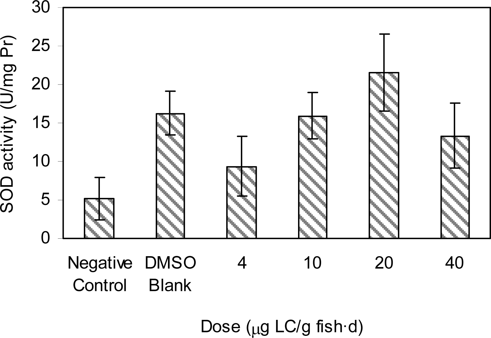
Figure 3.
Response of Se-GPx activity in fish livers to LC dose

Figure 4.
Response of GST activity in fish livers to LC dose

© MDPI. All rights reserved
Share and Cite
MDPI and ACS Style
An, R.; Li, Y.; Niu, X.; Yu, H. Responses of Antioxidant Enzymes in Catfish Exposed to Liquid Crystals from E-Waste. Int. J. Environ. Res. Public Health 2008, 5, 99-103. https://doi.org/10.3390/ijerph5020099
AMA Style
An R, Li Y, Niu X, Yu H. Responses of Antioxidant Enzymes in Catfish Exposed to Liquid Crystals from E-Waste. International Journal of Environmental Research and Public Health. 2008; 5(2):99-103. https://doi.org/10.3390/ijerph5020099
Chicago/Turabian StyleAn, Ran, Yadong Li, Xiaojun Niu, and Hongtao Yu. 2008. "Responses of Antioxidant Enzymes in Catfish Exposed to Liquid Crystals from E-Waste" International Journal of Environmental Research and Public Health 5, no. 2: 99-103. https://doi.org/10.3390/ijerph5020099