Article

Antibiotic Sensitivity Profiling and Virulence Potential of Campylobacter jejuni Isolates from Estuarine Water in the Eastern Cape Province, South Africa

Microbial Pathogenicity and Molecular Epidemiology Research Group (MPMERG), Department of Biochemistry and Microbiology, Department of Biochemistry & microbiology, University of Fort Hare, Private Bag X1314, Alice 5700, South Africa
*
Author to whom correspondence should be addressed.
Int. J. Environ. Res. Public Health 2018, 15(5), 925; https://doi.org/10.3390/ijerph15050925
Submission received: 10 April 2018 / Revised: 30 April 2018 / Accepted: 3 May 2018 / Published: 6 May 2018
(This article belongs to the Special Issue Recreational Water Illnesses)

Abstract

:
Campylobacter jejuni (CJ) is a zoonotic microbe and a major causative organism of diarrheal infection in humans that often has its functional characteristics inactivated in stressed conditions. The current study assessed the correlation between recovered CJ and water quality parameters and the drug sensitivity patterns of the pathogen to frontline antibiotics in human and veterinary medicine. Water samples (n = 244) from rivers/estuarines were collected from April–September 2016, and physicochemical conditions were recorded on-site. CJ was isolated from the samples using standard microbiological methods and subjected to sensitivity testing to 10 antibiotics. Mean CJ counts were between 1 and 5 logs (CFU/mL). Ninety-five isolates confirmed as CJ by PCR showed varying rates of resistance. Sensitivity testing showed resistance to tetracycline (100%), azithromycin (92%), clindamycin (84.2%), clarithromycin and doxycycline (80%), ciprofloxacin (77.8%), vancomycin (70.5%), erythromycin (70%), metronidazole (36.8%) and nalidixic acid (30.5%). Virulence encoding genes were detected in the majority 80/95, 84.2%) of the confirmed isolates from cdtB; 60/95 (63.2%) from cstII; 49/95 (51.6%) from cadF; 45/95 (47.4%) from clpP; 30/95 (31.6%) from htrB, and 0/95 (0%) from csrA. A multiple resistance cmeABC active efflux pump system was present in 69/95 (72.6) isolates. The presence of CJ was positively correlated with temperature (r = 0.17), pH (r = 0.02), dissolved oxygen (r = 0.31), and turbidity (r = 0.23) but negatively correlated with salinity (r = −0.39) and conductivity (r = −0.28). The detection of multidrug resistant CJ strains from estuarine water and the differential gene expressions they possess indicates a potential hazard to humans. Moreover, the negative correlation between the presence of the pathogen and physicochemical parameters such as salinity indicates possible complementary expression of stress tolerance response mechanisms by wild-type CJ strains.

1. Introduction

Campylobacter spp. are of the epsilonproteobacteria class of microorganism [1]. They are slow growing, Gram-negative, spiral shaped, motile organisms, characterized by their microaerobic nature [2]. They have been reported to be detected in greater quantities in diarrhea infections in humans than any other enteric pathogen and they require less than 100 cells to infect a host [3]. Campylobacteriosis is a chronic enteric infection primarily caused by cytotoxin-producing Campylobacters that invade and colonize the gastrointestinal (GI) tract in humans [4]. It is a zoonotic disease mainly transmitted via the consumption of poultry products [2], contact with pets and livestock, ingestion of water contaminated with human faeces originating from sewage, septic tanks, latrines and even animal faeces or from raw milk [5]. In humans, the disease lasts between 4–7 days and is characterized by acute enteritis, fever, vomiting and abdominal pain [4], with the danger of possibly leading to some post-infectious neuropathic diseases, such as bacteraemia, Guillain–Barre syndrome (GBS), reactive arthritis (ReA), and abortion [4,6]. Campylobacter jejuni and C. coli are the two widely recognized pathogenic species of Campylobacter that cause diseases in humans [7]. A large number of recorded campylobacteriosis outbreaks have also been traced to the ingestion of untreated surface water contaminated through human activities or by avian wildlife faeces [8,9]. Recreational and potable water are potential reservoirs of Campylobacter infection [5]. Therefore, the role of water in the transmission of Campylobacter species is of significant importance.

1.1. Virulence Determinants

Campylobacter jejuni possesses some multiple cell surface expressive virulent factors responsible for its high prevalence and pathogenicity compared to other enteric bacteria [3,10]. The secreted toxin is either enterotoxic or cytotoxic in its mode of action [10]. The target sites of enterotoxins are the cells lining the gastrointestinal (GI) tract of the host where its virulent effect is felt, resulting in either cytotoxin-linked inflammatory diarrhea associated with fever or non-inflammatory diarrhea associated with enterotoxins, characterized by non-leukocyte watery stools [4].

1.1.1. Cytolethal Distending Toxin (CDT)

The Cytolethal Distending Toxin (CDT) is an apoptosis triggering toxin produced by a group of Gram negative bacteria including Campylobacter jejuni (CJ) [9]. This toxin plays an important role in the host mucosal inflammatory response for interleukin-8 (IL-8) released by intestinal cells [10]. CDT is suggested to have an AB2 tripartite structure with cdtB as the main effector, while cdtA and cdtC are makeup units associated with cell membrane binding [11]. The cdtA protein has a molecular mass of 27 KDa, CdtB has a molecular mass of 29 KDa, and cdtC has a molecular mass of 20 KDa [11]. Subunit A is the active unit directly responsible for DNA damage, while subunit B is a binding subunit that helps to bind the toxin to the specific target cells which inhibit cdc2, causing cellular distention and eventually death. The DNase activity of CDT is lethal—causing singular strand breakage with an estimated lethal dose (LD) of 50 pg/mL [11]. The pivotal role played by in cell and DNA degradation not only results in inflammatory diarrhea with faecal leukocytes but can potentially create lesions in fragmented DNA strands that can promote cancer [4,9]. CDT has high cross species sequence similarity and cdtB has the highest interspecies similarity [11]. It is, however, believed that some species lack cdtB but still have the potential to cause symptoms in children less than 3 years old [11].

1.1.2. Campylobacter Invasion Antigens (ciaB)

This is a protein synthesized by CJ which facilitates invasion to epithelial cells [10] of the gastrointestinal tract where it inflicts increased damage on the columnar epithelial cells, leading to swelling and rounding of invaded cells as a result of the cytotoxin and enterotoxin activities [8,10]. Campylobacter heat labile cytotonic (CTON) and cytotoxin (CTOX) are associated with non-inflammatory and inflammatory diarrhea, respectively [11]. Cia proteins are suggested to modify host cell regulatory pathways to promote CJ pathogenicity [12].

1.1.3. Fibronectin-Binding Protein (cadF)

This outer membrane conserved gene encodes a protein containing 326 amino acids of molar mass 37 kDa and plays a vital role in adherence to intestinal epithelial cells [13]. Internalization of the organism into its host is harnessed by the binding activity of cadF to the extracellular fibronectin [14]. However, studies have shown that there may be a reduction in cadF functionality when it assumes a defensive viable but non-culturable (VBNC) state [14].

1.1.4. Sialyltransferases (cstII)

This is an outer core structure carbohydrate called lipo-oligosaccharide (LOS) expressed by CJ that evades detection by mimicry of the human gangliosides [15]. The mechanism of action of this gene is providing the LOS with a protective barrier which facilitates its invasion of the epithelial cells by portraying a resemblance to the human ganglioside in the vertebrate nerve cells, allowing the host’s immune system to self-destruct its own ganglioside [15]. It is believed to trigger the development of autoimmune diseases, such as Guillain–Barre Syndrome [16].

1.1.5. Post Transcriptional Regulator (csrA)

csrA gene is a carbon starvation regulator gene linked to the encoding of protein regulation which plays a vital role in CJ’s ability to responsively regulate a stationary phase mechanism to withstand hostile conditions [17]. Other associated virulence expressed by this gene is related to oxidative stress survival, the adherence of intestinal epithelial cells and biofilm formation [18]. Biofilm formation is an adaptive mechanism which complements the fragility of an organism when exposed to stress conditions by triggering a switch into a VBNC state [19].

1.1.6. ATP-Dependent Endopeptidase Protease (clpP)

This protease subunit in the bacterial caseinolytic proteases (CLP) contributes to virulence via energy formation through the degradation of virulence regulators [20], while indirectly playing a triggering role in stress tolerance of the organism when subjected to stress conditions [21]. The association of clpP with clp ATPase subunits enhances the proteolytic activity of the enzyme in the presence of ATP, producing a catalytic action [21]. In many pathogens, clpP enhances protein induced growth, under conditions, such as high temperature and oxidative stress [22].

1.1.7. Periplasmic Chaperon (htrB)

This is a periplasmic chaperon gene that encodes an acyltransferase for lipid A synthesis [23,24]. Synthesis of this enzyme regulates the organism’s response to environmental changes [25]. It is, however, interesting to note that the Campylobacter species shows varying diversities of infection outcomes [26] attributable to differences in genetic composition [22].

1.2. Treatment and Drug Resistance

Campylobacter infection is, at times, regarded as self-limiting, but in the case of severe complications, antibiotics are commonly recommended, especially for immunodeficient patients. Antibiotic resistance among Campylobacter species has emerged as a global public health burden [4]. There are cases of growing resistance of Campylobacter spp. against the front line and alternative treatment therapies, such as macrolides (erythromycin), tetracycline, fluoroquinolones and aminoglycoside (gentamycin) [7,27]. The unregulated use of antimicrobial agents as food additives in livestock in order to prevent and control infections and enhance growth rates [28,29] has contributed to an increased resistance in microbes against multiple antibiotics [9]. The unregulated administration of fluoroquinolones to poultry has contributed to increased resistance of CJ to fluoroquinolones in industrialized regions [27].
A survey of the antimicrobial susceptibility of Campylobacter species isolated from poultry and pigs was carried out in the Western Cape and Gauteng provinces of South Africa and the results displayed clear traces of resistance to fluoroquinolones, macrolides and tetracycline antibiotics, while some of the isolates displayed multidrug resistance [30]. These characteristic drug resistances were prominent among two specific Campylobacter species, CJ and C. coli, which have very similar epidemiology, but require biochemical tests to distinguish between them [30]. Previous studies have reported variation in CJ sensitivity to erythromycin and ciprofloxacin; resistance rates of 79.2% were reported in Nigeria [31], 0% in Djibouti [14] and in Qatar, resistance rates of 63.2% to ciprofloxacin and 8.6% to erythromycin were shown [32]. Resistance to ciprofloxacin, another antimicrobial agent of consideration next to erythromycin, has also been recorded in some other parts of the world [33]. Multidrug resistance in Campylobacter is a widely studied area. Previous studies have suggested mutation as a factor responsible for the acquisition of this characteristic [34,35,36]. Campylobacters have an innate resistance trait in combination with externally acquired resistance traits to express virulence [35]. Mutation is believed to play a role in the evolution of the cmeABC operon [15,36] in the multidrug efflux system. Drug resistance has, however, been attributed to target modification-mediated enzymatic inactivation and enhanced efflux [37].

2. Materials and Methods
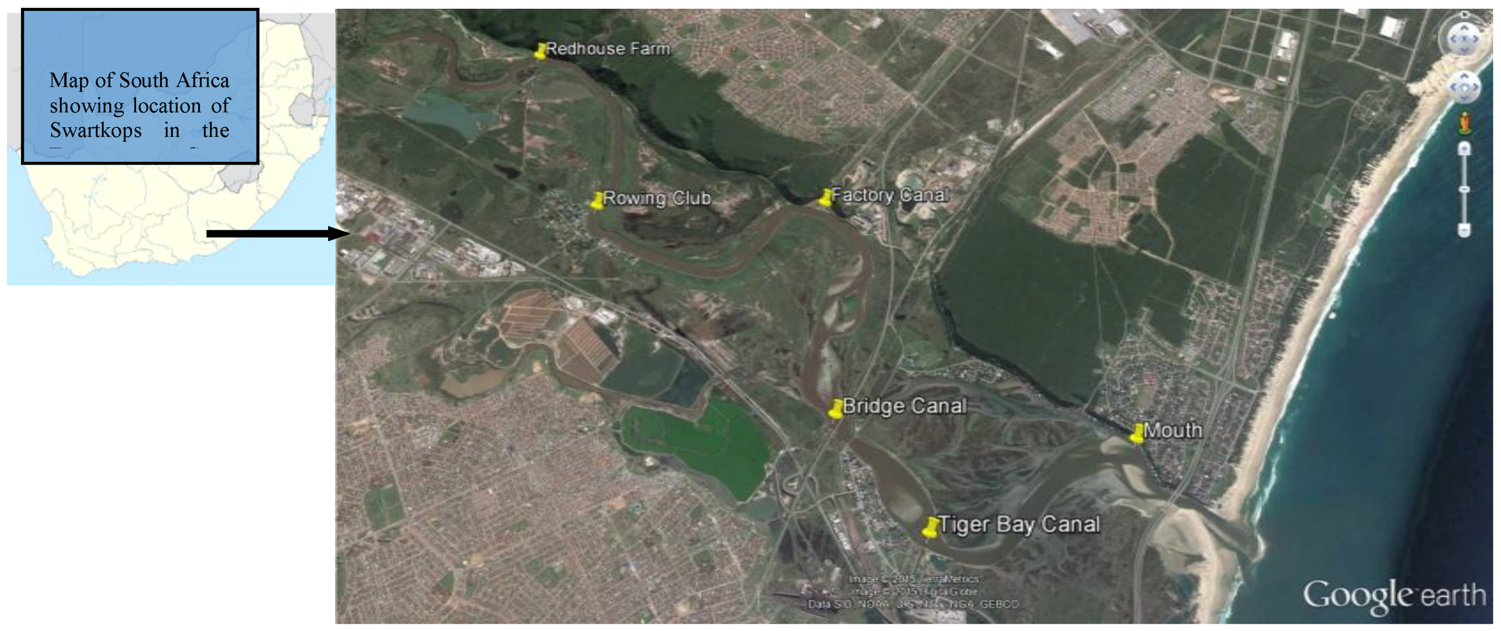
2.1. Study Area

The Swartkops estuary (33°52′ S; 25°38′ E) was selected for this study, and it is one of the most important estuaries in South Africa. It is also an important bird area (IBA) harbouring approximately 4000 migratory birds annually [38]. It is located close to the coastal city of Port Elizabeth in Nelson Mandela Bay Municipality of the Eastern Cape Province. The river is approximately 134 km long, while the estuary is approximately 16.4 km long with a permanent open connection into Algoa Bay in the Indian Ocean [39]. The total catchment area of the Swartkops River (including the tributary) is about 1360 km2 [39]. Surrounding areas in the catchment of the Swartkops River are used for agriculture, while the lower reaches of the river and the estuary are surrounded by extensive human development, including several industries [40].

2.2. Sampling and Isolation of CJ

Sampling and Isolation

The spot sampling method, as described by the JEEP92 project [41], was used. An Aestuaria Bandi 410 vessel was used for sampling a total distance of 12.775 km of the Swartkops river estuary between the six sample points (Figure 1). Triplicate water samples were collected against water flow from surface level and at a depth below (3 m) using sterile bottles from each sampling point over a 6-month period (April–September 2016) covering three seasons (autumn, winter and spring) of the year and transported at 4 °C to the laboratory and analyzed within 5 h after collection. Physicochemical parameters (temperature, pH, electrical conductivity, salinity and turbidity) of sample stations were recorded in-situ [42] using the YSI 650 MDS multi-parameter reader at two levels (surface and bottom) from each sampling point.
Bacteria cells were concentrated on a microfilter (0.65 µm pore size cellulose ester Millipore) from raw water (100 mL; 10−1; 10−2) samples. The concentrated filter was aseptically folded and enriched in 20 mL nutrient broth supplemented with Preston Campylobacter selective supplement (SR0117-Oxoid) with 5% lysed horse blood and incubated microaerobically at 37 °C for 48 h. One hundred microlitres of enrichment culture was sub-cultured to Campylobacter blood-free agar (CCDA; CM739; Oxoid) containing CCDA selective supplement (SR155E; Oxoid) and incubated microaerobically using a campy gas pack (5% O2, 10% CO2, 85% N2, CampyGen Oxoid) at 37 °C for 72 h. All plating was carried out in duplicate. Distinct presumptive colonies on each plate were counted by the surface count method to determine the total viable Campylobacter counts (TVCC). Then, 8–10 different colonies per plate were picked and subcultured on the selective medium for purity. A positive control (Campylobacter jejuni ATCC 33560 strain) was included with each set of tests. Identification of positive isolates was based on colony morphology, Gram-stain, no-growth in aerobic condition, hippurate hydrolysis and oxidase tests.

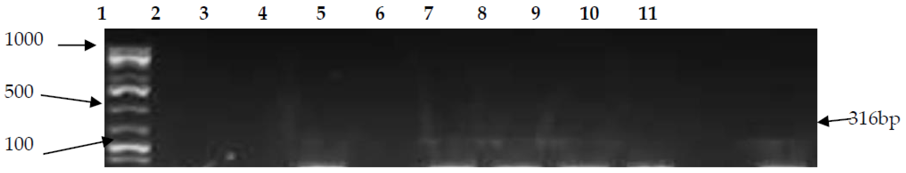
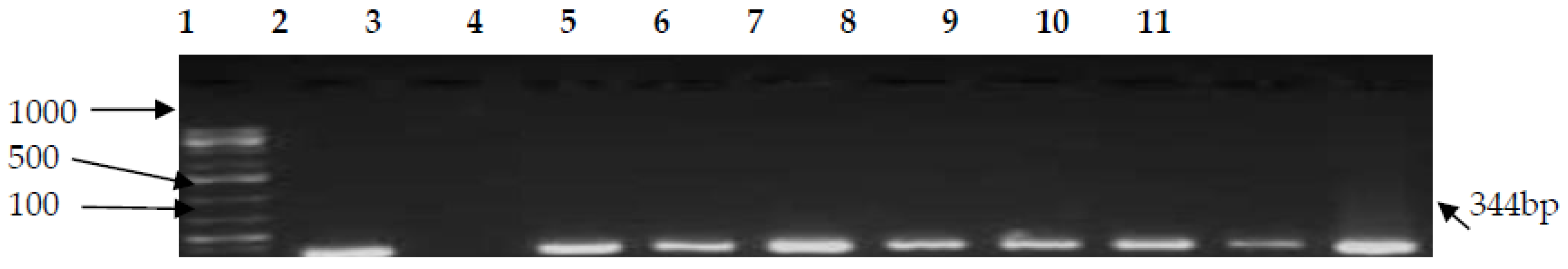
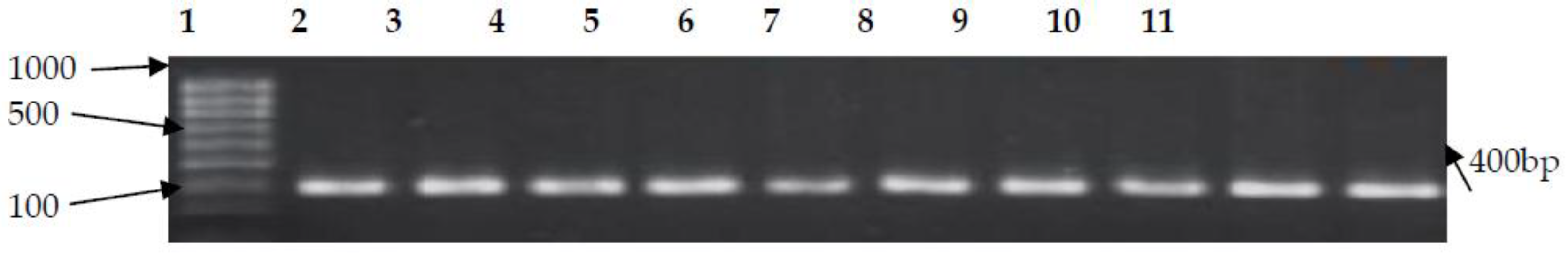
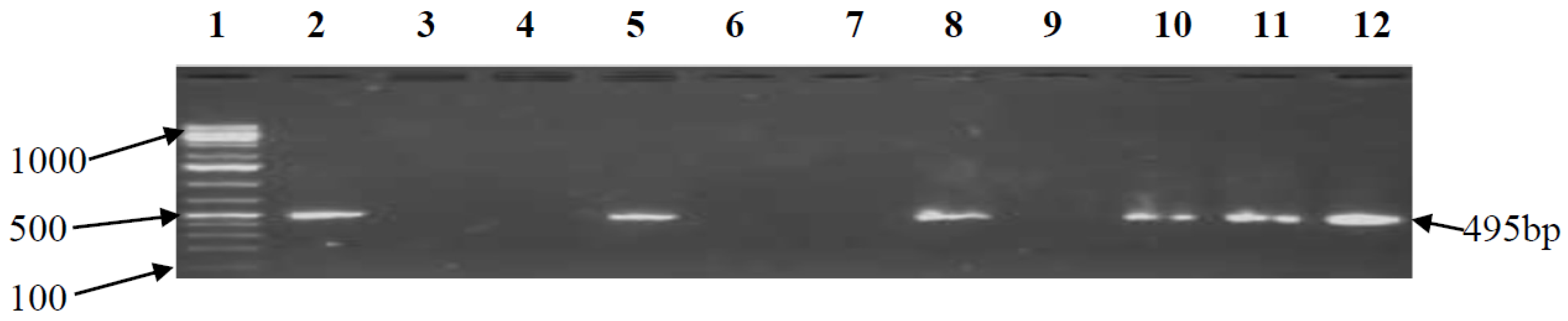
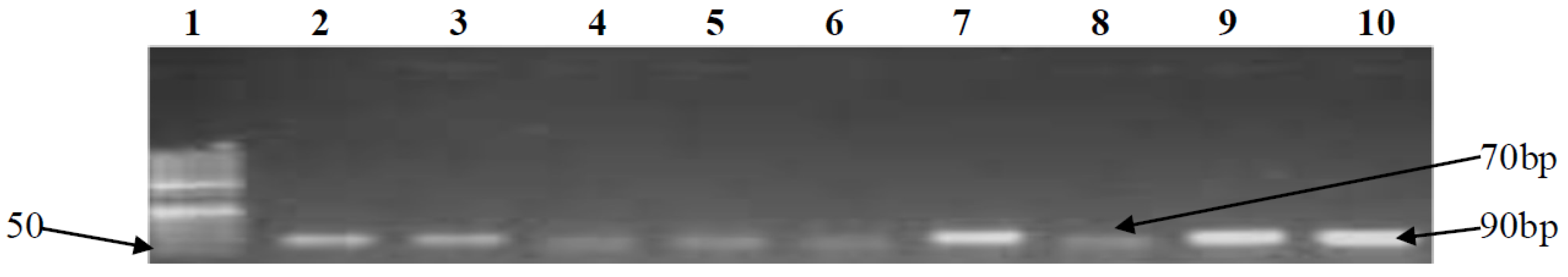
2.3. PCR Confirmation of CJ and Detection of Virulence Genes

Genus confirmation was performed using the 23S rRNA gene [43], and species confirmation was done using the hipO gene [44]. Screening for the pathogenic virulence genes, cdtB, cadF, cstII, csrA, htrB and clpP, was performed on the CJ confirmed isolates. Genomic isolation from the confirmed isolates was carried out using a commercial genomic DNA isolation kit (Qiagen Kit, Invitrogen, Thermo Fisher Scientific, USA) according to the manufacturer’s instructions. Table 1 shows the primer sequences (Inqaba biotech, South Africa), amplicon sizes and cycling conditions of the various genes used in this study. The final concentration of the 25 µL PCR reaction consisted of 12.5 µL of the 2X master mix (Sybrselect, USA) 0.5 µL forward and reverse primers, 6.5 µL molecular grade water and 5 µL template DNA. The cycling conditions were as follows: initial denaturation was at 94 °C for 5 min, and then 94 °C for 30 s, with modifications in annealing temperatures specific to the primer pair (as given in Table 1) for 5 min and extension at 72 °C for 50 s. All PCR products were analyzed by electrophoresis on 1.5% agarose gels (CSL-AG100, Cleaver Scientific Ltd. Warwickshire, UK) except for htrB and clpP genes which were analyzed on 2% agarose gels. The gels were stained with ethidium bromide and visualized with a UV transilluminator and photographed (Alliance 4.7).

2.4. Antimicrobial Sensitivity Testing

Confirmed CJ isolates were subjected to antimicrobial sensitivity testing with 10 antimicrobial agents. The Kirby–Bauer disk diffusion method on Mueller–Hinton agar supplemented with 5% horse blood, in accordance with Clinical and Laboratory Standards Institute guidelines [38] was used to perform the antimicrobial profiling. An inoculum of each bacterial isolate was emulsified in 3 mL of sterile normal saline (0.9%) in test tubes and the density was adjusted to 0.5 McFarland standard (0.5 mL of 1% w/v BaCl2 and 99.5 mL of 1% v/v H2SO4), equivalent to 1.0 × 10⁸ cfu/mL. The bacterial suspension was evenly spread on the Mueller–Hinton agar plates using sterile swab sticks and allowed to dry. Antibiotic discs (Mast Diagnostics Ltd., UK) with the following drug concentrations were selected for the assay: nalidixic acid (30 µg), ciprofloxacin (5 µg), azithromycin (15 µg), doxycycline (30 µg), erythromycin (15 µg), clarithromycin (15 µg), vancomycin (15 µg), tetracycline (30 µg), clindamycin (2 µg) and metronidazole (15 µg). Plates were incubated at 37 °C under microaerophilic conditions (5% O2, 10% CO2, 85% N2) with gas generator envelopes (CampyGen; 2.5 L Thermo Scientific, UK) for 48 h, and the diameter zones of inhibition were measured, and the results were interpreted in accordance with the CLSI unit [48].

Detection of Multidrug Resistance Genes (cmeA, cmeB and cmeC)

Detection of the cmeA, cmeB and cmeC genes was determined by PCR, as described by [49], with slight modifications. Table 2 shows the primer sequences (Inqaba biotech, South Africa). The final concentration of the 25 µL PCR reaction consisted of 12.5 µL of the 2X master mix (Sybrselect, USA) 0.5 µL forward and reverse primers, 6.5 µL molecular grade water and 5 µL template DNA. The cycling conditions were as follows: initial denaturation at 94 °C for 7 min, followed by 94 °C for 1 min, annealing temperatures (see Table 2) for 1.5 min, extension at 72 °C for 3 min and then, final extension at 72 °C for 5 min for 30 cycles. All PCR products were analyzed by electrophoresis on 1% agarose gel (CSL-AG100, Cleaver Scientific Ltd. Warwickshire, UK). The gels were stained with ethidium bromide and visualized with a UV transilluminator and photographed (Alliance 4.7). Campylobacter jejuni ATCC 33560 strains were used as the positive control.

3. Results

3.1. Physicochemical Analyses

The mean water temperature in the Swartkops river estuary for the sampled months was between 14.7 °C and 15.6 °C with the Despatch Mouth (DM) recording the highest mean temperature, while the Rowing Club station (RC) had the lowest mean temperature. No clear-cut difference in pH values was recorded for the sample stations, as the mean pH for all the stations ranged between 8.27 and 8.33. However, a high level of variation in the salinity level was recorded at all stations, with the mean salinity ranging between 13.92 practical salinity units (psu) and 32.77 psu. Station E, which is the dispatch point of Swartkops River to Algoa Bay recorded the highest salinity out of all the sampled months, while the Rowing Club (RC) station recorded the lowest salinity. The average dissolved oxygen (DO) concentrations were 53.83 mg/L and 62.9 mg/L, respectively. Station RC also recorded the lowest DO reading, while the Tiger Bay (TB) station recorded the highest overall reading. In terms of turbidity, the Swartkops water was very turbid during the sampled seasons, ranging, on average, between 4.2 Nephelometric Turbidity Units (NTU) and 66.9 NTU. The Factory Dam (FD) station recorded the highest average turbidity (66.9 NTU), especially in July and August, while DM presented more pristine water for all sample periods. The conductivity was between 20.9 ms/cm and 30 ms/cm on average. A low coefficient of variability was observed for all sampled sites (Table 3).

3.2. PCR Confirmation of CJ and Detection of Virulence Genes

One hundred and twenty isolates were phenotypically confirmed as Campylobacteracea (Figure 2). Further screening at the species level confirmed 95 isolates as C. jejuni (Figure 3) and the other 25 identified as C. coli (18) and C. upselensis (7). Determination of the occurrence of virulence genes in the confirmed CJ isolates revealed the cdtB gene in 80/95 (84.2%) of the isolates, an indication that the toxin production gene (cdtB) was the most prevalent virulence determinant. Forty-nine (52%) of the isolates were identified as having adherence virulence genes (cadF), while 60/95 (63.2%) isolates tested positive for the intestinal epithelial invasive virulence gene (cstII). This gene is also linked to the risk of Gullian–Barre Syndrome (GBS) development. Thirty (31.6%) of the isolates were positive for the lipid A synthesis gene (htrB) responsible for the adjustment of organisms to stressful external environmental changes, while 45/95 (47.4%) of the isolates were identified as having the ATP dependent protease gene (clpP), which is responsible for the degradation of damaged proteins due to unfavourable conditions. The carbon starvation regulator gene (csrA), which is linked to cell division and the formation of biofilm, was absent in all isolates. Isolates recovered from the Redhouse Farm (RF) and Bridge Canal (BC) sampling sites were confirmed as housing all but the csrA gene which was absent in all isolates. Table 4 shows the number of genes detected in confirmed CJ isolates.

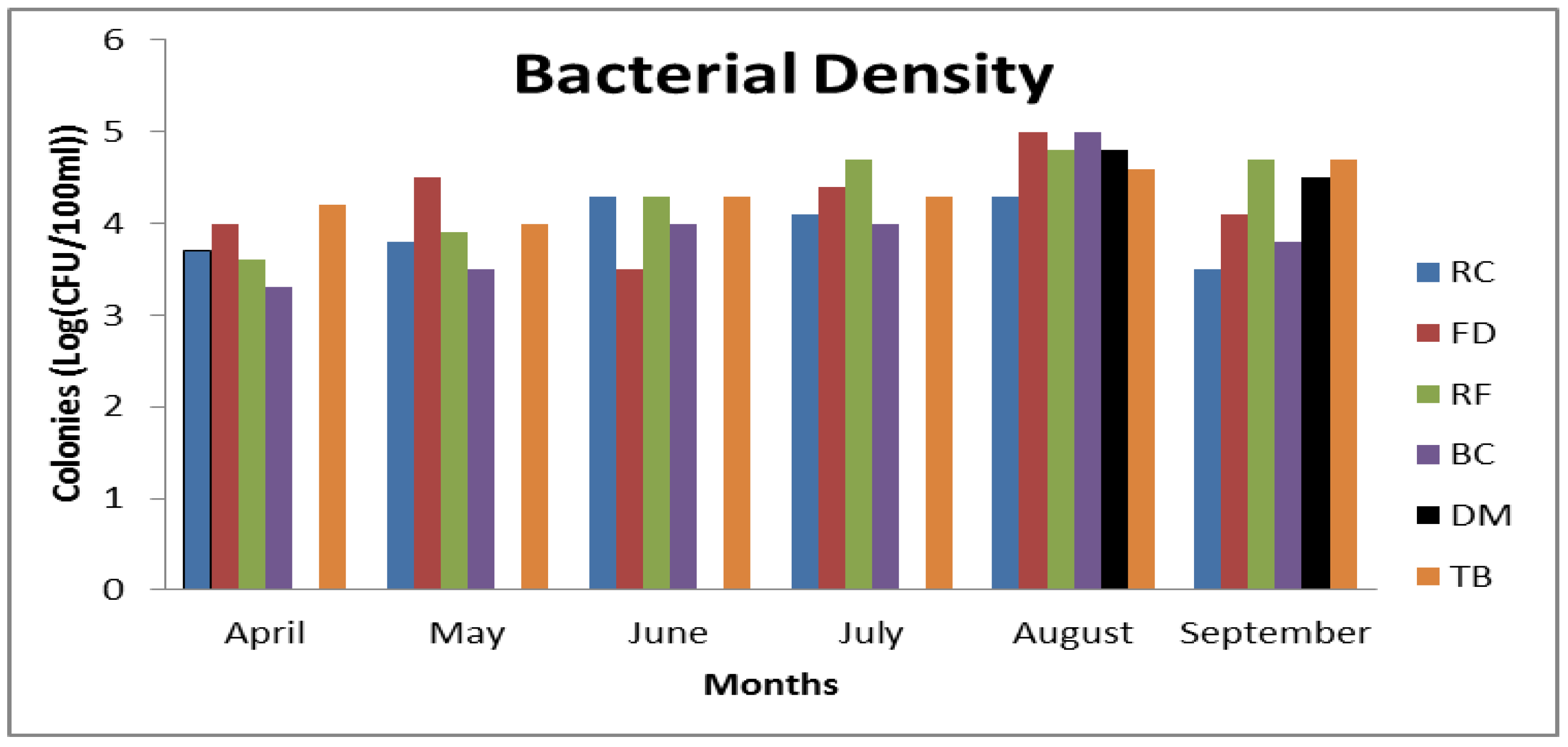
3.3. Frequency of CJ Isolation

The frequency of bacterial isolation frequency at all sample sites for the sampling period was recorded (Figure 4). The Despatch Mouth (DM) was the least Campylobacter-contaminated site, and the most pristine with an isolation frequency of 33%. No Campylobacter count was recorded at DM for April, May, June or July (autumn and winter), but Campylobacter were recorded in August and September (spring). The Tiger Bay (TB) site appeared to be the most Campylobacter-contaminated site in the Swartkops estuary with an isolation frequency of 100% during the sampled seasons. Campylobacter counts were recorded for all sampling months, with higher readings in July, August and September. However, the overall highest average Campylobacter counts were recorded at the Factory Dam (FD) and Bridge Canal (BC) sites for the month of August. Consequently, very low counts were recorded in April, May, and June (winter) for all sites.

3.4. Physicochemical Parameters and Occurrence of C. jejuni

No significant positive correlation was observed between the population density of CJ and temperature (r = 0.17), pH (r = 0.02), dissolved oxygen (r = 0.31), and turbidity (r = 0.23). A negative correlation was observed with salinity (r = −0.39) and conductivity (r = −0.28). The correlation values were not statistically different for temperature, dissolved oxygen, salinity, turbidity and conductivity, while they were statistically different for pH (Table 5).

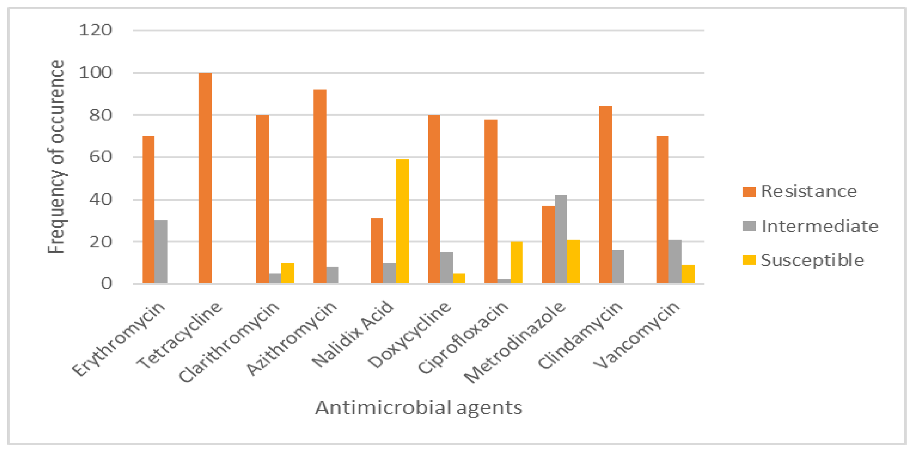
3.5. Antimicrobial Sensitivity Testing and Prevalence of Multidrug Resistance (MDR) Efflux Pump Genes

The antibiotic sensitivity of 95 CJ isolates was profiled and revealed a higher degree of resistance to tested antimicrobial agents. The highest resistance level, 95/95 (100%), was recorded for tetracycline, followed by azithromycin (87/95, 92%), clindamycin (80/95, 84.2%), clarithromycin and doxycycline (76/95, 80%), ciprofloxacin (78/95, 77.8%), vancomycin (67/95, 70.5%), and erythromycin (67/95, 70%), with the lowest resistance levels recorded for metronidazole (35/95, 36.8%) and nalidixic acid (29/95, 30.5%). Nalidixic acid was the most effective antibiotic with a susceptibility of 57/95 (59%) (Figure 5). The percentage incidences of multidrug resistance (MDR) efflux pump genes of C. jejuni are shown in Table 6. A high (69/95, 72.6%) number of CJs expressed MDR efflux pump genes with only 6/95 (6.3%) not expressing them. Twenty isolates showed a disruption in the expression of all tripartite efflux systems, which could trigger a malfunction of the cmeABC system.

4. Discussion

4.1. Survival of Organism

Estuaries are confluent ecosystems where a mixture of salty sea waters and rivers meet with freshwater [42]. They are transition points from land to sea and freshwater to salt water and are rich in organic contents [5]. The dynamic physicochemical nature of the ecosystem is peculiarly detrimental to the survival of fastidious microbes such as Campylobacter [14]. However, Campylobacter spp. display complex survival mechanisms by transiting to a stationary viable, but non-culturable, form (VBNC) for survival [11]. The recovered strains in this study displayed differential gene expression, which could be peculiarto wild-type CJ strains, and morphological evidence showed 66/95 (69%) without flagella. This could be due to the expression of the flagella gene being switched off in a process known as phase variation [49]. The carbon starvation regulator gene (csrA) which is linked to the encoding of protein regulation was not detected in the study. The absence of csrA could have been complimented by the presence of htrB and clpP genes. Other associated virulence expressed by this gene is related to oxidative stress survival, adherence of intestinal epithelial cells and in biofilm formation [18,45]. Although the pathogenic potential of wild-type strains is debatable due to the adverse stress conditions which can reduce their colonization and invasive abilities, their ability to live asymptomatically in their hosts may not be fully ruled out, especially as this concerns externally acquired genes from surrounding environs. This is indicated by the presence of the cstII gene in some of the strains (Figure 6). Some of the test isolates lacked cdtB, and cadF genes and could, therefore, be considered non-pathogenic. Most of the isolates lacked motility potential at the point of analysis, inferring the inactivation of the racR gene [11]. The results as shown in Figure 7 strongly correlate with the inference drawn in a past study suggesting that CJ could still retain its cadF adhesion functionality under stressed conditions [14].
The prevalence of the cytotoxin production gene (cdtB) in the confirmed isolates (Figure 8) shows that the organism is capable of retaining its toxin production ability even in a starved state. The presence of htrB and clpP genes (Figure 9) provides an extra boost in the organism’s aero-tolerance survival in environmental waters. However, the survival of microorganisms in oxidative stress environments has been attributed to their ability to develop specialized defensive mechanisms [11]. The existence of CJ in the Swartkops could be as a result of oceanic effects rather than continental effects. The FD sample site was the most polluted site in the Swartkops due to effluent discharges from the factory directly to the river. Between FD and BC is the shallowest area; this may be due to high land surface run-off into the river. This area has the highest population of migratory birds and fishing activities with the highest CJ density which strongly indicates that avian species are major reservoirs of the organism. Seasonality, on the other hand, could also play a pivotal role in the survival and existence of the organism, as larger population densities of the organism were recorded during spring and the lowest population densities were recorded during winter [2,9]. The Swartkops river estuary has a strong nutrient zonation, which is typified by the variation in salinity distribution in the estuary as a result of upstream shift of salt and fresh waters, which may be responsible for the abundance and distribution of species. Hence, this was the major reason for choosing to investigate the effect of the physicochemical parameters on the survival of the organism. The salinity reading at DM was the highest total average reading of all seasons, but barely affected the survival of the organism.

4.2. Drug Resistance

Multiple resistances have been reported globally in both CJ and C. coli in human and animal isolates. The resistance pattern was observed notably in tetracyclines, macrolides and fluoroquinolones [1,7]. In South Africa, antimicrobials with broad-spectrum activity such as tetracyclines are used in both the poultry and pig industries, as they are affordable and easy to administer in food and water [30]. In a recent study, a high level of resistance to tetracyclines was also revealed in Campylobacter spp. isolated from broilers and hens [1]. In this study, multidrug resistance was observed in more than 70% of the isolates (Figure 10). The isolates harbored the Campylobacter multidrug efflux pump (cmeABC) genes responsible for multidrug resistance [7]. Previous studies have shown variation in the resistance pattern of CJ in South Africa [7,30]. Some studies revealed a high resistance of the organism to fluoroquinolones, macrolides and tetracyclines [30], while some showed high susceptibility to fluoroquinolones [1]. Resistance to fluoroquinolone is believed to develop more rapidly in Campylobacter spp, than in other Gram-negative bacteria, mainly attributed to single-step point mutations in gyrA [2]. However, these studies were carried out on CJ isolated from avians, porcines and bovines. In this study, multidrug resistance was shown for fluoroquinolones, macrolides and tetracyclines. A high resistance to tetracycline (100%) was observed and the highest susceptibility (59%) was shown to nalidixic acid. Previous studies have suggested that aquatic environments (surface and groundwater bodies) are perfect for horizontal gene exchange of mobile genetic elements (MGEs) which results in antibiotic resistance [2].

5. Conclusions

This is the first study to report the occurrence of differential gene expressions in wild-type CJ isolated from the Swartkops in the Eastern Cape Province. The results showed that the estuarine water could potentially harbour multiple resistant CJ strains of public health concern among estuarine users. Although the extent of their pathogenicity is not fully ascertained, it could be assumed that pathogens with similar traits are likely to be found in other similar ecosystems.

6. Future Direction of Study

This study focused on the detection of active virulence-inducing genes and antimicrobial sensitivity profiling of environmentally recovered CJ strains. More elaborate future studies should include a comparative genomic analysis using whole genome sequencing (WGS) for other related water sources of high importance around the Eastern Cape Province to fully understand the pathophysiological mechanisms of recovered wild-type Campylobacter strains. Moreover, a comparative study of the antimicrobial profile and analysis of expressed virulence of wild-type isolates and clinical strains should be investigated. Other important virulence determinants which were not investigated in this study such as racR and flaA genes associated with motility and cosR, rrpA or rrpB associated with the oxidative stress response should be studied in wild-type isolates. It is also necessary to conduct the same study on other closely-related organisms, such as Arcobacter species, to document their pathogenicity.

Author Contributions

A.C.O. carried out the project experimental design, sample collection, performed the experiments, analysed data, and drafted the manuscript, H.A.N. and J.F. contributed to data analysis, study design and proof reading of manuscript, O.E.A. assisted in sample collection and proof reading of manuscript, A.M.C. contributed to the study design and organisation of sampling.

Acknowledgments

This work was supported by the National Research Foundation (NRF) and the South African Institute for Aquatic Biodiversity (SAIAB) under the NRF/SAIAB innovative scholarship.

Conflicts of Interest

The authors declare no conflict of interest.

References

  1. Bester, L.; Essack, S. Prevalence of antibiotic resistance in Campylobacter isolates from commercial poultry suppliers in KwaZulu-Natal, South Africa. J. Antimicrob. Chemother. 2008, 62, 1298–1300. [Google Scholar] [CrossRef] [PubMed]
  2. Nichols, G.; Richardson, J.; Sheppard, S.; Lane, K.; Sarran, C. Campylobacter epidemiology: A descriptive study reviewing 1 million cases in England and Wales between 1989 and 2011. BMJ 2012, 2, e001179. [Google Scholar] [CrossRef] [PubMed]
  3. Croinin, O.; Backert, S. Host epithelial cell invasion by Campylobacter jejuni: Trigger of zipper mechanism? Research Advances in the study of Campylobacter, Helicobacter and related organisms. Front. Cell. Infect. Microbiol. 2012, 2, 25. [Google Scholar] [CrossRef] [PubMed]
  4. World Health Organization. The Increasing Incidence of Human Campylobacteriosis Report and Proceedings of a WHO Consultation of Experts, Copenhagen, Denmark 21–25 November 2000; World Health Organization: Copenhagen, Denmark; pp. 21–25.
  5. EFSA (European Food Safety Authority); ECDC (European Centre for Disease Prevention and Control). The European Union summary report on trends and sources of zoonoses, zoonotic agents and food-borne outbreaks in 2012. EFSA J. 2014, 12, 3547. [Google Scholar] [CrossRef]
  6. Skirrow, M.; Blaser, M. Clinical aspects of Campylobacter infection. In Campylobacter, 2nd ed.; Nachamkin, I., Blaser, M.J., Eds.; ASM Press: Washington, DC, USA, 2000; pp. 69–88. [Google Scholar]
  7. Engberg, J.; Aarestrup, F.; Taylor, D.; Gerner-Smidt, P.; Nachamkin, I. Quinolone and macrolide resistance in Campylobacter jejuni and C. coli: Resistance mechanisms and trends in human isolates. Emerg. Infect. Dis. 2001, 7, 24–34. [Google Scholar] [CrossRef] [PubMed]
  8. Sopwith, W.; Birtles, A.; Matthews, M.; Fox, A.; Gee, S.; Painter, M. Campylobacter jejuni multilocus sequence types in humans, northwest England, 2003–2004. Emerg. Infect. Dis. 2006, 12, 1500–1507. [Google Scholar] [CrossRef] [PubMed]
  9. DefFraites, F.; Sanchez, L.; Brandt, A.; Kadlec, P.; Haberberger, L.; Lin, J.; Taylor, N. An outbreak of Campylobacter enteritis associated with a community water supply on a US military installation. MSMR 2014, 21, 10–15. [Google Scholar]
  10. Perera, V.; Nachamkin, I.; Ung, H.; Patterson, J.; McConville, M. Molecular mimicry in Campylobacter jejuni: Role of the lipo-oligosaccharide core oligosaccharide in inducing anti-ganglioside antibodies. FEMS Immunol. Med. Microbiol. 2007, 50, 27–36. [Google Scholar] [CrossRef] [PubMed]
  11. Samosornsuk, W.; Asakura, M.; Yoshida, E.; Taguchi, T.; Eampokalap, B.; Chaicumpa, W.; Yamasaki, S. Isolation and characterization of Campylobacter strains from diarrheal patients in Bangkok and its suburb in Thailand. Jpn. J. Infect. Dis. 2015, 68, 209–215. [Google Scholar] [CrossRef] [PubMed]
  12. Saouaf, J.; Li, B.; Zhang, G.; Shen, Y.; Furuuchi, N.; Hancock, W.; Greene, I. Deacetylase inhibition increases regulatory T cell function and decreases incidence and severity of collagen-induced arthritis. Exp. Mol. Pathol. 2009, 87, 99–104. [Google Scholar] [CrossRef] [PubMed]
  13. Sails, A.; Bolton, F.; Fox, A.; Wareing, D.; Greenway, D. Detection of Campylobacter jejuni and Campylobacter coli in environmental waters by PCR enzyme- linked immunosorbent assay. Appl. Environ. Microbiol. 2002, 68, 1319–1324. [Google Scholar] [CrossRef] [PubMed]
  14. Patrone, V.; Campana, R.; Vallorani, L.; Dominici, S.; Federici, S.; Casadei, L.; Gioacchini, A.M.; Stocchi, V.; Baffone, W. CadF expression in Campylobacter jejuni strains incubated under low-temperature water microcosm conditions which induce the viable but non-culturable (VBNC) state. Antonie Van Leeuwenhoek 2013, 103, 979–988. [Google Scholar] [CrossRef] [PubMed]
  15. Pérez-Boto, D.; López-Portolés, J.; Simón, C.; Valdezate, S.; Echeita, M. Study of the molecular mechanisms involved in high-level macrolide resistance of Spanish Campylobacter jejuni and Campylobacter coli strains. J. Antimicrob. Chemother. 2010, 65, 2083–2088. [Google Scholar]
  16. Bach, J. Infections and autoimmune diseases. J. Autoimmun. 2005, 25, 74–80. [Google Scholar] [CrossRef] [PubMed]
  17. Fields, J.; Thompson, S. Campylobacter jejuni CsrA mediates oxidative stress responses, biofilm formation, and host cell invasion. J. Bacteriol. 2008, 190, 3411–3416. [Google Scholar] [CrossRef] [PubMed]
  18. González-Hein, G.; Huaracán, B.; García, P.; Figueroa, G. Prevalence of virulence genes in strains of Campylobacter jejuni isolated from human, bovine and broiler. Braz. J. Microbiol. 2013, 44, 1223–1229. [Google Scholar]
  19. Thakur, S.; Zhao, S.; McDermott, P.; Harbottle, H.; Abbott, J.; Gebreyes, W.; White, D. Antimicrobial resistance, virulence, and genotypic profile comparison of Campylobacter jejuni and Campylobacter coli isolated from humans and retail meats. Foodborne Pathog. Dis. 2010, 7, 835–844. [Google Scholar] [CrossRef] [PubMed]
  20. Hlaváček, O.; Vachova, L. ATP- dependent proteinases in bacteria. Folia Microbiol. 2002, 47, 203–212. [Google Scholar] [CrossRef]
  21. Frees, D.; Brøndsted, L.; Ingmer, H. Bacterial proteases and virulence. In Regulated Proteolysis in Microorg; Springer Science: Dordrecht, The Netherlands, 2013; pp. 161–192. [Google Scholar]
  22. Hughes, R.; Cornblath, D. Guillain-Barre´ syndrome. Lancet 2005, 366, 1653–1666. [Google Scholar] [CrossRef]
  23. Parkhill, J.; Wren, B.W.; Mungall, K.; Ketley, J.M.; Churcher, C.; Basham, D.; Chillingworth, T.; Davies, R.M.; Feltwell, T.; Holroyd, S.; et al. The genome sequence of the food-borne pathogen Campylobacter jejuni reveals hypervariable sequences. Nature 2000, 403, 665–668. [Google Scholar] [CrossRef] [PubMed]
  24. Hausdorf, L.; Neumann, M.; Bergmann, I.; Sobiella, K.; Mundt, K.; Fröhling, A.; Schlüter, O.; Klocke, M. Occurrence and genetic diversity of Arcobacter spp. in a spinach processing plant and evaluation of two Arcobacter-specific quantitative PCR assays. Syst. Appl. Microbiol. 2013, 36, 235–243. [Google Scholar] [CrossRef] [PubMed]
  25. Mihaljevic, R.; Sikic, M.; Klancnik, A.; Brumini, G.; Mozina, S.; Abram, M. Environmental stress factors affecting survival and virulence of Campylobacter jejuni. Microb. Pathog. 2007, 43, 120–125. [Google Scholar] [CrossRef] [PubMed]
  26. Sheppard, S.; Dallas, J.; Strachan, N.; MacRae, M.; McCarthy, N.; Wilson, D.; Gormley, F.J.; Falush, D.; Ogden, I.; Maiden, M.; et al. Campylobacter genotyping to determine the source of human infection. Clin. Infect. Dis. 2009, 48, 1072–1078. [Google Scholar] [CrossRef] [PubMed]
  27. Koolman, L.; Whyte, P.; Burgess, C.; Bolton, D. Distribution of virulence- associated genes in a selection of Campylobacter isolates. Foodborne Pathol. Dis. 2016, 12, 424–432. [Google Scholar] [CrossRef] [PubMed]
  28. Igimi, S.; Okada, Y.; Ishiwa, A.; Yamasaki, M.; Morisaki, N.; Kubo, Y.; Asakura, H.; Yamamoto, S. Antimicrobial resistance of Campylobacter: Prevalence and trends in Japan. Food Addit. Contam. 2008, 25, 1080–1083. [Google Scholar] [CrossRef]
  29. Rożynek, E.; Dzierżanowska-Fangrat, K.; Szczepańska, B.; Wardak, S.; Szych, J.; Konieczny, P.; Albrecht, P.; Dzierżanowska, D. Trends in antimicrobial susceptibility of Campylobacter isolates in Poland (2000–2007). Polskie Tow. Mikrobiol. Pol. Soc. Microbiol. 2009, 58, 111–115. [Google Scholar]
  30. Jonker, A.; Picard, J. Antimicrobial susceptibility in thermophilic Campylobacter spp. isolated from pigs and chickens in South Africa. J. S. Afr. Vet. Assoc. 2010, 81, 228–236. [Google Scholar] [CrossRef] [PubMed]
  31. Coker, O.; Adefeso, O. The changing patterns of Campylobacter jejuni/coli in Lagos, Nigeria after ten years. East African Med. J. 1994, 71, 437–440. [Google Scholar]
  32. Ghunaim, H.; Behnke, J.; Aigha, I.; Sharma, A.; Doiphode, S.; Deshmukh, A.; Abu-Madi, M. Analysis of Resistance to Antimicrobials and Presence of Virulence/Stress Response Genes in Campylobacter Isolates from Patients with Severe Diarrhoea. PLoS ONE 2015, 10, e0119268. [Google Scholar] [CrossRef] [PubMed]
  33. Nachamkin, I.; Mishu-Allos, B.; Ho, T. Campylobacter species and Guillain-Barre´ Syndrome. J. Clin. Microbiol. 1998, 11, 555–567. [Google Scholar]
  34. Vacher, S.; Menard, A.; Bernard, E.; Santos, A.; Megraud, F. Detection of mutation associated with macrolide resistance in thermophilic Campylobacter spp. By real-time PCR. Microb. Drug Resist. 2005, 11, 40–47. [Google Scholar] [CrossRef] [PubMed]
  35. Qin, S.; Wang, Y.; Zhang, Q.; Chen, X.; Shen, Z.; Deng, F. Identification of a novel genomic island conferring resistance to multiple amino glycoside antibiotics in Campylobacter coli. Antimicrob. Agents Chemother. 2012, 56, 5332–5339. [Google Scholar] [CrossRef] [PubMed]
  36. Iovine, N. Resistance mechanisms in Campylobacter jejuni. Virulence 2013, 4, 230–240. [Google Scholar] [CrossRef] [PubMed]
  37. Gibreel, A.; Taylor, D.E. Macrolide resistance in Campylobacter jejuni and Campylobacter coli. J. Antimicrob. Chemother. 2006, 58, 243–255. [Google Scholar] [CrossRef] [PubMed]
  38. Department of Water Affairs. Revision of general authorizations in terms of Section 39 of the National Water Act, 1998 (At No. 36 of 1998); Published under Government Notice 665 in Government Gazette 36820 No. 665; Department of Water Affairs: Cape Town, South Africa, 2013.
  39. Scharler, M.; Baird, D. A comparison of selected ecosystem attributes of three South African estuaries with different freshwater inflow regimes using network analysis. J. Mar. Syst. 2005, 56, 283–308. [Google Scholar] [CrossRef]
  40. Baird, D.; Marais, J.; Martin, P. The Swartkops Estuary: Proceedings of a Symposium at the University of Port Elizabeth; Foundation for Research Development: Pretoria, South Africa, 2007; p. 8. [Google Scholar]
  41. Heip, C.; Herman, P. Major biological processes in European tidal estuaries: A synthesis of the JEEP-92 Project. Hydrobiologia 1995, 311, 1–7. [Google Scholar] [CrossRef]
  42. EPA. Guidelines for Water Reuse; EPA/625/R-04/108; Environmental Protection Agency, Municipal Support Division Office of Wastewater Management Office of Water: Washington, DC, USA; Agency for International Development: Washington, DC, USA, 2012.
  43. Vacher, S.; Menard, A.; Bernard, E.; Mégraud, F. PCR-Restriction Fragment Polymorphism analysis for detection of point mutations associated with macrolide resistance in Campylobacter spp. Antimicrob. Agents Chemother. 2003, 47, 1125–1128. [Google Scholar] [CrossRef] [PubMed]
  44. Persson, S.; Olsen, K.E. Multiplex PCR for identification of Campylobacter coli and Campylobacter jejuni from pure cultures and directly on stool samples. J. Med. Microbiol. 2005, 54, 1043–1047. [Google Scholar] [CrossRef] [PubMed]
  45. Phongsisay, V. Campylobacter jejuni and the Guillain-Barré Syndrome. Ph.D. Thesis, RMIT University, Melbourne, Australia, 2006. [Google Scholar]
  46. Cohn, M.; Ingmer, H.; Mulholland, F.; Jørgensen, K.; Wells, J.; Brøndsted, L. Contribution of conserved ATP-dependent proteases of Campylobacter jejuni to stress tolerance and virulence. Appl. Environ. Microbiol. 2007, 73, 7803–7813. [Google Scholar] [CrossRef] [PubMed]
  47. Lin, J.; Overbye Michel, L.; Zhang, Q. CmeABC functions as a multidrug efflux system in Campylobacter jejuni. Antimicrob. Agents Chemother. 2002, 46, 2124–2131. [Google Scholar] [CrossRef] [PubMed]
  48. CLSI. Methods for Antimicrobial Dilution and Disk Susceptibility Testing of infrequently Isolated or Fastidious Bacteria, Approved Guidelines-2nd ed.; CLSI: Wayne, PA, USA, 2012. [Google Scholar]
  49. De Vries, S.P.; Gupta, S.; Baig, A.; L’Heureux, J.; Pont, E.; Wolanska, D.P.; Maskell, D.J.; Grant, A.J. Motility defects in Campylobacter jejuni defined gene deletion mutants caused by second-site mutations. Microbiology 2015, 161, 2316–2327. [Google Scholar] [CrossRef] [PubMed]
Figure 1. Location and GPS coordinates of the study sites in the Swartkops River estuary, Port Elizabeth. Sampling locations within the estuary; Rowing Club (RC) (33°50’14.60 S; 25°34’14.37 E); Factory Dam (FD) (33°50’12.16 S; 25°35’41.24 E); Redhouse Farm (RF) (33°49’10.56 S; 25°33’37.44 E); Bridge Canal (BC) (33°51’25.99 S; 25°35’49.99 E); Despatch Mouth (DM) (33°51’32.22 S; 25°37’31.33 E); Tiger Bay Canal (TB) (33°52’0.26 S; 25°36’22.32 E) (https://www.google.com.au/maps/).
Ijerph 15 00925 g001
Figure 2. PCR detection of 23S rRNA gene (316 bp). Lane 1 is themarker, Lanes 2–9 are test isolates, Lane 10 is the negative control, and Lane 11 is the positive control.
Ijerph 15 00925 g002
Figure 3. PCR detection of hipO gene (344 bp) for CJ confirmation. Lane 1 is the marker, Lane 2 is the positive control, Lane 3 is the negative control, and Lanes 4–11 are the test isolates.
Ijerph 15 00925 g003
Figure 4. Bacterial density at all sampling sites. * Mean values are expressed in log (cfu per 100 mL).
Ijerph 15 00925 g004
Figure 5. Antimicrobial sensitivity Profile.
Ijerph 15 00925 g005
Figure 6. PCR detection of cstII gene (570 bp) of CJ. Lane 1 is the marker, Lane 2 is the positive control, Lane 3 is the negative control, and Lanes 4–13 are test isolates.
Ijerph 15 00925 g006
Figure 7. PCR detection of cadF gene (400 bp) of CJ: Lane 1 is the marker, Lane 2 is the positive control, and Lanes 3–11 are test isolates.
Ijerph 15 00925 g007
Figure 8. PCR detection of cdtB gene (495 bp) of CJ. Lane 1 is the marker, Lane 2 is the positive control, Lane 3 is the negative control, and Lanes 4–12 are test isolates.
Ijerph 15 00925 g008
Figure 9. PCR detection of htrB (70bp) and clpP genes (90bp) of C. jejuni. Lane 1 is the marker, Lane 2 is the positive control, Lanes 3, 7, 9 and 10 are clpP positive isolates, and Lanes 4, 5, 6 and 8 are htrB positive isolates.
Ijerph 15 00925 g009
Figure 10. PCR detection of cmeABC in isolates. Lane 1 is the marker, Lane 2 is the positive control, Lane 8 is the CmeA positive isolate (435 base pair, Lane 4 is the negative control, Lanes 5 and 6 are cmeB positive isolates (444 bp), and Lane 3 is the cmeC positive isolate (431 bp).
Ijerph 15 00925 g010
Table 1. PCR primer sizes and annealing temperatures.
Name of GeneSequence (5′-3′)Product (bp)Annealing Temperature (°C)References
Campy 23SF-AATTGATGGGGTTAGCATTAGC
R-CAACAATGGCTCATATACAACTGG
31655[43]
hipOF-AGAGTTTGATCCTGGCTCAG
R-ACGGCTACCTTGTTACGACTT
34458[44]
cdtBF-CAC GGT TAA AAT CCC CTG CT
R-GCA CTT GGA ATT TGC AAG GC
49552[18]
htrBF-CGC ACC CAA TTT GAC ATA GAA
R-TTT TTA GAG CGC TTA GCA TTT GTC T
7052[45]
clpPF-TCG GAG CAT TTT TGC TTA GTT G
R-CTC CAC CTA AAG GTT GAT GAA TCA T
9052[46]
csrAF-CAC AGT CAG TGA AGG TGC TT
R-ACT CGC ACA ATC GCT ACT TC
87852[47]
cstIIF-CAG CTT TCT ATT GCC CTT GC
R-ACA CAT ATA GAC CCC TGA GG
57052[18]
cadFF-TTGAAGGTAATTTAGATATG
R-CTAATACCTAAAGTTGAAAC
40042[10]
Table 2. Primers used in the study.
Target GenesPrimer Sequences 5’-3’Annealing Temp (°C)Amplicon Size (bp)
CmeAF-TAGCGGCGTAATAGTAAATAAAC
R-ATAAAGAAATCTGCGTAAATAGGA
50435
CmeBF-AGGCGGTTTTGAAATGTATGTT
R-TGTGCCGCTGGGAAAAG
50444
CmeCF-CAAGTTGGCGCTGTAGGTGAA
R-CCCCAATGAAAAATAGGCAGAGTA
52431
Table 3. Physicochemical parameters of water at different sample stations.
Sample StationsParametersTemp (°C)pHSalinity (psu)DO (mg/L)Turbidity (NTU)Conductivity (ms/cm)
RCM14.78.3213.953.837.220.9
Cv0.160.020.520.680.850.49
FDM14.88.3319.260.566.925.7
Cv0.160.010.40.670.740.43
RFM14.98.2714.955.348.422
Cv0.170.010.150.70.780.13
BCM14.98.3121.456.931.926.3
Cv0.150.020.50.690.40.55
DMM15.68.2932.858.94.226.3
Cv0.130.010.190.690.70.23
TBM14.98.3625.462.924.230
Cv0.150.010.380.681.50.44
M = mean; Cv = coefficient of variability; DO = dissolved oxygen.
Table 4. Detection of pathogenic genes in confirmed C. jejuni (CJ) isolates.
Sample SourceNo. of SamplesNo. of Isolates Confirmed as Campylobacter Genus for Samples (%)No. of Isolates Confirmed as CJ (%)Genes Detected in CJ Isolates (% Positive)
Estuarine water24423S rRNAHipOcdtBcadFcstIIcsrAhtrBClpP
120 (49.2)95 (79.2)80 (84.2)49 (51.6)60 (63.2)0 (0)30 (31.6)45 (47.4)
Table 5. Correlation (Bravais Pearson) matrix between temperature, bacterial contamination, pH, salinity, turbidity, dissolved oxygen and conductivity.
TempPHSalinityDOTurbidityConductivityCJ
Temp1
pH−0.301
Salinity−0.600.461
DO0.190.07−0.311
Turbidity−0.08−0.03−0.12−0.431
Conductivity−0.530.480.87−0.320.081
CJ0.170.02−0.390.210.23−0.281
Table 6. Incidences of Campylobacter multidrug resistance (MDR) efflux pump genes.
GenesNo. of Isolates (n = 95)
a (%)b (%)
cmeA11/20 (55)9/20 (45)
cmeB18/20 (90)2/20 (20)
cmeC14/20 (70)7/20 (30)
cmeABC69/95 (72.6)6/95 (6.3)
a = positive isolates; b = negative isolates.

