Article

An Application of the Multivariate Linear Mixed Model to the Analysis of Shoulder Complexity in Breast Cancer Patients

1
Department of Mechanical Engineering an Mathematical Sciences, Oxford Brookes University, Wheatley Campus, Wheatley, Oxford OX33 1HX, UK
2
Leuven Biostatistics and Statistical Bioinformatics Centre (L-BioStat), Kapucijnenvoer 35 blok D, B-3000 Leuven, Belgium
3
Department of Primary Care and Public Health, Imperial College London, Charing Cross Hospital, London W6 8RP, UK
4
Clinical Research Centre, University of Cape Town, Old Main Building, L51. Groote Schuur Hospital Observatory, Cape Town 7700, South Africa
*
Author to whom correspondence should be addressed.
Int. J. Environ. Res. Public Health 2016, 13(3), 274; https://doi.org/10.3390/ijerph13030274
Submission received: 21 November 2015 / Revised: 16 February 2016 / Accepted: 18 February 2016 / Published: 2 March 2016

Abstract

:
In this study, four major muscles acting on the scapula were investigated in patients who had been treated in the last six years for unilateral carcinoma of the breast. Muscle activity was assessed by electromyography during abduction and adduction of the affected and unaffected arms. The main principal aim of the study was to compare shoulder muscle activity in the affected and unaffected shoulder during elevation of the arm. A multivariate linear mixed model was introduced and applied to address the principal aims. The result of fitting this model to the data shows a huge improvement as compared to the alternatives.

1. Introduction

Wide local excision (WLE) with adjuvant radiotherapy is the standard treatment of breast cancer. However, despite the use of less extensive surgery, there is still morbidity affecting the shoulder [1,2,3]. Local radiotherapy has several known effects on lung parenchyma, vascular and connective tissues [4,5,6]. Findings with respect to the latter suggest that thickening of the tissues may restrict movement of surrounding areolar tissues held within fascial planes. This limited ability to expand together with ischaemia due to changes in the vascular network could have an effect on the efficacy of muscle contraction [7,8,9].
Thus far, the exact nature of shoulder morbidity and its relationship to pain has only been described in terms of glenohumeral movement; ignoring the critical effect of scapulo-thoracic motions. Scapular movements are a product of the delicate inter-play between the rotator cuff muscles; thus, deficiency in one can alter all movements due to associations. The pattern of these associations is not known and has been investigated here.
Scapular movements such as abduction, adduction, anterior/posterior tilt, and medial/lateral rotations are a product of the delicate inter-play between the rotator cuff muscles. Deficiency in one may change all other movements. The pattern of these associations is not known and needs to be investigated.
Four muscles (pectoralis major, serratus anterior, upper trapezius and rhomboid major) acting on the scapula were investigated in patients who had been treated in the last six years for unilateral carcinoma of the breast. Muscle activity was assessed by electromyography (EMG) during abduction and adduction of the ipsilateral (affected) and contralateral (unaffected) sides.
The principal aims of the study were:
(1)
to compare shoulder muscle activity in the affected and unaffected shoulder during elevation of the arm;
(2)
to explore the relationship between any observed differences in muscle activity and patients report of pain and dysfunction;
(3)
to explore the relationship between any observed differences in muscle activity and key clinical variables.
Section 2 introduces the collected data set and its procedure. In Section 3, the multivariate linear mixed models are described which address the above principal aims. Section 4 presents the results of the fit of the model to the data. In Section 5, some final conclusions and a discussion of future analyses are given.

2. Data Set

Two hundred and two patients who had been treated from 2005 to 2010 for unilateral carcinoma of the breast were included in a shoulder morbidity study. The measurement procedure is detailed in our previous work [10]. Patients with any previous history of shoulder or neck problems in either arm were excluded from the study. Humeral elevation in degrees was measured using the Polhemus Fastrak™ motion analysis system, and concurrent EMG readings were taken at 10° increments of humeral elevation of each shoulder for each patient. The average of three EMG readings at each elevation point was taken as a response (dependent) variable for each patient.
All patients filled in a Shoulder Pain and Disability Index (SPADI) questionnaire immediately prior to data collection. The SPADI questionnaire is a known and valid measure of pain and disability for shoulder dysfunction with high levels of sensitivity and reliability [11], it is scored on a visual analogue scale with 13 items (five for pain and eight for disability). All subjects gave their informed consent for inclusion before they participated in the study. The study was conducted in accordance with the Declaration of Helsinki, and the protocol was approved by Oxford Brookes University ethics committee (HREC no: A02,064)
Pain scores range from a minimum of 0 to a maximum of 500 mm and for disability 0 to 800 mm, where 0 represents no symptoms of pain or disability. Figure 1 represents the distribution of pain and disability scores. Given the skewness of these distributions, each variable is log-transformed in subsequent analyses.
Key clinical variables include: affected side, dominant hand, degree of arm elevation, treatment protocol (Wide Local Excision (WLE) or other), duration after surgery in days, age in years, physiotherapy and exercise level on affected side, receiving chemotherapy, and the Shoulder Pain and Disability Index [10].
Bilateral EMG measurements of the four shoulder muscles were taken for each patient. Each muscle activity is recorded in millivolts (mV) at 10° increments of humeral elevation during upward and downward arm movement. At each increment point, for each shoulder and each movement, four EMG measures were obtained (one for each of the four muscles), creating potentially a correlated or multivariate response structure. Incremental measurements at different arm elevations per muscle created a multivariate response structure that can also be regarded as “repeated measures” dimension of the data. Summarised, four correlated, repeatedly measured responses for each shoulder and each movement were generated. In Figure 2, typical observations of a patient are given, for humeral elevation between 10° and 150°, this figure represents the observations for an upward arm movement of the left arm. This graph clearly shows that the EMG reading at each given point very much depends on the previous reading within the same movement. Hence, this fact should be implemented in the modelling strategy in Section 3.

3. Statistical Hypotheses Testing and Statistical Modelling

3.1. Statistical Test of Clinical Hypotheses

Shamley et al. previously demonstrated [10] that the activities of muscles controlling the movement of the scapula are linked; if one muscle is compromised, then other muscles might become more active to compensate for the lost movement. However, muscle activity can be influenced by several demographic (age, dominance, gender) and clinical (pain, radiotherapy, chemotherapy, etc.) variables. In this study, we are interested to explore the previous findings in detail and describe the rationale of whether the clinical variables have the same effect on all muscles, or whether the effect of these variables is different for different muscles under different conditions. Hence, an advanced joint multivariate approach must be implemented to take these associations into consideration whilst investigating the following four main null hypotheses.
  • H01: The altered muscle activity is not associated with the patient’s report of pain and dysfunction;
  • H02: Clinical risk factors have no effect on the activities of the muscles;
  • H03: The effect of clinical risk factors is the same on the affected arm as with that on the unaffected arm.
  • H04: The activity of the four key muscles acting at the scapula on the affected arm is the same as from those acting on the unaffected arm.

3.2. The Multivariate Linear Mixed Model

Let Yiksm denote the ni × 1 vector of EMG readings at different humeral elevation points from the ith patient (i = 1,…, N), ni is the number of elevation points for that patient on the kth muscle (k = 1,…, 4), of the affected arm (s = 1) or unaffected arm (s = 2) during upward (m = 1) or downward (m = 2) movements.
We assume for the ni measurements, Yiksm, a multivariate normal distribution over the elevations. It is in fact the usual multivariate multiple regression, i.e.,:
Y i k s m N ( X i k s m β k s m , Σ k )
where Xiksm is a ni × p matrix of covariates (p is the number of covariates), βksm is a p × 1 vector of regression coefficients and Σk is a ni × ni covariance matrix.
Obviously, when the humeral elevations are closer, the correlation should be stronger, therefore, after testing a few other correlation structures, we sensibly assumed an autoregressive structure of order one, i.e., AR(1) for Σk, i.e.,
Σ k = σ k 2 ( 1 ρ k ρ k 2 ρ k 1 ρ k ρ k 2 ρ k 2 ρ k 1 ρ k : : : : ρ k n i 1 : ρ k n i 1 ρ k 1 )
A simplifying assumption could be to assume that the responses are independent among patients and muscles, that is
Y i k = [ Y i k 11 Y i k 12 Y i k 21 Y i k 22 ] N [ ( X i k 11 β k 11 X i k 12 β k 12 X i k 21 β k 21 X i k 22 β k 22 ) , Σ k k = ( Σ k 0 0 0 0 Σ k 0 0 0 0 Σ k 0 0 0 0 Σ k ) ]
It is generally unrealistic to assume that all important risk factors (covariates) are measured and included in the model explanatory matrix (Xiksm). The unobserved or unmeasured risk factors of a muscle, which are usually known as the “unobserved muscle’s specific effect”, causes the measurements of a muscle to be more correlated. To improve Model (3), one could introduce a “muscle specific effect” or random effect into the model. This term usually is known as frailty, so that, conditional on this muscle’s specific effect, νik, we can assume
Y i k | v k ~ N ( X i k β k + v i k , Σ k k )
with, Xik, the stacked design matrix X i k T = [ X i k 11 T X i k 12 T X i k 21 T X i k 22 T ] and β k T = [ β k 11 T , β k 12 T , β k 21 T , β k 22 T ] .
Coull and Agresti [12] presented similar models for multivariate binomial logit-normal. Fieuws et al. [13] discussed similar models in more detail.
Furthermore, we assumed that the frailty terms of different muscles are correlated. That is, we assumed that the vector of frailty terms ν i T = ( ν i 1 , ν i 2 , ν i 3 , ν i 4 ) has the following distribution:
ν i = [ ν i 1 ν i 2 ν i 3 ν i 4 ] N [ ( 0 0 0 0 ) , D   =   ( d 11 d 12 d 13 d 14 d 12 d 22 d 23 d 24 d 13 d 23 d 33 d 34 d 14 d 24 d 34 d 44 ) ] .
We assume that the joint model of Y i T = ( Y i 1 , Y i 2 , Y i 3 , Y i 4 ) follows a conditional multivariate normal distribution
Y i | ν i N [ ( X i 1 β 1 + ν i 1 , X i 2 β 2 + ν i 2 , X i 3 β 3 + ν i 3 , X i 4 β 4 + ν i 4 , ) , Σ = ( Σ 1 0 0 0 0 Σ 2 0 0 0 0 Σ 3 0 0 0 0 Σ 4 ) ]
with the normal distribution of the frailty vector which adds the covariance structure to the frailties across muscles. The joint model defined by Model (6) is a multivariate linear mixed model with multivariate random effects νi.
As for the univariate linear mixed model with a univariate normal random effect, the marginal likelihood in Model (6) is also analytically tractable. We used SAS™ [14] 9.2, PROC MIXED (SAS, Cary, NC, USA) to estimate the model parameters.
In this model, the correlation structure among the muscles is presented by the correlation structure among their specific frailties given by the variance-covariance matrix (D); if the muscles are not significantly correlated, all off-diagonal entities in the variance-covariance matrix D will be zero. Significant non-zero off-diagonal entities of variance-covariance matrix D are showing the correlations among the muscles.

4. Empirical Results

4.1. Model Comparison

Table 1 presents the result of analyzing the natural log transformation of EMG activities of each muscle using Model (3), i.e., a multivariate normal distribution for ln(emgiksm) = Yiksm with block diagonal variance-covariance matrix of Σk. The likelihood ratio test suggests that the effect of arm movement (MOVE_UP/MOVE_DOWN) is best presented by a dummy variable indicating different intercepts for the upward and downward movement. Interactions between arm movements with all other clinical risk factors (covariates) were not collectively significant at the 5% level.
Contrariwise, the effect of affected shoulder and unaffected shoulder cannot be modelled by considering only different intercepts for affected/unaffected shoulder. This is due to the fact that some of the risk factors are specific to the affected shoulder, such as physiotherapy and exercise. This implies that the effect of clinical risk factors might well be different on the affected shoulder compared to the unaffected shoulder. Therefore, a full interaction model (all main effects and all interactions) initially was employed to assess the effects of the affected shoulder on all clinical risk factors; the highly insignificant interactions were subsequently dropped from the model. The likelihood ratio test suggests that the included interactions are highly significant (change in deviance of 717.2 for 32 degrees of freedom).
The values in bold correspond to significant effects (at the 5% level) for each muscle. The last column (“p-value”) indicates whether the clinical risk factor is collectively (the effect on all four muscles collectively with four degrees of freedom) significant or not.
A significant value for the interaction terms shows that, over and above the overall effect of the clinical risk factor on electrical activity of the muscles, the effect of clinical risk factor on the affected side is different to the unaffected side. The autoregressive correlation (ρk) and the variance of each muscle ( σ k 2 ) are also presented.
The result of fitting a multivariate normally distributed model with AR(1) structure for Σk, presented in Table 1, controls the autoregressive structure of the measurements. The high positive value of ρ shows that the electrical activities are more alike at humeral elevation angles that are closer together.
This model not only ignores associations among the four muscles but also ignores any possible associations between measurements of the same muscle, i.e., affected/unaffected shoulders and downwards/upwards movement. In other words, this model assumes independence between the four muscles and between all observations of the same muscle at the same humeral elevation. This is not a realistic assumption as the four muscles are from the same individual and are likely to be correlated, and all measurements of the same muscle at the same humeral elevation are also likely to be correlated as they are from the same muscle. Table 2 is the result of fitting a univariate linear mixed Model (4) with independent random intercept νk across muscles and residual block diagonal variance-covariance matrix with blocks Σk assumed to follow the auto-regressive process of order one. Model (4) controls the association between the measurements of the same muscles at the same humeral elevation point by including a muscle specific random effect into the model.
This model suggests that humeral elevation of the arm, the upwards move, affected side and left shoulder will increase the electrical activity generally for all four muscles irrespective of which side is affected, whilst treatment with wide local excision and receiving chemotherapy are associated with generally decreased electrical activity.
Interaction analysis suggests that the electrical activity is significantly different for duration after surgery and SPADI pain. Doing exercise and having physiotherapy on the affected shoulder are significantly associated with electrical activity of the affected muscles. The deviance (−2 × log likelihood) for this model is 27,645.1. The change in deviance compared to Model (3) is 1536.1 for four degrees of freedom (p-value < 0.0001), which is highly statistically significant.
Univariate Linear Mixed Model with AR(1) structure for Σk controls the autoregressive structure of the measurements and accounts for possible associations between measurements of the same muscle at the same elevation point. The disadvantage of Model (4) is that it ignores any possible associations among the four muscles within patients. Table 3 is the result of fitting a full multivariate linear mixed model with AR(1) structure for Σk given in Model (6).
The result of fitting a full multivariate Linear Mixed Model with AR(1) structure for Σk, as expected, is almost similar to that of Model (4). This model suggests that humeral elevation of the arm, the upwards move, affected side, left shoulder and longer duration from the surgery will increase the electrical activity generally for all four muscles irrespective of the side affected—whilst treatment with wide local excision and receiving chemotherapy are associated with generally decreased electrical activity
Interaction analysis suggests that electrical activity is significantly different for duration after surgery and SPADI pain. Once again, doing exercise and having physiotherapy on the affected shoulder are significantly associated with electrical activity of the affected muscles. SPADI pain increases the muscle activity in the affected arm, but the affected arm slightly dilutes the positive effect of duration since surgery on the muscle activity. The deviance for this model is 26833.2. The change in deviance compared to Model (4) is 811.9 for six degrees of freedom, and, compared to Model (3), the change in deviance is 2348 for 10 degrees of freedom. Both suggest statistically significant improvement (both p-values < 0.0001) in model fitting.
Hence, using a multivariate normal distribution for natural log of muscle activities with AR(1) structure for Σk, for each muscle and ignoring the existing associations between the measurements leads to unrealistic inferences for important clinical variables.
Applying a univariate Linear Mixed Model with AR(1) structure for Σk (4) while ignoring the associations between muscle specific effects leads to clinically much more sensible parameter estimates. Subsequently, comparing the likelihoods of Models (3) and (4) confirms the presence of strong muscle specific effects.
A full multivariate Linear Mixed Model with AR(1) structure for Σk (6) assesses the presence of significant association between muscle-specific random effects. A deviance difference of 811.9 for six degrees of freedom suggests the use of a joint multivariate linear mixed model with residual block diagonal variance-covariance matrix, which assumed to following auto-regressive process of order one, is more appropriate.
In this model, the associations between measurements of the same movement at different elevation points is given by ρ for each muscle. The estimated value of ρ is about 0.8, which shows a strong association between measurements of the same movement. However, the associations between muscles where modelled via their unobserved specific random effects. The estimated variance-covariance matrix of this association is given in the following matrix D ^ :
D ^   =   ( 0.33 0.23 0.38 0.21 0.18 0.29 0.24 0.25 0.22 0.35 )
A likelihood ratio test comparing Models (4) and (6) shows that the associations between muscles are very important and cannot be ignored. The estimated variance-covariance matrix D ^ indicates that all estimated off-diagonal entities are positive, which demonstrates that all correlations among the muscles are significantly positive.

4.2. Statistical Inference of Clinical Hypotheses

The result of Model (6), which is presented in Table 3, can be explored and refitted to assess the main clinical hypotheses.
H01: The activity of four key muscles acting at the scapula on the affected arm is not different to those acting on the unaffected arm.
The null hypothesis H01 should be rejected as the activity of four key muscles acting at the scapula on the affected arm is significantly different to the unaffected arm. The gain in deviance between models with and without affected side interactions is 689.6 for 16 degrees of freedom returning a p-value < 0.0001, highly statistically significant.
H02: Altered muscle activity is not associated with patients’ report of pain or dysfunction.
SPADI pain has a significant effect on activity of the affected arm p-value < 0.0001); however, report of pain and dysfunction had no significant effect in general.
H03: Clinical risk factors have no significant effect on the activity of muscles.
Clinical risk factors have a significant effect on muscle activities. For instance, WLE treatment (p-value = 0.027) and chemotherapy (p-value = 0.021), compared to their alternatives, have adverse effects on all muscle activities.
H04: The effects of clinical risk factors are not different on the affected arm as compared to the unaffected arm. The effects of clinical risk factors on muscle activity are different on the affected arm compared to the unaffected arm. For example, reports of pain and dysfunction (SPADI pain) had no significant effect on the unaffected arm (p-value = 0.422), but increasing SPADI pain is associated with increased muscle activity in the affected arm (p-value < 0.0001). Duration since surgery is associated with decreased muscle activity in the affected arm as compared to the unaffected arm (p-value < 0.0001).

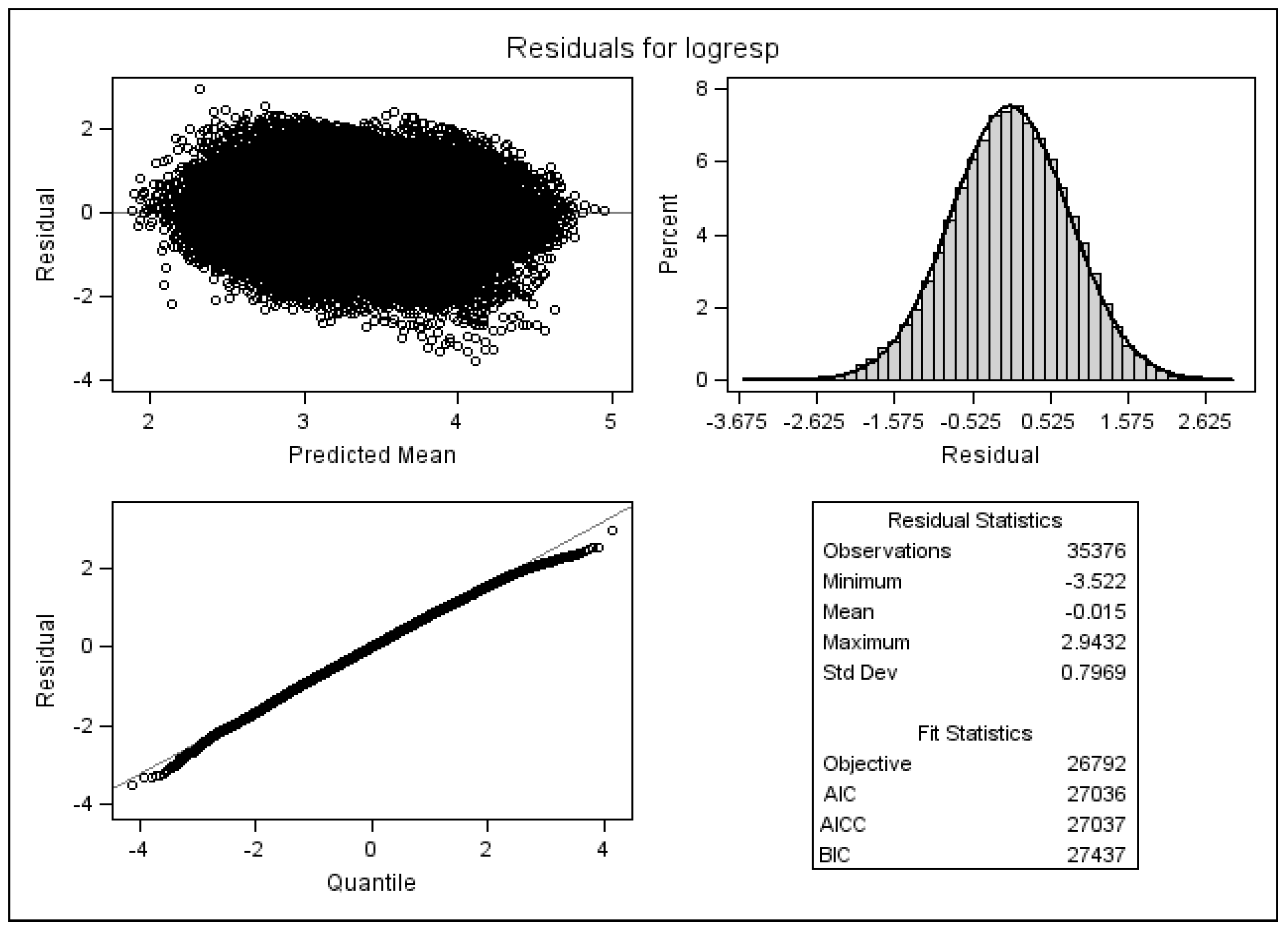
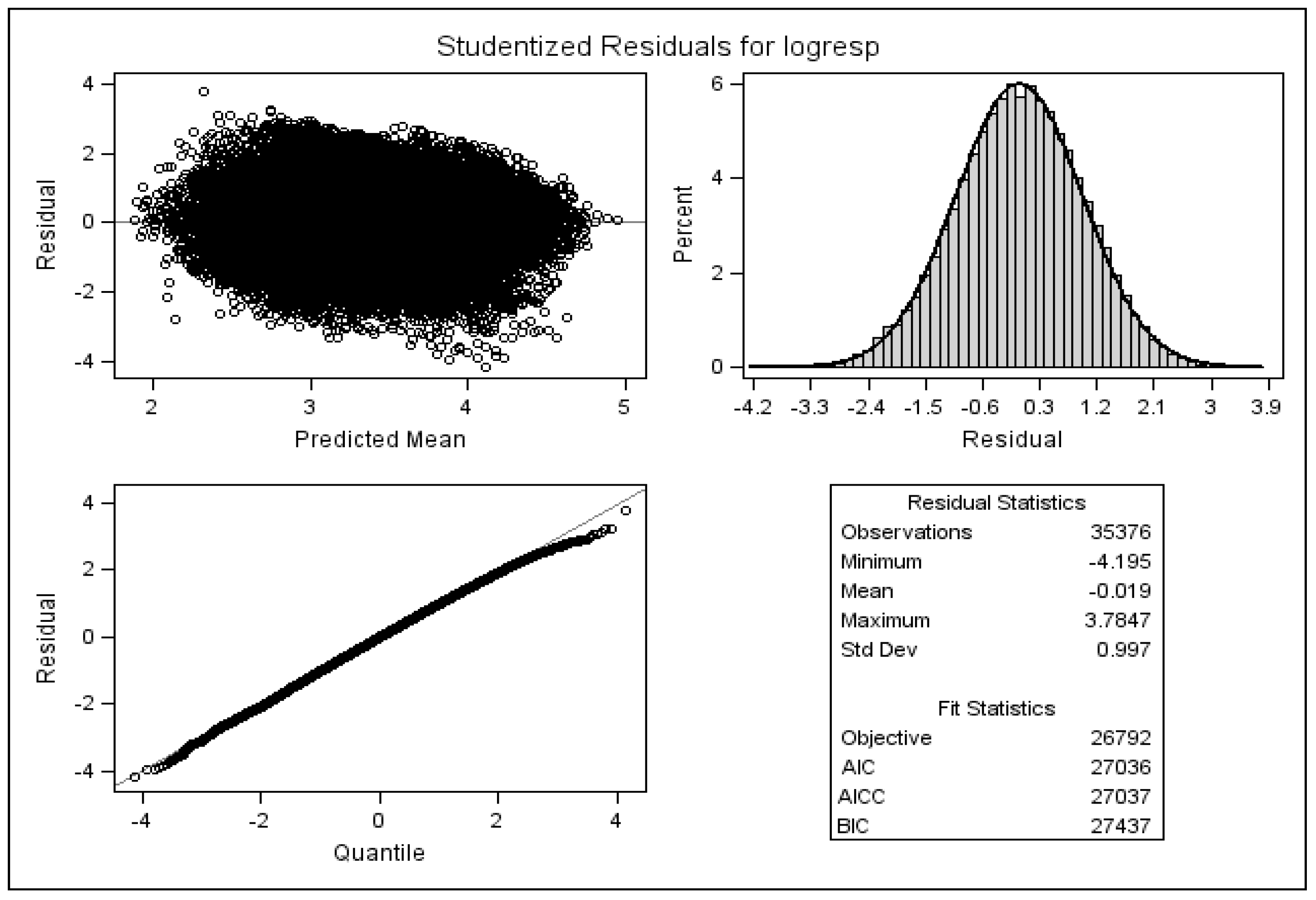
4.3. Checking Model Assumptions

Figure 3 is the result of a diagnostic analysis, and it shows the standardised residuals of the full multivariate mixed linear model with AR(1) variance-covariance structure. It is clear that most of the standardised residuals have an absolute value of less than 2.0, which could have easily occurred by chance. It is expected that 5% of standardised residuals should be greater than 2.0 in absolute value according to expected standard normal distribution of the standardised residuals. Hence, Figure 3 does not show any serious diversion from normality for residuals of the implemented multivariate linear mixed model given by Model (6).
Figure 4 may potentially detect influential observations. Here, residuals are presented similar to the standardised residuals except that they are calculated after deleting the ith observation. In other words, the figure shows the difference between the observed response values and the predicted response values excluding the ith observation from the regression.
The comparison between the two figures does not suggest any major influential observation that if removed has significant effect on parameter estimates.

5. Discussions

Our results demonstrated that the affected side is significantly different in terms of muscle activity than the unaffected side. This is understandable given the nature of the disease and treatment. The results of our model also demonstrate, however, several other important points.
Firstly, with respect to personal factors, only pain appears to play a significant role. Whilst no effect was observed with respect to SPADI pain in the unaffected arm, it was associated with increased muscle activity in the affected arm. This phenomenon has been previously reported and is hypothesised to be a functional adaptation in order to limit movements of the painful muscle, hence reduce overall pain [15].
Hand dominance had no significant effect on muscle activity, regardless of the operated side correlating well with other studies demonstrating either no difference in female subjects [16] or only significance in a few movements, primarily the flexion [17,18,19]. Still independent of the operative side, there is also greater activation and activity in shoulder muscles during abduction of the arm compared to adduction, with the magnitude of activity being proportional to the extent of abduction. The significant difference between abduction and adduction as displayed by the model is naturally expected due to the nature of gravity assisting adduction and thus activating of fewer muscle spindles. We hypothesise that if adduction was performed against resistance such as to simulate the force of gravity which muscles experience during abduction, then activity would be similar. The significance of these findings between healthy volunteers and breast cancer survivors should be assessed in a case-control study.
With respect to clinical factors, previous work had already demonstrated that surgical breast cancer treatment resulted in shoulder morbidity [10], with recent work also showing mastectomy causing greater degrees than WLE [20]. This study of WLE, controlling for chemotherapy and radiotherapy effects, also demonstrated a significant adverse effect on all muscles independent of the affected side, similar to previously reported results.
The effects of various variables on muscles are summarized in Table 4. The effect of time since surgery on muscle activity was not significant per muscle, although, overall, its effect was significant. The effect of chemotherapy similarly shows significant reduction in overall muscle activity on the ipsilateral side, although, individually, the difference was only significant for the serratus anterior. Chemotherapy induced peripheral neuropathy (CIPN) is a well-known complication of adjuvant therapy, and its incidence is closely linked to the agents used. Although primarily a sensory neuropathy, motor complications have also been reported [21] with greater impact on quality of life [22]. Taxanes, prolific agents used in breast cancer treatment [23], have been implicated in reduced compound muscle action potentials and myopathy, although less frequently than their sensory effects [24]. With respect to the observed difference on the contralateral side with serratus anterior, we could hypothesise that, due to the length of the long thoracic nerve, it may be more at risk from the neurotoxic effects of the chemotherapy agents. This is not without precedence, as the sural nerve, another similarly long nerve, has been reported as being particularly at risk of CIPN with platinum and paclitaxel based compounds [25].
With respect to rehabilitation, our model indicates that pain, physiotherapy and exercise result in an overall significant increase in muscle activity on the affected shoulder (Table 5). Specifically, exercise results in independent increases for the pectoralis major, upper trapezius and serratus anterior whilst physiotherapy results in similar findings for the latter two aforementioned muscles. With respect to the affected side, only our findings are summarised in Table 5.
Physiotherapy, either currently or at any point, was shown to improve muscle activity on the affected shoulder significantly. Multiple previous studies have demonstrated the beneficial effect of physiotherapy on shoulder function; maintenance of movement ranges, reduced pain, improved muscle strength and overall better quality of life are all reported [1,26,27].

6. Conclusions

Shoulder EMG activity and, therefore, muscle activation depends on both clinical and personal variables. Clinically the use of chemotherapy, WLE and time since surgery all decrease muscle activation, whilst physiotherapy increases it. Pain, a personal subjective influence, serves to increase muscle activation and is likely to limit movement and prevent further pain. This work builds on previously published data and offers new insights into the variables that affect shoulder morbidity with an effective model. This study further adds to the understanding of modelling multi-dimensional data and illustrates the risk of ignoring potential associations in the data structure. Several modelling approaches were discussed and compared, and the more suitable analysis method was identified and applied to the data.

Acknowledgments

This work is dedicated to Marek Molas who was part of the research team but sadly passed away in 2013. We would like to also acknowledge the reviewers for their very clear and succinct commentary on our paper. We were very appreciative of their obviously informed and expert input and were delighted to be able to make the adjustments they recommended.

Author Contributions

Gholamreza Oskrochi was responsible for the original idea and its development, statistical analysis, manuscript writing and editing. Emmanuel Lesaffre handled supervision and statistical analysis. Youssof Oskrochi was responsible for clinical analysis, manuscript writing, and editing. Delva Shamley handled data collection, clinical analysis, and manuscript editing

Conflicts of Interest

The authors declare no conflict of interest.

References

  1. Box, R.C.; Reul-Hirche, H.M.; Bullock-Saxton, J.E.; Furnival, C.M. Shoulder movement after breast cancer surgery: Results of a randomised controlled study of postoperative physiotherapy. Breast Cancer Res. Treat. 2002, 75, 35–50. [Google Scholar] [CrossRef] [PubMed]
  2. Rietman, J.S.; Dijkstra, P.U.; Hoekstra, H.J.; Eisma, W.H.; Szabo, B.G.; Groothoff, J.W.; Geertzen, J.H.B. Late morbidity after treatment of breast cancer in relation to daily activities and quality of life: A systematic review. Eur. J. Surg. Oncol. 2003, 29, 229–238. [Google Scholar] [CrossRef] [PubMed]
  3. Gosselink, R.; Rouffaer, L.; Vanhelden, P.; Piot, W.; Troosters, T.; Christiaens, M.-R. Recovery of upper limb function after axillary dissection. J. Surg. Oncol. 2003, 83, 204–211. [Google Scholar] [CrossRef] [PubMed]
  4. Rezvani, M.; Hopewell, J.W.; Robbins, M.E. Initiation of non-neoplastic late effects: The role of endothelium and connective tissue. Stem. Cells Dayt. Ohio 1995, 13 (Suppl. 1), 248–256. [Google Scholar] [CrossRef] [PubMed]
  5. Wedgwood, K.R.; Benson, E.A. Non-tumour morbidity and mortality after modified radical mastectomy. Ann. R. Coll. Surg. Engl. 1992, 74, 314–317. [Google Scholar] [PubMed]
  6. Soulen, R.L.; Romero, J.A.; Chuba, P.J.; Evelhoch, J.L.; Simpson, R.E.; Forman, J.D. Musculoskeletal complications of neutron therapy for prostate cancer. Radiat. Oncol. Investig. 1997, 5, 81–91. [Google Scholar] [CrossRef]
  7. Ryttov, N.; Blichert-Toft, M.; Madsen, E.L.; Weber, J. Influence of adjuvant irradiation on shoulder joint function after mastectomy for breast carcinoma. Acta Radiol. Oncol. 1983, 22, 29–33. [Google Scholar] [CrossRef] [PubMed]
  8. Bentzen, S.M.; Overgaard, M.; Thames, H.D. Fractionation sensitivity of a functional endpoint: Impaired shoulder movement after post-mastectomy radiotherapy. Int. J. Radiat. Oncol. Biol. Phys. 1989, 17, 531–537. [Google Scholar] [CrossRef]
  9. Aitken, R.J.; Gaze, M.N.; Rodger, A.; Chetty, U.; Forrest, A.P. Arm morbidity within a trial of mastectomy and either nodal sample with selective radiotherapy or axillary clearance. Br. J. Surg. 1989, 76, 568–571. [Google Scholar] [CrossRef] [PubMed]
  10. Shamley, D.R.; Srinanaganathan, R.; Weatherall, R.; Oskrochi, R.; Watson, M.; Ostlere, S.; Sugden, E. Changes in shoulder muscle size and activity following treatment for breast cancer. Breast Cancer Res. Treat. 2007, 106, 19–27. [Google Scholar] [CrossRef] [PubMed]
  11. Roach, K.E.; Budiman-Mak, E.; Songsiridej, N.; Lertratanakul, Y. Development of a shoulder pain and disability index. Arthritis Care Res. Off. J. Arthritis Health Prof. Assoc. 1991, 4, 143–149. [Google Scholar] [CrossRef]
  12. Coull, B.A.; Agresti, A. Random effects modeling of multiple binomial responses using the multivariate binomial logit-normal distribution. Biometrics 2000, 56, 73–80. [Google Scholar] [CrossRef] [PubMed]
  13. Fieuws, S.; Verbeke, G.; Molenberghs, G. Random-effects models for multivariate repeated measures. Stat. Methods Med. Res. 2007, 16, 387–397. [Google Scholar] [CrossRef] [PubMed]
  14. SAS Institute. SAS; SAS Inst.: Cary, NC, USA, 2011. [Google Scholar]
  15. Graven-Nielsen, T.; Svensson, P.; Arendt-Nielsen, L. Effects of experimental muscle pain on muscle activity and co-ordination during static and dynamic motor function. Electroencephalogr. Clin. Neurophysiol. Mot. Control. 1997, 105, 156–164. [Google Scholar] [CrossRef]
  16. Cahalan, T.D.; Johnson, M.E.; Chao, E.Y. Shoulder strength analysis using the Cybex II isokinetic dynamometer. Clin. Orthop. 1991, 271, 249–257. [Google Scholar] [CrossRef] [PubMed]
  17. Hughes, R.E.; Johnson, M.E.; O’Driscoll, S.W.; An, K.N. Age-related changes in normal isometric shoulder strength. Am. J. Sports Med. 1999, 27, 651–657. [Google Scholar] [PubMed]
  18. Murray, M.P.; Gore, D.R.; Gardner, G.M.; Mollinger, L.A. Shoulder motion and muscle strength of normal men and women in two age groups. Clin. Orthop. 1985, 192, 268–273. [Google Scholar] [CrossRef] [PubMed]
  19. Otis, J.C.; Warren, R.F.; Backus, S.I.; Santner, T.J.; Mabrey, J.D. Torque production in the shoulder of the normal young adult male. The interaction of function, dominance, joint angle, and angular velocity. Am. J. Sports Med. 1990, 18, 119–123. [Google Scholar] [CrossRef] [PubMed]
  20. Shamley, D.; Lascurain-Aguirrebeña, I.; Oskrochi, R.; Srinaganathan, R. Shoulder morbidity after treatment for breast cancer is bilateral and greater after mastectomy. Acta Oncol. 2012, 51, 1045–1053. [Google Scholar] [CrossRef] [PubMed]
  21. Perry, M.C. Perry’s The Chemotherapy Source Book, 5th Revised ed.; Lippincott Williams and Wilkins: Philadelphia, PA, USA, 2012; p. 848. [Google Scholar]
  22. Binda, D.; Vanhoutte, E.K.; Cavaletti, G.; Cornblath, D.R.; Postma, T.J.; Frigeni, B.; Alberti, P.; Bruna, J.; Velasco, R.; Argyriou, A.A.; et al. Rasch-built Overall Disability Scale for patients with chemotherapy-induced peripheral neuropathy (CIPN-R-ODS). Eur. J. Cancer 2013, 49, 2910–2918. [Google Scholar] [CrossRef] [PubMed]
  23. Giordano, S.H.; Lin, Y.-L.; Kuo, Y.F.; Hortobagyi, G.N.; Goodwin, J.S. Decline in the use of anthracyclines for breast cancer. J. Clin. Oncol. Off. J. Am. Soc. Clin. Oncol. 2012, 30, 2232–2239. [Google Scholar] [CrossRef] [PubMed]
  24. Argyriou, A.A.; Kyritsis, A.P.; Makatsoris, T.; Kalofonos, H.P. Chemotherapy-induced peripheral neuropathy in adults: A comprehensive update of the literature. Cancer Manag. Res. 2014, 6, 135–147. [Google Scholar] [CrossRef] [PubMed]
  25. Argyriou, A.A.; Polychronopoulos, P.; Koutras, A.; Iconomou, G.; Iconomou, A.; Kalofonos, H.P.; Chroni, E. Peripheral neuropathy induced by administration of cisplatin- and paclitaxel-based chemotherapy. Could it be predicted? Support. Care Cancer 2005, 13, 647–651. [Google Scholar] [CrossRef] [PubMed]
  26. Gomide, L.B.; Matheus, J.P.C.; Candido dos Reis, F.J. Morbidity after breast cancer treatment and physiotherapeutic performance. Int. J. Clin. Pract. 2007, 61, 972–982. [Google Scholar] [CrossRef] [PubMed]
  27. Beurskens, C.H.; Uden, C.J.; van Strobbe, L.J.; Oostendorp, R.A.; Wobbes, T. The efficacy of physiotherapy upon shoulder function following axillary dissection in breast cancer, a randomized controlled study. BMC Cancer 2007, 7, 166. [Google Scholar] [CrossRef] [PubMed]
Figure 1. Frequency distribution for pain and disability (N = 202).
Ijerph 13 00274 g001
Figure 2. Typical observations from a patient’s arm movement.
Ijerph 13 00274 g002
Figure 3. Standardised residuals of the multivariate mixed linear model.
Ijerph 13 00274 g003
Figure 4. Standardised residuals with the ith observation removed.
Ijerph 13 00274 g004
Table 1. Results of the analysis using a multivariate normal distribution with autoregressive of order one (AR(1)) structure for Σk.
Clinical MeasuresParameter Estimatesp-Value
Ln(PM)Ln(UT)Ln(SA)Ln(RH)Overall Effect
Intercept2.98123.072.79052.1944<0.0001
Humeral elevation × 1000.50530.58680.84030.7097<0.0001
Move up0.11750.2870.12860.1746<.0001
Affected−0.18970.028960.4124−0.09950.1855
Handleft0.29080.16770.19590.1686<0.0001
Dominant0.073380.12330.078650.077440.4888
Agex100−0.000370.4736−0.5030.48880.0386
Duration × 100−0.010.00240.01190.01250.0286
Spadipai × 100−0.146−0.156−0.061−0.0950.0005
Spadidis × 1000.05910.07260.0707−0.0420.1284
Wle−0.2556−0.1913−0.2544−0.2267<0.0001
Chemocat−0.1107−0.0803−0.4658−0.1233<0.0001
Inter_humeral elevation * × 1000.01610.08370.0190.11940.2037
Inter_dominant *−0.2211−0.1212−0.3452−0.2614<0.0001
Inter_duration * × 1000.0037−0.004−0.021−0.0070.1410
Inter_spadipai * × 1000.10830.1046−0.0510.09480.0239
Exercise 6 m **−0.00577−0.0644−0.059410.016780.0349
Exercise now **0.068080.055040.086550.01458<0.0001
Physio now **0.3070.55230.15830.30820.1207
Physio ever **−0.01245−0.06630.1235−0.14390.4765
ρ0.890.920.900.91N/A
σ20.620.730.610.63N/A
–2 log likelihood of the model with interaction terms
–2 log likelihood of the model without interaction
29,181.2
29,898.4
* Interaction with affected side, ** Only on affected side.
Table 2. Result of the analysis using univariate Linear Mixed Model with AR(1) structure for Σk.
Clinical MeasuresParameter Estimatesp-Value
Ln(PM)Ln(UT)Ln(SA)Ln(RH)Overall Effect
Intercept3.06313.14682.83392.2456<0.0001
Humeral elevation × 1000.49990.57330.82880.6937<0.0001
Move up0.13570.29850.1360.2008<0.0001
Affected–0.061450.075790.55620.14570.0113
Handleft0.28250.1540.1990.1627<0.0001
Dominant0.073790.10440.073930.077660.6258
Agex100–0.0880.4317−0.5420.43610.5757
Duration × 100–0.0110.00220.01130.01190.2323
Spadipai × 100–0.147–0.155–0.063–0.1040.1053
Spadidis × 1000.05320.06760.0589–0.0350.8000
Wle–0.2831−0.199−0.2626–0.2262<0.0001
Chemocat–0.1611–0.1034–0.4759−0.13650.0231
Inter_humeral elevation * × 1000.00290.07740.00470.10910.1146
Inter_dominant *–0.2326–0.1192–0.3466–0.27460.0654
Inter_duration * × 1000.0022–0.007–0.024−0.011<0.0001
Inter_spadipai * × 1000.10420.1195−0.0230.1221<0.0001
Exercise 6 m **–0.02449–0.04658–0.059680.0038090.0142
Exercise now **0.069140.019970.05287–0.03337<0.0001
Physio now **0.012010.47880.11070.59510.0060
Physio ever **0.002231–0.16510.01827–0.24810.0163
ρ0.790.850.820.80N/A
σ20.310.370.330.29N/A
Random effect variance0.330.380.280.35N/A
–2 log likelihood of the model with interaction terms
–2 log likelihood of the model without interaction
27,645.1
27,862.8
* Interaction with affected side, ** Only on affected side.
Table 3. Result of the analysis using multivariate Linear Mixed Model with AR(1) structure for Σk.
Clinical MeasuresParameter Estimatesp-Value
Ln(PM)Ln(UT)Ln(SA)Ln(RH)Overall Effect
Intercept3.06623.15662.84132.2559<0.0001
Humeral elevation × 1000.50120.57610.83140.694<0.0001
Move_up0.13570.2990.13620.201<0.0001
Affected–0.036530.12710.58370.13680.0054
Handleft0.28370.15370.19830.1608<0.0001
Dominant0.073910.10570.075860.076870.8796
Age × 100–0.0930.4211–0.5490.42920.1775
Duration × 100–0.0110.00210.01120.01190.0227
Spadipai × 100–0.148–0.155–0.064–0.1030.4220
Spadidis × 1000.05330.06790.0604–0.0370.5270
Wle–0.283–0.2028–0.2668–0.23060.0272
Chemocat–0.1598–0.1078–0.4812–0.14460.0205
Inter_humeral elevation * × 1005.07E-040.07370.00360.10960.1219
Inter_dominant *–0.2347–0.1247–0.3505–0.27640.2958
Inter_duration * × 1000.0015−0.008–0.024–0.011<0.0001
Inter_spadipai * × 1000.11150.1211–0.0230.1212<0.0001
Exercise 6 m **–0.02175–0.05333–0.062860.0063820.0049
Exercise now **0.058790.017270.05089–0.03237<0.0001
Physio now **0.085530.49070.11580.50010.0177
Physio ever **–0.0337–0.16690.02314–0.24840.0121
ρ0.790.850.820.80N/A
σ20.310.370.330.29N/A
Random effect variance0.330.380.290.35N/A
–2 log likelihood of the model with interaction terms
–2 log likelihood of the model without interaction
26,833.2
27,522.8
* Interaction with affected side, ** Only on affected side.
Table 4. Factors that significantly affect muscle activity, irrespective of affected shoulder.
VariableEMG Effect (Significant)
Pectoralis MajorUpper TrapeziusSerratus AnteriorRhomboid
Humeral Elevation↑ *↑ *↑ *↑ *
Left vs. Right Shoulder↑ *↑ *↑ *↑ *
Time Since Surgery (DURATION)
WLE (vs. Other Treatment Modalities)↓ *↓ *↓ *↓ *
Chemotherapy↓ *
* indicates, significant at the 5% level.
Table 5. Significant factors affecting muscle activity on the affected shoulder as compared to the unaffected shoulder.
VariableEMG Effect (Significant)
Pectoralis MajorUpper TrapeziusSerratus AnteriorRhomboid
Increasing Pain Score↑ *↑ *↑ *
Exercise Last 6 Months↓ *↓ *
Current Exercise↑ *↑ *↓ *
Current Physiotherapy↑ *↑ *
Time Since Surgery↓ *↓ *

