Review

Assessment of Infantile Mineral Imbalances in Autism Spectrum Disorders (ASDs)

La Belle Vie Research Laboratory, 8-4 Nihonbashi-Tomizawacho, Chuo-ku, Tokyo 103-0006, Japan
*
Author to whom correspondence should be addressed.
Int. J. Environ. Res. Public Health 2013, 10(11), 6027-6043; https://doi.org/10.3390/ijerph10116027
Submission received: 26 August 2013 / Revised: 31 October 2013 / Accepted: 6 November 2013 / Published: 11 November 2013
(This article belongs to the Special Issue The Environment Risk of Autism)

Abstract

:
The interactions between genes and the environment are now regarded as the most probable explanation for autism. In this review, we summarize the results of a metallomics study in which scalp hair concentrations of 26 trace elements were examined for 1,967 autistic children (1,553 males and 414 females aged 0–15 years-old), and discuss recent advances in our understanding of epigenetic roles of infantile mineral imbalances in the pathogenesis of autism. In the 1,967 subjects, 584 (29.7%) and 347 (17.6%) were found deficient in zinc and magnesium, respectively, and the incidence rate of zinc deficiency was estimated at 43.5% in male and 52.5% in female infantile subjects aged 0–3 years-old. In contrast, 339 (17.2%), 168 (8.5%) and 94 (4.8%) individuals were found to suffer from high burdens of aluminum, cadmium and lead, respectively, and 2.8% or less from mercury and arsenic. High toxic metal burdens were more frequently observed in the infants aged 0–3 years-old, whose incidence rates were 20.6%, 12.1%, 7.5%, 3.2% and 2.3% for aluminum, cadmium, lead, arsenic and mercury, respectively. These findings suggest that infantile zinc- and magnesium-deficiency and/or toxic metal burdens may be critical and induce epigenetic alterations in the genes and genetic regulation mechanisms of neurodevelopment in the autistic children, and demonstrate that a time factor “infantile window” is also critical for neurodevelopment and probably for therapy. Thus, early metallomics analysis may lead to early screening/estimation and treatment/prevention for the autistic neurodevelopment disorders.

1. Introduction

ASDs are a group of neural development disorders characterized by impairments in social interaction and communication, and by the presence of restricted and repetitive behaviours [1,2]. Clarification of the pathogenesis and effective treatment of autism spectrum disorders (ASDs) is one of the challenges today. ASDs continue to increase in prevalence up to 1 in 88 children [1,2,3] and are known to be highly heritable (~90%), and some related genes have been reported [4,5,6,7,8]. However, the underlying genetic determinants are still not clarified [1,9], and the interaction of heritable factors with uncertified lifestyle and environmental factors seem play a significant role in the pathogenesis. For example, organic mercury had been claimed one of environmental candidates causing autistic disorders [10,11,12], but its relationship remains to be established. Recently, epigenetic alteration of gene expression by environmental factors is considered one of key events in the pathogenesis of genetic diseases [13,14], and some toxic elements such as cadmium and arsenic have been reported to be candidate factors that induce epigenetic alterations [15,16,17,18,19] and neurodevelopmental disorders [20].
Recent great advances in high-sensitive and reliable trace element analysis method using inductively coupled plasma mass spectrometry (ICP-MS) have enabled it to be applied for forensic medical research and estimating chronic toxic metal burden and mineral deficiency in the human body [21,22]. Thus, the clinical application of reliable hair mineral analysis methods based on ICP-MS has been tried to investigate the association of some diseases/symptoms with trace bio-element kinetics including toxic metals and essential minerals [23,24,25,26,27,28].
For the last seven years, we have examined the association of toxic metal burdens with autistic disorders, and reported that some of the autistic children have suffered from high accumulation of toxic metals such as cadmium, lead or aluminium [29,30,31], and recently demonstrating the association with infantile zinc deficiency [32,33].
In this overview article in which human scalp hair concentrations of 26 trace elements have been examined for 1,967 children with autistic disorders aged 0–15 years, we demonstrate that many of the patients, especially in the infants aged 0–3 years-old, are suffering from marginal to severe zinc- and magnesium-deficiency and/or high burdens of several toxic metals such as aluminium, cadmium and lead, indicating the presence of a critical term “infantile window” in neurodevelopment and probably for therapy.

2. Mineral Disorders in Autism

2.1. Infantile Zinc Deficiency

The histogram of hair logarithmic zinc concentrations for 1,967 autistic children diagnosed by their physicians was non-symmetric with tailing in lower range, and 584 in 1,967 subjects (29.7%) were found to have a lower zinc concentration than—2 S.D. (standard deviation) level of the reference range (86.3–193 ppm; geometric mean = 129 ppm), estimated as zinc deficiency. The incidence rates of zinc deficiency in the age groups of 0–3, 4–9 and 10–15 years-old were estimated 43.5%, 28.1% and 3.3% in male and 52.5%, 28.7% and 3.5% in female, and a significant correlation of zinc concentration with age (r = 0.367, p < 0.0001) was observed (Figure 1), suggesting that infants are more liable to zinc deficiency than elder children. The minimum zinc concentration of 10.7 ppm was detected in a 2-year-old boy, corresponding to about 1/12 of the mean reference level. The zinc concentration of only one 0-year-old case (11 months-old) was 173 ppm in the normal range and seem to be a suspected case, because she was suffered from high burdens of aluminium (52.5 ppm), lead (9.1 ppm), iron (12.8 ppm) and copper (134 ppm). There was little marked gender difference in hair zinc concentration and incidence rate of zinc deficiency.
Figure 1. Relation of logarithmic zinc concentration with age in autistic children [33].
Ijerph 10 06027 g001
The association of hair logarithmic zinc concentration with age in autistic children (N = 1,967) is shown. Each point represents the corresponding age and logarithmic zinc concentration (ng/g hair: ppb) of the individual child. The dotted horizontal lines represent the ±2 S.D. levels of the reference range (86.3–193 ppm) of hair zinc concentrations. A significant relation of the zinc concentration with age (r = 0.367, p < 0.0001) in the autistic children is shown [33].

2.2. Infantile Magnesium/Calcium Deficiency

Following to zinc deficiency, magnesium and calcium deficiency was observed in 347 (17.6%) and 114 (5.8%) individuals in the autistic children, and for the other essential metals such as iron, chromium, manganese, copper and cobalt, their incidence rates of deficiency were 2.0% or less (Table 1). The incidence rates of magnesium deficiency in the age groups of 0–3, 4–9 and 10–15 years-old were 27.0%, 17.1% and 4.2% in male and 22.9%, 12.7% and 4.3% in female subjects, and a significant correlation of magnesium concentration with age (r = 0.362, p < 0.0001) was observed, suggesting that infants are also liable to magnesium deficiency than elder children. The minimal magnesium concentration of 3.88 ppm was detected in a 2-year-old girl, corresponding to almost 1/10 of the mean reference level (39.5 ppm). Considerable calcium deficiency rate was observed only in lower age groups less than 10 years-old.
Table 1. Prevalence of mineral deficiency in autistic children [33].
MineralNumber of Cases with DeficiencyRate (%) of Deficiency
Zn58429.7
Mg34717.6
Ca1145.8
Co402.0
Fe170.9
Cr120.6
Mn40.2
Cu40.2
The number and incidence rate of individuals with mineral deficiency (lower than −2 S.D.) in 1,967 autistic children (1,553 males and 414 females) are shown in the table [33].

2.3. Toxic Metal Burdens

In contrast to essential metals, high body burdens of some toxic metals such as aluminium, cadmium and lead of over their +2 S.D. levels were observed in 339 (17.2%), 168 (8.5%) and 94 (4.8%) individuals, respectively, and their incidence rates of high burden were higher than that of mercury and arsenic (2.8% and 2.6%) (Table 2).
Table 2. Prevalence of high toxic metal burden and the maximum level in autistic children [33].
Toxic MetalNumber of Cases with High BurdenRate (%) of High BurdenMaximum (ppm)Ratio to Reference
Al33917.279.421.1
Cd1688.55.5782.0
Pb944.824.957.4
Hg562.836.39.3
As522.61.733.5
The number and incidence rate of individuals with high toxic metal burden (higher than +2 S.D.) in 1,967 autistic children (1,553 males and 414 females) and the maximum concentration are tabled [33].
High toxic metal burdens were more frequently observed in the infants aged 0–3 years-old: that is, the incidence rate was 20.6%, 12.1%, 7.5%, 3.2% and 2.3% for aluminium, cadmium, lead, arsenic and mercury. The detected maximal concentration of aluminium, cadmium, lead, mercury and arsenic was 79.4 ppm, 5.47 ppm, 24.9 ppm, 36.3 ppm and 1.7 ppm, respectively, corresponding to 21-, 782-, 57-, 9- and 33-fold of each mean reference level.
A high significant inverse relationship between zinc and lead concentrations (r = −0.339, p < 0.0001; Figure 2), and also aluminium (r = −0.247) and cadmium (r = −0.198) concentrations, was observed, suggesting that these toxic metal burdens associate with infantile zinc deficiency.
Figure 2. Inverse relation of zinc and lead concentration in autistic children.
Ijerph 10 06027 g002
The inverse relationship between hair logarithmic zinc and lead concentration in autistic children (N = 1,967) is shown. Each point represents the corresponding logarithmic zinc and lead concentration of the individual child. A high significant inverse relationship between zinc and lead concentrations (r = −0.339, p < 0.0001) in the autistic children is shown.

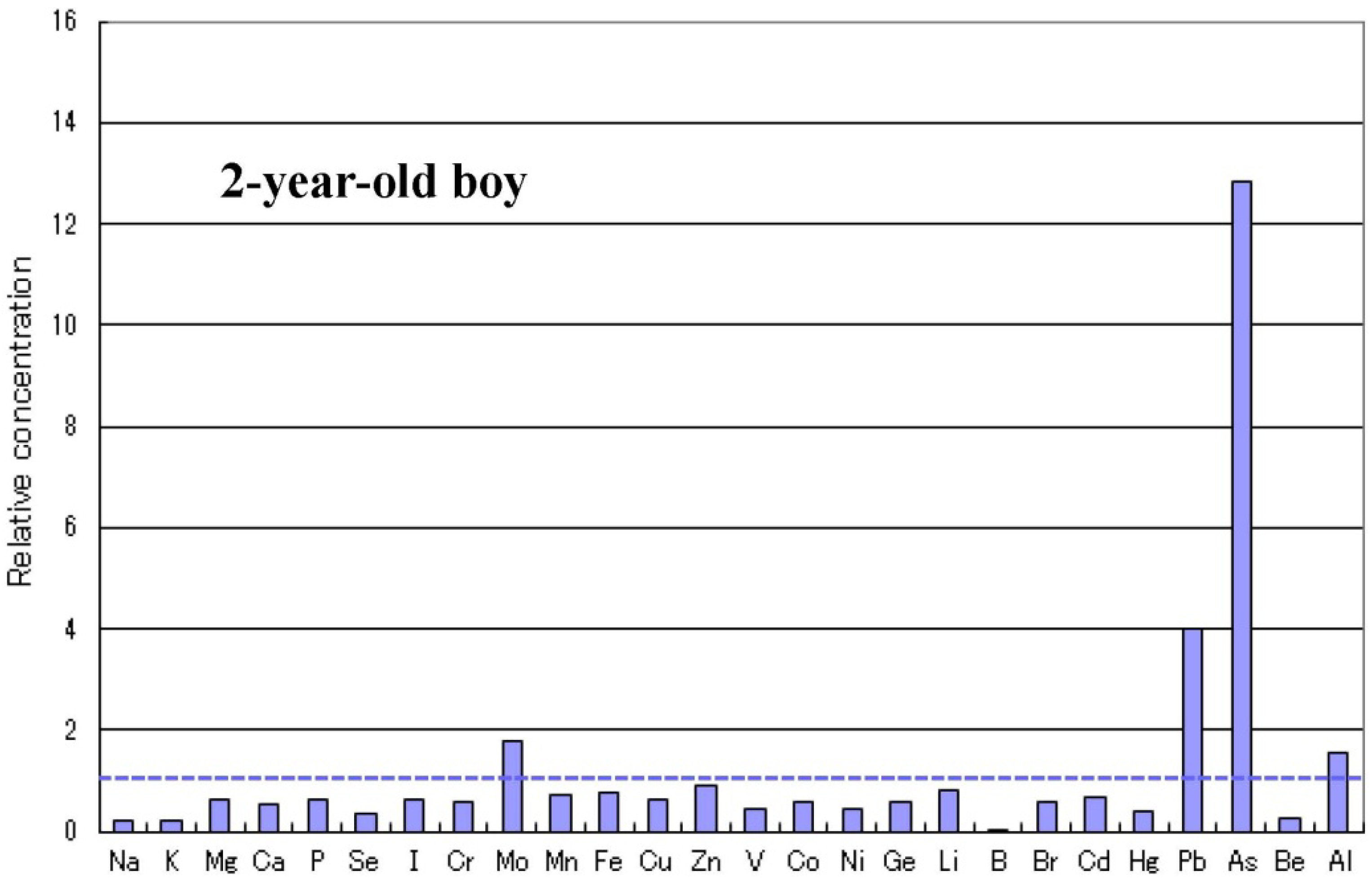
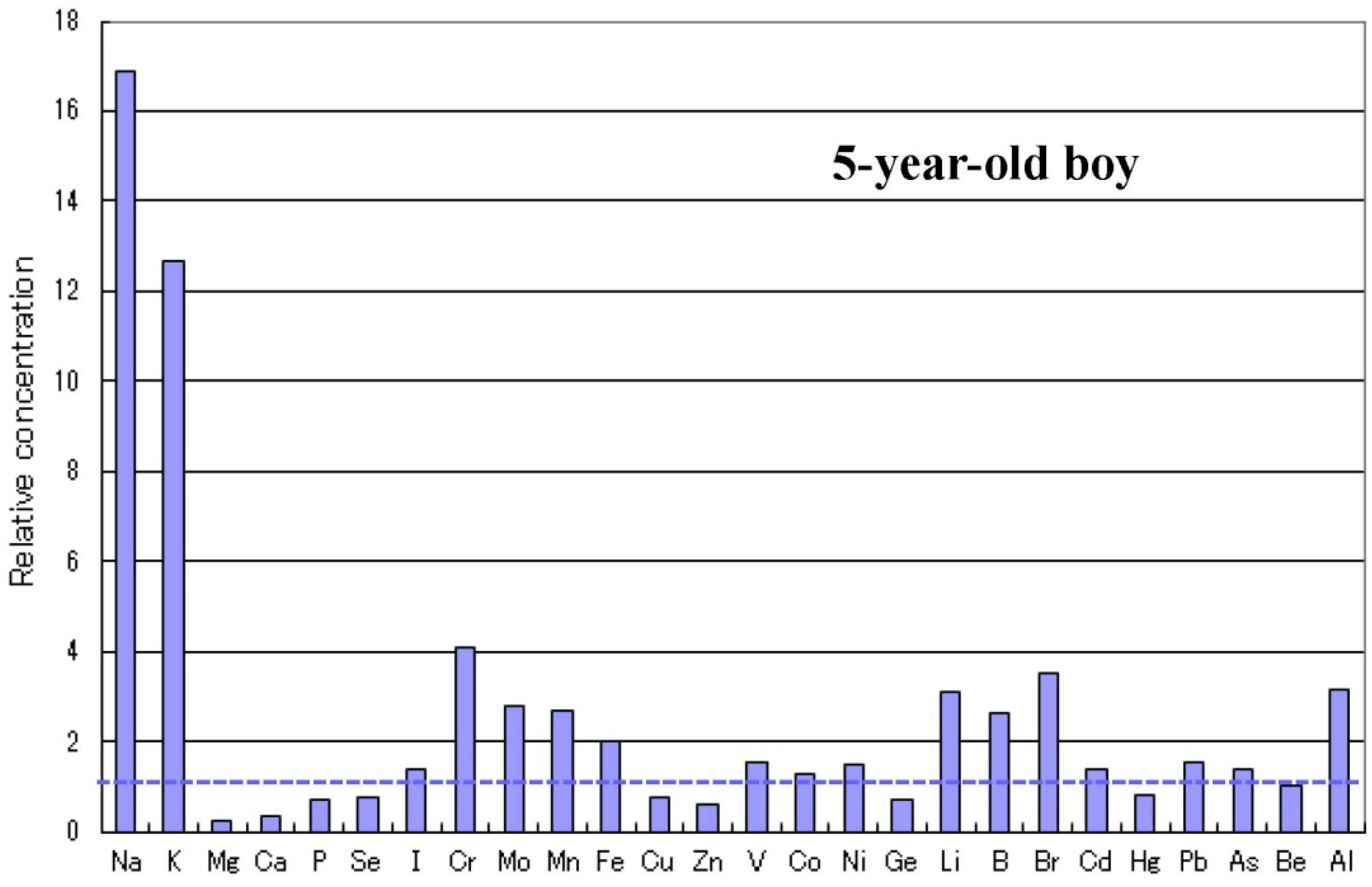
2.4. Metallomics Profiles in Autistic Infants

There are some sub-types observed in the metallomics profiles characteristic in autistic children. Figure 3 shows a representative autistic profile in a 1-year-old boy suffering from severe zinc- and magnesium-deficiency and simultaneous high burdens of cadmium and lead. The other autistic metallomics profiles with high burdens of aluminium, mercury or arsenic are shown in Figure 4, Figure 5 and Figure 6. Figure 7 shows a unique profile with high sodium and potassium concentrations, a characteristic profile detectable in hair specimens. It remains to be clarified which type of metallomcs profiles corresponds to which type of autism spectrum disorders.
Figure 3. Metallomics profile of an autistic child with high cadmium and lead burdens [33].
Ijerph 10 06027 g003
A representative metallomics profile of a 1-year-old boy with autism is shown, exhibiting severe zinc- and magnesium-deficiency and simultaneous high burdens with cadmium (107 ppb) and lead (8.11 ppm). Each bar represents the relative concentration of the respective trace element in his scalp hair specimen. The dotted horizontal line at 1.0 represents the reference control level of each trace element [33].
Figure 4. Metallomics profile of an autistic child with high aluminium burden.
Ijerph 10 06027 g004
A representative metallomics profile of 4-year-old girl with autism is shown, exhibiting simultaneous high burdens with aluminium (53.7 ppm), manganese (702 ppb), iron (37.0 ppm) and lithium (12.2 ppb). Each bar represents the relative concentration of the respective trace element in her scalp hair specimen. The dotted horizontal line at 1.0 represents the reference control level of each trace element.
Figure 5. Metallomics profile of an autistic child with high mercury burden.
Ijerph 10 06027 g005
A metallomics profile of a 3-year-old boy with autism is shown, exhibiting a high burden with mercury of over 10-fold level (40.1 ppm). Each bar represents the relative concentration of the respective trace element in his scalp hair specimen. The dotted horizontal line at 1.0 represents the reference control level of each trace element.
Figure 6. Metallomics profile of an autistic child with high arsenic burden.
Ijerph 10 06027 g006
A metallomics profile of a 2-year-old boy with autism is shown, exhibiting a high burden with arsenic of over 12-fold level (655 ppb). Each bar represents the relative concentration of the respective trace element in his scalp hair specimen. The dotted horizontal line at 1.0 represents the reference control level of each trace element.
Figure 7. Metallome profile of an autistic child with high sodium and potassium levels.
Ijerph 10 06027 g007
A metallome profile of 5-year-old boy with autism is shown, exhibiting simultaneous high levels of sodium (335 ppm) and potassium (210 ppm). Each bar represents the relative concentration of the respective trace element in his scalp hair specimen. The dotted horizontal line at 1.0 represents the reference control level of each trace element.

2.5. Infantile Time Window in Neurodevelopment and for Therapy

The age at final diagnosis of autism spectrum disorders ranges from 3 to 6 years, although most cases of autism are diagnosed by the age of three and as early as 14 months [34]. In facts, zinc deficiency was detected in many of the infantile patients in the first 3 years of life (Figure 1), and high toxic metal burdens were also detected in the autistic subjects, especially in the younger children (Table 2). Thus, for treatment/prevention of autism spectrum disorders, its early screening and estimation is necessary and it is desirable to early check any metabolic and/or mineral disorders for the infants and children with autistic symptoms, though there are serious limitations of diagnosis of the younger children. It should be considered that the pathogenesis of neurodevelopment disorders might start in prenatal phase and be progressive within the time window for diagnosis.

2.6. Autism-Related Genes and Epigenetic Alteration by Mineral Disorders

Zinc is a structural component of zinc-finger proteins and a transcriptional regulator, and influences some candidate genes reported to be associated with the development of autism, such as MTF1 (metal-responsive transcription factor 1), metallothionein, ZnT5 (zinc transporter 5), COMMD1 (COMM domain-containing protein 1), ERK1 (extracellular signal-regulated kinase 1), TrkB (tyrosine-related kinase B), and ProSAP/Shank (proline-rich synapse-associated protein/SH and multiple ankyrin repeat domains) that themselves are involved in zinc signalling and homeostasis [35,36,37,38,39,40]. Thus, zinc deficiency observed in the autistic subjects (Figure 1) might induce critical epigenetic alterations to provide a central mechanism of gene/environment interaction to interfere with neuronal maturation during early development [32,38,39].
In addition, high toxic metal burdens detected in the autistic patients (Table 2) might contribute to the mechanism of gene/environment interaction, because cadmium and arsenic have been reported to be candidate factors that induce epigenetic alterations [15,16,17,18,19] and neurodevelopmental disorders [20].

3. Discussion

In this metallomics study of human scalp hair concentrations of 26 trace elements for 1,967 children with autistic disorders aged 0–15 years, we demonstrate that many of the patients, especially in the infants aged 0–3 year-old, are suffering from marginal to severe zinc- and magnesium-deficiency and/or high burdens of several toxic metals such as aluminium, cadmium and lead.
Zinc is well-accepted as essential trace element that plays important roles in nucleic acid/protein synthesis, cell replication, tissue growth and repair, especially in pregnant women and infants. In fact, zinc ions function as the active centers in more than 300 kinds of enzymes, and about 10% in the total gene-coded proteins have been known to have zinc-finger sequences [38,39,40,41,42], emphasizing the physiological importance of this trace element. In brain, especially in the hippocampus, zinc is co-stored with glutamate in pre-synaptic vesicles in the excitatory neuron terminal, is released from them and controls the activity of excitatory glutamate receptors on the post-synaptic excitable membrane [43,44]. Thus, zinc deficiency is known associated with not only various pathological conditions, including dysgeusia, delayed wound healing, impaired immunity and retarded growth, but also neurodegenerative diseases and neurodevelopment disorders [45,46,47,48,49].
Recently we reported that many infants with autistic disorders are suffering from marginal to severe zinc deficiency, suggesting considerable relationship of infantile zinc deficiency with autism [32]. Furthermore, we have determined scalp hair concentrations of 26 trace elements for 1,967 subjects with autism spectrum disorders and demonstrated that infantile autistic children are liable to deficiency in magnesium and calcium next to zinc, but not in the other essential metals (Table 1) [33]. These findings suggest that autistic infants and probably infants generally have a characteristic liability to zinc- and magnesium-deficiency, because larger amounts of the essential metals (per kg body weight) are needed for the development and growth.
There are numerous studies with the same theme reporting nutritional status and mineral deficiencies in autistic children [50,51,52,53,54]. However, the conclusions of their studies, in which the restricted age (over 4-years-old) of children and number of minerals were examined, were not consistent, and the critical environmental factors remained to be established. In our metallomics analysis study for the 1,967 autistic children aged 0–15 years-old, we were able to demonstrate not only the critical and environmental epigenetic factor (zinc- and magnesium-deficiency and high burdens of aluminium, cadmium, lead and so on) but also the presence of another critical factor, “infantile window” in neurodevelopment and probably for therapy [32,33].
Recently, Gebremedhin et al. [55] reported that compared to pregnant women aged 15–24 years, those aged 25–34 and 35–49 years had 1.57 (95% CL: 1.04–2.34) and 2.18 (95% CL: 1.25–3.63) times higher risk of zinc deficiency, respectively. Their study may demonstrate that old age pregnancy is negatively associated to zinc status, maybe suggesting that one of the origins of the high incidence rate of infantile zinc deficiency may be higher age pregnancy of their mothers. Recently, Kurita et al. [56] reported that zinc deficiency in utero induces foetal epigenetic alterations of histone modifications in metallothionein 2 promoter region having metal responsive elements in 1-day-old and 5-week-old mice, of which pregnant mother were fed low zinc diet from gestation day 8 until delivery.
Arnold et al. [57] reported that mean serum zinc level in children was significantly lower in attention-deficit/hyperactivity disorder (ADHD) group, and that serum zinc level correlated inversely with parent- and teacher-rated inattention in ADHD children. Furthermore, zinc treatment was reported significantly superior to placebo in reducing symptoms of hyperactivity, impulsivity and impaired socialization in ADHD patients [58,59]. Another preliminary human study showed that many children with ADHD have lower zinc concentration in comparison to healthy children and zinc supplement as an adjunct to methylphenidate has favourable effects in the treatment of ADHD children, pointing to the possible association of zinc deficiency and ADHD pathophysiology [60].
Kozielec et al. [61] have reported that in 116 hyperactive children with ADHD, magnesium deficiency was found in 95% of the subjects, most frequently in hair (77.6%), next in red-blood cells (58.6%) and in blood serum (33.6%). Furthermore, they reported that in the group of ADHD children given 6 months of magnesium supplementation, a significant decrease of hyperactivity and increase in hair magnesium contents has been achieved [62]. Mousain-Bosc et al. [63] also reported that 52 hyper-excitable children have low intra-erythrocyte magnesium levels with normal serum magnesium values, and that magnesium/vitamin B6 supplementation can restore the erythrocyte magnesium levels to normal and improve their abnormal behaviours. They also reported that thirty-three children with clinical symptoms of pervasive developmental disorder or autism (PDD) exhibit significantly lower red blood cell magnesium values, and that the combination therapy with magnesium/vitamin B6 for 6 months improved significantly PDD symptoms in 23/33 children (p < 0.0001) with concomitant increases in intra-erythrocyte magnesium values [64].
Recently, Ochi et al. [65] found that hair magnesium concentration, but not its serum level, was significantly (p < 0.01) inversely-associated with left ventricular hypertrophy in hemodialysis patients, suggesting that hair magnesium concentration is a useful intracellular biomarker independent of its serum level. In a preliminary metallomics study for healthy volunteers, we have observed a high significant correlation between whole blood levels and scalp hair levels of trace elements, but little relation between their serum levels and whole blood levels (unpublished observation). These findings suggest that as biomarker specimen representing mineral dynamics in human body, whole blood/erythrocyte and hair samples are superior to extra-cellular fluids such as serum or plasma for metallomics analysis, although it is necessary to consider that there is a problem of contamination of some trace-elements due to artificial hair treatment such as permanent and colouring.
Recently, dietary restriction-induced zinc deficiency has been reported to up-regulate intestinal zinc-importer (ZIP4) and induce the increase in ZIP4 protein located to the plasma membrane of enterocytes [66,67]. This adoptive response to zinc deficiency is known to lead to increasing in the risk of high-uptake of toxic metals such as cadmium and lead [68]. Thus, infants with zinc deficiency are liable to increased risk of absorbing high amount of toxic metals and retaining them in their bodies, as shown in Figure 4, which demonstrates a high significant inverse relationship between zinc and lead level. These findings suggest that the increased toxic metal burdens concomitant with zinc deficiency may also epigenetically contribute to the pathogenesis of this disorder.
Deficiency in magnesium/calcium seems further enhance the toxic effects of lead (Pb) on cognitive and behavioural development in children [69]. A significant inverse relationship between dietary calcium intake and blood lead concentrations was found in 3,000 American children examined as part of NHANES II [69]. Elevated blood lead levels are found in some children diagnosed with autism and are associated with the development of ADHD [70,71].
About 250,000 children in the U.S.A. were reported to have high blood lead (Pb) levels over the current Level of Concern of 10 µg/dL [72], despite significant progresses over the past half century in reducing child lead poisoning rates [73]. Therefore, the U.S. Centers for Disease Control and Prevention (CDC) has lowered the Level of Concern from 10 µg/dL to 5 µg/dL [74]. This major change in national policy is based on a large and growing body of evidence showing that even single-digit blood Pb levels have significant impacts on Intelligence Quotients, risk for Attention Deficit Hyperactivity Disorder (ADHD), cardiovascular disease, and kidney function [75,76,77,78,79].
The most common lead exposure pathway for children are ingestion or inhalation of lead-bearing road dusts, whether in the household or outdoor environment [80,81,82], and its most common sources are fossil fuels, asphalt and paints (lead chromate or lead carbonate) [82,83,84]. In addition, maternal cigarette smoking has been reported to be associated with lower zinc and higher cadmium and lead concentrations in their neonates [85,86]. These toxic metals accumulated in the maternal bone tissues are co-transferred with calcium to foetal and new-born bodies through activated bone-resorption during pregnancy and lactation [85,86,87,88]. In fact, a recent birth cohort study for new-borns in Nepal shows that the motor cluster score was inversely associated with the cord blood levels of lead and arsenic, suggesting that high exposures to Pb and As during the prenatal period may induce retardation during in-utero neurodevelopment [89].
For mercury and arsenic, the maximum burden levels of 9.3- and 33.5-fold of the reference level (Table 2) may also epigenetically play a pathogenic role in the respective autistic individuals, even though their incidence rates were 2.8% or less. It remains to be established that these mineral disorders induce the epigenetic deficits in autism-related candidate genes. In near future, we hope it will be clarified what type of metallomics profiles is associated with what disorder in various behaviour/neurological deficits in autism spectrum disorders.
It is demonstrated that many autistic infants are suffering from marginal to severe zinc- and magnesium-deficiency and/or high toxic metal burdens of aluminium, cadmium, lead and so on. These findings suggest that infantile autistic patients with mineral disorders may respond to a novel evidence-based nutritional approach which supplements deficient nutrients and detoxifies accumulated toxic metals. This evidence-based nutritional approach may yield a new vista into early screening/assessment and treatment/prevention of infantile patients with autism spectrum disorders including the suspects. Well-controlled intervention studies for this novel nutritional therapy are desired to establish the epigenetic roles of infantile mineral imbalances in the pathogenesis of neurodevelopment disorders and to develop an early screening and therapy of neurodevelopment disorders such as autism spectrum disorders, ADHD and learning disorder.

4. Conclusions

This overview demonstrates that many of infantile patients with autism spectrum disorders suffer from marginal to severe zinc- and magnesium-deficiency and/or high toxic metal burdens, and that these mineral disorders (mineral imbalances) in bodies may play principal, epigenetic roles as environment factors in the pathogenesis of the neurodevelopment disorders. In addition, it is suggested that there is a critical time window “infantile window” in neurodevelopment and probably for treatment and prevention of these disorders. In near future, an introduction of innovative clinical tests such as metabolomics and metallomics analysis is desired for early estimation and treatment of neurodevelopment disorders.

Acknowledgments

The authors appreciate the autistic subjects and their relatives for collaboration to this study. Hiroshi Yasuda conceived this review article and wrote the manuscript with the help of Toyoharu Tsutsui.

Conflicts of Interest

The author declares no conflict of interest.

References

  1. Weintraub, K. Autism counts. Nature 2011, 479, 22–24. [Google Scholar]
  2. Pinto, D.; Pagnamenta, A.T.; Klei, L.; Anney, R.; Merico, D.; Regan, R.; Conroy, J.; Magalhaes, T.R.; Correia, C.; Abrahams, B.S.; et al. Functional impact of global rare copy number variation in autism spectrum disorders. Nature 2010, 466, 368–372. [Google Scholar] [Green Version]
  3. Chakrabarti, S.; Fombonne, E. Pervasive developmental disorders in preschool children: Confirmation of high prevalence. Amer. J. Psychiat. 2005, 162, 1133–1141. [Google Scholar] [CrossRef]
  4. Bailey, A.; le Couteur, A.; Gottesman, I.; Bolton, P.; Simonoff, E.; Yuzda, E.; Rutter, M. Autism as a strongly genetic disorder: Evidence from a British twin study. Psychol. Med. 1995, 25, 63–77. [Google Scholar] [CrossRef]
  5. Marshall, C.R.; Noor, A.; Vincent, J.B.; Lionel, A.C.; Feuk, L.; Skaug, J.; Shago, M.; Moessner, R.; Pinto, D.; Ren, Y.; et al. Structural variation of chromosomes in autism spectrum disorder. Amer. J. Hum. Genet. 2008, 82, 477–488. [Google Scholar] [CrossRef]
  6. Kim, S.J.; Silva, R.M.; Flores, C.G.; Jacob, S.; Guter, S.; Valcante, G.; Zaytoun, A.M.; Cook, E.H.; Badner, J.A. A quantitative association study of SLC25A12 and restricted repetitive behavior traits in autism spectrum disorders. Mol. Autism 2011, 2. [Google Scholar] [CrossRef]
  7. Murdoch, J.D.; State, M.W. Recent developments in the genetics of autism spectrum disorders. Curr. Opin. Genet. Dev. 2013, 23, 310–315. [Google Scholar] [CrossRef]
  8. Huguet, G.; Ey, E.; Bourgeron, T. The genetic landscapes of autism spectrum disorders. Ann. Rev. Genomics Hum. Genet. 2013, 14, 191–213. [Google Scholar] [CrossRef]
  9. Hughes, V. Autism: Complex disorder. Nature 2012, 491, S2–S3. [Google Scholar] [CrossRef]
  10. Dufault, R.; Schnoll, R.; Lukiw, W.J.; Leblanc, B.; Cornett, C.; Patrick, L.; Wallinga, D.; Gilbert, S.G.; Crider, R. Mercury exposure, nutritional deficiencies and metabolic disruption may affect learning in children. Behav. Brain Funct. 2009, 5, 44–58. [Google Scholar] [CrossRef]
  11. Palmer, R.F.; Blanchard, S.; Wood, R. Proximity to point sources of environmental mercury release as a predictor of autism prevalence. Health Place 2009, 15, 18–24. [Google Scholar] [CrossRef]
  12. Majewska, M.D.; Urbanowicz, E.; Rok-Bujko, P.; Namyslowska, I.; Mierzejewsky, P. Age-dependent lower or higher levels of hair mercury in autistic children than in healthy controls. Acta Neurobiol. Exp. 2010, 70, 196–208. [Google Scholar]
  13. O’Rahilly, S. Human genetics illuminates the paths to metabolic disease. Nature 2009, 462, 307–314. [Google Scholar] [CrossRef]
  14. James, S.J.; Shpyleva, S.; Melnyk, S.; Pavliv, O.; Pogribny, I.P. Complex epigenetic regulation of engrailed-2 (EN-2) homeobox gene in the autism cerebellum. Transl. Psychiat. 2013, 3, e232. [Google Scholar] [CrossRef]
  15. Jin, Y.H.; Clark, A.B.; Slebos, R.J.; Al-Refai, H.; Taylor, J.A.; Kunkel, T.A.; Resnick, M.A.; Gordenin, D.A. Cadmium is a mutagen that acts by inhibiting mismatch repair. Nat. Genet. 2003, 34, 239–241. [Google Scholar] [CrossRef]
  16. Takiguchi, M.; Achanzar, W.E.; Qu, W.; Li, G.; Waalkes, M.P. Effects of cadmium on DNA-(cytosine-5) methyltransferase activity and DNA methylation status during cadmium-induced cellular transformation. Exp. Cell Res. 2003, 286, 355–365. [Google Scholar] [CrossRef]
  17. Arita, A.; Costa, M. Epigenetics in metal carcinogenesis: Nickel, arsenic, chromium and cadmium. Metallomics 2009, 1, 222–228. [Google Scholar] [CrossRef]
  18. Perera, F.; Herbstman, J. Prenatal environmental exposure, epigenetics, and disease. Reprod. Toxicol. 2011, 31, 363–373. [Google Scholar] [CrossRef]
  19. Jakovcevski, M.; Akbarian, S. Epigenetic mechanisms in neurological disease. Nat. Med. 2012, 18, 1194–1204. [Google Scholar] [CrossRef]
  20. Ciesielski, T.; Weuve, J.; Bellinger, D.C.; Schwartz, J.; Lanphear, B.; Wright, R.O. Cadmium exposure and neurodevelopmental outcomes in U.S. children. Environ. Health Perspect. 2012, 120, 758–763. [Google Scholar] [CrossRef]
  21. Rodushkin, I.; Axelsson, M.D. Application of double focusing sector field ICP-MS for multi elemental characterization of human hair and nails. Part I. Analytical methodology. Sci. Total Environ. 2000, 250, 83–100. [Google Scholar] [CrossRef]
  22. Goulle, J.P.; Mahieu, L.; Castermant, J.; Neveu, N.; Bonneau, L.; Lainé, G.; Bouige, D.; Lacroix, C. Metal and metalloid multi-elementary ICP-MS validation in whole blood, plasma, urine and hair: Reference values. Forensic Sci. Int. 2005, 153, 39–44. [Google Scholar] [CrossRef]
  23. Wang, C.T.; Chang, W.T.; Zeng, W.F.; Lin, C.H. Concentrations of calcium, copper, iron, magnesium, potassium, sodium and zinc in adult female hair with different body mass indexes in Taiwan. Clin. Chem. Lab. Med. 2005, 43, 389–393. [Google Scholar]
  24. Munakata, M.; Onuma, A.; Kobayashi, Y.; Haginoya, K.; Yokoyama, H.; Fujiwara, I.; Yasuda, H.; Tsutsui, T.; Iinuma, K. A preliminary analysis of trace elements in the scalp hair of patients with severe motor disabilities receiving enteral nutrition. Brain Dev. 2006, 28, 521–525. [Google Scholar] [CrossRef]
  25. Yasuda, H.; Yonashiro, T.; Yoshida, K.; Ishii, T.; Tsutsui, T. Mineral imbalance in children with autistic disorders. Biomed. Res. Trace Elem. 2005, 16, 285–291. [Google Scholar]
  26. Yasuda, H.; Yonashiro, T.; Yoshida, K.; Ishii, T.; Tsutsui, T. Relationship between body mass index and minerals in male Japanese adults. Biomed. Res. Trace Elem. 2006, 17, 316–321. [Google Scholar]
  27. Yasuda, H.; Yoshida, K.; Segawa, M.; Tokuda, R.; Tsutsui, T.; Yasuda, Y.; Magara, S. Metallomics study using hair mineral analysis and multiple logistic regression analysis: Relationship between cancer and minerals. Environ. Health Prev. Med. 2009, 14, 261–266. [Google Scholar] [CrossRef]
  28. Ochi, A.; Ishimura, E.; Tsujimoto, Y.; Kakiya, R.; Tabata, T.; Mori, K.; Tahara, H.; Shoji, T.; Yasuda, H.; Nishizawa, Y.; et al. Elemental concentrations in scalp hair, nutritional status and health-related quality of life in haemodialysis patients. Ther. Apher. Dial. 2012, 16, 127–133. [Google Scholar] [CrossRef]
  29. Yasuda, H.; Yonashiro, T.; Yoshida, K.; Ishii, T.; Tsutsui, T. High toxic metal levels in scalp hair of infants and children. Biomed. Res. Trace Elem. 2005, 16, 39–45. [Google Scholar]
  30. Yasuda, H.; Yoshida, K.; Segawa, M.; Tokuda, R.; Yasuda, Y.; Tsutsui, T. High accumulation of aluminium in hairs of infants and children. Biomed. Res. Trace Elem. 2008, 19, 57–62. [Google Scholar]
  31. Yasuda, H.; Yoshida, K.; Yasuda, Y.; Tsutsui, T. Two age-related accumulation profiles of toxic metals. Curr. Aging Sci. 2012, 5, 105–111. [Google Scholar] [CrossRef]
  32. Yasuda, H.; Yoshida, K.; Yasuda, Y.; Tsutsui, T. Infantile zinc deficiency: Association with autism spectrum disorders. Sci. Rep. 2011, 1. [Google Scholar] [CrossRef]
  33. Yasuda, H.; Yasuda, Y.; Tsutsui, T. Estimation of autistic children by metallomics analysis. Sci. Rep. 2013, 3, ep01199. [Google Scholar] [CrossRef]
  34. Landa, R.J. Diagnosis of autism spectrum disorders in the first 3 years of life. Nat. Clin. Pract. Neurol. 2008, 4, 138–147. [Google Scholar] [CrossRef]
  35. Huang, Y.Z.; Pan, E.; Xiong, Z.Q.; McNamara, J.O. Zinc-mediated transactivation of TrkB potentiates the hippocampal mossy fiber-CA3 pyramidal synapse. Neuron 2008, 57, 546–558. [Google Scholar] [CrossRef]
  36. O’Roak, B.J.; Deriziotis, P.; Lee, C.; Vives, L.; Schwartz, J.J.; Girirajan, S.; Karakoc, E.; Mackenzie, A.P.; Ng, S.B.; Baker, C.; et al. Exome sequencing in sporadic autism spectrum disorders identifies severe de novo mutations. Nat. Genet. 2011, 43, 585–589. [Google Scholar] [CrossRef]
  37. Sanders, S.J.; Murtha, M.T.; Gupta, A.R.; Murdoch, J.D.; Raubeson, M.J.; Willsey, A.J.; Ercan-Sencicek, A.G.; DiLullo, N.M.; Parikshak, N.N.; Stein, J.L.; et al. De novo mutations revealed by whole exome sequencing are strongly associated with autism. Nature 2012, 485, 237–241. [Google Scholar] [CrossRef]
  38. Grabrucker, A.M. Environmental factors in autism. Front. Psychist. 2013, 3, 1–13. [Google Scholar]
  39. Dufault, R.; Lukiw, W.J.; Crider, R.; Schnoll, R.; Wallinga, D.; Deth, R. A macroepigenetic approach to identify factors responsible for the autism epidemic in the United States. Clin. Epigenetics 2012, 4, 6. [Google Scholar] [CrossRef]
  40. Grabrucker, A.M. A role for synaptic zinc in ProSAP/Shank PSD scaffold malformation in autism spectrum disorders. Dev. Neurobiol. 2013. [Google Scholar] [CrossRef]
  41. Fukada, T.; Yamasaki, S.; Nishida, K.; Murakami, M.; Hirano, T. Zinc homeostasis and signalling in health and diseases: Zinc signalling. J. Biol. Inorg. Chem. 2011, 16, 1123–1134. [Google Scholar] [CrossRef]
  42. Andreini, C.; Banci, L.; Bertini, I.; Rosato, A. Counting the zinc-proteins encoded in the human genome. J. Proteome Res. 2006, 5, 196–201. [Google Scholar] [CrossRef]
  43. Takeda, A. Movement of zinc and its functional significance in the brain. Brain Res. Rev. 2000, 34, 137–148. [Google Scholar] [CrossRef]
  44. Takeda, A.; Nakamura, M.; Fujii, H.; Tamano, H. Synaptic Zn(2+) homeostasis and its significance. Metallomics 2013, 5, 417–423. [Google Scholar] [CrossRef]
  45. Prasad, A.S. Impact of the discovery of human zinc deficiency on health. J. Am. Coll. Nutr. 2009, 28, 257–265. [Google Scholar] [CrossRef]
  46. Arnold, L.E.; di Silvestro, R.A. Zinc in attention-deficit/hyperactivity disorder. J. Child Adolesc. Psychopharmacol. 2005, 15, 619–627. [Google Scholar] [CrossRef]
  47. di Girolamo, A.M.; Raminez-Zea, M. Role of zinc in maternal and child mental health. Amer. J. Clin. Nutr. 2009, 89, S940–S945. [Google Scholar] [CrossRef]
  48. Scheplyagina, L.A. Impact of the mother’s zinc deficiency on the woman’s and new-borns health status. J. Trace Elem. Med. Biol. 2005, 19, 29–35. [Google Scholar] [CrossRef]
  49. Plum, L.M.; Rink, L.; Haase, H. The essential toxin: Impact of zinc on human health. Int. J. Environ. Res. Public Health 2010, 7, 1342–1365. [Google Scholar] [CrossRef]
  50. Yorbik, O.; Akay, C.; Sayal, A.; Cansever, A.; Sohmen, T.; Cavdar, A.O. Zinc status in autistic children. J. Trace Elem. Exp. Med. 2004, 17, 101–107. [Google Scholar] [CrossRef]
  51. Fido, A.; Al-Saad, S. Toxic trace elements in the hair of children with autism. Autism 2005, 9, 290–298. [Google Scholar] [CrossRef]
  52. Adams, J.B.; Holloway, C.E.; George, F.; Quig, D. Analyses of toxic metals and essential minerals in the hair of Arizona children with autism and associated conditions, and their mothers. Biol. Trace Elem. Res. 2006, 110, 193–209. [Google Scholar] [CrossRef]
  53. Faber, S.; Zinn, G.M.; Kern, J.C.; Kingston, H.M. The plasma zinc/serum copper ratio as a biomarker in children with autism spectrum disorders. Biomarkers 2009, 14, 171–180. [Google Scholar] [CrossRef]
  54. Priya, M.D.L.; Geetha, A. Level of trace elements (copper, zinc, magnesium and selenium) and toxic elements (lead and mercury) in the hair and nail of children with autism. Biol. Trace Elem. Res. 2011, 142, 148–158. [Google Scholar] [CrossRef]
  55. Gebremedhin, S.; Enquselassie, F.; Umeta, M. Prevalence of prenatal zinc deficiency and its association with socio-demographic, dietary and health care related factors in Rural Sidama, Southern Ethiopia: A cross-sectional study. BMC Public Health 2011, 11, 898–907. [Google Scholar] [CrossRef]
  56. Kurita, H.; Ohsako, S.; Hashimoto, S.; Yoshinaga, J.; Tohyama, C. Prenatal zinc deficiency-dependent epigenetic alterations of mouse metallothionein-2 gene. J. Nutr. Biochem. 2013, 24, 256–266. [Google Scholar] [CrossRef]
  57. Arnold, L.E.; Bozzolo, H.; Hollway, J.; Cook, A.; di Silvestro, R.A.; Bozzolo, D.R.; Crowl, L.; Ramadan, Y.; Williams, C. Serum zinc correlates with parent- and teacher- rated inattention in children with attention-deficit/hyperactivity disorder. J. Child Adoles. Psychopharmacol. 2005, 15, 628–636. [Google Scholar] [CrossRef]
  58. Yorbik, O.; Ozdag, M.F.; Olgun, A.; Senol, M.G.; Bek, S.; Akman, S. Potential effects of zinc on information processing in boys with attention deficit hyper-activity disorder. Prog. Neuropsychopharmacol. Biol. Psychiatry 2008, 32, 662–667. [Google Scholar] [CrossRef]
  59. di Girolamo, A.M.; Ramirez-Zea, M.; Wang, M.; Flores-Ayala, R.; Martorell, R.; Neufeld, L.M.; Ramakrishnan, U.; Sellen, D.; Black, M.M.; Stein, A.D. Randomized trial of the effect of zinc supplementation on the mental health of school-age children in Guatemala. Am. J. Clin. Nutr. 2010, 92, 1241–1250. [Google Scholar] [CrossRef]
  60. Akhondzadeh, S.; Mohammadi, M.R.; Khademi, M. Zinc sulfate as an adjunct to methylphenidate for the treatment of attention deficit hyperactivity disorder in children: A double blind and randomised trial [ISRCTN64132371]. BMC Psychiatry 2004, 4, 9–14. [Google Scholar] [CrossRef]
  61. Kozielec, T.; Starobrat-Hermelin, B. Assessment of magnesium levels in children with attention deficit hyperactivity disorder (ADHD). Magnes. Res. 1997, 10, 143–148. [Google Scholar]
  62. Starobrat-Hermelin, B.; Kozielec, T. The effects of magnesium physiological supplementation on hyperactivity in children with attention deficit hyperactivity disorder (ADHD). Positive response to magnesium oral loading test. Magnes. Res. 1997, 10, 149–156. [Google Scholar]
  63. Mousain-Bose, M.; Roche, M.; Rapin, J.; Bali, J.P. Magnesium VitB6 intake reduces central nervous system hyperexcitability in children. J. Amer. Coll. Nutr. 2004, 23, S545–S548. [Google Scholar] [CrossRef]
  64. Mousain-Bose, M.; Roche, M.; Polge, A.; Pradal-Prat, D.; Rapin, J.; Bali, J.P. Improvement of neurobehavioral disorders in children supplemented with magnesium-vitanin B6 II. Pervasive developmental disorder-autism. Magnes. Res. 2006, 19, 53–62. [Google Scholar]
  65. Ochi, A.; Ishimura, E.; Tsujimoto, Y.; Kakiya, R.; Tabata, T.; Mori, K.; Fukumoto, S.; Tahara, H.; Shoji, T.; Yasuda, H.; et al. Hair magnesium, but not serum magnesium, is associated with left ventricular wall thickness in hemodialysis patients. Circ. J. 2013. [Google Scholar] [CrossRef]
  66. Dufner-Beattie, J.; Kuo, Y.M.; Gitschier, J.; Andrews, G.K. The adaptive response to dietary zinc in mice involves the differential cellular localization and zinc regulation of the zinc transporters ZIP4 and ZIP5. J. Biol. Chem. 2004, 279, 49082–49090. [Google Scholar] [CrossRef]
  67. Lichten, L.A.; Cousins, R.J. Mammalian zinc transporters: Nutritional and physiologic regulation. Ann. Rev. Nutr. 2009, 29, 153–176. [Google Scholar] [CrossRef]
  68. Goyer, R.A. Toxic and essential metal interactions. Ann. Rev. Nutr. 1997, 17, 37–50. [Google Scholar] [CrossRef]
  69. Mahaffey, K.R.; Gartside, P.S.; Glueck, C.J. Blood lead levels and dietary calcium intake in 1 to 11 year-old children: The second national health and nutrition examination survey, 1976 to 1980. Pediatrics 1986, 78, 257–262. [Google Scholar]
  70. Shannon, M.; Graef, J.W. Lead intoxication in children with pervasive developmental disorders. J. Toxicol. Clin. Toxicol. 1996, 34, 177–181. [Google Scholar] [CrossRef]
  71. Eubig, P.A.; Agular, A.; Schantz, S.L. Lead and PCBs as risk factors for attention deficit/hyperactivity disorder. Environ. Health Perspect. 2010, 118, 1654–1667. [Google Scholar] [CrossRef]
  72. Centers for Disease Control and Prevention (CDC). CDC National Surveillance Data (1997–2009), National Centers for Environmental Health 2012. Available online: http://www.cdc.gov/nceh/lead/data/national.htm. (accessed on 6 November 2013).
  73. Jones, R.L.; Homa, D.M.; Meyer, P.A.; Brody, D.J.; Caldwell, K.L.; Pirkle, J.L.; Brown, M.J. Trends in blood lead levels and blood testing among US children aged 1 to 5 years, 1988–2004. Pediatrics 2009, 123, 376–385. [Google Scholar] [CrossRef]
  74. Advisory Committee on Childhood Lead Poisoning Prevention, of the Centers for Disease Control and Prevention. Low Level Lead Exposure Harms Children: A Renewed Call for Primary Prevention: Report to the CDCP; ACCLPP: Atlanta, GA, USA, 2012; pp. 1–54.
  75. Needleman, H.L.; Schell, A.; Bellinger, D.; Leviton, A.; Allred, E.N. The long-term effects of exposure to low doses of lead in childhood. An 11-year follow up report. N. Engl. J. Med. 1990, 322, 83–88. [Google Scholar] [CrossRef]
  76. Binns, H.J.; Campbell, C.; Brown, M.J. Interpreting and managing blood lead levels of less than 10 micro g/dL in children and reducing childhood exposure to lead: Recommendations of the centers for disease control and prevention advisory committee on childhood lead poisoning prevention. Pediatrics 2007, 120, e1285–e1298. [Google Scholar] [CrossRef]
  77. Bellinger, D.C. Lead neurotoxicity and socioeconomic status: Conceptual and analytical issues. Neurotoxicology 2008, 29, 828–832. [Google Scholar] [CrossRef]
  78. Gump, B.B.; Stewart, P.; Reihman, J.; Lonky, E.; Darvill, T.; Parsons, P.J.; Granger, D.A. Low-level prenatal and postnatal blood lead (Pb) exposure and adrenocortical responses to acute stress in children. Environ. Health Perspect. 2008, 116, 249–255. [Google Scholar]
  79. Nigg, J.T.; Knottnerus, G.M.; Martel, M.M.; Nikoas, M.; Cavanagh, V.; Karmaus, W.; Rappley, M.D. Low blood lead levels associated with clinically diagnosed attention-deficit/hyperactivity disorder and mediated by weak cognitive control. Biol. Psychiatry 2008, 63, 325–331. [Google Scholar] [CrossRef]
  80. Dixon, S.L.; Gaitens, J.M.; Jacobs, D.E.; Strauss, W.; Nagaraja, L.; Pivetz, T.; Wilson, J.W.; Ashley, P.J. Exposure of U.S. children to residential dust lead, 1999–2004: II. The contribution of lead-contaminated dust to children’s blood lead levels. Environ. Health Perspect. 2009, 117, 468–474. [Google Scholar]
  81. American Academy of Pediatrics. Lead exposure in children: Prevention, detection, and management: Statement of policy reaffirmation. Psychiatry 2009, 123, 1421–1422. [Google Scholar]
  82. Deocampo, D.M.; Reed, P.J.; Kalenuik, A.P. Road dust lead (Pb) in two neighbourhoods of urban Atlanta, (GA, USA). Int. J. Environ. Res. Public Health 2012, 9, 2020–2030. [Google Scholar] [CrossRef]
  83. Hall, G.; Tinklenberg, J. Determination of Ti, Zn, and Pb in lead-based house paints by EDXRF. J. Anal. At. Spectrom. 2003, 18, 775–778. [Google Scholar] [CrossRef]
  84. Lewis, P.A. Inorganic Colored Pigments. In Paint and Coating Testing Manual, 14th ed.; Koleske, J.V., Ed.; ASTM: West Conshohocken, PA, USA, 1995; pp. 1–950. [Google Scholar]
  85. Symanski, E.; Hertz-Picciotto, I. Blood lead levels in relation to menopause, smoking, and pregnancy history. Amer. J. Epidemiol. 1995, 141, 1047–1058. [Google Scholar]
  86. Razagui, I.B.; Ghribi, I. Maternal and neonatal scalp hair concentrations of zinc, cadmium, and lead: Relationship to some lifestyle factors. Biol. Trace Elem. Res. 2005, 106, 1–28. [Google Scholar] [CrossRef]
  87. Gulson, B.L.; Jameson, C.W.; Mahaffey, K.R.; Mizon, K.J.; Korsch, M.J.; Vimpani, G. Pregnancy increases mobilization of lead from maternal skeleton. J. Lab. Clin. Med. 1997, 130, 51–62. [Google Scholar] [CrossRef]
  88. Sanders, A.P.; Flood, K.; Chiang, S.; Herring, A.H.; Wolf, L.; Fry, R.C. Towards prenatal biomonitoring in North Carolina: Assessing arsenic, cadmium, mercury and lead levels in pregnant women. PLoS One 2012, 7, e31354. [Google Scholar] [CrossRef]
  89. Parajuli, R.P.; Fujiwara, T.; Umezaki, M.; Watanabe, C. Association of cord blood levels of lead, arsenic, and zinc with neurodevelopmental indicators in newborns: A birth cohort study in Chitwan Valley, Nepal. Environ. Res. 2012, 10, 509–519. [Google Scholar]

