Article

Paulomycin G, a New Natural Product with Cytotoxic Activity against Tumor Cell Lines Produced by Deep-Sea Sediment Derived Micromonospora matsumotoense M-412 from the Avilés Canyon in the Cantabrian Sea

1
Departamento de Biología Funcional, Área de Microbiología, and Instituto Universitario de Oncología del Principado de Asturias, Universidad de Oviedo, 33006 Oviedo, Spain
2
Fundación MEDINA, Centro de Excelencia en Investigación de Medicamentos Innovadores en Andalucía, Avda, del Conocimiento 34, Parque Tecnológico de Ciencias de la Salud, E-18016 Granada, Spain
3
Departamento de Biología de Organismos y Sistemas, Área de Ecología, Universidad de Oviedo, 33006 Oviedo, Spain
4
Departamento de Ingeniería Química y Tecnología del Medio Ambiente, Área de Ingeniería Química, Universidad de Oviedo, 33006 Oviedo, Spain
*
Authors to whom correspondence should be addressed.
Mar. Drugs 2017, 15(9), 271; https://doi.org/10.3390/md15090271
Submission received: 15 June 2017 / Revised: 22 August 2017 / Accepted: 23 August 2017 / Published: 28 August 2017
(This article belongs to the Collection Marine Compounds and Cancer)

Abstract

:
The present article describes a structurally novel natural product of the paulomycin family, designated as paulomycin G (1), obtained from the marine strain Micromonospora matsumotoense M-412, isolated from Cantabrian Sea sediments collected at 2000 m depth during an oceanographic expedition to the submarine Avilés Canyon. Paulomycin G is structurally unique since—to our knowledge—it is the first member of the paulomycin family of antibiotics lacking the paulomycose moiety. It is also the smallest bioactive paulomycin reported. Its structure was determined using HRMS and 1D and 2D NMR spectroscopy. This novel natural product displays strong cytotoxic activities against different human tumour cell lines, such as pancreatic adenocarcinoma (MiaPaca_2), breast adenocarcinoma (MCF-7), and hepatocellular carcinoma (HepG2). The compound did not show any significant bioactivity when tested against a panel of bacterial and fungal pathogens.

Graphical Abstract

1. Introduction

Paulomycins are glycosylated natural products featuring a pauloate residue with pharmacological interest due to their antibiotic activities. Paulomycins A and B are antibiotics with very potent activity against Gram-positive bacteria (Staphylococcus aureus and Bacillus cereus) and are of therapeutic use in the treatment of gonococcal and Chlamydia infections [1]. Initially described in Streptomyces paulus [2] and later in Streptomyces albus J1074 [3], a series of paulomycins with various modifications at the two-carbon branched chain of paulomycose were subsequently isolated from these Streptomyces species [3,4,5]. The biosynthetic pathway of paulomycins is also the subject of active research [6,7], and no chemical synthesis has been reported.
In oceans, sediments are one of the most-studied marine sources for actinobacterial isolation [8,9]. Previous work in the Cantabrian Sea (Biscay Bay, Northeast Atlantic), have revealed that bioactive actinobacteria—mainly Streptomyces species—are associated to corals and other invertebrates living up to 4700 m depth in the submarine Avilés Canyon [10,11,12]. Actinobacteria displaying a wide repertoire of chemically diverse secondary metabolites with different antibiotic or antitumor activities have been isolated from coral reefs ecosystems from the Avilés Canyon [13]. Recently, new natural products with antibiotic and cytotoxic activities have been reported in this Canyon [14,15]. Paulomycins A and B have been reported to be produced by a ubiquitous Streptomyces albidoflavus strain widely distributed among terrestrial, marine, and atmospheric environments in the Cantabrian Cornice [12].
Herein, we report the discovery of a novel natural product, paulomycin G (1), obtained from Micromonospora matsumotoense M-412, isolated from deep sea sediments collected at 2000 m depth during an oceanographic expedition to the submarine Avilés Canyon. The presence of a new paulomycin not previously reported was identified in the extract by LC-UV-MS and LC-HRMS chemical dereplication [16], and further efforts were focused on the isolation, structural elucidation, and biological properties of this new molecule. Paulomycin G is also the first member of the family displaying strong cytotoxic activity against different human tumour cell lines, such as pancreatic adenocarcinoma (MiaPaca_2), breast adenocarcinoma (MCF-7), and hepatocellular carcinoma (HepG2).

2. Results and Discussion

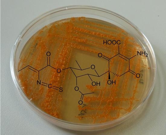
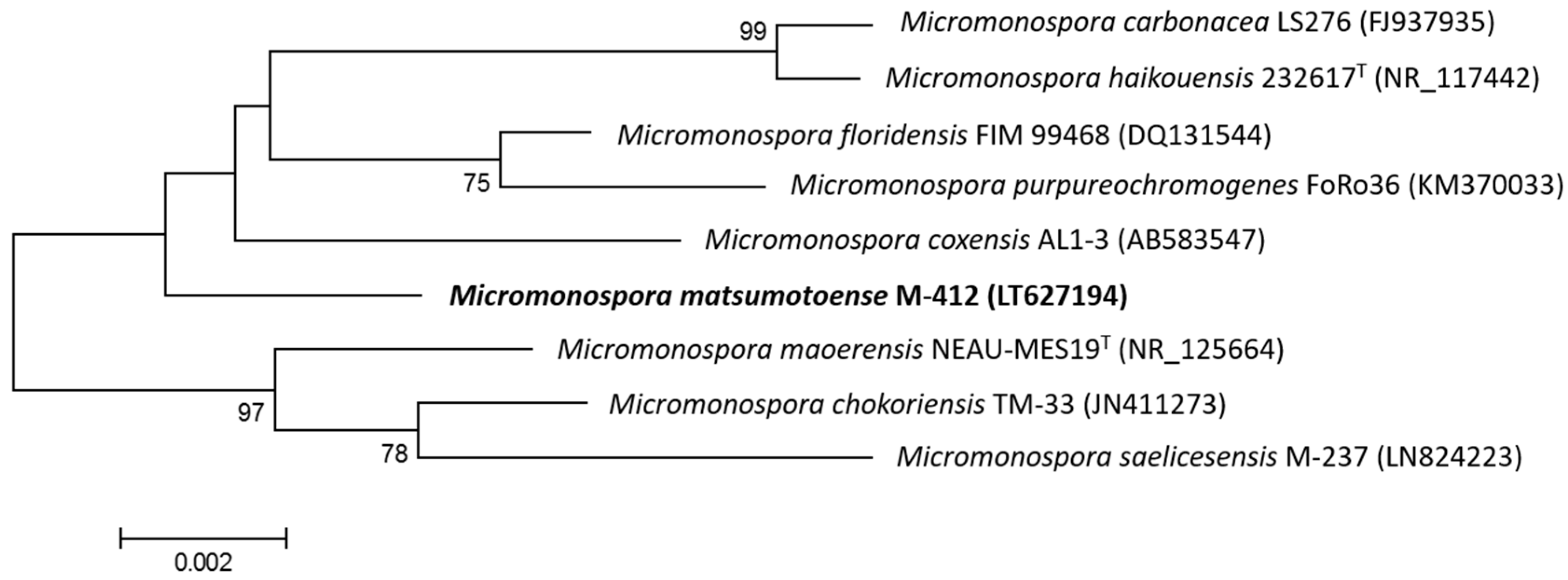
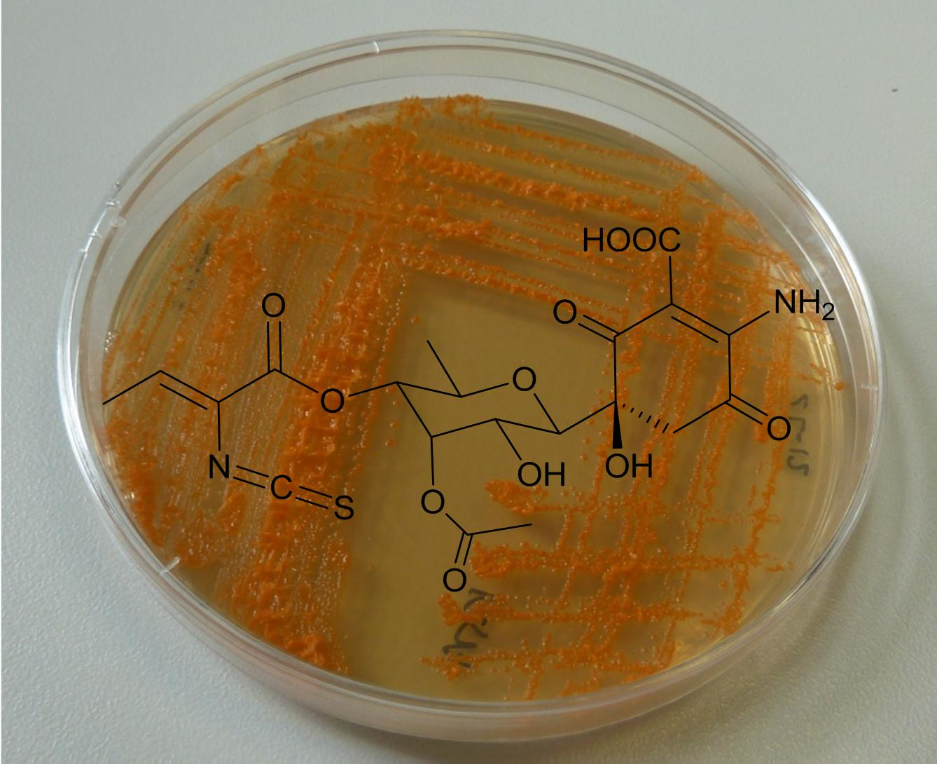
2.1. Taxonomy and Phylogenetic Analysis of the Strain

The 16S rDNA of producing strain M-412 was amplified by polymerase chain reaction (PCR) and sequenced. After Basic Logic Alignment Search Tool (BLAST) sequence comparison, strain M-412 showed 100% identity to Micromonospora matsumotoense (Accession number NR_025015); thus, this strain was designated as Micromonospora matsumotoense M-412 (EMBL Sequence number LT627194). The phylogenetic tree generated by a neighbour-joining method based on 16S rDNA sequence clearly revealed the evolutionary relationship of strain M-412 with a group of known Micromonospora species (Figure 1). To our knowledge, all known paulomycin compounds have only been produced by Streptomyces; thus, paulomycin G is the first member of the family produced by a Micromonospora species.

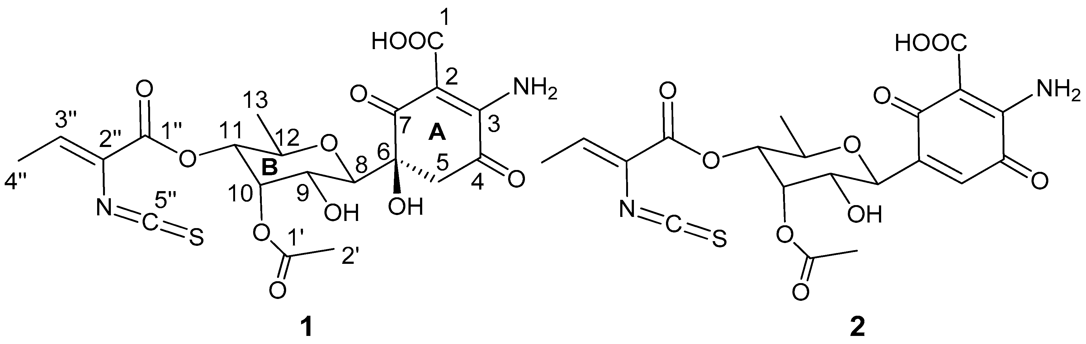
2.2. Structure Determination

Compound 1 was isolated as a pale yellow solid. LC-UV-HRMS analysis of the isolated sample revealed a purity of 83.5% (UV at 210 nm), and indicated the presence of a major impurity in the sample (16.5%), which was identified as the dehydrated paulomycinone 2 based on its UV and HRMS spectra (see Figures S1, S4 and S5). Dehydration of paulomycins has been described to occur easily, even when leaving the compounds in solution in aqueous media at neutral pH, and is therefore difficult to avoid [17]. The major compound of the mixture, paulomycin G, had a molecular formula of C20H22N2O11S according to ESI-TOF MS measurements (m/z 499.1015 [M + H]+, calcd. for C20H23N2O11S+, 499.1017) and the presence of 20 signals in its 13C NMR spectrum. Its UV spectrum displayed maxima at 238, 276, and 320 nm, in agreement with a paulomycin-like structure [2,3]. An intense absorption band at 2041 cm−1 in its IR spectrum confirmed the presence of an isothiocyanate group in the molecule, present in the pauloate moiety of all paulomycins. NMR spectra (Table 1) confirmed the presence of this pauloate moiety, with signals for a methyl group (δH 1.89, δC 14.6 ppm) coupled to an sp2 proton (δH 6.71, δC 136.9 ppm) in the COSY spectrum. HMBC correlations from the latter proton to carbons at δC 159.8 (α,β-unsaturated carbonyl group C1′′), 122.3 ppm (sp2 quaternary carbon C2′′), 141.6 ppm (isothiocyanate carbon C5′′, four-bond distance correlation), and 14.6 ppm (methyl C4′′) completed the structural assignment of the pauloate moiety. Additionally, signals for an aliphatic methylene at δH 3.22 and 3.17 that correlated in the HMBC spectrum with carbon signals at δC 159.4 (C3), 188.8 (C4), 77.5 (C6 and C8), and 197.5 (C7), and the presence of two additional signals at δC 169.0 and 99.1 ppm accounted for the presence in the molecule of the 6-substituted 2-amino-5-hydroxy-3,6-dioxocyclohex-1-enecarboxylic acid substructure present in the paulomycin family of compounds. On the other hand, signals for five oxygenated methines at δH 3.69, 3.61, 5.29, 4.51, and 3.79 ppm, and one aliphatic methyl group at 0.88 ppm conformed a spin system according to COSY correlations that accounted for ring B in the structure of the molecule. Connection between carbon C6 of ring A and carbon C8 in ring B was additionally confirmed via HMBC correlations observed between H8 and C5, C6, and C7. Finally, signals for an acetyl functionality were observed (δH 2.10 ppm, δC 170.1, and 20.8 ppm). This acetyl group was placed at C10 based on HMBC correlations of H2′ and H10 to the carbonyl carbon C1′ and the low field chemical shift of H10. The pauloate moiety was similarly placed at C11 based on an HMBC correlation observed between H11 and C1′′. The relative configuration proposed around the chiral centres in ring B was based on the large coupling constants observed between H8 and H9 (9.8 Hz) and between H11 and H12 (9.9 Hz), indicating the axial orientation of all these protons. The two small coupling constants measured for H10 strongly suggested an equatorial orientation for this proton, and finally, correlations observed in the ROESY spectrum between H10, H9, and H11, and between H8 and H12, proved a relative configuration for ring B as depicted in Figure 2. Based on biogenetic considerations, the absolute configuration was assumed to be the same as in other compounds of the paulomycin series.
Paulomycin G is a novel natural product, structurally unique since it constitutes the first member of the paulomycin family of antibiotics lacking the paulomycose moiety and having a methyl group at position C-13. It is also the smallest bioactive paulomycin reported to-date.

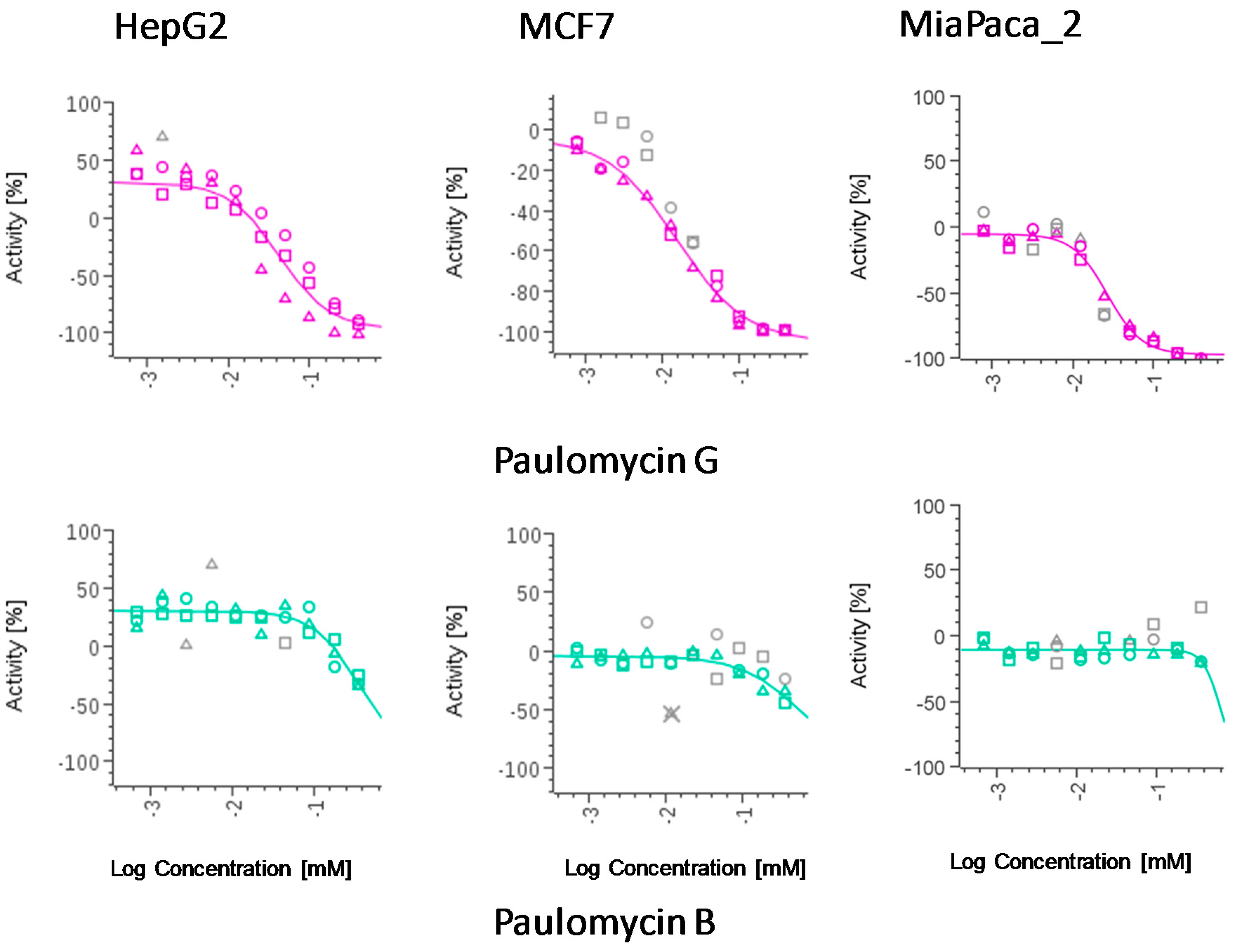
2.3. Cytotoxic Activity of Paulomycin G

Cytotoxic activity was observed for the compound against human breast adenocarcinoma (MCF-7), pancreatic adenocarcinoma (MiaPaca_2), and hepatocellular carcinoma (HepG2) cell lines (Table 2). Considering the lack of biological activity reported for compounds having the quinone moiety present in compound 2 [18], it is reasonable to assume that the biological activity reported herein for the isolated sample is mostly due to paulomycin G. Paulomycin B did not display any cytotoxic activity against the three cell lines when tested in parallel. Figure 3 represents the dose-response curves of paulomycins B and G against the different tumour cell lines.

2.4. Antimicrobial Activity of Paulomycin G

The antimicrobial activity of Paulomycin G was tested against a panel of pathogenic bacteria and fungi, including Gram-negative (Pseudomonas aeruginosa, Acinetobacter baumannii, Escherichia coli, and Klebsiella pneumoniae) and Gram-positive bacteria (methicillin-resistant Staphylococcus aureus, MRSA) and fungi (Aspergillus fumigatus and Candida albicans). Paulomycin B was tested in parallel against the same panel of pathogens. One of the E. coli strains tested (MB5746) was the only pathogen whose growth was inhibited by the action of paulomycin G, with an MIC90 of 38 μg/mL. Paulomycin B displayed activity against E. coli MB5746 and MRSA, with MIC90 values of 4.5 and 50 μg/mL, respectively.

3. Materials and Methods

3.1. General Experimental Procedures

Semipreparative HPLC was performed with an Alliance chromatographic system (Waters Corporation, Mildford, MA, USA) and an Atlantis C18 column (10 μm, 10 × 150 mm, Waters). For UPLC analysis an Acquity UPLC equipment (Waters) with a BEH C18 column (1.7 μm, 2.1 × 100 mm, Waters) was used. Optical rotations were determined with a JASCO P-2000 polarimeter (JASCO Corporation, Tokyo, Japan). IR spectrum was measured with a JASCO Fourier transform infrared (FT/IR)-4100 spectrometer (JASCO Corporation) equipped with a PIKE MIRacleTM single reflection ATR accessory. NMR spectra were recorded on a Bruker Avance III spectrometer (500 and 125 MHz for 1H and 13C NMR, respectively) equipped with a 1.7 mm TCI MicroCryoProbeTM (Bruker Biospin, Fällanden, Switzerland), using the signal of the residual solvent as internal reference (δH 2.50 and δC 39.5 ppm for DMSO-d6). ESI-TOF MS spectra were acquired with a Bruker maXis QTOF spectrometer (Bruker Daltonik GmbH, Bremen, Germany).

3.2. Microorganism and Fermentation Conditions

Strain M-412 was isolated from a deep-sea sediment sample collected from the Cantabrian Sea at a depth of 2000 m, as previously described [13]. GHSA medium (1% glucose, 1% soy bean flour, 0.05% yeast extract, 2.1% MOPS, 0.06% MgSO4·7H2O, 0.2% of a trace elements solution from R5A medium [19], pH 6.8) was selected for paulomycin G production. After autoclaving, the medium was supplemented with 0.4% of a 5 M solution CaCl2·2H2O and 3% DMSO. 20 Erlenmeyer flasks (250 mL), each containing 50 mL of GHSA medium, were inoculated with spores and incubated in an orbital shaker at 28 °C and 250 rpm during 7 days.

3.3. Isolation and Purification of Paulomycin G

The culture broths were centrifuged, and the pellets were extracted with ethyl acetate acidified with 1% formic acid. The supernatants were filtered and applied to a solid-phase extraction cartridge (Sep-Pak Vac C18, 10 g, Waters) that was eluted using a gradient of methanol and 0.05% TFA in water from 0 to 100% methanol in 60 min, at a flow rate of 5 mL/min. Fractions were collected every 5 min and analysed by UPLC using chromatographic conditions previously described [5]. A peak corresponding to an unknown paulomycin was detected in fractions eluting between 40 and 50 min. These fractions were pooled, partially dried in vacuo, and applied to a solid-phase extraction cartridge (Sep-Pak Vac C18, 2 g, Waters). The cartridge was washed with water and the retained compounds were eluted with methanol and dried in vacuo. The residue was subsequently redissolved in a small volume of acetonitrile and DMSO (2:1). The same peak of unknown paulomycin was also found in the organic extract of the culture pellets, which was dried and redissolved as above. Paulomycin G was eventually purified by semipreparative HPLC using an Atlantis C18 column (10 μm, 10 × 150 mm, Waters) in two isocratic elution steps, employing a mixture of 50% acetonitrile and water in the first step and 55% methanol and water in the second step, with a flow of 5 mL/min. In both cases, the solution containing the collected peak was evaporated and finally lyophilized, resulting in 2.7 mg of compound 1 (83.5% purity according to LC-UV analysis at 210 nm).
Paulomycin G (1). pale yellow solid; [ α ] D 20 +10.6° (c 0.18, MeOH); UV (DAD) λmax 238, 276 y 320 nm; IR (ATR) νmax 3359, 3229, 2979, 2933, 2041, 1736, 1695, 1626, 1571, 1442, 1381, 1260, 1227, 1129, 1025, 909, 751 cm−1; for 1H and 13C NMR data see Table 1; ESI-TOF MS m/z 521.0828 [M + Na]+ (calcd. for C20H22N2O11SNa+, 521.0837) 516.1278 [M + NH4]+ (calcd. for C20H26N3O11S+, 516.1283), 499.1015 [M + H]+ (calcd. C20H23N2O11S+, 499.1017).

3.4. Phylogenetic Analysis (Taxonomy) of the Producer Microorganism

Phylogenetic analysis based on 16S rRNA sequences was performed with strain Micromonospora matsumotoense M-412 using MEGA version 6.0 [20] after multiple alignment of data by Clustal Omega [21]. Distances (distance options according to the Kimura two-parameter model [22]) and clustering with the neighbour-joining method [23] were evaluated using bootstrap values based on 1000 replications [24].

3.5. Cytotoxic Activity of Compound 1

The MTT (3-(4,5-dimethylthiazol-2-yl)-2,5-diphenyltetrazolium bromide) colorimetric assay—which measures mitochondrial metabolic activity—was performed using three tumour cell lines obtained from the ATCC, namely human breast adenocarcinoma (MCF-7), pancreatic adenocarcinoma (MiaPaca_2), and hepatocellular carcinoma (HepG2), using previously-described methodology [15]. Methyl methanesulphonate at a concentration of 8 mM and 0.5% DMSO in water were used as positive and negative controls, respectively.

3.6. Antimicrobial Activity of Compound 1

Antibacterial and antifungal activity tests of paulomycins G and B were performed against the pathogenic strains P. aeruginosa PAO1, Acinetobacter baumannii MB5973, Escherichia coli MB5746 and MB2884, K. pneumoniae ATCC700603, methicillin-resistant Staphylococcus aureus MB5393, Aspergillus fumigatus ATCC46645, and Candida albicans ATCC64124, as previously described [25].

4. Conclusions

In summary, a new member of the paulomycin family that we have designated as paulomycin G has been obtained from cultures of Micromonospora matsumotoense M-412, isolated from deep-sea sediments collected at 2000 m depth in the submarine Avilés Canyon. Paulomycin G is a novel natural product, structurally unique since—to our knowledge—it is the first member of the paulomycin family of antibiotics lacking the paulomycose moiety, being the smallest bioactive paulomycin reported. This new natural product displayed strong cytotoxic activity against human cancer cell lines such as pancreatic adenocarcinoma (MiaPaca_2), breast adenocarcinoma (MCF-7), and hepatocellular carcinoma (HepG2). Based on its cytotoxic activities, paulomycin G deserves to be considered as a candidate to perform further studies assessing its anticancer potential. Besides its unique structural features, paulomycin G might also be of interest in biosynthetic studies and useful for future paulomycin biosynthesis research or as a core structure in the generation of novel derivatives through combinatorial biosynthesis to generate structural diversity in the paulomycin family. Additionally, the isolation of a compound of the paulomycin family from a non-Streptomyces actinomycete is particularly noteworthy and suggests horizontal gene transfer between Micromonospora and Streptomyces species.

Supplementary Materials

The following are available online at www.mdpi.com/1660-3397/15/9/271/s1. Figure S1: HPLC trace of the sample isolated; Figure S2: UV spectrum of compound 1; Figure S3: ESI-TOF spectra of compound 1; Figure S4: UV spectrum of compound 2; Figure S5: ESI-TOF spectra of compound 2; Figure S6: 1H NMR spectrum (DMSO-d6, 500 MHz) of compound 1; Figure S7: 13C NMR spectrum (DMSO-d6, 125 MHz) of compound 1; Figure S8: COSY spectrum (DMSO-d6) of compound 1; Figure S9: HSQC spectrum (DMSO-d6) of compound 1; Figure S10: HMBC spectrum (DMSO-d6) of compound 1; Figure S11: ROESY spectrum (DMSO-d6) of compound 1; Figure S12: Picture of Micromonospora matsumotoense M-412.

Acknowledgments

This study was financially supported by Gobierno del Principado de Asturias (SV-PA-13-ECOEMP-62). The polarimeter, IR, and NMR equipment used in this work were acquired via grants for scientific and technological infrastructures from the Ministerio de Ciencia e Innovación (Grants Nos. PCT-010000-2010-4 (NMR), INP-2011-0016-PCT-010000 ACT6 (polarimeter and IR)). The authors are grateful to all participants in the BIOCANT3 oceanographic expedition (DOSMARES) where Micromonospora matsumotoense M-412 was isolated. We are also grateful to J.A. Guijarro and J.L. Caso for continuous support. This is a contribution of the Asturias Marine Observatory.

Author Contributions

Aida Sarmiento-Vizcaíno isolated the strain and performed the phylogenetic analysis. Alfredo F. Braña and Aida Sarmiento-Vizcaíno identified and performed the purification of the compound. Ignacio Pérez-Victoria, Jesús Martín, and Fernando Reyes performed the structural elucidation of the compound. Nuria de Pedro, Mercedes de la Cruz, Caridad Díaz and Francisca Vicente performed the biological tests. José L. Acuña led the oceanographic expedition in which sediment samples were collected. Gloria Blanco and Fernando Reyes wrote the paper which was revised and approved by all the authors. Gloria Blanco and Luis A. García led and coordinated the research.

Conflicts of Interest

The authors declare no conflict of interest.

References

  1. Novak, E. Treating Chlamydia Infections with Paulomycin. Patent PCT/US1987/002420, 21 April 1988. [Google Scholar]
  2. Argoudelis, A.D.; Brinkley, T.A.; Brodasky, T.F.; Buege, J.A.; Meyer, H.F.; Mizsak, S.A. Paulomycins A and B. Isolation and characterization. J. Antibiot. 1982, 35, 285–294. [Google Scholar] [CrossRef] [PubMed]
  3. Majer, J.; Chater, K. Streptomyces albus G produces an antibiotic complex identical to paulomycins A and B. J. Gen. Microbiol. 1987, 133, 2503–2507. [Google Scholar] [CrossRef] [PubMed]
  4. Argoudelis, A.D.; Baczynskyj, L.; Haak, W.J.; Knoll, W.M.; Mizsak, S.A.; Shilliday, F.B. New paulomycins produced by Streptomyces paulus. J. Antibiot. 1988, 41, 157–169. [Google Scholar] [CrossRef] [PubMed]
  5. Braña, A.F.; Rodríguez, M.; Pahari, P.; Rohr, J.; García, L.A.; Blanco, G. Activation and silencing of secondary metabolites in Streptomyces albus and Streptomyces lividans after transformation with cosmids containing the thienamycin gene cluster from Streptomyces cattleya. Arch. Microbiol. 2014, 196, 345–355. [Google Scholar] [CrossRef] [PubMed]
  6. Li, J.; Xie, Z.; Wang, M.; Ai, G.; Chen, Y. Identification and analysis of the paulomycin biosynthetic gene cluster and titer improvement of the paulomycins in Streptomyces paulus NRRL 8115. PLoS ONE 2015, 10, e0120542. [Google Scholar] [CrossRef] [PubMed]
  7. González, A.; Rodríguez, M.; Braña, A.F.; Méndez, C.; Salas, J.A.; Olano, C. New insights into paulomycin biosynthesis pathway in Streptomyces albus J1074 and generation of novel derivatives by combinatorial biosynthesis. Microb. Cell Fact. 2016, 15. [Google Scholar] [CrossRef] [PubMed]
  8. Ward, A.C.; Bora, N. Diversity and biogeography of marine actinobacteria. Curr. Opin. Microbiol. 2006, 9, 279–286. [Google Scholar] [CrossRef] [PubMed]
  9. Hameş-Kocabaş, E.E.; Uzel, A. Isolation strategies of marine-derived actinomycetes from sponge and sediment samples. J. Microbiol. Methods 2012, 88, 342–347. [Google Scholar] [CrossRef]
  10. Braña, A.F.; Fiedler, H.P.; Nava, H.; González, V.; Sarmiento-Vizcaíno, A.; Molina, A.; Acuña, J.L.; García, L.A.; Blanco, G. Two Streptomyces Species Producing Antibiotic, Antitumor, and Anti Inflammatory Compounds Are Widespread Among Intertidal Macroalgae and Deep Sea Coral Reef Invertebrates from the Central Cantabrian Sea. Microb. Ecol. 2015, 69, 512–524. [Google Scholar] [CrossRef] [PubMed]
  11. Sarmiento-Vizcaíno, A.; González, V.; Braña, A.F.; Molina, A.; Acuña, J.L.; García, L.A.; Blanco, G. Myceligenerans cantabricum sp. nov., a barotolerant actinobacterium isolated from a deep cold water coral. Int. J. Syst. Evol. Microbiol. 2015, 65, 1328–1334. [Google Scholar] [PubMed]
  12. Sarmiento-Vizcaíno, A.; Braña, A.F.; González, V.; Nava, H.; Molina, A.; Llera, E.; Fiedler, H.P.; Rico, J.M.; García-Flórez, L.; Acuña, J.L.; et al. Atmospheric Dispersal of Bioactive Streptomyces albidoflavus Strains Among Terrestrial and Marine Environments. Microb. Ecol. 2016, 71, 375–386. [Google Scholar] [CrossRef] [PubMed]
  13. Sarmiento-Vizcaíno, A.; González, V.; Braña, A.F.; Palacios, J.J.; Otero, L.; Fernández, J.; Molina, A.; Kulik, A.; Vázquez, F.; Acuña, J.L.; et al. Pharmacological potential of phylogenetically diverse Actinobacteria isolated from deep-sea coral ecosystems of the submarine Avilés Canyon in the Cantabrian Sea. Microb. Ecol. 2017, 73, 338–352. [Google Scholar] [CrossRef] [PubMed]
  14. Braña, A.F.; Sarmiento-Vizcaíno, A.; Pérez-Victoria, I.; Otero, L.; Fernández, J.; Palacios, J.J.; Martín, J.; de la Cruz, M.; Díaz, C.; Vicente, F.; et al. Branimycins B and C, Antibiotics Produced by the Abyssal Actinobacterium Pseudonocardia carboxydivorans M-227. J. Nat. Prod. 2017, 80, 569–573. [Google Scholar] [CrossRef] [PubMed]
  15. Braña, A.F.; Sarmiento-Vizcaíno, A.; Osset, M.; Pérez-Victoria, I.; Martín, J.; de Pedro, N.; de la Cruz, M.; Díaz, C.; Vicente, F.; Reyes, F.; et al. Lobophorin K, a New Natural Product with Cytotoxic Activity Produced by Streptomyces sp. M-207 Associated with the Deep-Sea Coral Lophelia pertusa. Mar. Drugs 2017, 15, 144. [Google Scholar] [CrossRef] [PubMed]
  16. Pérez-Victoria, I.; Martín, J.; Reyes, F. Combined LC/UV/MS and NMR strategies for the dereplication of marine natural products. Planta Med. 2016, 82, 857–871. [Google Scholar] [CrossRef] [PubMed]
  17. Wiley, P.F.; Mizsak, S.A.; Baczynskyj, L.; Argoudelis, A.D.; Duchamp, D.J.; Watt, W. The Structure and Chemistry of Paulomycin. J. Org. Chem. 1986, 51, 2493–2499. [Google Scholar] [CrossRef]
  18. Argoudelis, A.D.; Baczynskyj, L.; Mizsak, S.A.; Shilliday, F.B.; Spinelli, P.A.; DeZwaan, J. Paldimycins A and B and antibiotics 273a and 273a synthesis and characterization. J. Antibiot. 1987, 40, 419–436. [Google Scholar] [CrossRef] [PubMed]
  19. Fernández, E.; Weissbach, U.; Sánchez Reillo, C.; Braña, A.F.; Méndez, C.; Rohr, J.; Salas, J.A. Identification of two genes from Streptomyces argillaceus encoding two glycosyltransferases involved in the transfer of a disaccharide during the biosynthesis of the antitrumor drug mithramycin. J. Bacteriol. 1998, 180, 4929–4937. [Google Scholar] [PubMed]
  20. Tamura, K.; Stecher, G.; Peterson, D.; Filipski, A.; Kumar, S. MEGA6: Molecular Evolutionary Genetics Analysis version 6.0. Mol. Biol. Evol. 2013, 30, 2725–2729. [Google Scholar] [CrossRef] [PubMed]
  21. Sievers, F.; Wilm, A.; Dineen, D.G.; Gibson, T.J.; Karplus, K.; Li, W.; Lopez, R.; McWilliam, H.; Remmert, M.; Söding, J.; et al. Fast, scalable generation of high-quality protein multiple sequence alignments using Clustal Omega. Mol. Syst. Biol. 2011, 7. [Google Scholar] [CrossRef] [PubMed]
  22. Kimura, M. A simple method for estimating evolutionary rates of base substitutions through comparative studies of nucleotide sequences. J. Mol. Evol. 1980, 16, 111–120. [Google Scholar] [CrossRef] [PubMed]
  23. Saitou, N.; Nei, M. The neighbour-joining method: A new method for reconstructing phylogenetic trees. Mol. Biol. Evol. 1987, 4, 406–425. [Google Scholar] [PubMed]
  24. Felsenstein, J. Conference limits on phylogenies: An approach using the bootstrap. Evolution 1985, 39, 783–791. [Google Scholar] [CrossRef] [PubMed]
  25. Audoin, C.; Bonhomme, D.; Ivanisevic, J.; de la Cruz, M.; Cautain, B.; Monteiro, M.C.; Reyes, F.; Rios, L.; Perez, T.; Thomas, O.P. Balibalosides, an Original Family of Glucosylated Sesterterpenes Produced by the Mediterranean Sponge Oscarella balibaloi. Mar. Drugs 2013, 11, 1477–1489. [Google Scholar] [CrossRef] [PubMed]
Figure 1. Neighbour-joining phylogenetic tree obtained by distance matrix analysis of 16S rDNA sequences, showing Micromonospora matsumotoense M-412 position and most closely related phylogenetic neighbours. Numbers on branch nodes are bootstrap values (1000 resamplings; only values >70% are given). Bar indicates 0.2% sequence divergence.
Marinedrugs 15 00271 g001
Figure 2. Chemical structures of paulomycin G (1) and compound 2.
Marinedrugs 15 00271 g002
Figure 3. Dose-response curves of compounds against human breast adenocarcinoma (MCF-7), pancreatic adenocarcinoma (MiaPaca_2), and hepatocellular carcinoma (HepG2) cell lines. Compounds were tested per triplicate and the obtained results are indicated with triangles, rounds, and squares in every picture.
Marinedrugs 15 00271 g003
Table 1. 1H and 13C NMR (500 and 125 MHz in DMSO-d6) data for compound 1.
Positionδ 13Cδ (1H), (Mult, J in Hz)
1169.0-
299.1-
3159.4-
4188.8-
547.73.22 (d, 16.0), 3.17 (d, 16.0)
677.5-
7197.5-
877.53.69 (d, 9.9)
967.03.61 (br dt, 9.7, 3.3)
1069.85.29 (dd, 2.6, 2.6)
1173.44.51 (dd, 9.9, 2.6)
1269.93.79 (dq, 9.9, 6.2)
1316.30.88 (d, 6.2)
1′170.1-
2′20.82.10 (s)
1′159.8-
2′′122.3-
3′′136.96.71 (quart., 7.1)
4′′14.61.89 (d, 7.1)
5′′141.6-
NH2 (3)-9.71 (br s), 9.35 (br s)
OH (1)-14.21 (br s)
OH (6)-5.43 (s)
OH (9)-5.78 (d, 4.4)
Table 2. Cytotoxic activity of paulomycins B and G against different tumour cell lines.
Cell linePaulomycin G (IC50 μM)Paulomycin B (IC50 μM)
HepG24.30 ± 0.42>36
MCF-71.58 ± 0.12>36
MiaPaca_22.70 ± 0.25>36

