Communication

A New Dioic Acid from a wbl Gene Mutant of Deepsea-Derived Streptomyces somaliensis SCSIO ZH66

1
Key Laboratory of Marine Drugs, Ministry of Education, School of Medicine and Pharmacy, Ocean University of China, Qingdao 266003, China
2
CAS Key Laboratory of Marine Bio-resources Sustainable Utilization, Guangdong Key Laboratory of Marine Materia Medica, RNAM Center for Marine Microbiology, South China Sea Institute of Oceanology, Chinese Academy of Sciences, 164 West Xingang Road, Guangzhou 510301, China
3
Laboratory for Marine Drugs and Bioproducts of Qingdao National Laboratory for Marine Science and Technology, Qingdao 266237, China
*
Author to whom correspondence should be addressed.
Mar. Drugs 2016, 14(10), 184; https://doi.org/10.3390/md14100184
Submission received: 20 September 2016 / Revised: 1 October 2016 / Accepted: 8 October 2016 / Published: 17 October 2016

Abstract

:
The wblAso gene functions as a global regulatory gene in a negative manner in deepsea-derived Streptomyces somaliensis SCSIO ZH66. A new dioic acid (1) as well as two known butenolides (2 and 3) were isolated from the ΔwblAso mutant strain of S. somaliensis SCSIO ZH66. The structure of 1 was elucidated by a combination of spectroscopic analyses, including MS and NMR techniques. In the cell growth inhibitory evaluation, compound 3 exhibited moderate activity against the human hepatic carcinoma cell line (Huh7.5) with an IC50 value of 19.4 μg/mL, while compounds 1 and 2 showed null activity up to 100 μg/mL.

Graphical Abstract

1. Introduction

Marine-derived bioactive compounds and their novel chemical scaffolds have been shown to be attractive starting points for drug discovery programs [1,2]. Over the last few years, the actinomycetes isolated from marine environments have attracted considerable attention, because of their great potential for producing a large diversity of bioactive compounds [3,4]. However, discovery of structurally novel secondary metabolites from microbes is becoming more and more difficult, as the rate of rediscovery of known compounds is increasing [5,6]. Recent genome sequencing revealed that actinomycetes can genetically synthesize secondary metabolites beyond those that were isolated under standard cultivation conditions [7,8,9]. However, a number of gene clusters in actinomycetes are silent or only low-expressed and thus are defined as cryptic or orphan clusters [10].
Manipulation of global regulators is an effective strategy for the activation of cryptic gene clusters [11]. In the previous study, we obtained a series of anti-MRSA (methicillin-resistant Staphylococcus aureus) α-pyrone compounds (violapyrones A–C, H, and J) from the deepsea-derived S. somaliensis SCSIO ZH66 by disruption of the negative global regulatory gene wblAso [12]. In our continuous search for significantly enhanced compounds in the ΔwblAso mutant strain, one new dioic acid (1) together with two known butenolides (2 and 3) were isolated (Figure 1). We describe herein the isolation, structure elucidation, and biological evaluation of these accumulated compounds.

2. Results and Discussion

The ΔwblAso mutant of S. somaliensis SCSIO ZH66 was constructed in our previous study [12]. The fermentation broths of the wild-type and the ΔwblAso mutant strains were extracted with EtOAc, respectively, and were subsequently subjected to high-performance liquid chromatography (HPLC) analysis (Figure 2), in which we observed three significantly enhanced peaks (13) in the ΔwblAso mutant compared with those in the wild-type strain at wavelengths of 200 nm. With the massive fermentation of the ΔwblAso mutant, compounds 13 were isolated and further identified via NMR assignments.
Compound 1 was obtained as a white, amorphous solid. The molecular formula of 1 was established as C17H26O4 on the basis of HR-ESIMS data ([M + Na]+ at m/z 317.1723) (Figure S1). The structure of 1 was determined by 1D (1H, 13C) and 2D NMR (COSY, HSQC and HMBC) data (Table 1). The 1H-NMR spectrum of 1 disclosed three methyls (δH 1.55, 1.56, 2.07) and six methylenes (δH 2.26, 2.16, 2.02, 1.93, 2.12, 2.12), and the 13C-NMR spectrum showed 17 carbon signals. The 1H–1H COSY spectrum established the spin systems of H-2 (δH 2.26)/H-3 (δH 2.16), H-5 (δH 5.09)/H-6 (δH 2.02)/H-7 (δH 1.93) and H-9 (δH 5.07)/H-10 (δH 2.12)/H-11 (δH 2.12) (Figure 3); the HMBC correlations from H-3 to C-1 (δC 174.1) and C-5 (δC 124.0), from H-7 to C-9 (δC 123.1), from H-11 to C-13 (δC 116.2), and from H-13 (δH 5.58) to C-14 (δC 167.4) established the main aliphatic chain of 1. The HMBC correlations from the methyl protons H-15 (δH 1.55) to C-4 (δC 133.6), from H-16 (δH 1.56) to C-8 (δC 135.2), and from H-17 (δH 2.07) to C-12 (δC 158.4) confirmed the location of three methyl groups (Figure 3). Moreover, the configurations of three double bonds were confirmed to be trans by NOSEY correlations of H-3/H-5, H-7/H-9, H-11/H-13, H-15/H-6, and H-16/H-10 (Figure 4). The 13C chemical shifts of C-1 and C-14, together with HR-ESIMS data, revealed two carboxyl groups in 1. Thus, the structure of 1 was finally identified as 3,7,11-trimethyl-2,6,10-triene-1,14-tetradecyl dioic acid.
Compound 2 was isolated as a light yellow oil. The molecular formula of 2 was established as C13H22O3 on the basis of HR-ESIMS data ([M + Na]+ at m/z 249.1337) (Figure S2). Compound 2 was identified as (4S)-4,10-dihydroxy-10-methyl-dodec-2-en-1,4-olide by comparison of 1H and 13C-NMR data (Table S1) with those reported in [13].
Compound 3 was isolated as a light yellow oil. The molecular formula of 3 was established as C13H22O3 on the basis of HR-ESIMS data ([M + Na]+ at m/z 249.1336) (Figure S3). Compound 3 was identified as (4S)-4,11-dihydroxy-10-methyl-dodec-2-en-1,4-olide by comparison of 1H and 13C-NMR data (Table S1) with those reported in [13].
Butenolides are a family of α,β-unsaturated lactones, and their saturated analogs act as signaling substances in bacteria to enhance sporulation or induce metabolite production [14]. Some butenolides have been reported to show cytotoxicity [13] or antimicrobial activities [15,16]. In the evaluation for cell growth inhibitory effects, compound 3 exhibited moderate growth inhibition against the human hepatic carcinoma cell line (Huh7.5) with an IC50 value of 19.4 μg/mL, while compounds 1 and 2 showed null activity up to 100 μg/mL. In the test of antimicrobial activity, none of these compounds showed significant activity against multi-drug resistant strains (Staphylococcus aureus CCARM 3090, Escherichia coli CCARM 1009, Enterococcus faecalis CCARM 5172, Enterococcus faecium CCARM 5203, and Salmonella typhimurium CCARM 8250).

3. Experimental Section

3.1. General Experimental Procedures

HPLC was performed on an Agilent 1260 Infinity equipment with Diode Array Detector (DAD) (Agilent, Waldbronn, Germany). NMR spectra were recorded on Agilent DD2-500 spectrometers. Chemical shifts were reported with reference to the respective solvent peaks and residual solvent peaks (δH 2.50 and δC 39.5 ppm for DMSO-d6). HR-ESIMS data were obtained on a Q-TOF Ultima Global GAA076 LC-MS spectrometer (Waters Corporation, Milford, MA, USA).

3.2. Strains and Culture Conditions

The S. somaliensis SCSIO ZH66 (CGMCC NO. 9492) was isolated from the deep-sea sediment collected at a depth of 3536 m in the South China Sea (120°0.250′ E; 20°22.971′ N) [17]. The ΔwblAso mutant was obtained as described previously [12]. The strains were grown at 30 °C on a MS (mannitol-soy flour) medium for sporulation.

3.3. Fermentation, Extraction, and Isolation of the Compounds

For each fermentation, strain spores were inoculated into 200 mL of fermentation medium (1% soluble starch, 2% glucose, 4% corn syrup, 1% yeast extract, 0.3% beef extract, 0.05% MgSO4·7H2O, 0.05% KH2PO4, 0.2% CaCO3, and 3% sea salt, pH = 7.0), which was further supplemented with 1.5% XAD-16 resin when fermenting the ΔwblAso mutant, and incubated at 30 °C, 220 rpm for 7 days. Analytical HPLC was performed on an Eclipse C18 column (5 μm, 4.6 × 150 mm) developed with a linear gradient from 20% to 70% B/A in 40 min (phase A: 0.1% formic acid in H2O; phase B: 100% acetonitrile supplemented with 0.1% formic acid). A total of 20 L of culture was made by this method. The fermentation cultures were harvested via centrifugation, and the supernatant was extracted twice with an equal volume of ethyl acetate. The combined ethyl acetate extracts were concentrated in vacuo to afford residue A. The precipitated mycelia and XAD-16 resin were extracted twice with acetone. The extracts were combined, and acetone was evaporated in vacuo to yield residue B. Both residues from fermentation broths and mycelia cakes were combined to afford crude extract (6.0 g). They were applied to reversed-phase C18 open column, eluting with a gradient eluent of 20%–100% methanol to give five fractions (Fr.1~Fr.5). Compound 1 (3.8 mg) was obtained by further separation of Fr.4 eluting with gradient solvents (phase A: 0.1% formic acid in H2O; phase B: 100% acetonitrile supplemented with 0.1% formic acid, 2 mL/min, UV detection at 200 nm) using a semi-preparative HPLC column (YMC-Pack ODS-AA C18 column, 120 Å, 250 × 10 mm, 5 μm). Fr.3 was also further subjected to semi-preparative HPLC eluting with gradient solvents to afford compounds 2 (2.9 mg) and 3 (5.5 mg).

3.4. Biological Assays

Viabilities of human hepatic carcinoma cell line (Huh7.5) were measured with a MTT assay. Briefly, logarithmically growing cells were trypsinized from culture dishes and placed into 96-well plate and cultured at 37 °C for 24 h. Cells were treated with the varying concentrations of each compound, and then 20 μL of a MTT solution (5 mg/mL) were added to each well. After incubating for 4 h, the medium was removed, and 150 μL of DMSO were added to each well to dissolve purple crystals of formazan via shaking at 260 rpm for 10 min. Absorbance was measured at 490 nm by a multi-detection microplate reader (infinite M1000 Pro, TECAN, Mannedorf, Switzerland). The 50% inhibitory concentration (IC50) value was determined as the concentration that caused 50% inhibition of cell proliferation [18,19].
The antibacterial activity against five multi-drug resistant (MDR) strains (S. aureus CCARM 3090, E. coli CCARM 1009, E. faecalis CCARM 5172, E. faecium CCARM 5203, and S. typhimurium CCARM 8250) was tested by the radial diffusion assay as previously described [12].

4. Conclusions

A new dioic acid (1) and two known butenolides (2 and 3) were isolated from the ΔwblAso mutant strain of deepsea-derived S. somaliensis SCSIO ZH66. In the evaluation for cell growth inhibitory effects, compound 3 showed moderate activity against the human hepatic carcinoma cell line (Huh7.5) with an IC50 value of 19.4 μg/mL. Isolation of a novel dioic acid (1) in this study indicated that the manipulation of global regulators can be used as an effective method for the accumulation of novel secondary metabolites.

Supplementary Materials

The following are available online at https://www.mdpi.com/1660-3397/14/10/184/s1. Figure S1: Spectral data of compound 1; Figure S2: Spectral data of compound 2; Figure S3: Spectral data of compound 3; Table S1: 1H and 13C-NMR Data of compound 2 and 3.

Acknowledgments

This work was supported by grants from the NSFC (31171201, 81561148012 & 41506157), the NSFC-Shandong Joint Fund for Marine Science Research Centers (U1406402), and the National High Technology Research and Development Program of China (2012AA092104). We would like to thank Chaomin Sun (The Institute of Oceanology, Chinese Academy of Sciences) for his help in the evaluation of cell growth inhibitory activity.

Author Contributions

H.H. performed the experiments and wrote the manuscript. H.L. was involved in NMR analysis and manuscript revision. Y.Q. and L.H. performed the experiments. J.J. provided S. somaliensis SCSIO ZH66. W.L. supervised the whole work, and revised and edited the manuscript. All authors read and approved the final manuscript.

Conflicts of Interest

The authors declare no conflict of interest.

References

  1. Haefner, B. Drugs from the deep: Marine natural products as drug candidates. Drug Discov. Today 2003, 8, 536–544. [Google Scholar] [CrossRef]
  2. Gupta, L.; Talwar, A.; Chauhan, P.M. Bis and tris indole alkaloids from marine organisms: New leads for drug discovery. Curr. Med. Chem. 2007, 14, 1789–1803. [Google Scholar] [CrossRef] [PubMed]
  3. Lane, A.L.; Moore, B.S. A sea of biosynthesis: Marine natural products meet the molecular age. Nat. Prod. Rep. 2011, 28, 411–428. [Google Scholar] [CrossRef] [PubMed]
  4. Zotchev, S.B. Marine actinomycetes as an emerging resource for the drug development pipelines. J. Biotechnol. 2012, 158, 168–175. [Google Scholar] [CrossRef] [PubMed]
  5. Wolfender, J.L.; Marti, G.; Ferreira, Q.E. Advances in techniques for profiling crude extracts and for the rapid identification of natural products: Dereplication, quality control and metabolomics. Curr. Org. Chem. 2010, 14, 1808–1832. [Google Scholar] [CrossRef]
  6. Wang, J.; Lin, W.; Wray, V.; Lai, D.; Proksch, P. Induced production of depsipeptides by co-culturing Fusarium tricinctum and Fusarium begoniae. Tetrahedron Lett. 2013, 54, 2492–2496. [Google Scholar] [CrossRef]
  7. Bentley, S.D.; Chater, K.F.; Cerdeno-Tarraga, A.M.; Challis, G.L.; Thomson, N.R.; James, K.D.; Harris, D.E.; Quail, M.A.; Kieser, H.; Harper, D.; et al. Complete genome sequence of the model actinomycete Streptomyces coelicolor A3 (2). Nature 2002, 417, 141–147. [Google Scholar] [CrossRef] [PubMed]
  8. Ikeda, H.; Ishikawa, J.; Hanamoto, A.; Shinose, M.; Kikuchi, H.; Shiba, T.; Sakaki, Y.; Hattori, M.; Omura, S. Complete genome sequence and comparative analysis of the industrial microorganism Streptomyces avermitilis. Nat. Biotechnol. 2003, 21, 526–531. [Google Scholar] [CrossRef] [PubMed]
  9. Ohnishi, Y.; Ishikawa, J.; Hara, H.; Suzuki, H.; Ikenoya, M.; Ikeda, H.; Yamashita, A.; Hattori, M.; Horinouchi, S. Genome sequence of the streptomycin-producing microorganism Streptomyces griseus IFO 13350. J. Bacteriol. 2008, 190, 4050–4060. [Google Scholar] [CrossRef] [PubMed]
  10. Nett, M.; Ikeda, H.; Moore, B.S. Genomic basis for natural product biosynthetic diversity in the actinomycetes. Nat. Prod. Rep. 2009, 26, 1362–1384. [Google Scholar] [CrossRef] [PubMed]
  11. Lu, C.; Liao, G.; Zhang, J.; Tan, H. Identification of novel tylosin analogues generated by a wblA disruption mutant of Streptomyces ansochromogenes. Microb. Cell Fact. 2015, 14, 173. [Google Scholar] [CrossRef] [PubMed]
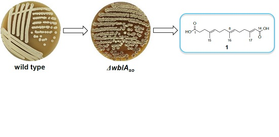
  12. Huang, H.; Hou, L.; Li, H.; Qiu, Y.; Ju, J.; Li, W. Activation of a plasmid-situated type III PKS gene cluster by deletion of a wbl gene in deepsea-derived Streptomyces somaliensis SCSIO ZH66. Microb. Cell Fact. 2016, 15, 116. [Google Scholar] [CrossRef] [PubMed]
  13. Li, D.; Zhu, T.; Liu, H.; Fang, Y.; Gu, Q.; Zhu, W. Four butenolides are novel cytotoxic compounds isolated from the marine-derived bacterium, Streptoverticillium luteoverticillatum 11014. Arch. Pharmacal. Res. 2006, 29, 624–626. [Google Scholar] [CrossRef]
  14. Mukku, V.J.; Speitling, M.; Laatsch, H.; Helmke, E. New butenolides from two marine streptomycetes. J. Nat. Prod. 2000, 63, 1570–1572. [Google Scholar] [CrossRef] [PubMed]
  15. Braun, D.; Pauli, N.; Séquin, U.; Zähner, H. New butenolides from the photoconductivity screening of Streptomyces antibioticus (Waksman and Woodruff) Waksman and Henrici 1948. FEMS Microbiol. Lett. 1995, 126, 37–42. [Google Scholar] [CrossRef] [PubMed]
  16. Wang, T.; Jiang, Y.; Ma, K.; Li, Y.; Huang, R.; Xie, X.; Wu, S. Two new butenolides produced by an Actinomycete Streptomyces sp. Chem. Biodiver. 2014, 11, 929–933. [Google Scholar] [CrossRef] [PubMed]
  17. Zhang, Y.; Huang, H.; Xu, S.; Wang, B.; Ju, J.; Tan, H.; Li, W. Activation andenhancement of Fredericamycin A production in deepsea-derived Streptomyces somaliensis SCSIO ZH66 by using ribosome engineering and response surface methodology. Microb Cell Fact. 2015, 14, 64. [Google Scholar] [CrossRef] [PubMed]
  18. Liu, G.; Kuang, S.; Wu, S.; Jin, W.; Sun, C. A novel polysaccharide from Sargassum integerrimum induces apoptosis in A549 cells and prevents angiogensis in vitro and in vivo. Sci. Rep. 2016, 6, 26722. [Google Scholar] [CrossRef] [PubMed]
  19. Lefranc, F.; Nuzzo, G.; Hamdy, N.A.; Fakhr, I.; Banuls, L.M.; Goietsenoven, G.V.; Villani, G.; Mathieu, V.; Soest, R.V.; Kiss, R.; et al. In vitro pharmacological and toxicological effects of norterpene peroxides isolated from the Red Sea sponge Diacarnus erythraeanus on normal and cancer cells. J. Nat. Prod. 2013, 76, 1541–1547. [Google Scholar] [CrossRef] [PubMed]
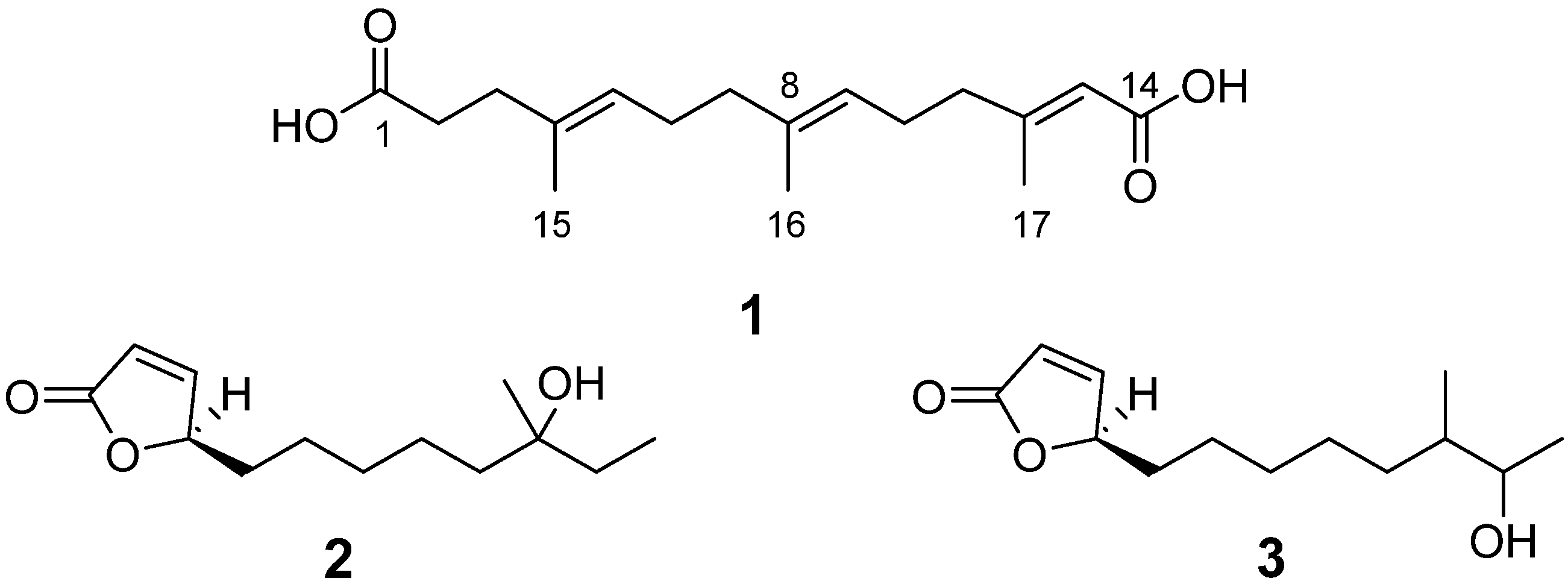
Figure 1. Structures of compounds 13.
Marinedrugs 14 00184 g001
Figure 2. Comparative HPLC analysis of the secondary metabolites in the culture extracts of wild-type S. somaliensis SCSIO ZH66 and its ΔwblAso mutant strain. The notably enhanced peaks (13) in the ΔwblAso mutant strain were numbered.
Marinedrugs 14 00184 g002
Figure 3. Key 1H–1H COSY and HMBC correlations of compound 1.
Marinedrugs 14 00184 g003
Figure 4. Key NOSEY correlations of compound 1.
Marinedrugs 14 00184 g004
Table 1. 1H and 13C-NMR data of compound 1 in DMSO-d6 (δ in ppm, J in Hz).
PositionδHδC
1-174.1
22.26 (2H, m)32.7
32.16 (2H, t, 8.4)34.2
4-133.6
55.09 (1H, m)124.0
62.02 (2H, m)26.1
71.93 (2H, t, 7.8)39.1
8-135.2
95.07 (1H, m)123.1
102.12 (2H, m)25.5
112.12 (2H, m)40.0
12 158.4
135.58 (1H, s)116.2
14 167.4
151.55 (3H, s)15.8
161.56 (3H, s)15.8
172.07 (3H, s)18.2

