Communication

A New Lyngbyatoxin from the Hawaiian Cyanobacterium Moorea producens

1
Department of Ocean Sciences, Tokyo University of Marine Science and Technology, Tokyo 108-8477, Japan
2
Division of Food Science and Biotechnology, Graduate School of Agriculture, Kyoto University, Kyoto 606-8502, Japan
3
National Research Institute of Fisheries Science, Yokohama 236-8648, Japan
4
Richard L. Roudebush VA Medical Center, Indianapolis, IN 46202, USA
*
Author to whom correspondence should be addressed.
Mar. Drugs 2014, 12(5), 2748-2759; https://doi.org/10.3390/md12052748
Submission received: 17 February 2014 / Revised: 6 April 2014 / Accepted: 8 April 2014 / Published: 12 May 2014

Abstract

:
Lyngbyatoxin A from the marine cyanobacterium Moorea producens (formerly Lyngbya majuscula) is known as the causative agent of “swimmer’s itch” with its highly inflammatory effect. A new toxic compound was isolated along with lyngbyatoxin A from an ethyl acetate extract of M. producens collected from Hawaii. Analyses of HR-ESI-MS and NMR spectroscopies revealed the isolated compound had the same planar structure with that of lyngbyatoxin A. The results of optical rotation and CD spectra indicated that the compound was a new lyngbyatoxin A derivative, 12-epi-lyngbyatoxin A (1). While 12-epi-lyngbyatoxin A showed comparable toxicities with lyngbyatoxin A in cytotoxicity and crustacean lethality tests, it showed more than 100 times lower affinity for protein kinase Cδ (PKCδ) using the PKCδ-C1B peptide when compared to lyngbyatoxin A.

Graphical Abstract

1. Introduction

Moorea producens (formerly classified as Lyngbya majuscula) [1] a filamentous marine cyanobacterium can cause human skin irritation (seaweed dermatitis) known as “swimmer’s itch” [2]. The causative agents of “swimmer’s itch” have been reported to be lyngbyatoxin A, aplysiatoxin and their derivatives, which are produced by M. producens [3,4,5]. These toxins have been shown to possess potent tumor-promoting activity and ability to activate protein kinase C isozymes [6,7]. Fatal intoxication due to ingestion of lyngbyatoxin A contaminated flesh of the turtle Chelonia mydas has been reported [8,9]. In addition, aplysiatoxin and related toxins were revealed to be the causative agents of food poisoning by the red alga Gracilaria coronopifolia [10,11,12]. The true producer of these toxins involved in these poisoning cases was deduced to be M. producens [8,10]. Since these toxins produced by M. producens are suspected as fatal tumor-causing factors for marine animals, such as the green turtle and manatee [13,14], the study of toxins produced by M. producens is important from an ecotoxicological point of view. Additionally, M. producens is a rich source of unique compounds which has led to the extensive study of its bioactive compounds, that may lead to the discovery of novel therapeutics agents [15,16,17]. We examined the toxic components in the extracts of M. producens collected from Hawaii guided by the lethal activity toward crustaceans. A new lyngbyatoxin derivative (1, 12-epi-lyngbyatoxin A, Figure 1) was isolated along with lyngbyatoxin A (2, Figure 1). In this report, the isolation, structure and toxicities of compound 1 will be discussed.
Figure 1. Structures of compounds 15.
Marinedrugs 12 02748 g001
1: 12-Epi-lyngbyatoxin A (9S,12R,19R); 2: Lyngbyatoxin A (teleocidin A-1) (9S,12S,19R);
3: Teleocidin A-2 (9S,12S,19S); 4: Enantiomer of 2 (9R,12R,19S); 5: Enantiomer of 3 (9R,12R,19R).

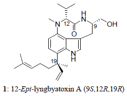
2. Results and Discussion
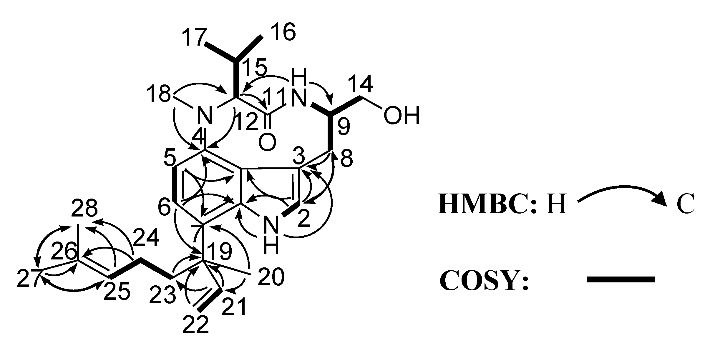
Compounds 1 and 2 were both isolated as pale yellow gummy solids. Compound 2 was identified to be lyngbyatoxin A which had been first isolated from the Hawaiian cyanobacterium [3]. HR-ESI-MS of compound 1 exhibited a molecular ion peak at m/z 438.3070 [M + H]+, consistent with the molecular formula of C27H39N3O2, which was the same molecular formula with that of lyngbyatoxin A (2). The presence of an indole ring was suggested from its UV spectrum (λmax (EtOH) nm (log ε) 231 (4.33), 301 (3.86)) comparing with that of 2.
Comparison of the 1H and 13C NMR data of 1 with those of 2, together with 2D NMR spectral analysis led us to elucidate the planar structure of the new compound as 1 (Figure 2). The planar structure of 1 was completely the same as that of lyngbyatoxin A (2). 1H and 13C NMR spectral data for 1 were shown in Table 1. On 1H NMR, most of the chemical shifts of 1 were closely similar to those of 2 (see Supplementary Information, Table S1). However, some proton chemical shifts (for example, H-9, H-12 and H-14) on a nine-membered lactam ring were somewhat different from those of 2. From these observations, 1 was deduced to have the same planar structure with 2. However, the absolute configuration around the nine-membered lactam ring appeared to be different between 1 and 2.
Figure 2. Key correlations of compound 1 in the COSY (bold line) and HMBC (arrow) spectra.
Marinedrugs 12 02748 g002
Lyngbyatoxin A (2) was first isolated as the causative agent of seaweed dermatitis from the cyanobacterium [3]. Later lyngbyatoxin A was found to be identical to teleocidin A-1 (2), the toxic metabolite from the actinomycete Streptomyces mediocidicus [18]. Teleocidin A-2 (3, Figure 1) of which C-19 had S configuration was also reported from S. mediocidicus [18]. The only structural difference between 2 and 3 was the configuration on C-19 in the linalyl group side chain. The circular dichroism (CD) spectra of compounds 2 and 3 showed only difference at around 230 nm (see Supplementary Information, Figure S22) [19,20]. The CD spectra around 230 nm of 2 and 3 showed upward and downward curves, respectively. Furthermore, CD spectra around 230 nm of 4 and 5 (synthesized compounds, Figure 1) showed downward and upward curves, respectively (see Supplementary Information, Figure S22) [20]. These results indicated the absolute configurations on C-19 of the linalyl group in lyngbyatoxin A derivatives were defined around 230 nm (CD spectra) as 19-R and 19-S configurations which resulted in upward and downward curves, respectively. CD spectra of 1 and 2 were shown in Figure 3. Both compounds showed upward curves around 230 nm, indicated that compound 1 had the same absolute configuration R at C-19 with that of 2. In addition, the CD spectra of compounds 2 and 5 showed differences at around 220 nm and 270 nm. The spectra around 220 nm and 270 nm of compound 2 showed both downward curves, while compound 5 showed both upward curves. The same spectral tendencies were observed in compounds 3 and 4 (see Supplementary Information, Figure S22) [20]. The both upward curves at 220 and 270 nm meant C-9 (R) and C-12 (R), while both downward curves meant C-9 (S) and C-12 (S). On the other hand, the CD spectra of compound 1 showed upward curve at 220 nm and downward curve at 270 nm. The results indicated that compound 1 had 9S,12R or 9R,12S configurations. Taking this into consideration, it was supposed that the absolute configurations of compound 1 were 9S,12R,19R or 9R,12S,19R.
Table 1. NMR spectroscopic data for compound 1 in CDCl3.
No.δC aδH (J in Hz) bHMBC
1 8.45, s3, 3a, 7a
2121.8 6.81, d (1.9)3, 3a, 7a, 8
3113.8
3a120.9
4146.7
5109.0 6.73, d (8.1)3, 3a, 4, 7
6119.8 6.95, d (8.1)4, 5, 7a, 19
7122.7
7a136.6
832.3 3.27, dd (15.6, 2.5)2, 3, 4, 9, 14
2.90, dd (15.6, 2.5)
957.6 3.84, br, m
10 7.45, br, s9, 12, 14
11175.2
1269.0 3.91, d (10.6)4, 11, 15, 17
1465.4 3.88, dd (10.4, 3.6)8, 9
3.82, dd (10.4, 6.5)
1528.1 2.61, m12, 16, 17
1620.0 0.67, d (6.6)12, 15, 17
1720.4 0.74, d (6.5)12, 15, 16
1831.6 3.09, s4, 12
1943.4
2024.71.44, s7, 19, 21, 23
21148.8 6.21, dd (17.7, 10.6)7, 19, 23
22112.2 5.29, dd (17.9, 1.4)19, 21
5.26, dd (10.7, 1.4)
2338.2 1.98, m7, 19, 21, 24, 25
1.81, m
2422.7 1.91, br, m23, 25, 26, 28
1.71, br, m
25124.6 5.08, t (7.1)23, 24, 27, 28
26131.5
2717.4 1.40, s25, 26, 28
2825.7 1.64, s25, 26, 27
OH on 14 Not observed
a Recorded at 200 MHz; b Recorded at 800 MHz. Coupling constants (Hz) are in parentheses. Abbreviations: s, singlet; d, doublet; t, triplet; m, multiplet; br, broad.
Figure 3. CD spectra of compounds 1 and 2.
Marinedrugs 12 02748 g003
The 1H NMR spectrum of 2 in CDCl3 showed that it existed as a mixture of trans and cis amide conformers. The conformational ratio of lyngbyatoxin A was about 1:3 (trans:cis) (see Supplementary Information, Figure S24). This conformational ratio was almost the same as that of (−)-indolactam-V (IL-V, 6, Figure 4) [21,22], which is regarded to be a core structure exhibiting tumor-promoting activity of teleocidins (lyngbyatoxins) [23]. IL-V (6), which has chiral carbon centers at C-9 and C-12 with the configurations of S and S, is a partial structure of 2. Therefore, the enantiomer (9R,12R) of IL-V should also exist as two conformers of which the ratio is about 1:3 (trans:cis) in CDCl3. In this study, it was shown that compound 1 had nearly one conformer on the 1H NMR spectrum (see Supplementary Information, Figure S23). It was reported that (+)-epi-IL-V (9S,12R) should have more than two conformers by the computational calculations [24]. However, it had been shown that the main conformation was dominant for (+)-epi-IL-V (9S,12R) on the 1H NMR spectrum as in the case of compound 1 [21,24]. Therefore, the absolute configurations at C-9 and C-12 in the new lyngbyatoxin derivative (1) were inferred to be S and R or R and S, respectively, which further certified the conclusion drawing from the analyses of the CD spectra.
IL-V (6) and a number of related compounds have been isolated from natural samples or synthesized [3,18,19,20,25,26,27,28,29,30,31,32,33]. Those reports showed that all the reported compounds containing 6 (9S,12S) so far had levorotatory optical rotations no matter what the terpene group was connected to the indole ring (see Supplementary Information, Figures S26 and S27). However, we should pay attention that the levorotatory optical rotation of IL-V is determined not only by the 9S,12S configuration, but also by the main conformer of cis amide in the solution [30,34]. The synthetic IL-Vs (9S,12S) and (9R,12S) showed levorotatory optical rotation while IL-Vs (9R,12R) and (9S,12R) had dextrorotatory optical rotation (see Supplementary Information, Figures S26 and S27) [26,28]. Since the optical rotation of 1 was dextrorotatory, the configurations of indolactam (IL-V) of 1 were deduced to be 9R,12R or 9S,12R. Moreover, the absolute configurations at C-9 and C-12 in compound 1 were inferred to be 9S,12R or 9R,12S from the results of CD analysis. The absolute configuration of C-19 has been deduced as R also from CD spectra. When taken these results together, it was deduced that compound 1 had 9S,12R,19R absolute configurations. Furthermore, the deduced absolute chemistry of indolactam of 1 (9S,12R) was also supported by the 1H NMR spectra of 1 in CD3OD which was superimposable with those of (+)-epi-indolactam V (see Supplementary Information, Figure S25, Table S2) [28]. Thus, leading us to conclude that compound 1 was 12-epi-lyngbyatoxin A. This is the first report of 12-epi-lyngbyatoxin A (1) from the nature. Compound 1 had been obtained during the synthetic study of lyngbyatoxin A (2, teleocidin A-1) as one of the inseparable mixtures [20].
Figure 4. The structure of (−)-indolactam-V (6).
Marinedrugs 12 02748 g004
6 (−)-indolactam-V.
In cytotoxic assays using L1210 leukemia cells, the IC50 values of 1 and 2 were 20.4 μM and 8.1 μM, respectively. The IC50 value of 1 was slightly higher than that of 2. Both compounds showed moderate cytotoxicity. Compound 2 was formerly reported to exhibit cytotoxic effects against HeLa and ACC-MESO-1 cells with IC50 of 35 μM and 11 μM, respectively [32]. These values are comparable to the cytotoxic activities of 1 and 2 obtained in this study.
In the crustacean lethal activity tests using the shrimp Palaemon paucidens, both 1 and 2 showed similar lethal activities. The LD100 value of 1 was 7.5 mg/kg, while 2 was 5 mg/kg. Injection of 1 and 2 caused paralysis on the tested shrimp. Paralysis was not observed with compound 2 if the dose was less than the lethal amount. However, it was observed that compound 1 caused paralysis with a dose of 0.9 mg/kg. These results may indicate that 1 and 2 have different toxicity pathways.
Lyngbyatoxin A (2, teleocidion A-1) and related compounds are known as potent tumor promoters [6]. These compounds bind to the cysteine-rich C1 domains (C1A and C1B) of protein kinase C (PKC) isozymes to activate them, possibly leading to tumor formation. Affinities of the isolated compounds for PKCδ using a synthetic PKCδ-C1B peptide were evaluated by competition binding assay with [3H]phorbol 12,13-dibutyrate (PDBu). The values of Ki for the inhibition of [3H]PDBu-binding were 17 nM and 0.11 nM for 1 and 2, respectively. Aplysiatoxin (ATX) and debromoaplysiatoxin (DATX) are 12-O-tetradecanoylphorbol-13-acetate (TPA)-type tumor promoters which activate PKC as with lyngbyatoxin A (2) [6,29]. It was reported that Ki values of ATX and DATX for binding to PKCδ-C1B are 0.41 nM [35,36] and 0.20 nM [37], respectively. These Ki values are comparable to that of compound 2 obtained in this study. On the other hand, the affinity for PKCδ-C1B binding of compound 1 was more than a hundred times weaker than that of compound 2. The results suggest the C12 configuration of the indolactam moiety of lyngbyatoxin A is very important for the binding with the PKCδ-C1B peptide.
Our results showed the Ki value for PKCδ-C1B peptide binding of lyngbyatoxin A (2) was similar to those of other PKC activators aplysiatoxin and debromoaplysiatoxin. Lyngbyatoxin A (2) has been reported to be a potent tumor promoter and have the ability to strongly induce PKC activity [6,7,29]. While 12-epi-lyngbyatoxin A (1) and lyngbyatoxin A (2) showed comparable cytotoxic and crustacean lethal activities, 12-epi-lyngbyatoxin A (1) had more than 100 times lower binding affinity for PKC compared to lyngbyatoxin A (2). The results revealed that the cytotoxic and lethal mechanism of lyngbyatoxin-type compounds might be mediated through a non-PKC activation pathway. In this connection, there was a report about no correlation between the cytotoxicity and ability to bind to and activate PKCδ when examining the contribution of PKCδ to the cytotoxicity of aplysiatoxin related compounds [38]. Therefore, other targets that offer alternatives to PKC isozymes might exist for the expression of toxic activities by lyngbyatoxin-type compounds. Further study is necessary to verify this hypothesis.
In addition, lyngbyatoxin A (2) was reported as the causative agent of human skin irritation and marine turtle poisoning [3,8,9]. Since the new lyngbyatoxin derivative (1) has the similar cytotoxicity and lethal activity with lyngbyatoxin A (2), it is supposed that 12-epi-lyngbyatoxin A (1) is also a causative agent possibly.

3. Experimental Section

3.1. General Experimental Procedures

HPLC was performed using a SHIMADZU HPLC (SHIMADZU Co., Kyoto, Japan) pump equipped with a SPD-M10A diode array detector. HR-ESI-MS spectra data were determined using Bruker micrOTOF QII (Bruker Co., Bremen, Germany) mass spectrometer. NMR spectra were recorded in CDCl3 at 800 MHz and 200 MHz for 1H and 13C on a Bruker AVANCE III 800 MHz (Bruker Co., Bremen, Germany) spectrometer or in CD3OD at 600 MHz for 1H on a Bruker AVANCE III 600 MHz spectrometer; the chemical shifts were reported in δ units (ppm) using CDCl3 solvent (δH at 7.3 ppm and δC at 77.2 ppm) or using CD3OD solvent (δH at 3.3 ppm) as the internal standard signals. Optical rotations were measured on a JASCO P-2100 series.CD spectra were measured on a JASCO J-715 (JASCO Co., Tokyo, Japan) and UV spectra were on a HITACHI U-3000 (Hitachi High-Tech Fielding Co., Tokyo, Japan) spectrometer.

3.2. Marine Cyanobacterium Moorea producens

Samples of a marine cyanobacterium, Moorea producens, were collected at Kahala Beach, Oahu island, Hawaii in 1998. After freeze-drying, samples were stored at −30 °C until experiments were performed.

3.3. Extraction and Isolation

The dried sample (823 g dry wt.) was extracted twice with ethanol, six times with methanol and five times with acetone (see Supplementary Information, Figure S28). After evaporation of the solvents, the extracts were combined and partitioned between hexane and 80% methanol. The 80% methanol layer was again evaporated and partitioned with a mixture of ethyl acetate (EtOAc) and distilled water. The fraction of distilled water was then dissolved with butanol and separated into two extracts. The ethyl acetate fraction was then subjected to an open glass column 20 × 300 mm packed with ODS resin (Pegasil Prep ODS, Senshu Co., Tokyo, Japan), with stepwise in 50%, 70%, 90% and 100% methanol. Each eluted fraction was tested with crustacean lethal toxicity. The most toxic fraction eluted with 90% methanol (1220 mg dry wt.) was purified by reversed phase liquid chromatography on a 20 × 250 mm column (SHISEIDO CAPCELL PAK-ODS, Shiseido Co., Tokyo, Japan) using an isocratic HPLC system (flow rate; 4 mL/min, detection; 190–800 nm) with 82% MeOH to yield the main toxic peak which was consisted with at least two compounds. A recycle chromatography HPLC system (system unit; Senshu Scientific SSC-1310 (Senshu Co., Tokyo, Japan), column; SHISEIDO CAPCELL PAK-ODS (Shiseido Co., Tokyo, Japan) 10 × 250 mm, solvent; 82% MeOH, flow rate; 1 mL/min, detection; 210 nm) was then utilized to completely isolate 12-epi-lyngbyatoxin A (1) (2.8 mg) and lyngbyatoxin A (2) (10.2 mg) from the main toxic fraction.

3.4. 12-Epi-Lyngbyatoxin A (1) and Lyngbyatoxin A (2)

12-Epi-lyngbyatoxin A (1): Gummy solid; [α]d18 = +85.5° (c 0.12, MeOH); Circular dichroism (CD, in MeOH), [θ]212.5nm −2760, [θ]215nm 970, [θ]225nm 33,900, [θ]235nm 41,200, [θ]249.5nm 1650, [θ]252.5nm −1650, [θ]258nm −4100, [θ]280nm −690, [θ]310nm 1470, [θ]330nm 630 (Figure 3); UV λmax (EtOH) nm (log ε) 231 (4.33), 301 (3.86); 1H NMR (800 MHz, CDCl3; 600 MHz, CD3OD) and 13C NMR (200 MHz, CDCl3) data; HRESIMS m/z [M + H]+ 438.3070 (calcd for C27H39N3O2, 437.3042).
Lyngbyatoxin A (2): Gummy solid; [α]d18 = −102.4° (c 0.13, MeOH); Circular dichroism (CD, in MeOH), [θ]215nm −660, [θ]221.5nm −6550, [θ]229.5nm 2670, [θ]234nm 12,200, [θ]237.5nm 2510, [θ]257nm −20,100, [θ]298nm −790, [θ]313nm 5520, [θ]333nm 470 (Figure 3); UV λmax (EtOH) nm (log ε) 230 (4.46), 301 (3.98); 1H NMR (800 MHz, CDCl3) and 13C NMR (200 MHz, CDCl3) data.

3.5. Cytotoxicity Assay

The cytotoxic activities of compounds 1 and 2 were tested toward L1210 mouse leukemia cells. The detailed methods were reported previously [39]. Differences with the reported methods [39] were (a) samples were dissolved with methanol; and (b) the methanol dissolved samples were applied onto each well and air-dried before the addition of L1210 cells.

3.6. Crustacean Lethality Test

The shrimp Palaemon paucidens (average weight, 0.5 g) was used for the crustacean lethality test. Tested samples were suspended with 1% Tween 20 (polyoxyethylene (20) sorbitan monolaurate). Two micro liter of each sample suspension was injected into the abdominal cavity of the shrimp (n = 3–5). The shrimp were monitored every 30 min for 4 h. Through the lethality tests, the lethal dose 100% values (LD100) were determined. One percent Tween 20 without sample was used as reference. LD100 was defined as weight of sample per unit weight of crustacean.

3.7. Binding Assay of PKC Ligands Using PKC-C1B Peptide

The binding of [3H]PDBu to the PKCδ-C1B peptide was evaluated by the procedure of Sharkey and Blumberg [40] with modifications as reported previously [41,42] using 50 mM tris-maleate buffer (pH 7.4 at 4 °C), 13.8 nM PKCδ-C1B peptide, 20 nM [3H]PDBu (18.7 Ci/mmol, Perkin-Elmer Life Sciences), 50 μg/mL 1,2-dioleoyl-sn-glycero-3-phospho-l-serine (Sigma, St. Louis, MO, USA), 3 mg/mL bovine γ-globulin (Sigma, St. Louis, MO, USA), and various concentrations of inhibitors. The concentration of the properly folded PKCδ-C1B peptide was estimated to be about 3 nM on the basis of Bmax values of Scatchard analyses reported previously [41,42]. Binding affinity was evaluated based on the concentration required to cause 50% inhibition of the specific binding of [3H]PDBu, the IC50, which was calculated with Microsoft Excel. The inhibition constant, Ki, was calculated by the method of Sharkey and Blumberg [40].

4. Conclusions

The lethal toxicity guided purification of an ethyl acetate extract from the cyanobacterium Moorea producens resulted in the isolation of lyngbyatoxin A (2) and a new compound 12-epi-lyngbyatoxin A (1). The absolute configuration of compound 1 was deduced from the analyses of the NMR, optical rotation and CD spectra. While 12-epi-lyngbyatoxin A (1) and lyngbyatoxin A (2) showed comparable cytotoxic and crustacean lethal activities, 12-epi-lyngbyatoxin A (1) had more than 100 times lower binding affinity for protein kinase Cδ compared to lyngbyatoxin A (2). This suggests that the cytotoxic and lethal mechanism of lyngbyatoxin-type compounds might be mediated through a non-PKC activation pathway.

Supplementary Files

  • Supplementary File 1:

    Supplementary Information (PDF, 1823 KB)

  • Acknowledgments

    This work was supported partly by JSPS Grant-in-Aid for Scientific Research Grant Number 24510294 (to Hiroshi Nagai).

    Author Contributions

    Hiroshi Nagai conceived and designed the research. Hiroshi Nagai and Kazuhiro Irie supervised the research. Hiroshi Nagai and Bryan Sakamoto contributed to the sample collection. Weina Jiang and Wei Zhou purified the compound. Ryuichi Watanabe, Toshiyuki Suzuki and Hajime Uchida performed the spectral measurement. Weina Jiang carried out the structural identification, toxicity assay. Masayuki Kikumori performed the binding assay. Weina Jiang, Kazuhiro Irie and Hiroshi Nagai analyzed the data and wrote the paper. Michiya Kamio and Bryan Sakamoto reviewed and edited the manuscript.

    Conflicts of Interest

    The authors declare no conflict of interest.

    References

    1. Engene, N.; Rottacker, E.C.; Kaštovský, J.; Byrum, T.; Choi, H.; Ellisman, M.H.; Komárek, J.; Gerwick, W.H. Moorea producens gen. nov., sp. nov. and Moorea bouillonii comb. nov., tropical marine cyanobacteria rich in bioactive secondary metabolites. Int. J. Syst. Evol. Microbiol. 2012, 62, 1171–1178. [Google Scholar] [CrossRef]
    2. Burja, A.M.; Banaigs, B.; Abou-Mansour, E.; Grant Burgess, J.; Wright, P.C. Marine cyanobacteria—Aprolific source of natural products. Tetrahedron 2001, 57, 9347–9377. [Google Scholar] [CrossRef]
    3. Cardellina, J.H.; Marner, F.J.; Moore, R.E. Seaweed dermatitis: Structure of lyngbyatoxin A. Science 1979, 204, 193–195. [Google Scholar]
    4. Moore, R.E.; Blackman, A.J.; Cheuk, C.E.; Mynderse, J.S.; Matsumoto, G.K.; Clardy, J.; Woodard, R.W.; Craig, J.C. Absolute stereochemistries of the aplysiatoxins and oscillatoxin A. J. Org. Chem. 1984, 49, 2484–2489. [Google Scholar] [CrossRef]
    5. Mynderse, J.S.; Moore, R.E.; Kashiwagi, M.; Norton, T.R. Antileukemia activity in the Osillatoriaceae: Isolation of debromoaplysiatoxin from Lyngbya. Science 1977, 196, 538–540. [Google Scholar]
    6. Fujiki, H.; Mori, M.; Nakayasu, M.; Terada, M.; Sugimura, T.; Moore, R.E. Indole alkaloids: Dihydroteleocidin B, teleocidin, and lyngbyatoxin A as members of a new class of tumor promoters. Proc. Natl. Acad. Sci. USA 1981, 78, 3872–3876. [Google Scholar] [CrossRef]
    7. Fujiki, H.; Tanaka, Y.; Miyake, R.; Kikkawa, U.; Nishizuka, Y.; Sugimura, T. Activation of calcium-activated, phospholipid-dependent protein kinase (protein kinase C) by new classes of tumor promoters: Teleocidin and debromoaplysiatoxin. Biochem. Biophys. Res. Commun. 1984, 120, 339–343. [Google Scholar] [CrossRef]
    8. Yasumoto, T. Fish poisoning due to toxins of microalgal origins in the Pacific. Toxicon 1998, 36, 1515–1518. [Google Scholar] [CrossRef]
    9. Ito, E.; Satake, M.; Yasumoto, T. Pathological effects of lyngbyatoxin A upon mice. Toxicon 2002, 40, 551–556. [Google Scholar] [CrossRef]
    10. Nagai, H.; Yasumoto, T.; Hokama, Y. Aplysiatoxin and debromoaplysiatoxin as the causative agents of a red alga Gracilaria. coronopifolia poisoning in Hawaii. Toxicon 1996, 34, 753–761. [Google Scholar] [CrossRef]
    11. Nagai, H.; Yasumoto, T.; Hokama, Y. Manauealides, some of the causative agents of a red alga Gracilaria. coronopifolia poisoning in Hawaii. J. Nat. Prod. 1997, 60, 925–928. [Google Scholar] [CrossRef]
    12. Nagai, H.; Kan, Y.; Fujita, T.; Sakamoto, B.; Hokama, Y. Manauealide C and anhydrodebromoaplysiatoxin, toxic constituents of the Hawaiian red alga, Gracilaria Coronopifolia. Biosci. Biotechnol. Biochem. 1998, 62, 1011–1013. [Google Scholar] [CrossRef]
    13. Arthur, K.; Limpus, C.; Balazs, G.; Capper, A.; Udy, J.; Shaw, G.; Keuper-Bennett, U.; Bennett, P. The exposure of green turtles (Chelonia mydas) to tumour promoting compounds produced by the cyanobacterium Lyngbya. majuscula and their potential role in the aetiology of fibropapillomatosis. Harmful Algae 2008, 7, 114–125. [Google Scholar] [CrossRef]
    14. Harr, K.E.; Szabo, N.J.; Cichra, M.; Phlips, J.E. Debromoaplysiatoxin in Lyngbya-dominated mats on manatees (Trichechus manatus latirostris) in the Florida King’s Bay ecosystem. Toxicon 2008, 52, 385–388. [Google Scholar] [CrossRef]
    15. Namikoshi, M.; Rinehart, K.L. Bioactive compounds produced by cyanobacteria. J. Ind. Microbiol. 1996, 17, 373–384. [Google Scholar] [CrossRef]
    16. Gademann, K.; Portmann, C. Secondary metabolites from cyanobacteria: Complex structures and powerful bioactivities. Curr. Org. Chem. 2008, 12, 326–341. [Google Scholar] [CrossRef]
    17. Molinski, T.F.; Dalisay, D.S.; Lievens, S.L.; Saludes, J.P. Drug development from marine natural products. Nat. Rev. Drug. Discov. 2008, 8, 69–85. [Google Scholar]
    18. Sakai, S.I.; Hitotsuyanagi, Y.; Aimi, N.; Fujiki, H.; Suganuma, M.; Sugimura, T.; Endo, Y.; Shudo, K. Absolute configuration of lyngbyatoxin A (teleocidin A-1) and teleocidin A-2. Tetrahedron Lett. 1986, 27, 5219–5220. [Google Scholar] [CrossRef]
    19. Aimi, N.; Odaka, H.; Sakai, S.I.; Fujiki, H.; Suganuma, M.; Moore, R.E.; Patterson, G.M.L. Lyngbyatoxins B and C, two new irritants from Lyngbya majuscula. J. Nat. Prod. 1990, 53, 1593–1596. [Google Scholar] [CrossRef]
    20. Muratake, H.; Okabe, K.; Natsume, M. Synthesis of teleocidins A, B and their congeners. Part 2. Synthesis of lyngbyatoxin A (teleocidin A-1), teleocidin A-2, pendolmycin, and (R,E)- and (S,E)-7-(3,7,11-trimethyl-1,6,10-dodecatrien-3-yl)-(−)-indolactams V. Tetrahedron 1991, 47, 8545–8558. [Google Scholar] [CrossRef]
    21. Endo, Y.; Shudo, K.; Okamoto, T. Molecular requirements for epigenetic modulators. Synthesis of active fragments of teleocidins and lyngbyatoxin. Chem. Pharm. Bull. 1982, 30, 3457–3460. [Google Scholar] [CrossRef]
    22. Irie, K.; Hirota, M.; Hagiwara, N.; Koshimizu, K.; Hayashi, H.; Murao, S.; Tokuda, H.; Ito, Y. The Epstein-Barr virus early antigen inducing indole alkaloids, (−)-indolactam-V and its related compounds, produced by Actinomycetes. Agric. Biol. Chem. 1984, 48, 1269–1274. [Google Scholar]
    23. Fujiki, H.; Suganuma, M.; Nakayasu, M.; Tahira, T.; Endo, Y.; Shudo, K.; Sugimura, T. Structure-activity studies on synthetic analogues (indolactams) of the tumor promoter teleocidin. Jpn. J. Cancer Res. 1984, 75, 866–870. [Google Scholar]
    24. Kawai, T.; Ichinose, T.; Takeda, M.; Tomioka, N.; Endo, Y.; Yamaguchi, K.; Shudo, K.; Itai, A. Prediction of ring conformations of indolactams. Crystal and solution structures. J. Org. Chem. 1992, 57, 6150–6155. [Google Scholar] [CrossRef]
    25. Sakai, S.I.; Aimi, N.; Yamaguchi, K.; Hitotsuyanagi, Y.; Watanabe, C.; Yokose, K.; Koyama, Y.; Shudo, K.; Itai, A. Elucidation of the structure of olivoretin A and D (teleocidin B). Chem. Pharm. Bull. 1984, 32, 354–357. [Google Scholar] [CrossRef]
    26. Endo, Y.; Shudo, K.; Furuhata, K.; Ogura, H.; Sakai, S.I.; Aimi, N.; Hitotsuyanagai, Y.; Koyama, Y. Synthesis of optically active teleocidin derivatives. Absolute configuration of teleocidin B and olivoretin A. Chem. Pharm. Bull. 1984, 32, 358–361. [Google Scholar] [CrossRef]
    27. Hitotsuyanagi, Y.; Yamaguchi, K.; Ogata, K.; Aimi, N.; Sakai, S.I.; Koyama, Y.; Endo, Y.; Shudo, K.; Itai, A.; Iitaka, Y. Elucidation of the structures of olivoretin B and C. Chem. Pharm. Bull. 1984, 32, 3774–3778. [Google Scholar] [CrossRef]
    28. Endo, Y.; Shudo, K.; Itai, A.; Hasegawa, M.; Sakai, S.I. Synthesis and stereochemistry of indolactam-V, an active fragment of teleocidins. Structural requirements for tumor-promoting activity. Tetrahedron 1986, 42, 5905–5924. [Google Scholar] [CrossRef]
    29. Fujiki, H.; Sugimura, T. New classes of tumor promoters: Teleocidin, aplysiatoxin, and palytoxin. Adv. Cancer Res. 1987, 49, 223–264. [Google Scholar] [CrossRef]
    30. Irie, K.; Kajiyama, S.-I.; Funaki, A.; Koshimizu, K.; Hayashi, H.; Arai, M. Biosynthesis of indole alkaloid tumor promoter teleocidins (I): Possible biosynthetic pathway of the monoterpenoid moieties of teleocidins. Tetrahedron 1990, 46, 2773–2788. [Google Scholar] [CrossRef]
    31. Irie, K.; Isaka, T.; Iwata, Y.; Yanai, Y.; Nakamura, Y.; Koizumi, F.; Ohigashi, H.; Wender, P.A.; Satomi, Y.; Nishino, H. Synthesis and biological activities of new conformationally restricted analogues of (−)-indolactam-V: Elucidation of the biologically active conformation of the tumor-promoting teleocidins. J. Am. Chem. Soc. 1996, 118, 10733–10743. [Google Scholar] [CrossRef]
    32. Izumikawa, M.; Khan, S.T.; Komaki, H.; Takagi, M.; Shin-ya, K. JBIR-31, a new teleocidin analog, produced by salt-requiring Streptomyces sp. NBRC 105896 isolated from a marine sponge. J. Antibiot. 2009, 63, 33–36. [Google Scholar]
    33. Pu, J.; Deng, K.; Butera, J.; Chlenov, M.; Gilbert, A.; Kagan, M.; Mattes, J.; Resnick, L. De novo synthesis of teleocidin B analogs. Tetrahedron 2010, 66, 1963–1972. [Google Scholar] [CrossRef]
    34. Irie, K.; Hagiwara, N.; Koshimizu, K. New probes for receptor analysis of tumor promoters synthesis of fluorescent derivatives of (−)-indolactam V, the basic ring-structure of teleocidins. Tetrahedron 1987, 43, 5251–5260. [Google Scholar] [CrossRef]
    35. Nakagawa, Y.; Yanagita, R.C.; Hamada, N.; Murakami, A.; Takahashi, H.; Saito, N.; Nagai, H.; Irie, K. A simple analogue of tumor-promoting aplysiatoxin is an antineoplastic agent rather than a tumor promoter: Development of a synthetically accessible protein kinase C activator with bryostatin-like activity. J. Am. Chem. Soc. 2009, 131, 7573–7579. [Google Scholar] [CrossRef]
    36. Yanagita, R.C.; Kamachi, H.; Tanaka, K.; Murakami, A.; Nakagawa, Y.; Tokuda, H.; Nagai, H.; Irie, K. Role of the phenolic hydroxyl group in the biological activities of simplified analogue of aplysiatoxin with antiproliferative activity. Bioorg. Med. Chem. Lett. 2010, 20, 6064–6066. [Google Scholar] [CrossRef] [Green Version]
    37. Kikumori, M.; Yanagita, R.C.; Tokuda, H.; Suzuki, N.; Nagai, H.; Suenaga, K.; Irie, K. Structure-activity studies on the spiroketalmoiety of a simplified analogue of debromoaplysiatoxin with antiproliferative activity. J. Med. Chem. 2012, 55, 5614–5626. [Google Scholar] [CrossRef]
    38. Hanaki, Y.; Kikumori, M.; Ueno, S.; Tokuda, H.; Suzuki, N.; Irie, K. Structure-activity studies at position 27 of aplog-1, a simplified analog of debromoaplysiatoxin with anti-proliferative activity. Tetrahedron 2013, 69, 7636–7645. [Google Scholar] [CrossRef]
    39. Kawabata, T.; Lindsay, D.J.; Kitamura, M.; Konishi, S.; Nishikawa, J.; Nishida, S.; Kamio, M.; Nagai, H. Evaluation of the bioactivities of water-soluble extracts from twelve deep-sea jellyfish species. Fish. Sci. 2013, 79, 487–494. [Google Scholar] [CrossRef]
    40. Sharkey, N.A.; Blumberg, P.M. Highly lipophilic phorbolesters as inhibitors of specific [3H]phorbol 12,13-dibutyrate binding. Cancer Res. 1985, 45, 19–24. [Google Scholar]
    41. Irie, K.; Oie, K.; Nakahara, A.; Yanai, Y.; Ohigashi, H.; Wender, P.A.; Fukuda, H.; Konishi, H.; Kikkawa, U. Molecular basis for protein kinase C isozyme-selective binding: The synthesis, folding, and phorbol ester binding of the cysteine-rich domains of all protein kinase C isozymes. J. Am. Chem. Soc. 1998, 120, 9159–9167. [Google Scholar] [CrossRef]
    42. Shindo, M.; Irie, K.; Nakahara, A.; Ohigashi, H.; Konishi, H.; Kikkawa, U.; Fukuda, H.; Wender, P.A. Toward the identification of selective modulators of protein kinase C (PKC) isozymes: Establishment of a binding assay for PKC isozymes using synthetic C1 peptide receptors and identification of the critical residues involved in the phorbolester binding. Bioorg. Med. Chem. 2001, 9, 2073–2081. [Google Scholar] [CrossRef]

    Share and Cite

    MDPI and ACS Style

    Jiang, W.; Zhou, W.; Uchida, H.; Kikumori, M.; Irie, K.; Watanabe, R.; Suzuki, T.; Sakamoto, B.; Kamio, M.; Nagai, H. A New Lyngbyatoxin from the Hawaiian Cyanobacterium Moorea producens. Mar. Drugs 2014, 12, 2748-2759. https://doi.org/10.3390/md12052748

    AMA Style

    Jiang W, Zhou W, Uchida H, Kikumori M, Irie K, Watanabe R, Suzuki T, Sakamoto B, Kamio M, Nagai H. A New Lyngbyatoxin from the Hawaiian Cyanobacterium Moorea producens. Marine Drugs. 2014; 12(5):2748-2759. https://doi.org/10.3390/md12052748

    Chicago/Turabian Style

    Jiang, Weina, Wei Zhou, Hajime Uchida, Masayuki Kikumori, Kazuhiro Irie, Ryuichi Watanabe, Toshiyuki Suzuki, Bryan Sakamoto, Michiya Kamio, and Hiroshi Nagai. 2014. "A New Lyngbyatoxin from the Hawaiian Cyanobacterium Moorea producens" Marine Drugs 12, no. 5: 2748-2759. https://doi.org/10.3390/md12052748

    Article Metrics

    Back to TopTop