Review

Functional Consequences of Mutations and Polymorphisms in the Coding Region of the PAF Acetylhydrolase (PAF-AH) Gene

Huntsman Cancer Institute and Department of Internal Medicine, University of Utah, Salt Lake City, UT 84112, USA
Pharmaceuticals 2009, 2(3), 94-117; https://doi.org/10.3390/ph2030094
Submission received: 27 October 2009 / Revised: 10 November 2009 / Accepted: 19 November 2009 / Published: 20 November 2009
(This article belongs to the Special Issue Biomarkers)

Abstract

:
In the past several years a number of alterations in the PAF-AH/PLA2G7/LpPLA2 gene have been described. These include inactivating mutations, polymorphisms in the coding region, and other genetic changes located in promoter and intronic regions of the gene. The consequences associated with these genetic variations have been evaluated from different perspectives, including in vitro biochemical and molecular studies and clinical analyses in human subjects. This review highlights the current state of the field and suggests new approaches that can be used to evaluate functional consequences associated with mutations and polymorphisms in the PAF-AH gene.

1. Introduction

The first step in the metabolism of two classes of inflammatory phospholipid mediators, Platelet-Activating Factor (PAF) and oxidatively-fragmented phospholipids (OxPL), is catalyzed by enzymatic activities known as PAF acetylhydrolases (PAF-AHs). This family of phospholipases A2 includes intracellular and secreted activities. The secreted form of PAF-AH (also known as lipoprotein-associated phospholipase A2, LpPLA2, PLA2G7) circulates in plasma as a complex with high- and low-density lipoproteins (HDL and LDL, respectively). While the physiological consequences of dual lipoprotein association remain to be completely identified, it is apparent that the location of the enzyme impacts its function and that aberrant distribution is associated with a variety of syndromes. The role of PAF-AH in human biology continues to be the subject of intense research as increasing numbers of laboratories around the world are utilizing complementary approaches to examine the function of this enzyme in vivo. The well-recognized fact that altered expression of PAF-AH correlates with the incidence and severity of vascular and other inflammatory diseases has sparked the interest of a wide range of scientists and clinicians in both the public and private sectors. In addition, recent elegant work by Vadas and co-workers showed direct correlations between serum PAF levels and severity of anaphylaxis [1]. Importantly, PAF-AH activity was inversely correlated with disease severity and it was significantly lower in patients with fatal anaphylactic reactions than in patients in the control groups [1]. Considerable effort has been devoted to evaluating whether genetic variations in the PAF-AH gene are associated with the incidence and severity of human diseases, and whether these alterations affect the levels of protein expression and/or its function. A number of PAF-AH loss-of-function mutations and single nucleotide polymorphisms (SNPs) have been identified and studied to various extents with the objective of elucidating their impact in physiology and disease. These analyses have provided new mechanistic and clinical insights that may have important translational utility, including the potential for personalized diagnostic and therapeutic applications.

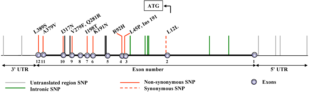
2. Naturally Occurring Genetic Alterations in the PAF-AH Gene

In humans, the PAF-AH gene harbors a number of mutations and polymorphisms within coding and non-coding regions (Figure 1).
Figure 1. Diagrammatic representation of mutations and polymorphisms in the PAF-AH gene.
Pharmaceuticals 02 00094 g001
The consequences of these genetic variations, particularly those that alter protein sequence, have been investigated in considerable detail in terms of the frequency with which they occur in both healthy and diseased subjects. One of the most thoroughly investigated variants is a loss-of-function mutation that occurs near the active site, and that has been observed mainly in Japanese [2], Taiwanese [3], Korean [4], and Chinese [5] populations. Three additional, but much less prevalent, loss-of-function mutations have been reported in Japanese subjects [6,7]. In addition, the coding region of the PAF-AH gene harbors several SNPs that moderately impact enzymatic activity of the gene product; these will be discussed in subsequent sections. Genetic alterations have also been described in untranslated regions [8]. This article summarizes current information on the incidence and functional consequences of non-synonymous variants in the coding region of the gene. A discussion of SNPs located in promoter, intronic, and 3’-untranslated regions of PAF-AH is beyond the scope of this review.

3. Inactivating Mutations

PAF-AH deficiency was initially characterized by Miwa and co-workers using functional approaches [2]. This trait is quite prevalent in Japan, as approximately 4% of the population lacks expression of the activity in plasma [2]. In most cases, PAF-AH deficiency occurs as a consequence of a missense mutation near the active site that results in a valine to phenylalanine transition (V279F, [9]). A much less frequent mutation, Q281R, also results in severe deficiency of PAF-AH [6,10]. Studies by Ishihara and co-workers revealed the occurrence of two additional loss-of-function variants, the incidence of which seems quite rare [7]. While PAF-AH deficiency alone has not been reported to have major physiological effects, several laboratories have demonstrated that a phenotype becomes evident when partial or complete deficiency is combined with other genetic defects or environmental factors, as is the case for many other gene products. Genetic studies in healthy and diseased populations indicate that PAF-AH deficiency affects the incidence and severity of a number of inflammatory diseases.

3.1. V279F (rs 16874954)

Most subjects who express virtually no plasma PAF-AH activity harbor two F279 alleles; heterozygous subjects express half the normal plasma activity [9]. Molecular studies revealed that the recombinant mutant protein is inactive [9]. The fact that a bulky residue (phenylalanine) replaces a smaller amino acid (valine) near the active site of the enzyme likely accounts for the loss of function associated with this mutation [9]. While the initial discovery of V279F was made in a Japanese cohort, the mutation was subsequently reported in Korean [4], Taiwanese [3], and Chinese [5] populations, and in subjects from Turkey, Azerbaijan, and Kyrgyzstan [11]. Allele frequency varies widely in these populations: the highest prevalence was reported in Japan and in Taiwan, followed by Korea and China (Table 1). Our studies, and those of others [12,13,14], revealed that complete PAF-AH deficiency does not occur in Caucasian subjects, and this has been confirmed in public databases. This variant likely originated in Japanese and Korean ancestors. The clinical consequences associated with complete and partial PAF-AH deficiency have been evaluated in patients suffering from a variety of inflammatory conditions in the vascular, respiratory, renal, and digestive systems (Table 1). These analyses clearly demonstrated that the incidence of the F279 allele correlates with several manifestations of vascular disease in Japanese subjects; multiple studies by Yamada and co-workers and by Unno et al. formed the bases to support this conclusion (Table 1).
Table 1. The V279F (rs16874954) polymorphism: allele frequency in control and diseased populations.
Population Clinical Status n Allele frequency, % p Authors [Ref.]
JapaneseControls26317.9Stafforini et al. [10]
Severe asthma26622.60.02
Control adults18821.0
JapaneseControl children14220.0Ito et al. [15]
Asthmatic children11831.00.007-0.004
JapaneseControls21721.7N. Satoh et al. [16]
Asthma27918.6N/S
JapaneseControls10612.7Unno et al. [17]
Abdominal aortic aneurysm13121.40.012
JapaneseControls10612.7Unno et al. [18]
Femoropopliteal bypass5021.4<0.001
JapaneseControls15819.0Unno et al. [19]
Peripheral artery occl. disease15013.00.031
JapaneseControls11413.0Unno et al. [20]
Atherosclerotic occl. disease10420.0<0.05
Controls22212.6
JapaneseNon fam. dilated cardiomyop.12221.30.003Ichihara et al. [21]
Controls1,68417.0
JapaneseRisk for atherosclerosis1,39818.0<0.001Yamada et al. [22]
Atherosclerosis85022.00.0019
Japanese menControls45212.7
Japanese womenMyocardial infarction37318.60.0002Yamada et al. [23]
Controls15016.3
Myocardial infarction8125.9N/S
Controls28416.0
JapaneseNonfamilial hypertrophic cardiomyopathy14227.00.004Yamada et al. [24]
KoreanControls67014.0Jang et al. [4]
CVD53210.20.005
Controls9095.6
Chinese HanCHD8085.0N/SHou et al. [5]
Myocardial infarction5025.2
TurkishControls1281.3Sekuri et al. [25]
Premature CAD1150N/S
TaiwaneseControls20017.0Liu et al. [3]
Myocardial infarction20016.0N/S
JapaneseControls13414.2Hiramoto et al. [26]
Stroke12023.80.01K. Satoh [27]
Controls27015.6
JapaneseCerebral hemorrhage Hypertension9924.2<0.01Yoshida et al. [28]
13819.9N/S
Chinese HanControls21511.2StatisticallyZhang et al. [29]
Cerebral Infarction10219.0significant
JapaneseType 2 diabetes, 40-59 y/o5014.0StatisticallyYamamoto et al. [30]
Type 2 diabetes, 60-79 y/o5030.0significant
JapaneseControls10016.0Tanaka et al. [31]
IgA nephropathy8917.0N/S
Controls10016.0
JapaneseSteroid responsive nephrotic syndrome10112.0N/SXu et al. [32]
JapaneseControls10016.0Xu et al. [33]
Hemolytic uremic syndrome5015.0N/S
Controls110
CaucasianUncomplicated infection with E. coli O157:H7520N/ASmith et al. [14]
Hemolytic uremic syndrome150
JapaneseControl10817.0Nakamura et al. [34]
Ulcerative colitis5322.5N/S
JapaneseControl18821.0Ohtsuki et al. [35]
Schizophrenia19119.0N/S
Controls21314.8
JapaneseConventional MS15112.6N/SOsoegawa et al. [36]
Opticospinal MS6516.9N/S
JapaneseControl10615.6Minami et al. [37]
Kawasaki disease7613.2N/S
CaucasianRandomly selected1,2020N/ASchnabel et al. [13]
This Table summarizes results from studies in various human populations genotyped for the V279F polymorphism. The incidence of rs16874954 is reported in terms of the percentage with which the allele was represented in healthy and diseased subjects. The “n” column refers to the number of participants in each study category and includes subjects harboring 0-1-2 polymorphic alleles. The “p” column depicts whether statistically significant differences were found in polymorphism incidence in control and diseased populations, where available/appropriate. CAD: coronary artery disease; CHD: coronary heart disease; CVD: cardiovascular disease; MS: multiple sclerosis; N/A: not applicable; N/S: Not statistically significant.
A study by Yamamoto and co-workers demonstrated that the intima media thickness in Japanese type 2 diabetics who harbored the F279 allele was significantly greater than that observed in age-matched patients who expressed wild-type PAF-AH [30]. This study suggested that F279 was associated with the development of carotid atherosclerosis in elderly diabetics. Wang and co-workers reported a significant increase in the oxLDL/LDL ratio in homozygous deficient subjects [38], suggesting that complete PAF-AH deficiency increases the susceptibility of LDL to oxidation, as previously shown in purified in vitro systems [39].
Some of these observations, however, were not confirmed in other laboratories. Reports by Liu et al. and by Hou and co-workers in Taiwanese and Chinese patients reported no association of F279 with myocardial infarction or CHD [3,5]. The relatively low frequency of the mutant allele in the Chinese population may explain results by Hou et al., as correctly pointed out by the authors [5]. In addition, the fact that neither study evaluated the results based on gender may have prevented the authors from identifying potential correlations. This is an important issue when one considers that PAF-AH deficiency has been shown to be more common in Japanese male, but not female, patients diagnosed with coronary artery disease, compared to healthy controls [23]. A study in Korean subjects reported that the frequency of F279 in this population is similar to that observed in Japan, and that this allele is less frequent in Korean men diagnosed with cardiovascular disease (Table 1, [4]). The reasons for this discrepancy are unknown, but it is possible that the criteria used for patient selection differed from those utilized in other studies.
Studies in Kei Satoh’s laboratory demonstrated that the incidence of F279 is associated with stroke and cerebral hemorrhage in Japanese subjects [26,27,28]. Interestingly, Zhang and co-workers also reported that F279 may be an independent risk for atherosclerotic cerebral infarction in Chinese subjects [29], suggesting that the clinical impact of F279 is not limited to Japanese populations. Studies in the respiratory system revealed similar trends. We found that the incidence and severity of asthma was higher in Japanese PAF-AH deficient subjects compared to healthy controls [10]. This finding was confirmed by another group [15] but not by N. Satoh et al. [16]. A limitation of the study by N. Satoh et al. is that the prevalence of the F279 allele in control subjects was higher than that detected in most other reports in Japanese populations (Table 1). It is possible that the higher-than-normal frequency of F279 in the controls accounted for the observations reported in this study.
Additional work in patients afflicted by a variety of diseases revealed potential contributions of F279 to the severity, but not the incidence, of a number of syndromes. In these analyses, F279 was shown to contribute to disease progression rather than onset. First, while F279 was reportedly unrelated to either susceptibility or severity of conventional multiple sclerosis, it increased the severity of female opticospinal multiple sclerosis [36]. Second, Nakamura and co-workers found no significant differences in genotypic frequency of ulcerative colitis patients compared to controls [34]. However, unresponsiveness to steroid therapy was significantly higher in patients who harbored one F279 allele compared to those who did not (66.7 vs. 27.6 percent, p = 0.019). The authors concluded that F279 could be a useful marker to predict responsiveness to steroid therapy [34]. Third, no significant differences have been detected in genotype frequency between Kawasaki Disease patients and controls [37]. However, patients who harbored one or two F279 alleles required additional intravenous immunoglobulin administration (52% vs 14%, p = 0.001). Three studies reported that F279 correlates with the severity of renal diseases (Table 1). Tanaka and co-workers showed that IgA nephritic children who carried the F279 allele excreted more protein in the urine compared to patients who harbored only the wild-type allele [31]. In addition, the percentage of glomeruli with mesangial cell proliferation was significantly greater in F279 carriers, suggesting that the mutation may increase the severity of childhood IgA nephropathy [31]. Second, patients diagnosed with steroid-responsive nephritic syndrome and who harbored one F279 allele had more relapses during the first year after disease onset compared to patients who carried only the wild-type allele. This observation suggests that genotyping at this locus could facilitate identification of children likely to have a disease relapse [32]. Finally, the mean duration of oligoanuria was significantly longer in heterozygous compared to homozygous wild- type patients diagnosed with E. coli O157-associated hemolytic uremic syndrome (p = 0.012, [33]). Most (75%) heterozygous subjects required dialysis, while most (63%) patients who harbored only the wild-type allele did not. Interestingly, aberrant PAF production has been reported in hemolytic uremic syndrome patients. A study in Caucasians who did not harbor F279, showed a rise in PAF levels following infection with E. coli O157, and decreased levels of the mediator during development of the disease [14]. It is possible that the initial increase in PAF levels is followed by resolution without full disease development in subjects who can rapidly and effectively inactivate PAF. Unfortunately, this study did not assess the levels of PAF-AH activity, so it is not possible to draw firm conclusions at this point. It is tempting to speculate, however, that subjects deficient in PAF-AH activity may have more severe forms of hemolytic uremic syndrome owing to impaired abilities to inactivate PAF. In summary, results from independent studies indicate that the presence of F279 increases the severity of several renal disorders and that identification of subjects who harbor this mutation could have significant clinical value.

3.2. Q281R, I317N and Ins 191

A relatively uncommon mutation (Q281R) located two amino acids downstream of the most prevalent inactivating PAF-AH mutation was shown to result in severe, but not complete, loss of enzymatic activity ([6,10], Figure 1). The precise incidence of this variant is likely quite low, even in Japanese populations. Previous studies suggested an allele frequency of approximately 1.0 % for this variant [7,40]. The Q281R mutation is near the active site of PAF-AH and is therefore likely to affect proper folding in the region, thus impairing enzymatic activity. A woman who harbored two R281 alleles suffered from coronary artery disease and hypertension [6]. In addition, a patient harboring F279 and R281 alleles had severe asthma [10]. Finally, Kujiraoka et al. identified a diabetic subject who was heterozygous for the Q281R mutation [40]. These isolated observations are consistent with findings in subjects harboring the F279 allele and they suggest that impaired PAF-AH function correlates with the incidence of several human diseases.
Ishihara and co-workers described two rare mutations in the coding region of PAF-AH. An insertion of adenine at nucleotide 191 in exon 3 (Ins191) creates a premature termination at codon 63 and impairs PAF-AH function ([7], Figure 1). In addition, a novel mutation in which adenine is substituted for thymine at nucleotide 950 in exon 10, results in substitution of N for I at codon 317 (I317N, Figure 1). This mutation could theoretically create a new N-linked glycosylation site on PAF-AH, but this has not been confirmed experimentally. Ins191 and I317N impair PAF-AH secretion by macrophages isolated from subjects carrying the mutant alleles [7].

3.3. Consequences associated with complete versus partial deficiency of PAF-AH

An issue that merits consideration is related to the functional consequences associated with partial versus complete deficiency of PAF-AH. In certain settings, partial deficiency of the enzymatic activity may result in a disease phenotype intermediate between that of subjects harboring two wild-type alleles and that of individuals harboring two mutant alleles. However, this may not always be the case. In fact, a 50% reduction in PAF-AH levels such as that typical of subjects harboring one F279 allele, may not significantly affect the extent of substrate hydrolysis [41]. When substrate levels are relatively low (i.e., nanomolar range), the rate-limiting step of the reaction is likely determined by factors other than total enzyme activity levels. Instead, the type of lipoprotein with which the enzyme associates, substrate levels, accessibility of substrates to the active site, and/or their solubility may determine the extent of hydrolysis [41]. A corollary of this observation is that partial deficiency of PAF-AH due to genetic, pharmacologic, or other causes may have limited functional consequences in vivo. Wang and co-workers correctly pointed out that it may not be appropriate to jointly analyze heterozygous and homozygous deficient subjects in clinical studies aimed at assessing the consequences of PAF-AH deficiency [38]. To rigorously determine the impact of aberrant PAF-AH expression in human diseases, it will be essential to independently compare cohorts of subjects harboring 0, 1, and 2 functional PAF-AH alleles.

4. Other Non-Synonymous and Synonymous Amino Acid Substitutions

In contrast to V279F, the incidence of which seems limited to defined populations, three relatively well studied non-synonymous polymorphisms [R92H, I198T, and A379V (Figure 1, Table 2)] occur in all populations studied so far. Affinities of the purified variants for PAF are summarized in Table 2.
Table 2. Affinity of wild-type, human PAF-AH and several variants for PAF.
ConstructAmino acid at position 92Amino acid at position 198Amino acid at position 379Km μM [Kruse et al., (42)]Km, μM (our work)
Wild typeRIA7.014.5
R92HHIA9.012.0
I198TRTA42.024.0
A379VRIV14.014.0
R92H-I198THTA11.028.9
R92H-A379VHIV8.012.0
I198T-A379VRTV50.030.0

4.1. R92H (rs1805017)

The presence of arginine at codon 92 as the major allele in plasma PAF-AH is observed only in humans and in mice (Figure 2). This indicates that absolute conservation of this residue is not essential for proper function. The role played by the amino acid present at this position has been analyzed in a variety of subjects from different populations. The largest study conducted thus far (3234 subjects) detected modest but statistically significant decreases in plasma activity levels in German subjects harboring 1 or 2 H92 alleles [43]. Similar results were recently reported by Li et al. who analyzed a cohort of ARDS patients [44]. In contrast, studies in Chinese and European subjects revealed no effects of H92 on total plasma PAF-AH activity [5,45]. These observations are consistent with kinetic studies by Kruse et al. [42] who expressed, purified, and tested the recombinant mutant protein. These investigators found that purified preparations of R92H and wild-type PAF-AH displayed similar affinities for PAF and hydrolyzed the substrate at comparable rates. While susceptibility of the mutant to substrate inhibition was much higher than that of the wild-type protein, it is clear that this was not a reflection of increased substrate affinity. Our own studies revealed that the presence of histidine at position 92 does not affect binding of PAF-AH to LDL or HDL in vitro (Figure 2, [46]), and does not alter substrate affinity (Table 2).
Figure 2. The R92H variant binds to human HDL and LDL in vitro.
Pharmaceuticals 02 00094 g002
These combined observations indicate that the presence of histidine at position 92 of PAF-AH has modest or no impact on optimized plasma activity levels, does not significantly affect classic biochemical properties of the protein, and does not seem to alter location. However, it would be of importance to complement these studies with lipoprotein distribution profiles in fresh plasma samples from subjects that harbor 0-1-2 H92 alleles as these analyses would definitively establish whether H92 contributes to the ability of PAF-AH to bind to lipoprotein particles in vivo.
Genetic studies revealed that the allele frequency of H92 varies among populations. A method of base-quenched probe for polymerase chain reaction genotyping that requires only a pair of primers and one fluorescent probe was recently described for the detection of this polymorphism [47]. In public databases, the lowest frequency (13.5%) has been reported in a Japanese cohort and the highest prevalence (35.0%) was observed in a primarily Caucasian cohort (http://www.ncbi.nlm.nih.gov/). Clinical studies centered on the contribution of H92 to disease incidence revealed similar frequencies of this polymorphism in healthy subjects compared to atopic and asthmatic patients (Table 3, [42]) and in patients with schizophrenia (Table 3, [12]). In addition, there seem to be no changes in the frequency of this polymorphism in patients with coronary artery diseases or myocardial infarction, compared to healthy subjects.
Table 3. The R92H (rs1805017) polymorphism: allele frequency in control and diseased populations.
PopulationClinical StatusnAllele frequencypAuthors [Ref.]
BritishControls15023.2 Kruse et al. [42]
Atopic asthma15023.4N/S
GermanNonatopic3324.1N/S
Atopic7225.4(IgE levels)
BritishControls14624.7 Bell et al. [12]
Schizophrenia29825.2N/S
CaucasianControls69325.8 Hoffmann et al. [43]
CAD2,54126.0N/S
GermanControls48422.4 Ninio et al. [45]
CAD1,30327.10.015
Caucasian + African American + American IndianControls26734.0 Sutton et al. [8]
CAD (< 56y/o)59928.00.01-0.04
CAD (> 56 y/o)20721.00.0001-0.0002
Myocardial infarction42528.00.0008-0.002
ChineseControls89617.2 Hou et al. [5]
CHD80618.2N/S
Myocardial infarction49920.1N/S
CaucasianARDS4120.7N/ALi et al. [44]
African AmericanARDS1729.4N/A
CaucasianControls35527.5 Limou et al. [48]
AIDS (slow progressors)16826.8N/S
AIDS (rapid progressors)5429.6N/S
JapaneseRandomly selected1,87821.1N/AKokubo et al. [49]
CaucasianRandomly selected1,18326.8N/ASchnabel et al. [50]
This Table summarizes results from studies in various human populations genotyped for the R92H polymorphism. The incidence of rs1805017 is reported in terms of the percentage with which the allele was represented in healthy and diseased subjects. The “n” column refers to the number of participants in each study category and includes subjects harboring 0-1-2 polymorphic alleles. The “p” column depicts whether statistically significant differences were found in polymorphism incidence in control and diseased populations, where available/appropriate. AIDS: acquired immune deficiency syndrome; ARDS: acute respiratory distress syndrome; CAD: coronary artery disease; CHD: coronary heart disease; N/A: not applicable; N/S: not statistically significant.
Two exceptions include a study by Ninio et al. describing increased risk of coronary artery disease associated with H92 [45] and a report by Sutton et al. [8] that found the opposite trend, that is, a protective effect of the minor allele (Table 3). The suitability of control cohorts utilized in these two studies has been questioned owing to the fact that the frequency of the H92 allele (22.4 and 34%, respectively) differed from that observed in other studies in similar populations (Table 3) and in an unaffected cohort (n=718) of GENECARD [43]. In addition, the report by Sutton et al. indicated that no differential gene expression was observed in diseased arteries and showed no influence of the polymorphism on disease burden in the aorta [8]. Given that a larger recent study [43] failed to confirm the contradictory associations reported in these analyses [8,45], it is tempting to conclude that H92 does not modulate, or has a modest impact, on vascular disease.
Recent work by Li and co-workers [44] showed that ARDS patients carrying the H92 allele were mechanically ventilated for a longer time period when compared to non-carriers. This patient group expressed 25.8% lower plasma PAF-AH activity compared to patients harboring the wild-type allele. This interesting study also reported increased survival in patients who expressed higher plasma PAF-AH levels seven days following diagnosis. The results indicate that inter-individual variability in plasma PAF-AH activity may have prognostic value and suggest that PAF-AH genotyping and assessment of plasma activity levels in critically ill patients may help in the selection of therapeutic approaches that can be tailored to the needs of individual patients.

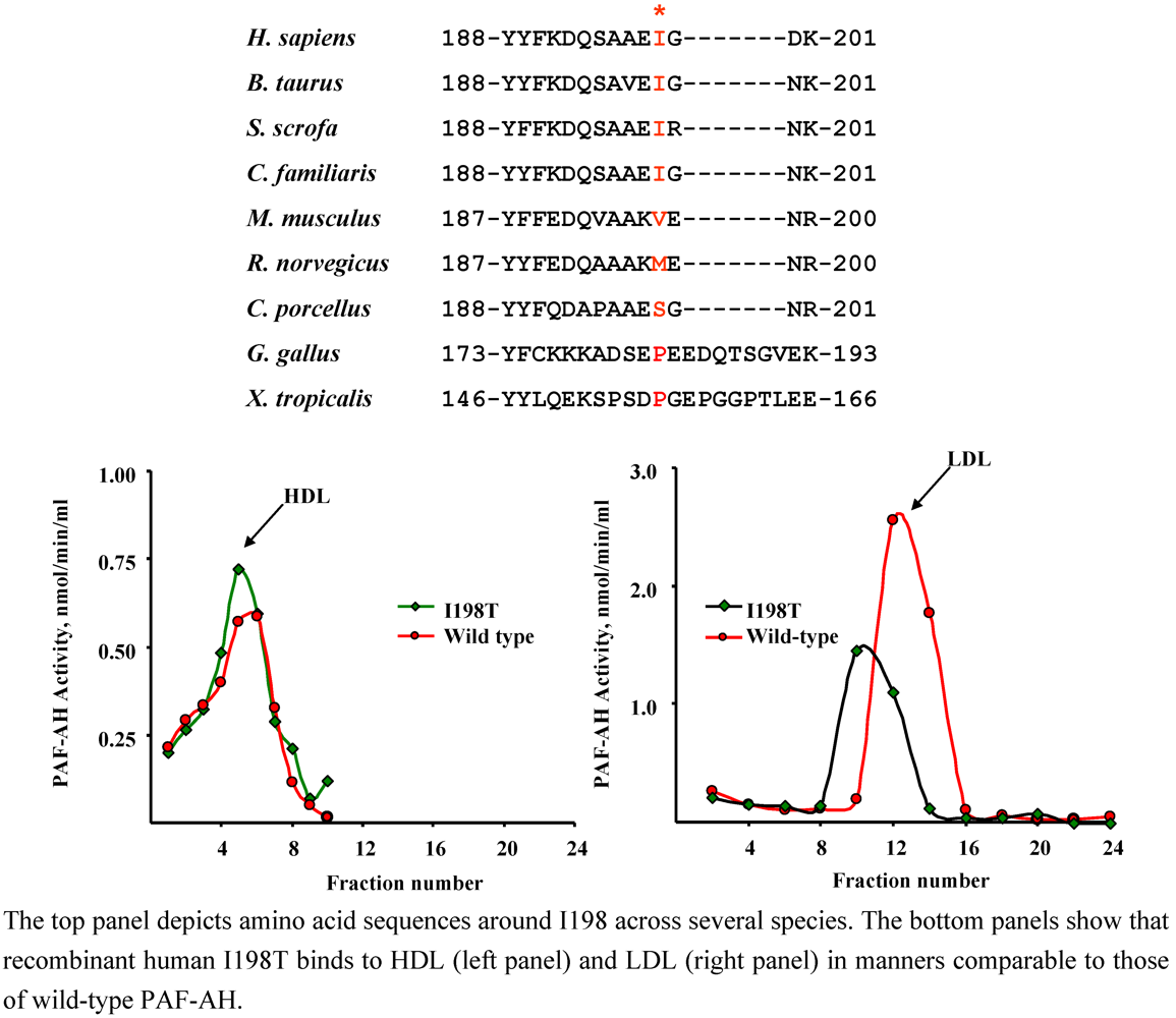
4.2. I198T (rs 1805018)

Individuals harboring a threonine at position 198 of PAF-AH were first described by Bell and co-workers [12]. The degree of conservation of this residue across species is moderate (Figure 3). Expression of the recombinant variant revealed that enzymatic activity was preserved [42]. However, the mutant protein displayed decreased affinity for PAF (Table 2, [42]). Our studies revealed that the presence of T198 did not affect binding to LDL or HDL in vitro (Figure 3, [46]). As in the case of R92H, it would be of interest to complement these studies with lipoprotein distribution profiles in fresh plasma samples from subjects that harbor 0-1-2 T198 alleles, as these analyses would establish whether T198 contributes to the ability of PAF-AH to associate with lipoprotein particles in vivo.
Individuals homozygous for the I198T polymorphism are rare, so most of the work in humans has been conducted in heterozygous subjects. There is controversy on the impact of T198 on the levels of expression of plasma PAF-AH protein and activity. Three investigations revealed no statistically significant effect of T198 on enzymatic activity [43,44,45]. However, a recent study by Hou et al. showed that subjects harboring this variant expressed substantially lower levels of PAF-AH activity [5]. The haplotypes of these subjects differed from those of the control subjects in more than the I198T polymorphism [5] and it is not clear whether these additional genetic variations contributed to the observed decreases in activity. This is particularly important as PAF-AH risk alleles may have an additive effect that leads to the generation of dysfunctional protein products [8]. Sutton et al. reported a trend towards decreased PAF-AH expression in the aorta of subjects harboring T198 [8]. These observations leave open the possibility that at least in some populations, T198 affects activity and/or protein levels, or protein stability, in addition to its effect on substrate affinity.
Public databases (http://www.ncbi.nlm.nih.gov/) report that the lowest T198 allele frequency occurs in Caucasian (4.2–12.0%) and Asian (4.3–11.0%, excluding Japanese) cohorts. The frequency is much higher in Japanese subjects (19.7%), in African Americans (23.5%), and in sub-Saharan African populations (28.5%).
Figure 3. The I198T variant binds to human HDL and LDL in vitro.
Pharmaceuticals 02 00094 g003
Kruse and co-workers found the T198 variant to be highly associated with total IgE concentrations in an atopic population and with atopic asthma in an asthmatic population (Table 4, [42]). Interestingly, T198 seems to be somewhat more prevalent in ARDS patients [44] compared to control populations with similar ethnic backgrounds (Table 4), suggesting that additional, well-controlled studies related to the impact of T198 on ARDS incidence and severity may be warranted.
The prevalence of the T198 polymorphism has been found to be weakly associated with the incidence of schizophrenia ([12], Table 4). Mechanistically, it is possible that the variant contributes to these syndromes as a consequence of decreased protein expression, at least in some populations [5]. Alternatively, since substrate concentration and/or accessibility to the active site tend to be rate limiting in vivo [41], decreased substrate affinity (i.e, increased Km [42]) may account for the observed effects.
A variety of studies have assessed the incidence of T198 in patients with cardiovascular disease (Table 4). These analyses have led to conflicting results possibly owing to the size and heterogeneity of the patient populations and/or the type of statistical analyses utilized. Sutton et al. used logistic regression modeling and found no significant association of T198 with CAD outcome [8]. Similarly, Ninio et al. reported that the incidence of cardiovascular events was not significantly associated with survival outcome by univariate analyses [45]. In contrast, Hoffmann and co-workers reported lower incidence of T198 in a CAD cohort. The larger size of the populations studied by Hoffmann et al. may explain why this correlation did not become evident in other studies. Kokubo and co-workers reported that the presence of T198 was associated with hypertension in women when the data were adjusted for several factors and analyzed by multivariate logistic regression analyses [49]. In summary, while there is no absolute agreement on the precise role of the somewhat rare T198 polymorphism on activity and/or disease incidence, it seems that additional large-scale studies should further evaluate whether, and how, T198 contributes to inflammatory and vascular disorders. These analyses should include activity measurements as at least one study [5] showed a remarkably high inhibitory effect of this polymorphism on PAF-AH levels. It seems that analyses in Japanese cohorts would be ideal for this purpose, owing to the reportedly high prevalence of the polymorphism in this population.
Table 4. The I198T (rs1805018) polymorphism: Allele frequency in control and diseased populations.
PopulationClinical StatusnAllele frequency,%pAuthors [Ref.]
BritishControls1507.6 Kruse et al. [42]
Atopic asthma15011.70.008
GermanNonatopic330.8
Atopic723.70.0087 (IgE levels)
BritishControls992.5 Bell et al. [12]
Schizophrenia2046.40.04
CaucasianControls6936.1 Hoffmann et al. [43]
CAD2,5414.40.009
GermanControls4845.7 Ninio et al. [45]
CAD1,3115.4N/S
Caucasian + African American + American IndianControls2676.0 Sutton et al. [8]
CAD (< 56y/o)5999.0N/S
CAD (> 56 y/o)2077.0N/S
Myocardial infarction4258.0N/S
ChineseControls9098.7 Hou et al. [5]
CHD8089.8N/S
Myocardial infarction5029.9N/S
CaucasianARDS419.8N/ALi et al. [44]
African AmericanARDS1711.8N/A
CaucasianRandomly selected1,2025.2N/ASchnabel et al. [13]
JapaneseRandomly selected1,87820.7N/AKokubo et al. [49]
JapaneseControls9628.1N/AJinnai et al. [51]
CaucasianRandomly selected1,2025.2N/ASchnabel et al. [13]
This Table summarizes results from studies in various human populations genotyped for the I198T polymorphism. The incidence of rs1805018 is reported in terms of the percentage with which the allele was represented in healthy and diseased subjects. The “n” column refers to the number of participants in each study category and includes subjects harboring 0-1-2 polymorphic alleles. The “p” column depicts whether statistically significant differences were found in polymorphism incidence in control and diseased populations, where available/appropriate. ARDS: acute respiratory distress syndrome; CAD: coronary artery disease; CHD: coronary heart disease; N/A: not applicable; N/S: not statistically significant.

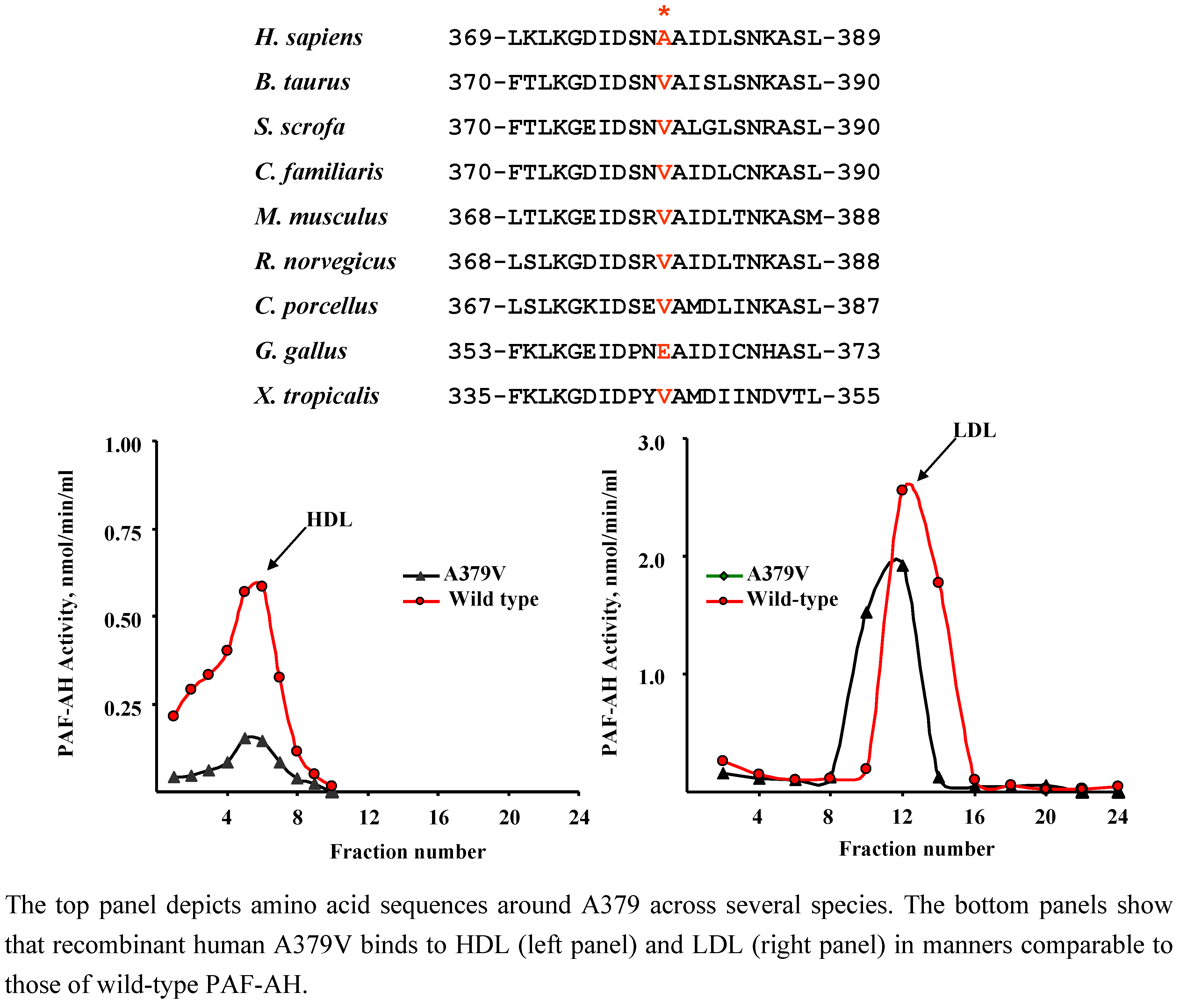
4.3. A379V (rs 1051931)

Individuals harboring a valine at position 379 of PAF-AH were described a few years ago [12]. While humans express predominantly an alanine residue at this position, most species harbor a valine residue (Figure 4). This feature forecasts limited impact of V379 on enzyme function in humans. Purified recombinant A379V displays increased maximal velocity and a two-fold decrease in substrate affinity compared to the wild-type protein ([42], Table 2). We found that the A379V variant associated with LDL and HDL in fashions comparable to those of the human protein (Figure 4, [46]). However, a systematic study to characterize the lipoprotein distribution of PAF-AH in subjects homozygous for the V379 polymorphism has not, to my knowledge, been performed.
Figure 4. The A379V variant binds to human HDL and LDL in vitro.
Pharmaceuticals 02 00094 g004
As in the case of other polymorphisms, there is controversy on the impact of V379 on plasma PAF-AH protein and activity levels. Hoffmann and co-workers reported increases in PAF-AH plasma levels with a clear gene-dose effect [43]. Similarly, Ninio and collaborators found that the presence of V379 was significantly associated with increased PAF-AH activity in a co-dominant fashion (p = 0.02, [45]). Schnabel and co-workers reported higher PAF-AH activity in subjects carrying two V379 alleles and extended the observation to a large community-based cohort [50]. While the observed differences were statistically significant in these two instances, the increases in activity were modest (<10 %). Three unrelated studies found no statistically significant effect of V379 on enzymatic activity in ARDS patients (Table 5), in a Chinese cohort, and in a Caucasian control group [5,44,52]. An interesting study in a Taiwanese population showed dramatic decreases in activity in subjects homozygous for the V379 allele [3]. The fact that this is the only study reporting such large effects suggests that an additional factor(s) peculiar to this group of subjects likely contributed to the results. In summary, the presence of V379 may confer a very modest increase in plasma PAF-AH activity levels in some populations. These effects are likely mediated by the enzyme itself, because studies that evaluated properties of the purified recombinant A379V variant in a lipoprotein-free assay system recapitulated the increased enzymatic activity that was observed in some clinical studies [42].
Table 5. The A379V (rs1051931) polymorphism: allele frequency in control and diseased populations.
PopulationClinical StatusnAllele frequencypAuthors [Ref.]
BritishControls15015.20.038Kruse et al. [42]
Atopic asthma15021.6
GermanNonatopic3310.30.0017
Atopic7221.9
GermanControls48424.30.0007Ninio et al. [45]
CAD129819.5
Caucasian + African American + American IndianControls26715.0Sutton et al. [8]
CAD (< 56y/o)59920.00.05
CAD (> 56 y/o)20726.00.002
Myocardial infarction42519.00.01
TaiwaneseControls20021.00.01Liu et al. [3]
Myocardial infarction20033.0
CaucasianControls69320.9N/SHoffmann et al. [43]
CAD254121.4
ChineseControls90415.9Hou et al. [5]
CHD80816.6N/S
Myocardial infarction50315.5N/S
KoreanControls67014.6N/SJang et al. [4]
CVD53215.5
EuropeanControls55624.0--Abuzeid et al. [53]
Myocardial infarction52722.0
CaucasianMale controls35920.2Wootton et al. [52]
Male CHD10421.2N/S
Female controls24420.9
Female CHD5023.0N/S
Caucasian Early ARDS4113.4N/ALi et al. [44]
African American Early ARDS1711.8N/A
BritishControls9325.8Bell et al. [12]
Schizophrenia19118.80.06
BritishControls12321.5N/AWootton et al. [54]
British CaucasianControls269519.6N/ARudd et. al. [55]
MixedRandomly selected810519.4N/ASchnabel et al. [50]
DutchRandomly selected357519.0N/AVan den Berg et al. [56]
JapaneseRandomly selected1,87810.8N/AKokubo et al. [49]
JapaneseControls964.2N/AJinnai et al. [51]
This Table summarizes results from studies in various human populations genotyped for the A379V polymorphism. The incidence of rs1051931 is reported in terms of the percentage with which the allele was represented in healthy and diseased subjects. The “n” column refers to the number of participants in each study category and includes subjects harboring 0-1-2 polymorphic alleles. The “p” column depicts whether statistically significant differences were found in polymorphism incidence in control and diseased populations, where available/appropriate. ARDS: acute respiratory distress syndrome; CAD: coronary artery disease; CHD: coronary heart disease; CVD: cardiovascular disease; N/A: not applicable; N/S: not statistically significant.
The prevalence of the V379 allele varies among populations. The lowest frequency (9.2–9.5%) has been reported in Japanese populations and the highest prevalence (26.7–32.6%) was observed in Sub-Saharan African subjects. A number of groups have investigated whether V379 is associated with the incidence of various diseases and/or physiological responses (Table 4). Bell and co-workers found no significant differences in allele frequency between patients with schizophrenia and control subjects, although a trend (p = 0.057) towards increased incidence in the patients was noted [12]. In contrast, V379 was found to be highly associated with specific sensitization in a German atopic population and with atopic asthma in a British cohort (Table 5, [42]). Wootton and co-workers reported that after ten weeks of intensive physical exercise, individuals homozygous for the V379 allele showed decreased percentage of adipose tissue compared to AV and AA genotype groups (p = 0.01, [52]). A potentially interesting observation by Li and co-workers is that ARDS patients seem to have a lower incidence of the V379 polymorphism compared to similar Caucasian cohorts [13.4% versus 19.6–25.8% in control Caucasian subjects, excluding reference [42], Table 5]. Unfortunately, this analysis only evaluated ARDS patients, so a comparison with healthy control subjects can only be made across studies. Nonetheless, the fact that ARDS survivors expressed higher plasma PAF-AH activity [44], combined with the fact that the prevalence of the V379 allele was lower in at least one ARDS cohort, suggests that V379 may offer some protection from this disease.
Eight studies investigated whether V379 contributes to vascular disease [3,4,5,8,43,45,52,53]. Four of these analyses reported no statistically significant differences in allele frequency of control subjects compared to patients with coronary heart disease or myocardial infarction (Table 5). Three studies found statistically significant, but opposite, relationships between these parameters [3,8,45]. The suitability of control cohorts utilized in two of these reports [8,45] was questioned by others [43], as indicated above. The lack of consensus among these analyses could be due to the criteria utilized for patient selection, combined with the size and nature of the patient populations studied. The mechanism by which potential V379-mediated effects contribute to inflammatory diseases could include direct effects of the polymorphism on enzymatic activity, but this contribution is likely to be relatively modest [45]. The possibility that V379 is in linkage disequilibrium with an un-identified etiological variant cannot be ruled out.

4.4. L12L (rs 35142331), L45P (rs 45521937), K191N (rs 45454695), and L389S (rs 34159425)

These variants have only been reported in public databases and they occur with various frequencies in human populations. One of the variants (rs 35142331) results in no amino acid change and is thus unlikely to impact protein function. The frequency of the remaining variants is either unknown or very rare in Caucasians. The functional consequences associated with these polymorphisms are currently unknown.

5. Additional Measurements May Be Necessary to Elucidate Functional Consequences of PAF-AH Polymorphisms

5.1. Rate of hydrolysis at sub-saturating substrate levels and effect of lipoprotein environment

We previously showed that the kinetics of PAF hydrolysis are dependent on the ratio between enzyme and substrate, and that at low substrate levels the rate of hydrolysis deviates from that predicted by Michaelis-Menten kinetics [41]. We investigated the effect of sequentially introducing naturally occurring PAF-AH polymorphisms on the kinetics of PAF hydrolysis at sub-saturating substrate levels. This analysis revealed that single, double, and triple mutants hydrolyzed PAF with similar efficiencies compared to the wild-type protein (Figure 5, left panel).
Figure 5. Kinetics of PAF (10-9 M) hydrolysis by free (left panel) and LDL-associated (right panel) PAF-AH variants.
Pharmaceuticals 02 00094 g005
In these assays, we utilized equal amounts of PAF-AH activity as determined by our standard, optimized, enzyme assay. Thus, the results were qualitatively comparable in both assay types, that is, we observed no major differences in the rate of PAF hydrolysis by PAF-AH variants. However, the experimental design in these studies did not recapitulate the environment in which the enzyme functions in vivo. This is an important issue because lipoproteins alter the efficiency of hydrolysis of phospholipid substrates, presumably by modulating access to the active site, altering substrate presentation, or changing solubility. To investigate this, we incorporated wild-type and PAF-AH variants into LDL, and then assessed the ability of the lipoprotein-associated variants to hydrolyze sub-saturating levels of PAF. Our studies showed that sequential introduction of each polymorphism increased the half–life of PAF in LDL (Figure 5, right panel). These results suggested that in experimental settings modeled to mimic conditions likely to occur in vivo, that is, when substrate is limited, enzyme levels are in excess, and the enzyme functions in its natural environment, factors other than absolute enzyme levels impact the rate of hydrolysis. These studies revealed potentially important functional consequences associated with naturally occurring polymorphisms in PAF-AH. In addition, the results illustrate the importance of complementing traditional biochemical analyses with assays that take into consideration properties peculiar to the gene product and reaction being evaluated.

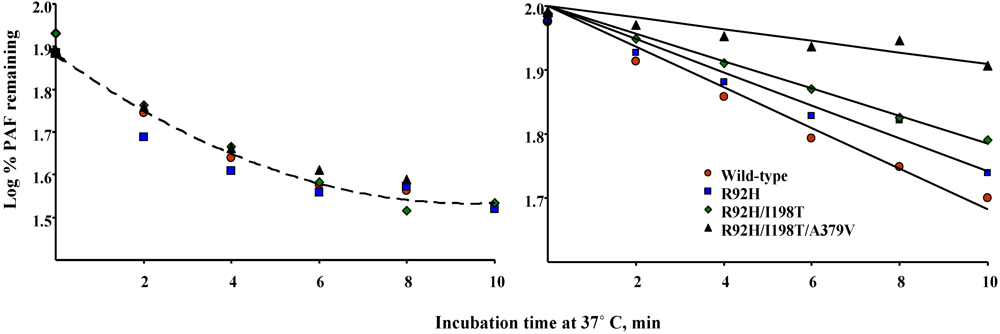
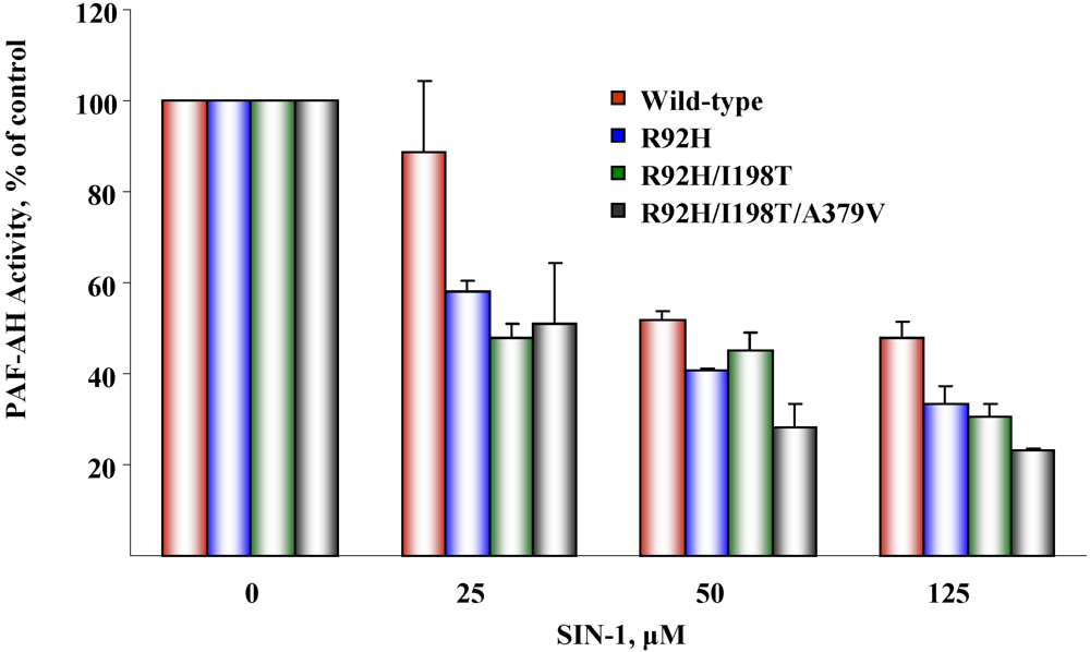
5.2. Susceptibility to oxidative inactivation

Previous work demonstrated that PAF-AH is susceptible to inactivation by oxidants, and that key methionine and tyrosine residues confer susceptibility to oxidant attack [57]. We investigated whether naturally occurring PAF-AH variants were differentially affected by oxidants, as this could provide additional mechanistic insight into the functional consequences of genetic alterations. We expressed single, double, and triple mutants harboring R92H, I198T, and A379V, and then investigated if these polymorphic forms of PAF-AH were differentially affected by SIN-1, a peroxynitrite-generating agent [57]. We found that sequential replacement of these residues increased susceptibility to oxidative inactivation (Figure 6). These findings suggested that subjects harboring these polymorphisms may accumulate PAF-AH substrates in settings of elevated oxidant stress that are characteristic of acute inflammation and other syndromes.
Figure 6. Susceptibility of PAF-AH variants to oxidative inactivation mediated by SIN-1
Pharmaceuticals 02 00094 g006

6. Conclusions

In this article, I have presented current information that summarizes functional and clinical consequences associated with naturally occurring variations in the PAF-AH gene. Many studies have attempted to demonstrate that genetic diversity of PAF-AH affects features related to vascular and other diseases. It is clear that mutations that compromise the normal function of the enzyme (i.e., V279F) increase the incidence and/or severity of several conditions, including vascular disease, in defined populations. Superficially, this relationship may appear contrary to studies showing that elevated PAF-AH expression correlates with vascular disease incidence and/or severity in Western populations. However, these two sets of observations can be reconciled from a physiological perspective if increased PAF-AH expression is viewed as a compensatory, not causal, mechanism to limit damage caused by pro-inflammatory phospholipids generated in settings of high oxidant stress. PAF-AH polymorphisms that have more subtle effects in standard laboratory assays have also been reported to modulate severity or incidence of a number of syndromes and diseases. It is possible that evaluating additional enzyme properties, location, and/or functions may help elucidate whether, and how, genetic variations within the coding region of PAF-AH impact its in vivo function. The effect of lipoprotein location on the function of PAF-AH remains to be completely elucidated, but the fact that altered distribution has been observed in a variety of human syndromes suggests potentially important effects. This is supported by biochemical studies indicating that the environment provided by LDL allows for higher efficiency of substrate hydrolysis compared to that of HDL. Studies aimed at characterizing whether PAF-AH polymorphisms have a physiological impact should evaluate haplotypes in individual subjects rather than the prevalence of each variant in the sample set studied. This approach could have potential clinical utility as it may help identify patients most likely to benefit from therapies that target this pathway. A number of the studies cited here have used this strategy and it is likely that the approach will become standard practice. SNPs located in the promoter and other regulatory regions of PAF-AH, while not the focus of this review, may have important functional consequences as well. These variants have received limited attention and it would be interesting to evaluate whether they modulate expression levels, and/or the incidence or severity of inflammatory and vascular diseases. To establish meaningful relationships between genotype and function, and to identify the clinical impact of PAF-AH SNPs, it will be necessary to conduct systematic analyses that take into consideration individual genetic complexities, environmental influences, and the unique properties of this interesting enzyme.

Acknowledgements

I wish to express my gratitude to Alison Gardner, Ethan Reichert, L. Jeff Johnson, Damian Dayton and Michael R. Hinckley for excellent technical assistance and many valuable contributions over the years. I am always indebted to my long-time colleague and friend, Matthew K. Topham, for his constant support, advice, and helpful suggestions. This work was supported by grant HL35828 to DMS and by the Huntsman Cancer Foundation.

References and Notes

  1. Vadas, P.; Gold, M.; Perelman, B.; Liss, G.M.; Lack, G.; Blyth, T.; Simons, F.E.; Simons, K.J.; Cass, D.; Yeung, J. Platelet-activating factor, PAF acetylhydrolase, and severe anaphylaxis. N Engl. J. Med. 2008, 358, 28–35. [Google Scholar] [PubMed]
  2. Miwa, M.; Miyake, T.; Yamanaka, T.; Sugatani, J.; Suzuki, Y.; Sakata, S.; Araki, Y.; Matsumoto, M. Characterization of serum platelet-activating factor (PAF) acetylhydrolase. Correlation between deficiency of serum PAF acetylhydrolase and respiratory symptoms in asthmatic children. J. Clin. Invest. 1988, 82, 1983–1991. [Google Scholar] [CrossRef] [PubMed]
  3. Liu, P.Y.; Li, Y.H.; Wu, H.L.; Chao, T.H.; Tsai, L.M.; Lin, L.J.; Shi, G.Y.; Chen, J.H. Platelet-activating factor-acetylhydrolase A379V (exon 11) gene polymorphism is an independent and functional risk factor for premature myocardial infarction. J. Thromb. Haemost. 2006, 4, 1023–1028. [Google Scholar]
  4. Jang, Y.; Kim, O.Y.; Koh, S.J.; Chae, J.S.; Ko, Y.G.; Kim, J.Y.; Cho, H.; Jeong, T.S.; Lee, W.S.; Ordovas, J.M.; Lee, J.H. The Val279Phe Variant of the Lipoprotein-Associated Phospholipase A2 Gene Is Associated with Catalytic Activities and Cardiovascular Disease in Korean Men. J. Clin. Endocrinol. Metab. 2006, 91, 3521–3527. [Google Scholar]
  5. Hou, L.; Chen, S.; Yu, H.; Lu, X.; Chen, J.; Wang, L.; Huang, J.; Fan, Z.; Gu, D. Associations of PLA2G7 gene polymorphisms with plasma lipoprotein-associated phospholipase A2 activity and coronary heart disease in a Chinese Han population: the Beijing atherosclerosis study. Hum. Genet. 2009, 125, 11–20. [Google Scholar]
  6. Yamada, Y.; Yokota, M. Loss of activity of plasma platelet-activating factor acetylhydrolase due to a novel Gln281-->Arg mutation. Biochem. Biophys. Res. Commun. 1997, 236, 772–775. [Google Scholar]
  7. Ishihara, M.; Iwasaki, T.; Nagano, M.; Ishii, J.; Takano, M.; Kujiraoka, T.; Tsuji, M.; Hattori, H.; Emi, M. Functional impairment of two novel mutations detected in lipoprotein-associated phospholipase A2 (Lp-PLA2) deficiency patients. J. Hum. Genet. 2004, 49, 302–307. [Google Scholar]
  8. Sutton, B.S.; Crosslin, D.R.; Shah, S.H.; Nelson, S.C.; Bassil, A.; Hale, A.B.; Haynes, C.; Goldschmidt-Clermont, P.J.; Vance, J.M.; Seo, D.; Kraus, W.E.; Gregory, S.G.; Hauser, E.R. Comprehensive genetic analysis of the platelet activating factor acetylhydrolase (PLA2G7) gene and cardiovascular disease in case-control and family datasets. Hum. Mol. Genet. 2008, 17, 1318–1328. [Google Scholar]
  9. Stafforini, D.M.; Satoh, K.; Atkinson, D.L.; Tjoelker, L.W.; Eberhardt, C.; Yoshida, H.; Imaizumi, T.; Takamatsu, S.; Zimmerman, G.A.; McIntyre, T.M.; Gray, P.W.; Prescott, S.M. Platelet-activating factor acetylhydrolase deficiency. A missense mutation near the active site of an anti-inflammatory phospholipase. J. Clin. Invest. 1996, 97, 2784–2791. [Google Scholar]
  10. Stafforini, D.M.; Numao, T.; Tsodikov, A.; Vaitkus, D.; Fukuda, T.; Watanabe, N.; Fueki, N.; McIntyre, T.M.; Zimmerman, G.A.; Makino, S.; Prescott, S.M. Deficiency of platelet-activating factor acetylhydrolase is a severity factor for asthma. J. Clin. Invest. 1999, 103, 989–997. [Google Scholar]
  11. Balta, G.; Gurgey, A.; Kudayarov, D.K.; Tunc, B.; Altay, C. Evidence for the existence of the PAF acetylhydrolase mutation (Val279Phe) in non-Japanese populations: a preliminary study in Turkey, Azerbaijan, and Kyrgyzstan. Thromb. Res. 2001, 101, 231–234. [Google Scholar] [CrossRef] [PubMed]
  12. Bell, R.; Collier, D.A.; Rice, S.Q.; Roberts, G.W.; MacPhee, C.H.; Kerwin, R.W.; Price, J.; Gloger, I.S. Systematic screening of the LDL-PLA2 gene for polymorphic variants and case-control analysis in schizophrenia. Biochem. Biophys. Res. Commun. 1997, 241, 630–635. [Google Scholar]
  13. Schnabel, R.; Larson, M.G.; Dupuis, J.; Lunetta, K.L.; Lipinska, I.; Meigs, J.B.; Yin, X.; Rong, J.; Vita, J.A.; Newton-Cheh, C.; Levy, D.; Keaney, J.F., Jr.; Vasan, R.S.; Mitchell, G.F.; Benjamin, E.J. Relations of inflammatory biomarkers and common genetic variants with arterial stiffness and wave reflection. Hypertension 2008, 51, 1651–1657. [Google Scholar]
  14. Smith, J.M.; Jones, F.; Ciol, M.A.; Jelacic, S.; Boster, D.R.; Watkins, S.L.; Williams, G.D.; Tarr, P.I.; Henderson, W.R., Jr. Platelet-activating factor and Escherichia coliO157:H7 infections. Pediatr. Nephrol. 2002, 17, 1047–1052. [Google Scholar]
  15. Ito, S.; Noguchi, E.; Shibasaki, M.; Yamakawa-Kobayashi, K.; Watanabe, H.; Arinami, T. Evidence for an association between plasma platelet-activating factor acetylhydrolase deficiency and increased risk of childhood atopic asthma. J. Hum. Genet. 2002, 47, 99–101. [Google Scholar]
  16. Satoh, N.; Asano, K.; Naoki, K.; Fukunaga, K.; Iwata, M.; Kanazawa, M.; Yamaguchi, K. Plasma platelet-activating factor acetylhydrolase deficiency in Japanese patients with asthma. Am. J. Respir. Crit. Care Med. 1999, 159, 974–979. [Google Scholar]
  17. Unno, N.; Nakamura, T.; Mitsuoka, H.; Uchiyama, T.; Yamamoto, N.; Saito, T.; Sugatani, J.; Miwa, M.; Nakamura, S. Association of a G994 -->T missense mutation in the plasma platelet-activating factor acetylhydrolase gene with risk of abdominal aortic aneurysm in Japanese. Ann. Surg. 2002, 235, 297–302. [Google Scholar]
  18. Unno, N.; Nakamura, T.; Mitsuoka, H.; Saito, T.; Miki, K.; Ishimaru, K.; Sugatani, J.; Miwa, M.; Nakamura, S. Single nucleotide polymorphism (G994-->T) in the plasma platelet-activating factor-acetylhydrolase gene is associated with graft patency of femoropopliteal bypass. Surgery 2002, 132, 66–71. [Google Scholar]
  19. Unno, N.; Sakaguchi, T.; Nakamura, T.; Yamamoto, N.; Sugatani, J.; Miwa, M.; Konno, H. A single nucleotide polymorphism in the plasma PAF acetylhydrolase gene and risk of atherosclerosis in Japanese patients with peripheral artery occlusive disease. J. Surg. Res. 2006, 134, 36–43. [Google Scholar]
  20. Unno, N.; Nakamura, T.; Kaneko, H.; Uchiyama, T.; Yamamoto, N.; Sugatani, J.; Miwa, M.; Nakamura, S. Plasma platelet-activating factor acetylhydrolase deficiency is associated with atherosclerotic occlusive disease in japan. J. Vasc. Surg. 2000, 32, 263–267. [Google Scholar]
  21. Ichihara, S.; Yamada, Y.; Yokota, M. Association of a G994-->T missense mutation in the plasma platelet-activating factor acetylhydrolase gene with genetic susceptibility to nonfamilial dilated cardiomyopathy in Japanese. Circulation 1998, 98, 1881–1885. [Google Scholar]
  22. Yamada, Y.; Yoshida, H.; Ichihara, S.; Imaizumi, T.; Satoh, K.; Yokota, M. Correlations between plasma platelet-activating factor acetylhydrolase (PAF-AH) activity and PAF-AH genotype, age, and atherosclerosis in a Japanese population. Atherosclerosis 2000, 150, 209–216. [Google Scholar] [CrossRef] [PubMed]
  23. Yamada, Y.; Ichihara, S.; Fujimura, T.; Yokota, M. Identification of the G994--> T missense in exon 9 of the plasma platelet-activating factor acetylhydrolase gene as an independent risk factor for coronary artery disease in Japanese men. Metabolism 1998, 47, 177–181. [Google Scholar]
  24. Yamada, Y.; Ichihara, S.; Izawa, H.; Tanaka, M.; Yokota, M. Association of a G994 --> T (Val279 --> Phe) polymorphism of the plasma platelet-activating factor acetylhydrolase gene with myocardial damage in Japanese patients with nonfamilial hypertrophic cardiomyopathy. J. Hum. Genet. 2001, 46, 436–441. [Google Scholar]
  25. Sekuri, C.; Cam, F.S.; Tengiz, I.; Ercan, E.; Bayturan, O.; Berdeli, A. Association of platelet-activating factor acetylhydrolase gene polymorphism with premature coronary artery disease in Turkish patients. Anadolu Kardiyol Derg 2006, 6, 132–134. [Google Scholar]
  26. Hiramoto, M.; Yoshida, H.; Imaizumi, T.; Yoshimizu, N.; Satoh, K. A mutation in plasma platelet-activating factor acetylhydrolase (Val279-->Phe) is a genetic risk factor for stroke. Stroke 1997, 28, 2417–2420. [Google Scholar]
  27. Satoh, K. Plasma platelet-activating factor acetylhydrolase (PAF-AH) deficiency as a risk factor for stroke. Brain Nerve 2008, 60, 1319–1324. [Google Scholar]
  28. Yoshida, H.; Imaizumi, T.; Fujimoto, K.; Itaya, H.; Hiramoto, M.; Yoshimizu, N.; Fukushi, K.; Satoh, K. A mutation in plasma platelet-activating factor acetylhydrolase (Val279Phe) is a genetic risk factor for cerebral hemorrhage but not for hypertension. Thromb. Haemost. 1998, 80, 372–375. [Google Scholar]
  29. Zhang, X.; Yuan, C.L.; Zhang, H.Z.; Xu, J.; Wu, J.; Chen, B.L. Analysis of 994(G--> T) mutation in the plasma platelet-activating factor acetylhydrolase gene in the patients with cerebral infarction. Zhonghua Yi Xue Yi Chuan Xue Za Zhi 2005, 22, 450–452. [Google Scholar]
  30. Yamamoto, I.; Fujitsu, J.; Nohnen, S.; Igarashi, T.; Motomura, T.; Inaba, M.; Tsubakimori, S.; Azuma, J. Association of plasma PAF acetylhydrolase gene polymorphism with IMT of carotid arteries in Japanese type 2 diabetic patients. Diabetes. Res. Clin. Pract. 2003, 59, 219–224. [Google Scholar]
  31. Tanaka, R.; Iijima, K.; Xu, H.; Inoue, Y.; Murakami, R.; Shirakawa, T.; Nishiyama, K.; Miwa, M.; Shiozawa, S.; Nakamura, H.; Yoshikawa, N. Role of platelet-activating factor acetylhydrolase gene mutation in Japanese childhood IgA nephropathy. Am. J. Kidney. Dis. 1999, 34, 289–295. [Google Scholar]
  32. Xu, H.; Iijima, K.; Shiozawa, S.; Tanaka, S.S.; Inoue, Y.; Shirakawa, T.; Nishiyama, K.; Miwa, M.; Nakamura, H.; Yoshikawa, N. Platelet-activating factor acetylhydrolase gene mutation in Japanese nephrotic children. Kidney. Int. 1998, 54, 1867–1871. [Google Scholar]
  33. Xu, H.; Iijima, K.; Shirakawa, T.; Shiozawa, S.; Miwa, M.; Yamaoka, K.; Kawamura, N.; Nakamura, H.; Yoshikawa, N. Platelet-activating factor acetylhydrolase gene mutation in Japanese children with Escherichia coli O157-associated hemolytic uremic syndrome. Am. J. Kidney. Dis. 2000, 36, 42–46. [Google Scholar]
  34. Nakamura, T.; Sakaguchi, T.; Unno, N.; Sugatani, J.; Miwa, M.; Nakamura, S. Relationship between the platelet activating factor acetylhydrolase gene and intractability of ulcerative colitis. Dis. Colon. Rectum. 2002, 45, 389–393. [Google Scholar]
  35. Ohtsuki, T.; Watanabe, H.; Toru, M.; Arinami, T. Lack of evidence for associations between plasma platelet-activating factor acetylhydrolase deficiency and schizophrenia. Psychiatry Res 2002, 109, 93–96. [Google Scholar]
  36. Osoegawa, M.; Niino, M.; Ochi, H.; Kikuchi, S.; Murai, H.; Fukazawa, T.; Minohara, M.; Tashiro, K.; Kira, J. Platelet-activating factor acetylhydrolase gene polymorphism and its activity in Japanese patients with multiple sclerosis. J. Neuroimmunol. 2004, 150, 150–156. [Google Scholar]
  37. Minami, T.; Suzuki, H.; Takeuchi, T.; Uemura, S.; Sugatani, J.; Yoshikawa, N. A polymorphism in plasma platelet-activating factor acetylhydrolase is involved in resistance to immunoglobulin treatment in Kawasaki disease. J. Pediatr. 2005, 147, 78–83. [Google Scholar]
  38. Wang, T.; Karino, K.; Yamasaki, M.; Zhang, Y.; Masuda, J.; Yamaguchi, S.; Shiwaku, K.; Nabika, T. Effects of G994T in the Lp-PLA2 gene on the plasma oxidized LDL level and carotid intima-media thickness in Japanese: the Shimane study. Am. J. Hypertens. 2009, 22, 742–747. [Google Scholar]
  39. Stafforini, D.M.; Zimmerman, G.A.; McIntyre, T.M.; Prescott, S.M. The platelet-activating factor acetylhydrolase from human plasma prevents oxidative modification of low-density lipoprotein. Trans. Assoc. Am. Physicians 1992, 105, 44–63. [Google Scholar]
  40. Kujiraoka, T.; Iwasaki, T.; Ishihara, M.; Ito, M.; Nagano, M.; Kawaguchi, A.; Takahashi, S.; Ishi, J.; Tsuji, M.; Egashira, T.; Stepanova, I.P.; Miller, N.E.; Hattori, H. Altered distribution of plasma PAF-AH between HDLs and other lipoproteins in hyperlipidemia and diabetes mellitus. J. Lipid. Res. 2003, 44, 2006–2014. [Google Scholar]
  41. Stafforini, D.M.; Carter, M.E.; Zimmerman, G.A.; McIntyre, T.M.; Prescott, S.M. Lipoproteins alter the catalytic behavior of the platelet-activating factor acetylhydrolase in human plasma. Proc. Natl. Acad. Sci. USA 1989, 86, 2393–2397. [Google Scholar]
  42. Kruse, S.; Mao, X.Q.; Heinzmann, A.; Blattmann, S.; Roberts, M.H.; Braun, S.; Gao, P.S.; Forster, J.; Kuehr, J.; Hopkin, J.M.; Shirakawa, T.; Deichmann, K.A. The Ile198Thr and Ala379Val variants of plasmatic PAF-acetylhydrolase impair catalytical activities and are associated with atopy and asthma. Am. J. Hum. Genet. 2000, 66, 1522–1530. [Google Scholar]
  43. Hoffmann, M.M.; Winkler, K.; Renner, W.; Winkelmann, B.R.; Seelhorst, U.; Wellnitz, B.; Boehm, B.O.; Marz, W. Genetic variants and haplotypes of lipoprotein associated phospholipase A2 and their influence on cardiovascular disease (The Ludwigshafen Risk and Cardiovascular Health Study). J. Thromb. Haemost. 2009, 7, 41–48. [Google Scholar]
  44. Li, S.; Stuart, L.; Zhang, Y.; Meduri, G.U.; Umberger, R.; Yates, C.R. Inter-individual variability of plasma PAF-acetylhydrolase activity in ARDS patients and PAF-AH genotype. J. Clin. Pharm. Ther. 2009, 34, 447–455. [Google Scholar]
  45. Ninio, E.; Tregouet, D.; Carrier, J.L.; Stengel, D.; Bickel, C.; Perret, C.; Rupprecht, H.J.; Cambien, F.; Blankenberg, S.; Tiret, L. Platelet-activating factor-acetylhydrolase and PAF-receptor gene haplotypes in relation to future cardiovascular event in patients with coronary artery disease. Hum. Mol. Genet. 2004, 13, 1341–1351. [Google Scholar]
  46. Gardner, A.A.; Reichert, E.C.; Topham, M.K.; Stafforini, D.M. Identification of a domain that mediates association of platelet-activating factor acetylhydrolase with high density lipoprotein. J. Biol. Chem. 2008, 283, 17099–17106. [Google Scholar]
  47. Luo, G.; Zheng, L.; Zhang, X.; Zhang, J.; Nilsson-Ehle, P.; Xu, N. Genotyping of single nucleotide polymorphisms using base-quenched probe: a method does not invariably depend on the deoxyguanosine nucleotide. Anal. Biochem. 2009, 386, 161–166. [Google Scholar]
  48. Limou, S.; Coulonges, C.; Foglio, M.; Heath, S.; Diop, G.; Leclerc, S.; Hirtzig, T.; Spadoni, J.L.; Therwath, A.; Lambeau, G.; Gut, I.; Zagury, J.F. Exploration of associations between phospholipase A2 gene family polymorphisms and AIDS progression using the SNPlex method. Biomed. Pharmacother. 2008, 62, 31–40. [Google Scholar]
  49. Kokubo, Y.; Tomoike, H.; Tanaka, C.; Banno, M.; Okuda, T.; Inamoto, N.; Kamide, K.; Kawano, Y.; Miyata, T. Association of sixty-one non-synonymous polymorphisms in forty-one hypertension candidate genes with blood pressure variation and hypertension. Hypertens. Res. 2006, 29, 611–619. [Google Scholar]
  50. Schnabel, R.; Dupuis, J.; Larson, M.G.; Lunetta, K.L.; Robins, S.J.; Zhu, Y.; Rong, J.; Yin, X.; Stirnadel, H.A.; Nelson, J.J.; Wilson, P.W.; Keaney, J.F.; Vasan, R.S.; Benjamin, E.J. Clinical and genetic factors associated with lipoprotein-associated phospholipase A2 in the Framingham Heart Study. Atherosclerosis 2009, 204, 601–607. [Google Scholar]
  51. Jinnai, N.; Sakagami, T.; Sekigawa, T.; Kakihara, M.; Nakajima, T.; Yoshida, K.; Goto, S.; Hasegawa, T.; Koshino, T.; Hasegawa, Y.; Inoue, H.; Suzuki, N.; Sano, Y.; Inoue, I. Polymorphisms in the prostaglandin E2 receptor subtype 2 gene confer susceptibility to aspirin-intolerant asthma: a candidate gene approach. Hum. Mol. Genet. 2004, 13, 3203–3217. [Google Scholar]
  52. Wootton, P.T.; Stephens, J.W.; Hurel, S.J.; Durand, H.; Cooper, J.; Ninio, E.; Humphries, S.E.; Talmud, P.J. Lp-PLA2 activity and PLA2G7 A379V genotype in patients with diabetes mellitus. Atherosclerosis 2006, 189, 149–156. [Google Scholar]
  53. Abuzeid, A.M.; Hawe, E.; Humphries, S.E.; Talmud, P.J. Association between the Ala379Val variant of the lipoprotein associated phospholipase A2 and risk of myocardial infarction in the north and south of Europe. Atherosclerosis 2003, 168, 283–288. [Google Scholar]
  54. Wootton, P.T.; Flavell, D.M.; Montgomery, H.E.; World, M.; Humphries, S.E.; Talmud, P.J. Lipoprotein-associated phospholipase A2 A379V variant is associated with body composition changes in response to exercise training. Nutr. Metab. Cardiovasc. Dis. 2007, 17, 24–31. [Google Scholar]
  55. Rudd, M.F.; Webb, E.L.; Matakidou, A.; Sellick, G.S.; Williams, R.D.; Bridle, H.; Eisen, T.; Houlston, R.S. Variants in the GH-IGF axis confer susceptibility to lung cancer. Genome Res. 2006, 16, 693–701. [Google Scholar]
  56. van den Berg, S.W.; Dolle, M.E.; Imholz, S.; van der, A.D.; van 't Slot, R.; Wijmenga, C.; Verschuren, W.M.; Strien, C.; Siezen, C.L.; Hoebee, B.; Feskens, E.J.; Boer, J.M. Genetic variations in regulatory pathways of fatty acid and glucose metabolism are associated with obesity phenotypes: a population-based cohort study. Int. J. Obes. (London) 2009, 33, 1143–1152. [Google Scholar] [CrossRef]
  57. MacRitchie, A.N.; Gardner, A.A.; Prescott, S.M.; Stafforini, D.M. Molecular basis for susceptibility of plasma platelet-activating factor acetylhydrolase to oxidative inactivation. Faseb. J. 2007, 21, 1164–1176. [Google Scholar]

