Sunflower Plants as Bioindicators of Environmental Pollution with Lead (II) Ions

Abstract
:1. Introduction
2. Results and Discussion
2.1. Morphological changes
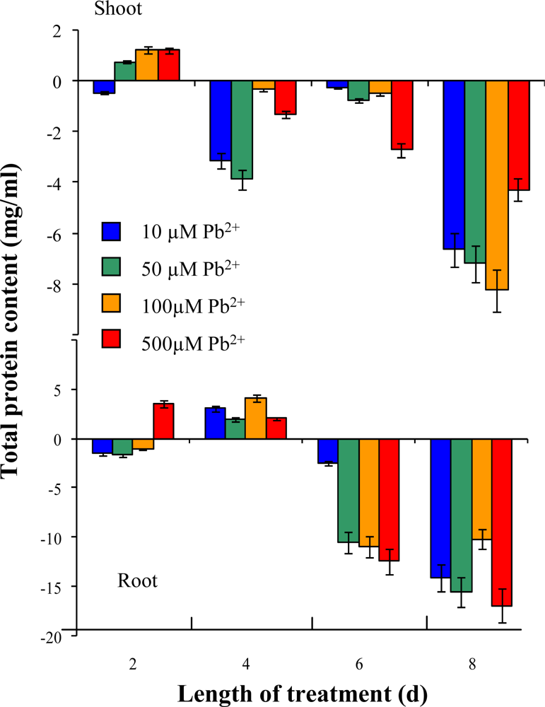
2.2. Total protein content
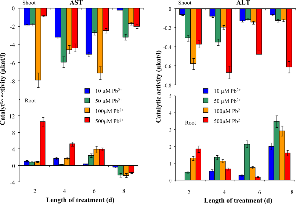
2.3. Determination of plant enzymes’ activity
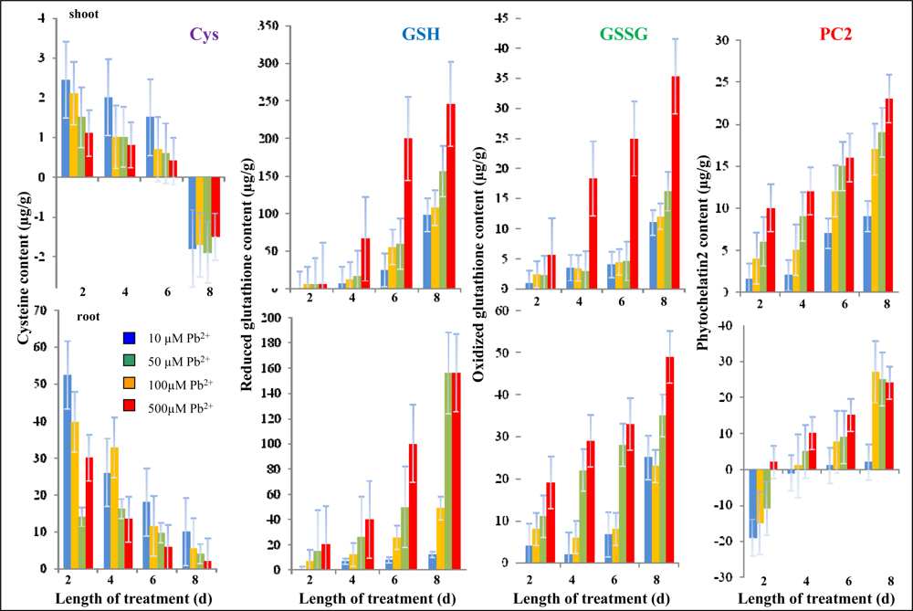
2.4. Content of low molecular mass thiols
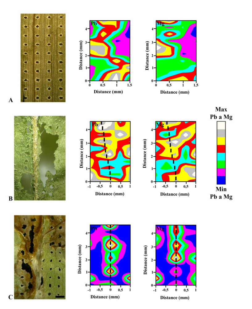
2.5. Monitoring of lead and magnesium distribution by LIBS
3. Material and Methods
3.1. Chemicals
3.2. Cultivation of plants and sample preparation
3.3. Sample preparation for thiol determination
3.4. High performance liquid chromatography with electrochemical detection
3.5. Automated spectrometric measurements
Urease activity determination – indophenol assay (Berthelot method)
ALT and AST activity determination
Bradford protein assay
3.6. Laser induced breakdown spectroscopy
4. Conclusions
Acknowledgments
References and Notes
- Macek, T.; Kotrba, P.; Svatos, A.; Novakova, M.; Demnerova, K.; Mackova, M. Novel roles for genetically modified plants in environmental protection. Trends Biotechnol 2008, 26, 146–152. [Google Scholar]
- Novakova, M.; Mackova, M.; Sylvestre, M.; Macek, T. Preparation of genetically modified plants containing bacterial dioxygenase – Tool for preferable phytoremediation. J. Biotechnol 2007, 131, S36–S36. [Google Scholar]
- Najmanova, J.; Mackova, M.; Macek, T.; Kotrba, P. Preparation of transgenic flax with enhanced metal tolerance. J. Biotechnol 2007, 131, S38–S39. [Google Scholar]
- Pavlikova, D.; Macek, T.; Mackova, M.; Sura, M.; Szakova, J.; Tlustos, P. The evaluation of cadmium, zinc and nickel accumulation ability of transgenic tobacco bearing different transgenes. Plant Soil Environ 2004, 50, 513–517. [Google Scholar]
- Pavlikova, D.; Macek, T.; Mackova, M.; Szakova, J.; Balik, J. Cadmium tolerance and accumulation in transgenic tobacco plants with a yeast metallothionein combined with a polyhistidine tail. Int. Biodeterior. Biodegrad 2004, 54, 233–237. [Google Scholar]
- Macek, T.; Mackova, M.; Pavlikova, D.; Szakova, J.; Truksa, M.; Cundy, S.; Kotrba, P.; Yancey, N.; Scouten, W.H. Accumulation of cadmium by transgenic tobacco. Acta Biotechnol 2002, 22, 101–106. [Google Scholar]
- Francova, K.; Macek, T.; Demnerova, K.; Mackova, M. Transgenic plants – A potential tool for decontamination of environmental pollutants. Chem. Listy 2001, 95, 630–637. [Google Scholar]
- Garbisu, C.; Alkorta, I. Phytoextraction: a cost-effective plant-based technology for the removal of metals from the environment. Bioresour. Technol 2001, 77, 229–236. [Google Scholar]
- Salt, D.E.; Blaylock, M.; Kumar, N.; Dushenkov, V.; Ensley, B.D.; Chet, I.; Raskin, I. Phytoremediation – A novel strategy for the removal of toxic metals from the environment using plants. Bio-Technology 1995, 13, 468–474. [Google Scholar]
- Fernandes, J.C.; Henriques, F.S. Biochemical, physiological, and structural effects of excess copper in plants. Bot. Rev 1991, 57, 246–273. [Google Scholar]
- Li, X.D.; Poon, C.S.; Liu, P.S. Heavy metal contamination of urban soils and street dusts in Hong Kong. Appl. Geochem 2001, 16, 1361–1368. [Google Scholar]
- Little, P.; Martin, M.H. Biological monitoring of heavy-metal pollution. Environ. Pollut 1974, 6, 1–19. [Google Scholar]
- Jarup, L. Hazards of heavy metal contamination. Br. Med. Bull 2003, 68, 167–182. [Google Scholar]
- Singh, R.P.; Tripathi, R.D.; Sinha, S.K.; Maheshwari, R.; Srivastava, H.S. Response of higher plants to lead contaminated environment. Chemosphere 1997, 34, 2467–2493. [Google Scholar]
- Sawidis, T. Effect of cadmium on pollen germination and tube growth in Lilium longiflorum and Nicotiana tabacum. Protoplasma 2008, 233, 95–106. [Google Scholar]
- Pandey, S.; Gupta, K.; Mukherjee, A.K. Impact of cadmium and lead on Catharanthus roseus – A phytoremediation study. J. Environ. Biol 2007, 28, 655–662. [Google Scholar]
- Doumett, S.; Lamperi, L.; Checchini, L.; Azzarello, E.; Mugnai, S.; Mancuso, S.; Petruzzelli, G.; Bubba, M. Heavy metal distribution between contaminated soil and Paulownia tomentosa, in a pilot-scale assisted phytoremediation study: influence of different complexing agents. Chemosphere 2008, 72, 1481–1490. [Google Scholar]
- Malkowski, E.; Kita, A.; Galas, W.; Karcz, W.; Kuperberg, J.M. Lead distribution in corn seedlings (Zea mays L.) and its effect on growth and the concentrations of potassium and calcium. Plant Growth Regul 2002, 37, 69–76. [Google Scholar]
- Kaiser, J.; Malina, R.; Galiova, M.; Novotny, K.; Diopan, V.; Adam, V.; Kizek, R. Employment of laser spectrometry in heavy metal analysis. Lis. Cukrov. Repar 2007, 123, 332–332. [Google Scholar]
- Stejskal, K.; Diopan, V.; Adam, V.; Zehnalek, J.; Trnkova, L.; Havel, L.; Galiova, M.; Malina, R.; Novotny, K.; Kaiser, J.; Kizek, R. Study of effects of lead ions on sugar beet. Lis. Cukrov. Repar 2008, 124, 116–119. [Google Scholar]
- Stejskal, K.; Supalkova, V.; Baloun, J.; Diopan, V.; Babula, P.; Adam, V.; Zehnalek, J.; Trnkova, L.; Havel, L.; Kizek, R. Affecting of sugar beet (Beta vulgaris var. Altissima) by lead chelate. Lis. Cukrov. Repar 2007, 123, 351–355. [Google Scholar]
- Krizkova, S.; Ryant, P.; Krystofova, O.; Adam, V.; Galiova, M.; Beklova, M.; Babula, P.; Kaiser, J.; Novotny, K.; Novotny, J.; Liska, M.; Malina, R.; Zehnalek, J.; Hubalek, J.; Havel, L.; Kizek, R. Multi-instrumental analysis of tissues of sunflower plants treated with silver (I) ions – Plants as bioindicators of environmental pollution. Sensors 2008, 8, 445–463. [Google Scholar]
- Supalkova, V.; Huska, D.; Diopan, V.; Hanustiak, P.; Zitka, O.; Stejskal, K.; Baloun, J.; Pikula, J.; Havel, L.; Zehnalek, J.; Adam, V.; Trnkova, L.; Beklova, M.; Kizek, R. Electroanalysis of plant thiols. Sensors 2007, 7, 932–959. [Google Scholar]
- Potesil, D.; Petrlova, J.; Adam, V.; Vacek, J.; Klejdus, B.; Zehnalek, J.; Trnkova, L.; Havel, L.; Kizek, R. Simultaneous femtomole determination of cysteine, reduced and oxidized glutathione, and phytochelatin in maize (Zea mays L.) kernels using high-performance liquid chromatography with electrochemical detection. J. Chromatogr. A 2005, 1084, 134–144. [Google Scholar]
- Vacek, J.; Petrek, J.; Kizek, R.; Havel, L.; Klejdus, B.; Trnkova, L.; Jelen, F. Electrochemical determination of lead and glutathione in a plant cell culture. Bioelectrochemistry 2004, 63, 347–351. [Google Scholar]
- Petrek, J.; Baloun, J.; Vlasinova, H.; Havel, L.; Adam, V.; Vitecek, J.; Babula, P.; Kizek, R. Image analysis and activity of intracellular esterases as new analytical tools for determination of growth and viability of embryonic cultures of spruce (Picea sp.) treated with cadmium. Chem. Listy 2007, 101, 569–577. [Google Scholar]
- Zitka, O.; Stejskal, K.; Kleckerova, A.; Adam, V.; Beklova, M.; Horna, A.; Supalkova, V.; Havel, L.; Kizek, R. Utilizing electrochemical techniques for detection of biological samples. Chem. Listy 2007, 101, 225–231. [Google Scholar]
- Petrlova, J.; Mikelova, R.; Stejskal, K.; Kleckerova, A.; Zitka, O.; Petrek, J.; Havel, L.; Zehnalek, J.; Adam, V.; Trnkova, L.; Kizek, R. Simultaneous determination of eight biologically active thiol compounds using gradient elution-liquid chromatography with Coul-Array detection. J. Sep. Sci 2006, 29, 1166–1173. [Google Scholar]
- Supalkova, V.; Petrek, J.; Baloun, J.; Adam, V.; Bartusek, K.; Trnkova, L.; Beklova, M.; Diopan, V.; Havel, L.; Kizek, R. Multi-instrumental investigation of affecting of early somatic embryos of spruce by cadmium (II) and lead (II) ions. Sensors 2007, 7, 743–759. [Google Scholar]
- Ryant, P.; Dolezelova, E.; Fabrik, I.; Baloun, J.; Adam, V.; Babula, P.; Kizek, R. Electrochemical determination of low molecular mass thiols content in potatoes (Solanum tuberosum) cultivated in the presence of various sulphur forms and infected by late blight (Phytophora infestans). Sensors 2008, 8, 3165–3182. [Google Scholar]
- Lima, P.R.; Santos, W.J.R.; Oliveira, A.B.; Goulart, M.O.; Kubota, L.T. Electrocatalytic activity of 4-nitrophthalonitrile-modified electrode for the L-glutathione detection. J. Pharm. Biomed. Anal 2008, 47, 758–764. [Google Scholar]
- Gutscher, M.; Pauleau, A.L.; Marty, L.; Brach, T.; Wabnitz, G.H.; Samstag, Y.; Meyer, A.J.; Dick, T.P. Real-time imaging of the intracellular glutathione redox potential. Nat. Methods 2008, 5, 553–559. [Google Scholar]
- Timur, S.; Odaci, D.; Dincer, A.; Zihnioglu, F.; Telefoncu, A. Biosensing approach for glutathione detection using glutathione reductase and sulfhydryl oxidase bienzymatic system. Talanta 2008, 74, 1492–1497. [Google Scholar]
- Korn, M.D.A.; de Andrade, J.B.; de Jesus, D.S.; Lemos, V.A.; Bandeira, M.; dos Santos, W.N.L.; Bezerra, M.A.; Amorim, F.A.C.; Souza, A.S.; Ferreira, S.L.C. Separation and preconcentration procedures for the determination of lead using spectrometric techniques: a review. Talanta 2006, 69, 16–24. [Google Scholar]
- Korn, M.D.A.; dos Santos, D.S.S.; Welz, B.; Vale, M.G.R.; Teixeira, A.P.; Lima, D.D.; Ferreira, S.L.C. Atomic spectrometric methods for the determination of metals and metalloids in automotive fuels – a review. Talanta 2007, 73, 1–11. [Google Scholar]
- Lin, T.J.; Chung, M.F. Using monoclonal antibody to determine lead ions with a localized surface plasmon resonance fiber-optic biosensor. Sensors 2008, 8, 582–593. [Google Scholar]
- Shaw, M.J.; Haddad, P.R. The determination of trace metal pollutants in enviromental matrices using ion chromatography. Environ. Int 2004, 30, 403–431. [Google Scholar]
- Yantasee, W.; Lin, Y.; Hongsirikarn, K.; Fryxell, G.E.; Addleman, R.; Timchalk, C. Electrochemical sensors for the detection of lead and other toxic heavy metals: the next generation of personal exposure biomonitors. Environ. Health Perspect 2007, 115, 1683–1690. [Google Scholar]
- Janssens, K.H.A.; Adams, F.C.V.; Rindby, A. X-ray fluorescence analysis; John Wiley & Sons: Chichester, UK, 2000. [Google Scholar]
- Jorks, S. X-ray microscopy. Instrumentation and biological application; Springer-Verlag: New York, NY, USA, 1987. [Google Scholar]
- Kaiser, J.; Reale, L.; Ritucci, A.; Tomassetti, G.; Poma, A.; Spano, L.; Tucci, A.; Flora, F.; Lai, A.; Faenov, A.; Pikuz, T.; Mancini, L.; Tromba, G.; Zanini, F. Mapping of the metal intake in plants by large-field X-ray microradiography and preliminary feasibility studies in microtomography. Eur. Phys. J. D 2005, 32, 113–118. [Google Scholar]
- Kaiser, J.; Samek, O.; Reale, L.; Liska, M.; Malina, R.; Ritucci, A.; Poma, A.; Tucci, A.; Flora, F.; Lai, A.; Mancini, L.; Tromba, G.; Zanini, F.; Faenov, A.; Pikuz, T.; Cinque, G. Monitoring of the heavy-metal hyperaccumulation in vegetal tissues by X-ray radiography and by femto-second laser induced breakdown spectroscopy. Microsc. Res. Tech 2007, 70, 147–153. [Google Scholar]
- Becker, J.S.; Su, J.; Zoriya, M.V.; Dobrowolska, J.; Matusch, A. Imaging mass spectrometry in biological tissues by laser ablation inductively coupled plasma mass spectrometry. Eur. J. Mass Spectrom 2007, 13, 1–6. [Google Scholar]
- DeLucia, F.C.; Samuels, A.C.; Harmon, R.S.; Walters, R.A.; McNesby, K.L.; LaPointe, A.; Winkel, R.J.; Miziolek, A.W. Laser-induced breakdown spectroscopy (LIBS): a promising versatile chemical sensor technology for hazardous material detection. IEEE Sens. J 2005, 5, 681–689. [Google Scholar]
- Martin, M.Z.; Wullschleger, S.D.; Garten, C.T.; Palumbo, A.V. Laser-induced breakdown spectroscopy for the environmental determination of total carbon and nitrogen in soils. Appl. Optics 2003, 42, 2072–2077. [Google Scholar]
- Russo, R.E.; Mao, X.L.; Gonzalez, J.J.; Mao, S.S. Femtosecond laser ablation ICP-MS. J. Anal. At. Spectrom 2002, 17, 1072–1075. [Google Scholar]
- Hubalek, J.; Hradecky, J.; Adam, V.; Krystofova, O.; Huska, D.; Masarik, M.; Trnkova, L.; Horna, A.; Klosova, K.; Adamek, M.; Zehnalek, J.; Kizek, R. Spectrometric and voltammetric analysis of urease - nickel nanoelectrode as an electrochemical sensor. Sensors 2007, 7, 1238–1255. [Google Scholar]
- Petrek, J.; Vitecek, J.; Vlasinova, H.; Kizek, R.; Kramer, K.J.; Adam, V.; Klejdus, B.; Havel, L. Application of computer imaging, stripping voltammetry and mass spectrometry to study the effect of lead (Pb-EDTA) on the growth and viability of early somatic embryos of Norway spruce (Picea abies/L./Karst.). Anal. Bioanal. Chem 2005, 383, 576–586. [Google Scholar]
- Vitecek, J.; Petrlova, J.; Petrek, J.; Adam, V.; Havel, L.; Kramer, K.J.; Kizek, R. Application of fluorimetric analysis of plant esterases to study of programmed cell death and effects of cadmium (II) ions. Biol. Plant 2007, 51, 551–555. [Google Scholar]
- Vitecek, J.; Adam, V.; Petrek, J.; Vacek, J.; Kizek, R.; Havel, L. Esterases as a marker for the growth of BY-2 tobacco cells and early somatic embryos of the norway spruce. Plant. Cell. Tiss. Org 2004, 79, 195–201. [Google Scholar]
- Vitecek, J.; Petrlova, J.; Adam, V.; Havel, L.; Kramer, K.J.; Babula, P.; Kizek, R. A fluorimetric sensor for detection of one living cell. Sensors 2007, 7, 222–238. [Google Scholar]
- Droge, W. Free radicals in the physiological control of cell function. Physiol. Rev 2002, 82, 47–95. [Google Scholar]
- Noctor, G.; Foyer, C.H. Ascorbate and glutathione: keeping active oxygen under control. Annu. Rev. Plant Physiol. Plant Molec. Biol 1998, 49, 249–279. [Google Scholar]
- Adam, V.; Zehnalek, J.; Petrlova, J.; Potesil, D.; Sures, B.; Trnkova, L.; Jelen, F.; Vitecek, J.; Kizek, R. Phytochelatin modified electrode surface as a sensitive heavy metal ion biosensor. Sensors 2005, 5, 70–84. [Google Scholar]
- Adam, V.; Petrlova, J.; Potesil, D.; Zehnalek, J.; Sures, B.; Trnkova, L.; Jelen, F.; Kizek, R. Study of metallothionein modified electrode surface behaviour in the presence of heavy metal ions-biosensor. Electroanalysis 2005, 17, 1649–1657. [Google Scholar]
- Adam, V.; Hanustiak, P.; Krizkova, S.; Beklova, M.; Zehnalek, J.; Trnkova, L.; Horna, A.; Sures, B.; Kizek, R. Palladium biosensor. Electroanalysis 2007, 19, 1909–1914. [Google Scholar]
- Das, A.K.; de la Guardia, M.; Cervera, M.L. Literature survey of on-line elemental speciation in aqueous solutions. Talanta 2001, 55, 1–28. [Google Scholar]
- Rizk, N.M.H.; Abbas, S.S.; Hamza, S.M.; El-Karem, Y.M.A. Thiopental and phenytoin as novel ionophores for potentiometric determination of lead (II) ions. Sensors 2009, 9, 1860–1875. [Google Scholar]
- Bondarenko, O.; Rolova, T.; Kahru, A.; Ivask, A. Bioavailability of Cd, Zn and Hg in soil to nine recombinant luminescent metal sensor bacteria. Sensors 2008, 8, 6899–6923. [Google Scholar]
- Prasek, J.; Adamek, M.; Hubalek, J.; Adam, V.; Trnkova, L.; Kizek, R. New hydrodynamic electrochemical arrangement for cadmium ions detection using thick-film chemical sensor electrodes. Sensors 2006, 6, 1498–1512. [Google Scholar]
- Galiova, M.; Kaiser, J.; Novotny, K.; Novotny, J.; Vaculovic, T.; Liska, M.; Malina, R.; Stejskal, K.; Adam, V.; Kizek, R. Investigation of heavy-metal accumulation in selected plant samples using laser induced breakdown spectroscopy and laser ablation inductively coupled plasma mass spectrometry. Appl. Phys. A-Mater. Sci. Process 2008, 93, 917–922. [Google Scholar]
- Kaiser, J.; Galiova, M.; Novotny, K.; Cervenka, R.; Reale, L.; Novotny, J.; Liska, M.; Samek, O.; Kanicky, V.; Hrdlicka, A.; Stejskal, K.; Adam, V.; Kizek, R. Mapping of lead, magnesium and copper accumulation in plant tissues by Laser-Induced Breakdown Spectroscopy and Laser-Ablation Inductively Coupled Plasma Mass Spectrometry. Spectrochim. Acta, Part B 2009, 64, 67–73. [Google Scholar]
- Kaiser, J.; Galiova, M.; Novotny, K.; Reale, L.; Stejskal, K.; Samek, O.; Malina, R.; Palenikova, K.; Adam, V.; Kizek, R. Utilization of the Laser Induced Plasma Spectroscopy for monitoring of the metal accumulation in plant tissues with high spatial resolution; Formatex: Badajoz, Spain, 2007; pp. 434–441. [Google Scholar]
- Witte, C.P.; Medina-Escobar, N. In-gel detection of urease with nitroblue tetrazolium and quantification of the enzyme from different crop plants using the indophenol reaction. Anal. Biochem 2001, 290, 102–107. [Google Scholar]
- Bradford, M.M. Rapid and sensitive method for quantitation of microgram quantities of protein utilizing principle of protein-dye binding. Anal. Biochem 1976, 72, 248–254. [Google Scholar]


© 2009 by the authors; licensee MDPI, Basel, Switzerland This article is an open access article distributed under the terms and conditions of the Creative Commons Attribution license (http://creativecommons.org/licenses/by/3.0/).
Share and Cite
Krystofova, O.; Shestivska, V.; Galiova, M.; Novotny, K.; Kaiser, J.; Zehnalek, J.; Babula, P.; Opatrilova, R.; Adam, V.; Kizek, R. Sunflower Plants as Bioindicators of Environmental Pollution with Lead (II) Ions. Sensors 2009, 9, 5040-5058. https://doi.org/10.3390/s90705040
Krystofova O, Shestivska V, Galiova M, Novotny K, Kaiser J, Zehnalek J, Babula P, Opatrilova R, Adam V, Kizek R. Sunflower Plants as Bioindicators of Environmental Pollution with Lead (II) Ions. Sensors. 2009; 9(7):5040-5058. https://doi.org/10.3390/s90705040
Chicago/Turabian StyleKrystofova, Olga, Violetta Shestivska, Michaela Galiova, Karel Novotny, Jozef Kaiser, Josef Zehnalek, Petr Babula, Radka Opatrilova, Vojtech Adam, and Rene Kizek. 2009. "Sunflower Plants as Bioindicators of Environmental Pollution with Lead (II) Ions" Sensors 9, no. 7: 5040-5058. https://doi.org/10.3390/s90705040