Communication

Electrochemical Detection of a Dengue-related Oligonucleotide Sequence Using Ferrocenium as a Hybridization Indicator

by
Fernando Rodrigues Ribeiro Teles
1,
Duarte Miguel França dos Prazeres
2,* and
José Luiz De Lima-Filho
1
1
Laboratório de Imunopatologia Keizo-Asami (LIKA), Universidade Federal de Pernambuco (UFPE), Av. Prof. Moraes Rego, S/N, Campus Universitário, Cidade Universitária, Recife, PE – CEP: 50670-901, Brazil
2
IBB–Institute for Biotechnology and Bioengineering, Centre for Biological and Chemical Engineering, Instituto Superior Técnico, Av. Rovisco Pais, 1049-001, Lisbon, Portugal
*
Author to whom correspondence should be addressed.
Sensors 2007, 7(11), 2510-2518; https://doi.org/10.3390/s7112510
Submission received: 25 September 2007 / Accepted: 26 October 2007 / Published: 30 October 2007

Abstract

:
A simple method for electrochemical detection of a synthetic 20-bp oligonucleotide sequence related with dengue virus genome was developed. A complimentary DNA probe sequence was electrostatically immobilized onto a glassy carbon electrode modified with chitosan. Electrochemical detection of hybridization between probe and target was performed by cyclic voltammetry, using ferrocene (Fc+) as a hybridization label. After hybridization, the peak current response of Fc+ oxidation increased around 26%. A higher voltammetric decay rate constant (kd) and a lower half-life period (t1/2) for the interaction of Fc+ with dsDNA compared to those with ssDNA quantitatively characterize the different strengths of interaction with both types of DNA. By combining the simplicity of DNA immobilization onto a chitosan film and suitable voltammetric detection of hybridization concomitant with ferrocene attachment, a good discrimination between ssDNA and dsDNA was obtained.

1. Introduction

Biosensors have found many applications in both research and industry, namely in clinics diagnosis, for detection of bacterial and viral nucleic-acids in biological samples. They usually make use of the peculiar ability of DNA complimentary chains to hybridize both in vivo as in vitro, thus allowing discrimination between a single and a double chain. Very often, the use of a previously labeled nucleic-acid probe is required. Enzymatic labeling with 32P or 125I radioisotopes, despite its high sensitivity, has been discarded due to the potential dangers of radioactivity. New labeling procedures, including avidin/biotin [1], digoxigenin [2], fluorescent dye [3] and a chemiluminescent agent [4], have replaced the radioactive method but, in general, they are complex and time-consuming. More recently, DNA biosensors with optical [5,6], piezoelectric [7,8,9], acoustic [10,11] and electrochemical transduction have been developed. In particular, electrochemical biosensors are rapid, sensitive, cheap and amenable for in-the-field detection. In parallel with the detection technique, the importance of surface-immobilization methods has increased for the design of electrochemical DNA biosensors, despite the persistent problems of DNA immobilization improvement and screening of highly sensitive hybridization indicators [12,13]. Since, as a whole, DNA is highly redox-inactive, an electrochemical hybridization indicator is usually employed (indirect method). They can bind DNA through reversible physical intercalation between base pairs or through electrostatic interaction, in well-defined binding sites [14]. The effect is a differential accumulation of the indicator in the DNA layer near the surface of the electrode when a ssDNA or a dsDNA is attached, which correlates with different voltammetric peak currents [15]. The most common indicators are heterocyclic dyes (e.g., ethidium bromide, methylene blue, anthracyclines, phenotiazines and acridine derivatives), anticancer drugs (e.g., daunomycin) and organomettalic complexes (mainly from Co, Fe, Os, Pt and Ru). Short, synthetic oligonucleotides are ideal chemical recognition elements for sequence-selective hybridization, because they do not suffer the complex conformational changes that occur in long-sized DNA. Cyclic voltammetry was used to detect dT-oligonucleotides enzymatically elongated with dG residues by covalent attachment to a glassy carbon electrode through N-hydroxysuccinimide and a water-soluble carbodiimide as coupling reagents, using the metallointercalators Co(bpy)33+ and Co(phen)33+ (bpy = 2,2′-bipyridine; phen = 1,10-phenanthroline) as electrochemical indicators [14]. The same group used a similar immobilization procedure onto a carbon paste electrode added with octadecylamine or stearic acid, which provided functional groups for the covalent attachment of a dT-polynucleotide elongated with dG residues, to build a biosensor for cystic fibrosis [15]. In another work, ethidium bromide was used as the electroactive intercalator to detect an oligonucleotide covalently immobilized in a graphite electrode chemically modified with a primary amino group [16]. Erdem et al. [17] detected short oligonucleotides related to the hepatitis B virus after electrochemical adsorption on a pretreated carbon paste electrode, the same immobilization strategy previously used by Wang et al. [18], working with carbon paste and strip electrodes, to detect short DNA sequences related to HIV by chronopotentiometry. Some works with gold electrodes include physical [13] and chemical [19] adsorption of DNA and covalent immobilization through a self-assembled monolayer (SAM) [20,21]. Interesting works were carried out by Xu's team towards sequence-specific voltammetric detection of PCR-derived DNA with graphite [22], platinum [12] and glassy carbon [23] electrodes, using ferrocene derivatives as hybridization markers that covalently label the DNA probes. Electrochemical accumulation of DNA onto a previously polarized (pretreated) electrode and DNA immobilization over a chitosan film-modified electrode were performed. The natural cationic chitosan polymer forms stable complexes with the polyanionic phosphodiester backbones of either native or denatured DNA [24], thus providing a very stable immobilization. In another approach, Ju et al. [25] used ferrocenium ion as a groove-binder to detect yeast DNA covalently attached to a gold electrode through a SAM. Fc+ is able to interact with the major grooves of immobilized dsDNA and also by electrostatic binding. The use of a non-covalent DNA hybridization indicator (intercalator, electrostatic binder or groove-binder) avoids the costly and labor-intensive procedures to synthesize the derivatives, label them to the DNA chain and separate the product. The present paper proposes a simple method to specifically detect a 20-bp oligonucleotide from the most conserved dengue genomic sequence, the 3′-noncoding region [26], thus envisaging detection of all the four antigenically-related dengue virus serotypes (DENV1-4). Due to the significant liability of native DNA under storage and operational conditions, the use of short synthetic oligonucleotides, as in the case of this work, becomes advantageous. One major novelty of this work is the combination of previously confirmed simplicity of DNA immobilization onto a chitosan-modified glassy carbon electrode with straightforward detection of the hybridization event through the oxidation peak current of Fc+ by cyclic voltammetry. Unlike in many other reports, we included the indicator into the voltammetric solution itself, thus avoiding a previous binding step.

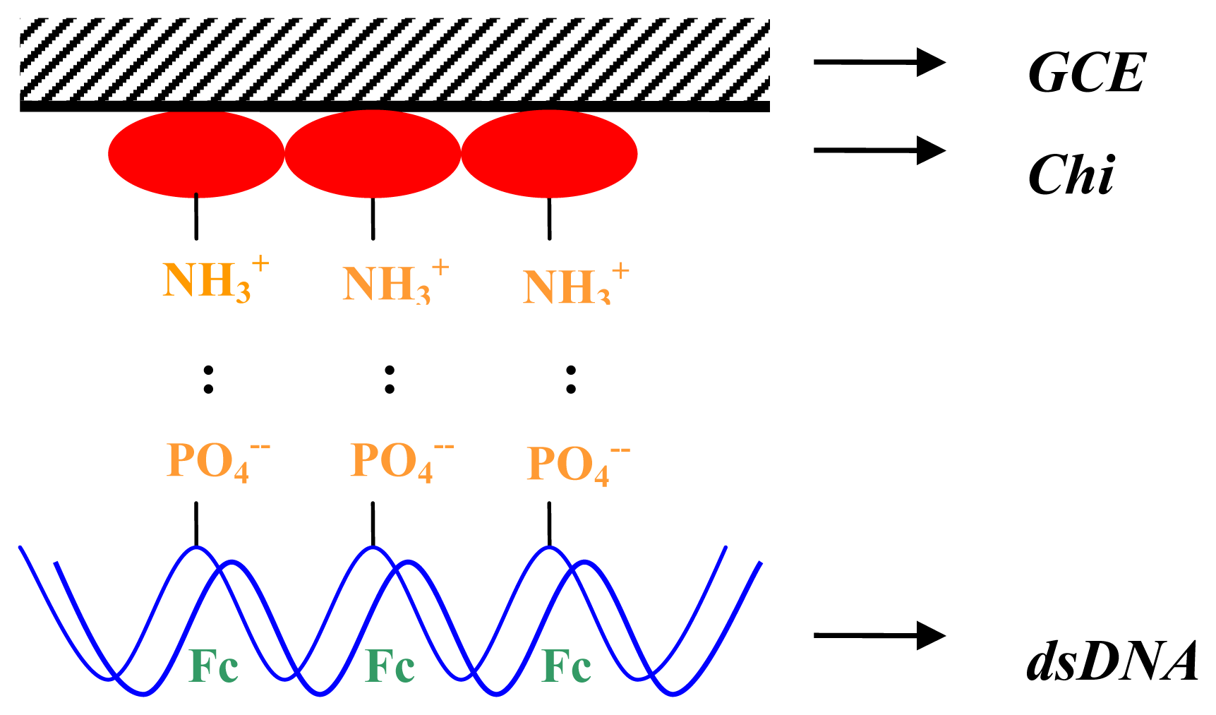
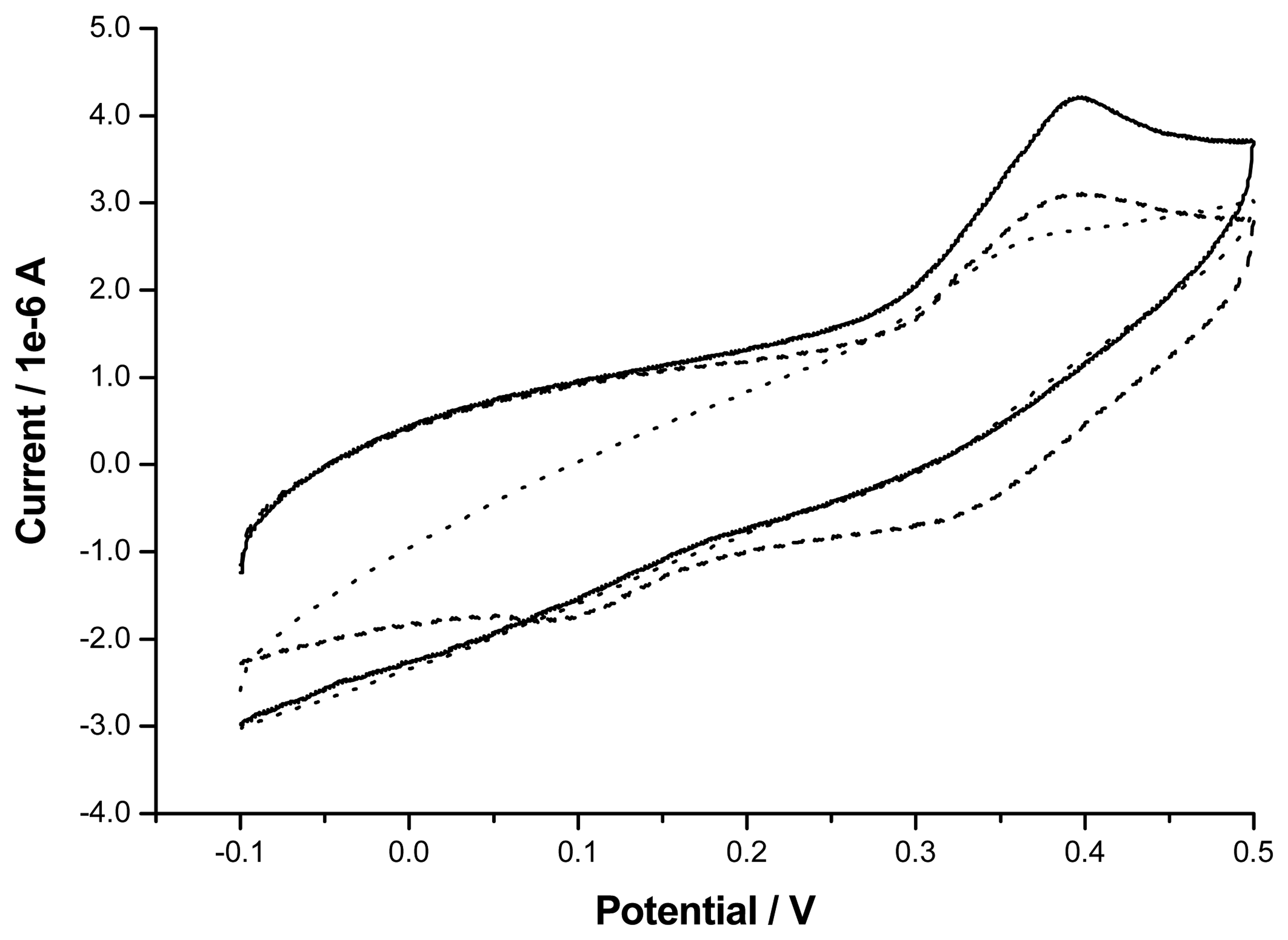
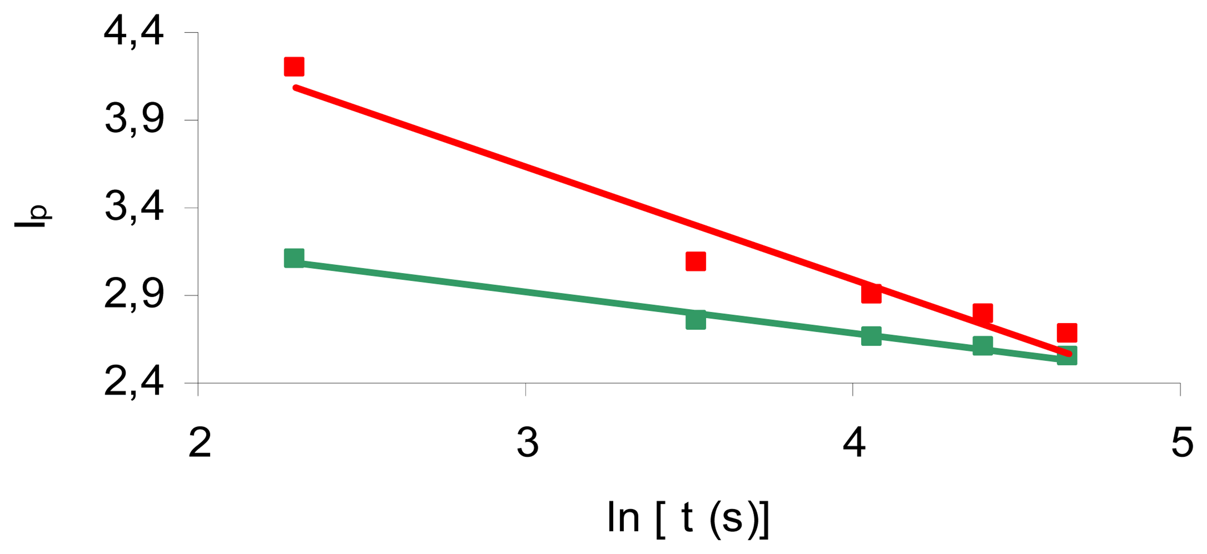
2. Results and Discussion

The principle of DNA electrochemical detection underlying this work relies on the fact that ferrocene is an electroactive DNA-binder, exhibiting greater affinity for dsDNA than for ssDNA due to its tight binding to the major grooves of the DNA double-chain. Since the different affinities to ssDNA and dsDNA provide different local Fc+ concentrations near the electrode, the presence of a single or a double DNA-chain correlates with distinctive voltammetric redox peak currents. According to the literature [12,23], a chitosan support film improves the efficiency of DNA immobilization onto glassy carbon electrode surfaces by electrostatic interaction between the polycationic chitosan oligomer and the polyanionic DNA backbone (Figure 1).
In this work, the immobilization and the hybridization event were distinguished through the voltammetric peak of Fc+ oxidation. In order to do so, we carried out cyclic voltammetry on a dsDNA-modified, a ssDNA-modified and a bare glassy carbon electrode at 50 mV s-1, between -0.1 V and 0.5 V. As can be seen in Figure 2, immobilization of the DNA probe yielded an oxidation peak current around 3.1 μA at 394 mV, probably due to the ferrocene directly adsorbed onto the ssDNA-modified electrode.
The nearby hybridization curve shows a 26% higher peak of 4.2 μA at 396 mV, which implies an increased affinity of ferrocene to dsDNA when compared to ssDNA. The reason for this is a higher Fc+ concentration available for oxidation near the electrode surface when dsDNA is attached instead of ssDNA. The figure also shows an oxidation shoulder of ferrocene in the bare electrode; despite being non-negligible, this shoulder is lower when compared with the two shoulders of the DNA-modified electrode. The higher inclination of the bare electrode voltammetric curve also indicates a higher hindrance of the electronic transport in the bare electrode, which is somewhat overwhelmed when the electrode is modified with ssDNA or dsDNA.
In order to assess the different interaction strengths of ferrocene with ssDNA and dsDNA, a logarithmic decay reaction kinetic model was used to assess a voltammetric decay rate constant, kd, which evaluates the decreasing tendency of voltammetric peak currents over time. The model equation is IP = - kd. ln(t) + constant, where t is the voltammetric decay period and Ip is the oxidation peak current at time t. This was performed by recording the IP values of consecutive scans in both the hybridization and the immobilization assays and plotting them against the logarithms of t (Figure 3).
The half-life period, t1/2 (the time necessary for the decay of IP until half of its maximum, initial value) for both the immobilization and the hybridization assays was also determined from these data. For the ssDNA-modified electrode, we estimated kd = 2.38.10-1 μA and t1/2 = 6.21.103 s and, for the dsDNA-modified electrode, kd = 6.41.10-1 μA and t1/2 = 2.21.102 s. According to the previous statements, the greater affinity of ferrocene to dsDNA than to ssDNA may be inferred from the higher voltammetric oxidation peak obtained, at nearly the same potential, in the former case. On the other hand, kd corresponds to the dissociation rate constant of ferrocene from the DNA molecule to the bulk solution. Its value is predictably higher with dsDNA than with ssDNA – as confirmed by the experimental results – since, according to theory, the decay of the ferrocene oxidation peak would not occur in the ssDNA-modified electrode if there was no ssDNA-bound ferrocene, which is not the case, as seen in Figure 2. The t1/2 depletion observed when passing from the ssDNA-modified electrode to the dsDNA-modified one is also explained by the existence of only a few ssDNA-bound ferrocene ions. According to theoretical predictions, t1/2 for the ssDNA-modified electrode should be nearly infinite.

3. Experimental Section

3.1 Apparatus

Cyclic voltammetry measurements were performed in a MQPG-01 potentiostat (Microquímica Indústria e Comércio, Brazil) coupled to a PC. Experiments were carried out in an electrochemical cell composed by a 0.5 ml acrylic vessel and three artisan, lab-made electrodes: a working glassy carbon electrode (tightly packed into a glass body) with 3.0 mm of diameter and an electrical contact provided by a copper wire, an Ag/AgCl (with 3M NaCl) reference electrode with 4.0 mm of diameter, also with a copper wire contact, and a platinum wire as the counter electrode (diameter of 0.5 mm). An USC750 ultrasonic bath (UltraSonic Cleaner, Brazil) and a thermostated bath (BioEng, Brazil) were also used.

3.2 Reagents

The synthetic dengue-related oligonucleotide sequence 5′-GGT TAG AGG AGA CCC CTC CC-3′ (target) and its complimentary counterpart, 5′-GGG AGG GGT CTC CTC TAA CC-3′ (probe), were purchased as a lyophilized powder from Invitrogen. Sodium hydroxide was purchased from Merck, ferrocenium hexafluorophosphate (FcPF6) was obtained from Aldrich, sodium chloride was acquired from Anidrol (Brazil) and EDTA was purchased from Grupo Química (Brazil). Chitosan and SDS were obtained from Sigma. Hydrochloric acid, hydroxymethyl aminomethane (Tris), glacial acetic acid and sodium citrate were acquired from Vetec (Brazil). Monobasic sodium phosphate (NaH2PO4.H2O) and dibasic sodium phosphate (Na2HPO4.7H2O), both used to prepare the phosphate buffer, were supplied by, respectively, CAQ - Casa da Química (Brazil) and Vetec. All reagents were of analytical grade. All solutions were prepared with deionized (milli-Q) and sterilized water. Alumina suspension with particle size of 0.5 μm and a polishing cloth were obtained from Fortel Indústria e Comércio (Brazil).

3.3. Methods

3.3.1 Electrode preparation

The surface of the glassy carbon electrode was polished with the alumina suspension using a polishing cloth to a mirror finish. The electrode surface was then cleaned in an ultrasonic bath for 15 minutes and washed with deionized water. The electrode was finally sterilized under UV-light for 1h. This procedure was repeated after each voltammetric experiment. Other materials, including glassware, containers, pipette tips and the electrochemical vessel, were heat-sterilized prior to use.

3.3.2 Probe immobilization

The electrode surface was uniformly coated with 2 μl of 1% chitosan solution in 1% acetic acid and air-dried, after which it was immersed in 50 μl of 0.1M NaOH (to stabilize the chitosan film [23]) and again air-dried. The electrode surface was then immersed in a 3.1 mg ml-1 of DNA-probe solution in 0.01 M TE buffer (10 mM Tris-HCl + 1 mM EDTA) pH=8.0 for 2h. After rinsing with 1 ml of 0.1 M phosphate buffer pH=7.0 with 0.1% SDS, to remove unbound DNA, the electrode surface was immersed in the TE buffer until further use.

3.3.3 Hybridization

The DNA-probe modified electrode was immersed in 2×SSC hybridization buffer (0.3 M NaCl + 0.03 M sodium citrate, pH=7.0) containing 2.6 mg ml-1 of target sequence. The set was incubated at 42°C in a water-bath for 1h so that hybridization could occur. The electrode was finally washed with 1 ml of 0.4 M NaOH with 0.1% SDS to remove adsorbed (unhybridized) DNA-target [22]. Control experiments of DNA hybridization (immobilization assays) were performed like the ‘true-hybridization’ assays, but with target-free 2×SSC hybridization buffer.

3.3.4 Indicator binding and electrochemical detection

Cyclic voltammetry assays, simultaneously with indicator-binding, were carried out at 25°C by immersing the DNA-modified electrode in 0.01 M TE buffer pH=8.0 with 0.4 mM FcPF6, at 50 mV s-1, in the scan range from -0.1 V to 0.5 V.

4. Conclusions

This paper describes a very simple method for electrochemical detection of a dengue-related oligonucleotide sequence with a chitosan-coated glassy carbon electrode. The method provided a good discrimination between dsDNA from ssDNA. Unlike in other reports, the oxidation instead of the reduction peak of Fc+ was used to detect DNA hybridization, as confirmed by a kd increase and t1/2 decrease when passing from ssDNA to dsDNA. The use of chitosan as an immobilization support and of Fc+ as a non-covalent DNA marker for hybridization allow avoiding the common drawbacks of covalent binding, namely complexity, morosity and expensiveness. A reported highlight of using a chitosan film to immobilize DNA is that a high ionic strength of the ssDNA solution may be employed to minimize nonspecific DNA adsorption [12]. In theory, the method may be used to specifically detect each one of the four dengue serotypes by appropriate choice of four serotype-specific oligonucleotide sequences amongst the viral genome. Being aware of the still early stages of biosensors' research and production for clinical purposes, the method here proposed may be a significant contribution for future diagnosis of dengue and other infectious pathogens envisaging rapidity, simplicity, low-cost and in-the-field detection with small and portable devices.

Acknowledgments

We acknowledge the Fundação para a Ciência e Tecnologia (FCT), Portugal, (through co-financing of Programa Operacional Ciência e Inovação – 2010, POCI 2010, and Fundo Social Europeu, FSE) for supporting this work with a PhD grant (ref.: SFRH/BD/6186/2001) to Fernando Teles, and to Financiadora de Estudos e Projetos (FINEP), Brazil, for partial support on the infrastructure. We also acknowledge Prof. Marcelo Navarro, from the Department of Fundamental Chemistry (DQF) of UFPE, for having gently lend us the glassy carbon electrode, as well as to Dr. Madalena Areias, also of DQF, and to Dr. Rosa Dutra, of our group, for helpful suggestions.

References

  1. Olejnik, J.; Olejnik, E.K.; Rotschild, K.J. Photocleavable aminotag phosphoramidites for 5′-termini DNA/RNA labeling. Nucleic Acids Res. 1998, 26, 3572–3576. [Google Scholar]
  2. Girotti, S.; Ferri, E.; Ghini, S.; Musiani, M.; Zerbini, M.L.; Gibellini, D.; Gentilomi, G. Direct quantitative chemiluminescent assays for the detection of viral DNA. Anal. Chim. Acta 1991, 255, 387–394. [Google Scholar]
  3. Livache, T.; Fouque, B.; Roget, A.; Marchand, J.; Bidan, G.; Teoule, R.; Mathis, G. Polypyrrole DNA chip on a silicon device: example of hepatitis C virus genotyping. Anal. Biochem. 1998, 255, 188–194. [Google Scholar]
  4. Xu, D.K.; Ma, L.R.; Liu, Y.Q.; Jing, Z.H. Development of chemiluminscent biosensing of nucleic acids based on oligonucleotide-immobilized gold surfaces. Analyst 1999, 124, 533–536. [Google Scholar]
  5. Mandenius, C.F.; Chollet, A.; Mecklenburg, M.; Lundström, I.; Mosbach, K. Optical surface methods for detection of nucleic acid binding. Anal. Letters 1989, 22, 2961–2973. [Google Scholar]
  6. Vo-Dinh, T.; Houck, K.; Stokes, D.L. Surface-enhanced Raman gene probes. Anal. Chem. 1994, 66, 3379–3383. [Google Scholar]
  7. Su, X.; Li, S.F.Y.; Liu, W.; Kwang, J. Piezoelectric quartz cristal based screening test for porcine reproductive and respiratory syndrome virus infection in pigs. Analyst 2000, 125, 725–730. [Google Scholar]
  8. Su, X.; Li, S.F.Y. Serological determination of Helicobacter pylori infection using sandwiched and enzymatically amplified piezoelectric biosensor. Anal. Chim. Acta 2001, 429, 27–36. [Google Scholar]
  9. Aizawa, H.; Kurosawa, S.; Tanaka, M.; Yoshimoto, M.; Miyake, J.; Tanaka, H. Rapid diagnosis of Treponema pallidum in serum using latex piezoelectric immunoassay. Anal. Chim. Acta 2001, 437, 167–169. [Google Scholar]
  10. Zhang, H.; Tan, H.; Wang, R.; Wei, W.; Yao, S. Immobilization of DNA on silver surface of bulk acoustic wave sensor and its application to the study of UV-C damage. Anal. Chim. Acta 1998, 374, 31–38. [Google Scholar]
  11. Su, H.; Kallury, K.M.R.; Thompson, M.; Roach, A. Interfacial nucleic acid hybridization studied by random primer 32P labeling and liquid-phase acoustic network analysis. Anal. Chem. 1994, 66, 769–777. [Google Scholar]
  12. Xu, C.; Cai, H.; Xu, Q.; He, P.; Fang, Y. Characterization of single-stranded DNA on chitosan-modified electrode and its application to the sequence-specific DNA detection. Fresenius J. Anal. Chem. 2001, 369, 428–432. [Google Scholar]
  13. Zhao, Y.-D.; Pang, D.-W.; Wang, Z.-L.; Cheng, J.-K.; Qi, Y.-P. DNA-modified electrodes. Part 2. Electrochemical characterization of gold electrodes modified with DNA. J. Electroanal. Chem. 1997, 431, 203–209. [Google Scholar]
  14. Millan, K.M.; Mikkelsen, S.R. Sequence-selective biosensor for DNA based on electroactive hybridization indicators. Anal. Chem. 1993, 65, 2317–2323. [Google Scholar]
  15. Millan, K.M.; Saraullo, A.; Mikkelsen, S.R. Voltammetric DNA biosensor for cystic fibrosis based on a modified carbon paste electrode. Anal. Chem. 1994, 66, 2943–2948. [Google Scholar]
  16. Liu, S.; Ye, J.; He, P.; Fang, Y. Voltammetric determination of sequence-specific DNA by electroactive intercalator on graphite electrode. Anal. Chim. Acta 1996, 335, 239–243. [Google Scholar]
  17. Erdem, A.; Kerman, K.; Meric, B.; Akarca, U.S.; Ozsoz, M. DNA electrochemical biosensor for the detection of short DNA sequences related to the hepatitis B virus. Electroanalysis 1999, 11, 586–588. [Google Scholar]
  18. Wang, J.; Cai, X.; Rivas, G.; Shiraishi, H.; Farias, P.A.M.; Dontha, N. DNA electrochemical biosensor for the detection of short DNA sequences related to the human immunodeficiency virus. Anal. Chem. 1996, 68, 2629–2634. [Google Scholar]
  19. Hashimoto, K.; Ito, K.; Ishimori, Y. Sequence-specific gene detection with a gold electrode modified with DNA probes and an electrochemically active dye. Anal. Chem. 1994, 66, 3830–3833. [Google Scholar]
  20. Sun, X.; He, P.; Liu, S.; Ye, J.; Fang, Y. Immobilization of single-stranded deoxyribonucleic acid on gold electrode with self-assembled aminoethanethiol monolayer for DNA electrochemical sensor applications. Talanta 1998, 47, 487–495. [Google Scholar]
  21. Kerman, K.; Oznan, D.; Kara, P.; Meric, B.; Gooding, J.J.; Ozsoz, M. Voltammetric determination of DNA hybridization using methylene blue and self-assembled alkanethiol monolayer on gold electrodes. Anal. Chim. Acta 2002, 462, 39–47. [Google Scholar]
  22. Xu, C.; He, P.; Fang, Y. Electrochemical labeled DNA probe for the detection of sequence-specific DNA. Anal. Chim. Acta 2000, 411, 31–36. [Google Scholar]
  23. Xu, C.; Cai, H.; He, P.; Fang, Y. Electrochemical detection of sequence-specific DNA using a DNA probe labeled with aminoferrocene and chitosan modified electrode immobilized with ssDNA. Analyst 2001, 126, 62–65. [Google Scholar]
  24. Hayatsu, H.; Tanaka, Y.; Negishi, K. Preparation of DNA-chitosan columns and their applications: binding of carcinogens to the column. Nucleic Acids Symp. Ser. 1997, 37, 139–140. [Google Scholar]
  25. Ju, H.; Ye, B.; Gu, J. Supermolecular interaction of ferrocenium with yeast DNA and application in electrochemical sensing for hybridization recognition of yeast DNA. Sensors 2004, 4, 71–83. [Google Scholar]
  26. Houng, H.H.-S.; Chen, R.C.-M.; Vaughn, D.W.; Kanesa-thasan, N. Development of a fluorogenic RT-PCR system for quantitative identification of dengue virus serotypes 1-4 using conserved and serotype-specific 3′ noncoding sequences. J. Virol. Methods 2001, 95, 19–32. [Google Scholar]
Figure 1. Scheme of the molecular mechanisms underlying electrochemical DNA detection with ferrocene. After chemical modification of the glassy carbon electrode (GCE) surface with chitosan (Chi), the free amino groups (NH3+) of chitosan monomers electrostatically bind the outer phosphate groups (PO4-) of a single-stranded DNA which, by this way, becomes immobilized on the surface of the electrode. Afterwards, a second, complimentary DNA chain hybridizes with the former and the ferrocene indicator (Fc) binds double-stranded DNA (dsDNA) through its major grooves or electrostatic interactions.
Sensors 07 02510f1
Figure 2. Cyclic voltammograms of 0.4 mM FcPF6 in 0.01 M TE buffer pH=8.0 at bare (dotted line), ssDNA-modified (dashed line) and dsDNA-modified (solid line) glassy carbon electrode, scanned between -0.1 V and 0.5 V (vs. Ag/AgCl), at 50 mV s-1.
Sensors 07 02510f2
Figure 3. Linearized time-dependence of oxidation peak currents (IP) of ferrocene in the ssDNA-modified (green dots) and in the dsDNA-modified (red dots) glassy carbon electrode.
Sensors 07 02510f3

