Article

Electrochemical Studies of the Inhibition and Activation Effects of Al (III) on the Activity of Bovine Liver Glutamate Dehydrogenase

1
College of Chemistry and Environment Science, Department of Environment Science, Nanjing Normal University, Nanjing, 210097, P. R. China
2
State Key Laboratory of Reuse and Control of Pollution, College of Environment Science, Nanjing University, Nanjing, 210093, P. R. China
*
Author to whom correspondence should be addressed.
Sensors 2005, 5(4), 235-244; https://doi.org/10.3390/s5040235
Submission received: 3 June 2004 / Accepted: 18 November 2004 / Published: 27 April 2005
(This article belongs to the Special Issue Papers presented at I3S2004, Nanjing)

Abstract

:
Since the study of Al3+ ion on the enzyme activity by using of electrochemical techniques was rarely found in available literatures, the differential-pulse polarography (DPP) technique was applied to study the effects of Al3+ ion on the glutamate dehydrogenase (GDH) activity in the catalytical reaction of α-KG +NADH+NH4+ ⇔ L-Glu+NAD++H2O by monitoring the DPP reduction current of NAD+. At the plant and animal physiologically relevant pH values (pH=6.5 and 7.5), the GDH enzyme activities were strongly depended on the concentrations of the metal ion in the assay mixture solutions. In the lower Al (III) concentration solutions (<30 μM), the inhibitory effects were shown, which are in accordance with the recently biological findings. With the increase of Al (III) concentrations (30∼80 μM), the enzyme GDH activities were activated. However, once the concentration of Al (III) arrived to near 0.1 mM level (>80μM), the inhibition effects of Al (III) were shown again. The cyclic voltammetry of NAD+ and NAD+-GDH in the presence of Al (III) can help to explain some biological phenomena. According to the differential-pulse polarography and cyclic voltammetry experiments, the present research confirmed that the electrochemical technique is a convenient and reliable sensor for accurate determination of enzyme activity in biological and environmental samples.

1. Introduction

The enzyme Glutamate dehydrogenase (GDH) [EC1.4.1.3] has been found in both higher and lower organisms, and is a pivotal link between carbohydrate and nitrogen metabolism. In the oxidative deamination reaction, GDH feeds the tricaboxylic acid cycle (TCA) by converting L-glutamate (L-Glu) to α-ketoglutarate (α-KG), whereas the reductive amination reaction with using dihydronicotinamide adenine dinucleotide (NADH) as coenzyme, supplies nitrogen for several biosynthetic pathways [1]. Since some of pathology of the disorders associated with GDH defects is restricted to the brain, the enzyme may be of particular importance not only in the plant biology, but also in the animal nervous system. It has, therefore, attracted considerable interests in the biological researches [2,3]. In the past, a variety of works have investigated cation, anion and nucleotides effects on this enzyme. For the cation effects, Ca2+ ion [4] and Mg2+ ion [5] have been reported to interact with the liver enzyme and produce effects on protein stability. Zn2+ ion [6,7] was reported to induce a conformation change and cause inhibition of the enzyme, and La3+ and Eu3+ ions [8] could activate the enzyme GDH because lanthanide ions can enhance the dissociation of the NAD+-GDH complex by combining preferentially to NAD+.
Aluminum (III) has been shown to interfere with many enzymatic key processes of the brain that basically control cell functions. Much less is known, however, about the mechanistic and structural aspects of these Al (III)-modulated enzymatic processes [9,10]. The mechanism of these actions will be an essential area of aluminum chemistry in the future [11]. It is recently demonstrated that Al (III) activates α-ketoglutarate dehydrogenase and succinate dehydrogenase. Meanwhile, aconitase and GDH exhibit a suppressed activity in the presence of this metal ion [12,13]. Accordingly, it is reasonable to assume that Al (III) could interfere with the bioenergetic process of mitochondria, which is affected by degenerative disorders related to biology ‘aging’ [14]. Moreover, the inhibitory effects are also happened in formation of glutamate in the transamination process, which may influence the amino acid synthesis and contribute to the Al (III) toxicity in higher plants [15]. Therefore, it is very interesting to investigate the interaction of Al (III) with the whole GDH enzyme system for further understanding the role of Al (III) in the dehydrogenase enzyme reaction process and helping to elucidate the effects of metal ions on enzyme catalytic reaction at a molecular level.
Since the GDH enzyme molecule consists of six identical subunits with a molecular mass of 56000Da each [1], it is quite difficult to investigate the enzymatic properties and the enzyme structure alteration in the presence of metal ions. Electrochemical analysis has always been recognized as a powerful tool for measuring trace metal ions and biomolecules in the biological system. Its remarkable sensitivity, simple instrumentation, lower costs, simple operation procedure, faster and reliable results and many other advantages make it satisfy many requires and extensively applied in different biological system analysis or in other fields [16]. In the past, we have investigated the interactions of Al (III) with the substrates (α-KG/ L-Glu) [17,18] and coenzyme I (NAD+/NADH) [19,20] of the GDH enzyme system. But the study of interaction between Al (III) with the whole GDH enzyme by using electrochemical techniques has not been found in the available literatures. Therefore, the present paper is the aim to assay glutamate dehydrogenase in reductive amination of α-KG with coenzyme as NADH by using the differential pulse polarography electrochemical technique. Some part of mechanism was proposed with the cyclic voltammetry of NAD+, NAD+-GDH in the absence and presence of Al (III). Such bioinorganic studies are performed to clarify the various aspects of aluminum potential toxicity, and to fill the gap between the knowledge concerning biochemical and biological phenomena [21].

2. Materials and methods

Bovine liver glutamate dehydrogenase was obtained from Sigma Co. (St. Louis. MO, USA) as a solution stored in 50% glycerol + 0.05M potassium phosphate buffer (pH=7.3) and was used directly. NAD+ and NADH (purity >98%) were also purchased from Sigma. L-Glu, α-KG and NH4Cl were the products of Acros Co. (New Jersey, USA). All solutions were prepared fresh daily with double-distilled water. The Al3+ stock solution was prepared by dissolving high purity metallic Al powder (99.99%) in hydrochloric acid. More dilute solutions were prepared by diluting this solution with double distilled water. To prevent hydrolysis of this metal ion, the Al3+ stock solutions were prepared at pH ≤2. Other chemicals were of analytical reagent grade. Necessary polyethylene vessels were used. All glass- wares were soaked in 10% HNO3 for at least 24 h, then washed carefully with double-distilled water. Buffer solutions were prepared by mixing appropriate amount of Tris aminomethane (hydroxymethyl) with HCl solutions and adjusted pH to 6.5 and 7.5, since these values were representative of the cytoplasmic pH in plants roots and animal physiologically relevant pH [12,22].
Differential pulse polarography and cyclic voltammetry experiments were performed by using a BAS-100B Electrochemical Analyzer (USA) with a three-electrode system, which could be used to determine the enzyme activities [8,23,24]. A hanging mercury drop electrode (HMDE) was used as the working electrode. A saturated calomel electrode (SCE) was used as a reference electrode and a platinum wire as an auxiliary electrode. The mixture solution was carried out in 100 mM Tris-HCl (pH 6.5 or 7.5) which contained 0.2 mM NADH, 10mM NH4Cl and 2mM α-KG and was stirred with a Teflon-coated magnetic stirring bar at 200 rpm. The final volume of the mixture solution was 20 ml. All the measurements were carried out at 25±0.10C. An inert gas, pure nitrogen, was used for degassing of solution at least 20 minutes prior to measurements. The pH value was measured with the HANNA instruments pH213 Microprocessor pH meter (Portugal) with a glass combination electrode.

3. Results and discussion

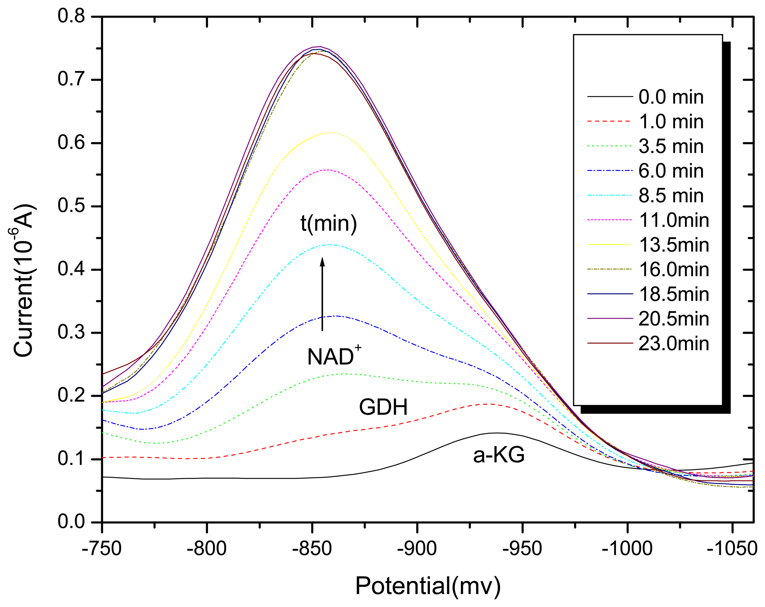
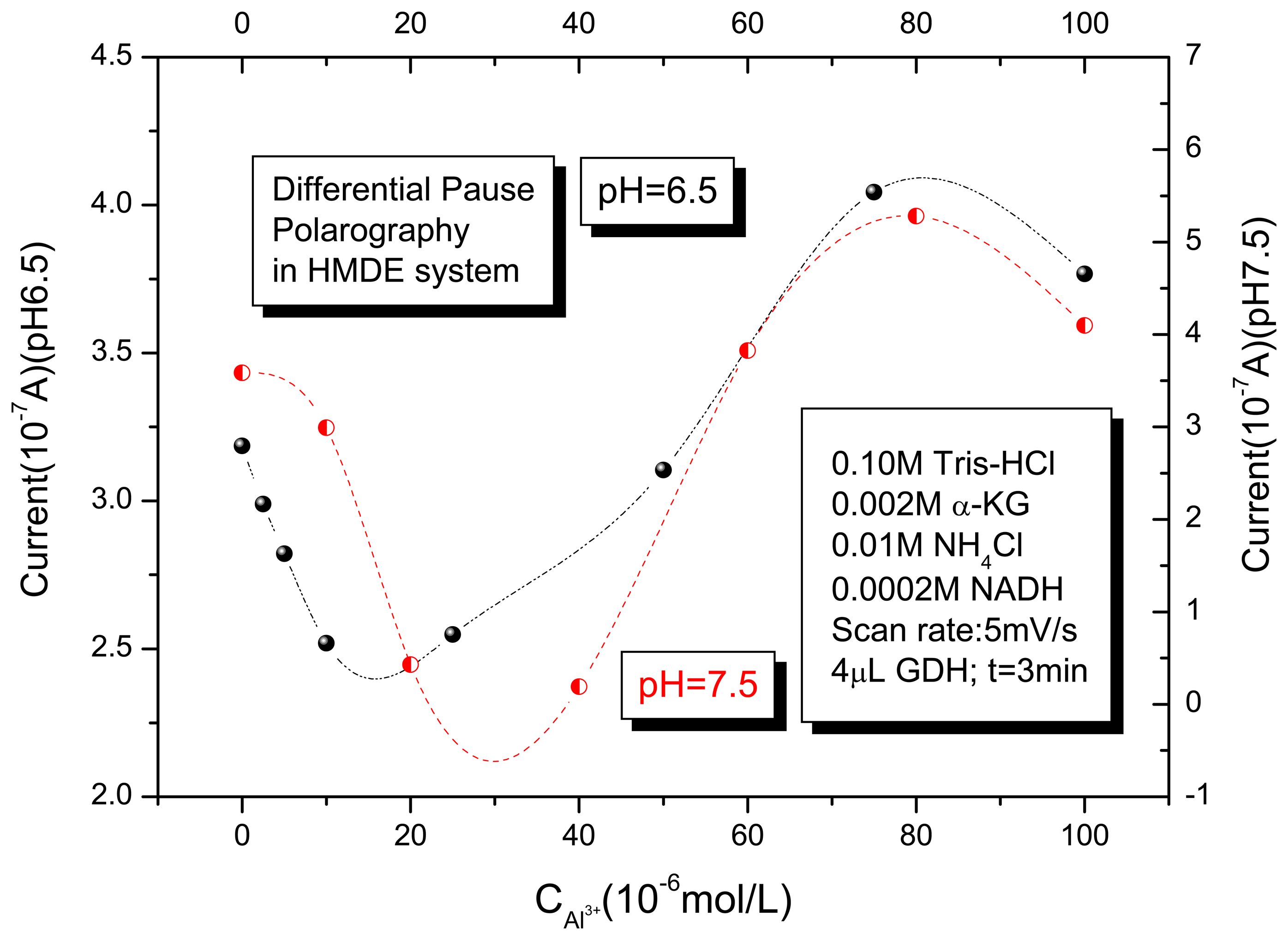
3.1. Differential pulse polarography of the GDH enzyme in the absence and presence of Al (III)

Since the differential pulse polarography is a well-stated electroanalytical technique, it is a widely used method for the routine metal ion analysis and the plant enzymatic activity measurement [23,24]. The study of the effects of Al (III) on the glutamate dehydrogenase activity in the catalytical reaction of α-KG +NADH+NH4+ ⇔ L-Glu +NAD++H2O can be performed by monitoring the DPP reduction current of NAD+. Figure 1 reports the differential-pulse polarography in a buffer solution of 100 mM Tris-HCl (pH 6.5)+2 mM α-KG +10 mM NH4Cl and 0.2 mM NADH with a final volume of 20 mL. It clearly shows that a well-defined higher differential-pulse peak potential of the NAD+ reduction current at –0.86V [23]. Meanwhile, a rather weaker peak was at –0.97 V for α-KG, which was also agreed with our previous finding for α-KG in case of the different potentials caused by different pH values [18]. With the addition of 4μL GDH enzyme into the assay solution, the peak current of NAD+ increased whereas that of α-KG decreased with the developing of the reaction time, which means that the GDH catalytical reaction started and finally arrived to the revisable equilibrium. It is, therefore, clearly revealed that the differential pulse polarography technique could accurately determine the GDH activity in an enzymatic catalytical reaction.
In the presence of Al (III) in the above enzyme system, the peak currents of NAD+, which were converted from NADH by GDH and measured at the same reaction time (3 min) and two pH value 6.5 and 7.5, are shown in Figure 2. It demonstrated that the effects of Al (III) modulates the GDH activity as a function of the metal ion concentrations. It showed that, at a lower concentration of Al (III) (<30μM), the metal ion inhibitory effect was observed. With the increase of the Al (III) concentration (30∼80μM), the activating effect of Al (III) on the enzyme GDH activity was exhibited. When the concentration of Al (III) arrived to near 0.1 mM level (>80μM), the curve reached to a high peak and started to fall. It reflected the Al (III) inactivation and inhibitory effects under these conditions. The inhibition effect observed in the first part of the curves was coincided with Zatta's biological report even though at a little different pH value [12]. However, the activating effect produced by Al3+ ions is a new finding, and it is therefore needed to be further studied.

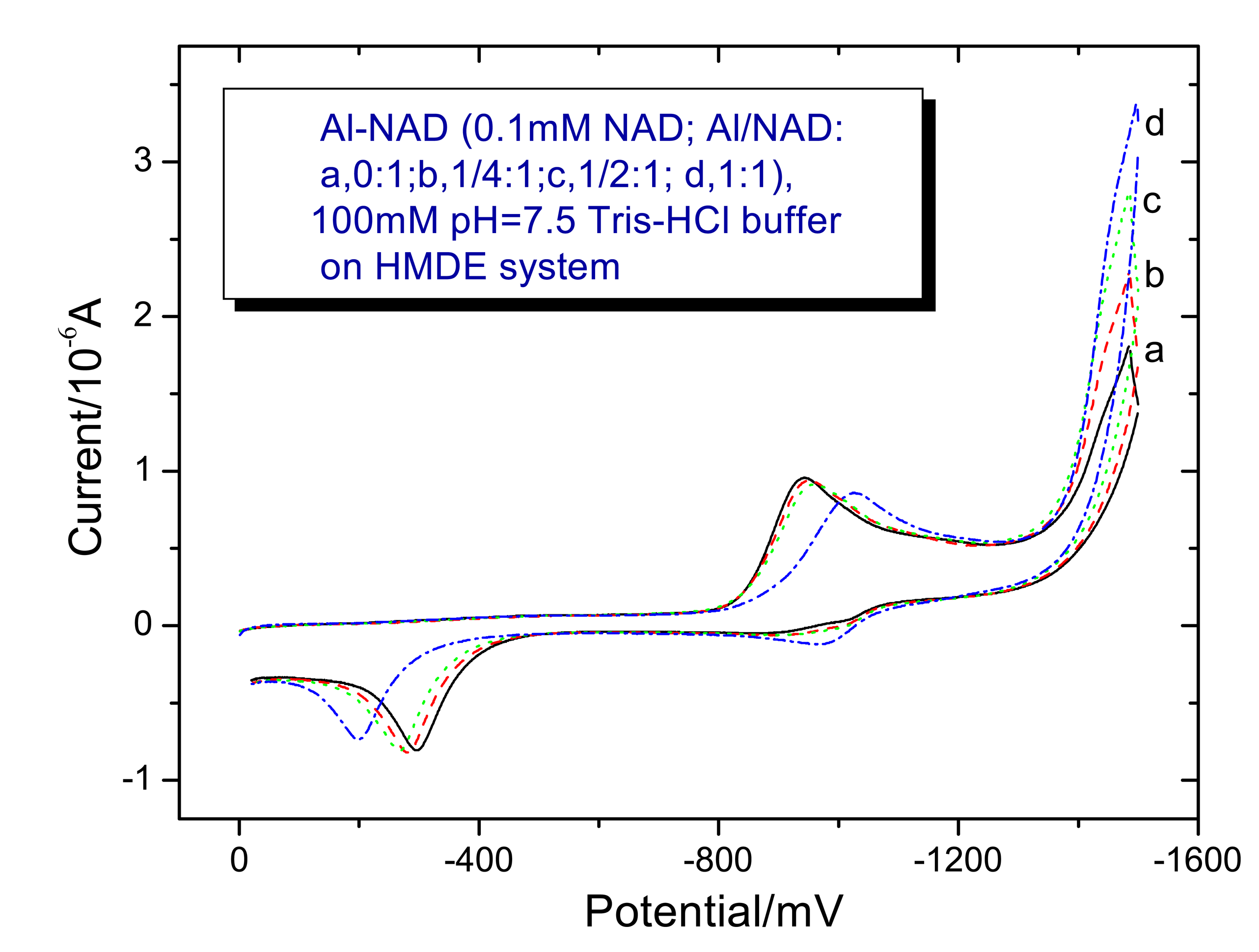
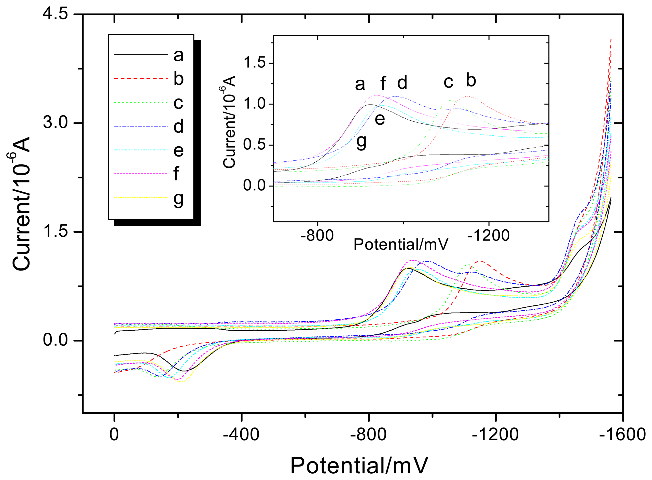
3.2. Voltammetry behaviors of Al (III) on the NAD+ and NAD+-GDH

In order to study the Al (III) effects on the bovine liver GDH system, the cyclic voltammetry technique was performed to elucidate the interaction between the Al (III) and NAD+. Figure 3 shows the cyclic voltammogram of 0.1 mM NAD+ in the presence of different Al/NAD+ ratios of Al3+ ion in a 100 mM pH 7.5 Tris-HCl buffer solution with a final volume of 20 mL. Curve (a) showed that NAD+ was reduced at approximately –0.93 V (vs. SCE) and its anodic wave was at –0.30V (vs. SCE). Meanwhile, curves (b) to (c) gave the voltammograms of NAD+ corresponding to different ratios of Al/NAD+. In the presence of the metal ion, the redox peak almost has not changed, however, the anodic peak has a little shifted positively. The result revealed that the Al (III) and NAD+ might form a weak binding through the phosphate moiety. With the increase of the Al3+ ion concentration (Al/NAD+ ratio 1:1), Fig. 3 curve (d) showed the cathodic peak shifted negatively and the anodic peak shifted positively. Under this concentration of Al (III), the metal ion might satisfy the phosphate groups and the residue part of Al (III) bonded with the adenine N7 function group of NAD+, which was in accordance with our previous report [19]. Therefore, it should influence the electroactive group adenine and cause an obvious change in the voltammogram.
In order to elucidate the effects of Al (III) on the binding of NAD+ to GDH, the cyclic voltammogram of 0.1mM NAD+ and 20μL GDH in the absence and presence of different Al/ NAD+ ratios of Al (III) were investigated in a 100 mM, pH 6.50 Tris-HCl buffer solution with a final volume of 20 mL. In Figure 4, curve (a) showed the voltammogram of NAD+, which was almost the same as that of the above experiment. With the addition of the GDH enzyme to the NAD+ solution, it showed that GDH can easily complex with NAD+, and the cathodic peak potential of NAD+ would negatively shift from –0.90 V to –1.15V (vs. SCE) (Fig.4b). It means that the adenine moiety of NAD+ might bind with GDH and caused an obvious electrochemical change in the voltammogram [8]. However, when the Al (III) was added to the GDH and NAD+ mixture solutions, the reduction peaks of the NAD+ of the NAD+-GDH complex (at –1.15V) gradually shifted positively. Meanwhile, the reduction peak of NAD+ appeared (Fig. 4d). Finally, the reduction peak of the NAD+-GDH complex completely disappeared and the peak curve was the same as the electrochemical behavior of pure NAD+ at pH 7.5 (at –0.90V) (Fig. 4g). It was clearly indicated that the presence of Al3+ ion would enhance the dissociation of the NAD+-GDH complex. These voltammograms with different Al (III) concentrations are shown in the Fig. 4 curves (c) to (g).

3.3. Proposal explanation of the inhibition and activation effects of Al (III) on GDH enzyme activities

The mechanism of the enzyme-catalyzed reaction can be explained by the formation of enzyme-substrate-coenzyme complex. The initial enzyme-catalyzed reaction steps are involved in the binding of the substrate and coenzyme to the enzyme surface and the enzyme will orient these reactants relative to each other. After the catalyzed reaction is completed, the enzyme opens up again to let the products leave, and to prepare for the next substrate [8]. According to the electrochemical behaviors of Al (III) on the GDH enzyme system, in the assay mixture solutions, the enzyme GDH activity was shown the strongly dependence of Al (III) concentrations. It can be divided into three parts in the above experiments. The electrochemical phenomena can be explained as the following points.
The reasons of the first inhibitory part at lower concentrations of Al (III) (<20 μmol/L) on the enzyme GDH are: (1). At the same concentration of Al (III) (20 μM) and pH value (pH=7.5), Zatta's biological finding [12] revealed that Al (III) can decrease the GDH activity. Our experimental result not only supported this biological phenomenon, but also proved that the electrochemical technique is an effective tool for the determination of the enzyme activity. (2). Michaelis-Menten equation showed that the inhibition of Al (III) with glutamate dehydrogenase activity is of the competitive type [12], which suggested that it might bind to the same active site. According to our previous reports [17-19], the binding ability of α-KG with Al (III) is even rather weaker and still stronger than others in the GDH enzyme system. Therefore, on the one hand, some Al-α-KG complex may complete with α-KG by occupying the GDH active site and affect the reactants GDH-NADH-α-KG attractive interactions (‘association’). On the other hand, it will block the important >C=O group converting to –NH2 group of L-Glu in the enzyme-catalyzed reaction system. (3). From our study [20], at biologically relevant pH and concentrations of Al (III) and NADH (pH=6.5, CAl = 1∼10 μM) [22], Al (III) could increase the amount of folded form conformations of NADH which will result in reducing the coenzyme NADH activity in dehydrogenases reaction systems.
The cause of the second activation part of Al (III) on the enzyme GDH is probably that: (1) At a higher concentration of Al (III), like the metal ion could decrease and increase the acetylcholinesterase (AChE) activity [25], Al (III) might also have this kind of “two phase effects” on the GDH enzyme activity. (2). In the reductive amination reaction, during converting the substrate α-KG to L-Glu and the coenzyme NADH was oxidized to NAD+, the step of dissociation of the NAD+-GDH complex is the rate-limiting step and the dissociation step of enzyme-coenzyme product complex is the key stage in the whole process [3,8]. The voltammetry studies indicated that Al3+ ion could interrupt the binding of NAD+ to GDH when the concentration of metal ion is increased. Meanwhile, the voltammetry studies of NAD+ in the presence of Al (III) also reveal that Al (III) can bind with the adenine N7 function group of NAD+ under the higher concentration of the metal ion, which will influence the adenine of NAD+ binding with GDH. Therefore, Al (III) could enhance the dissociation of the NAD+-GDH complex and activate GDH in this pH range.
For the third part of the curve in the DPP experiment, the inactivation or even inhibition of GDH enzyme activity was observed at the highest concentration of Al (III) (>0.1 mM). The reasons for that are rather unknown and we cannot make clearly explanation at this time. However, this electrochemical phenomenon is interestingly, and not surprisingly, the most Al (III) hydrolyzable species might show the lowest activating or even the inhibiting effect. This result is in accordance with the inactivation reported in Al (III)-AChE enzyme investigation [25], and it suggested that the ability of Al (III) to modulate the GDH activity is strongly dependent not only on the concentration of the metal ion but also on the metal ion actual chemical species. Furthermore, a recent biological study suggested that the inactivation of human GDH by aluminum was due to the conformational change induced by aluminum binding at micromolar concentrations [26].

4. Conclusions

In summary, although the effects of Al (III) in biological systems have been extensively described, direct information concerning the molecular basis of its effects on enzyme systems and cell culture is rather scant [9]. Since the study of metal ion influence to the substrate and coenzyme participated to the enzymatic system was rather complicated, the mechanistic and structural aspects of these Al (III)- modulated GDH enzymatic processes need to be further studied. However, from this study, the electrochemical technique was proven to be a convenient and reliable sensor for an accurate determination of enzyme activity because of its real-time, high sensitivity and continuous measurement of the change in coenzyme concentrations. Moreover, this study has achieved encouraging results and provided additional insight into the mechanism of actions of Al (III) on GDH activity. Therefore, this kind of electrochemical investigation could be attributed to the understanding of the toxicity of Al (III) in the plant enzyme-catalyzed reaction processes. Also, these results suggested a possibility that aluminum- induced alterations in enzymes of the glutamate system might be one of the causes of aluminum-induced human neurotoxicity.

Acknowledgments

Thanks for the project supported by the Research Funding from Natural Science Foundation of Jiangsu Province Education Administration of China (04KJB150067), Research Funding from Nanjing Normal University (2003103XGQ2B40) and the Research Funding from the State Key Laboratory of Reuse and Control of Pollution (NJUESKL03001).

References

  1. Peterson, P.E; Smith, T.J. The structure of bovine glutamate dehydrogenase provides insights into the mechanism of allostery. Structure 1999, 7, 769–782. [Google Scholar]
  2. Cho, S.W.; Cho, E.H.; Choi, S.Y. Activation of two types of brain glutamate dehydrogenase isoproteins by gabapetin. FEBS Lett. 1998, 426, 196–200. [Google Scholar]
  3. Pazhanisamy, S.; Maniscalco, S.J.; Singh, N.; Fisher, H.F. A kinetic mechanism of the allosteric control of enzyme-coenzyme binding: glutamate dehydrogenase-NADPH-phosphate acetate-hydrogen ion interactions. Biochemistry 1994, 33, 10381–10385. [Google Scholar]
  4. Jung, K.; Sokolowski, A.; Egger, E. Influence of calcium ions on the activity of human liver glutamate dehydrogenase. Hoppe-Seyler's Z. Physiol.Chem. 1973, 354, 101–103. [Google Scholar]
  5. McCarthy, A.D.; Tipton, K.F. The effects of magnesium ions on the interactions of ox brain and liver glutamate dehydrogenase with ATP and GTP. Biochem. J. 1984, 220, 853–856. [Google Scholar]
  6. Colman, R.F.; Foster, D.S.; Daniel, S. Absence of zinc in bovine liver glutamate dehydrogenase. J.Biol.Chem. 1971, 245, 6190–6195. [Google Scholar]
  7. Bell, E.T.; Stilwell, A.M.; Bell, J.E. Interaction of Zn2+ and Eu3+ with bovine liver glutamate dehydrogenase. Biochem.J. 1987, 246, 199–203. [Google Scholar]
  8. Zhuang, Q.K.; Dai, H.C.; Gao, X.X.; Xin, W.K. Electrochemical studies of the effect of lanthanide ions on the activity of glutamate dehydrogenase. Bioelectrochemistry 2000, 52, 37–41. [Google Scholar]
  9. Kiss, T.; Hollósi, M. Aluminum and Alzheimer's Disease The Science that Describes the Link; Exley, C., Ed.; Elivser: Amsterdam, 2001; p. p361. [Google Scholar]
  10. Louie, A.Y.; Meade, T.J. Metal complexes as enzyme inhibitors. Chem. Rev. 1999, 99, 2711–2734. [Google Scholar]
  11. Atwood, A.Y.; Yearwood, B.C. The future of aluminum chemistry. J. Organomet. Chem. 2000, 600, 186–197. [Google Scholar]
  12. Zatta, P.; Lain, E.; Cagnolini, C. Effects of aluminum on activity of Krebs cycle enzymes and glutamate dehydrogenase in rat brain homogenate. Eur. J. Biochem. 2000, 267, 3049–3055. [Google Scholar]
  13. Hamel, R.D; Appanna, V.D. Modulation of TCA cycle enzymes and aluminum stress in Pseudomonas fluorescens. J. Inorg. Biochem. 2001, 81, 1–8. [Google Scholar]
  14. Swegert, C.V.; Dave, K.R.; Katyare, S.S. Effects of aluminium-induced Alzheimer like condition on oxidative energy metabolism in rat liver,brain and heart mitochondria. Mech. Ageing Dev. 1999, 112, 27–42. [Google Scholar]
  15. Ma, J.F.; Ryan, P.R.; Delhaize, E. Aluminium tolerance in plants and the complexing role of organic acids. Trends Plant Sci. 2001, 6, 273–278. [Google Scholar]
  16. Xin, W.K.; Gao, X.X. Study of the effect of lanthanide ions on the kinetic of glutamate dehydrogenase by a chronoamoerometric method. Analyst 1996, 121, 687–690. [Google Scholar]
  17. Yang, X.D.; Bi, S.P.; Wang, X.L.; Liu, J.; Bai, Z.P. Multimethod characterization the interaction of aluminum ion with α-ketoglutaric acid in acidic aqueous solutions. Anal. Sci. 2003, 19, 273–279. [Google Scholar]
  18. Yang, X.D.; Tang, Y.Z.; Bi, S.P; Yang, G.S.; Hu, J. Multimethod study the complexation of aluminum (III) with L-glutamate in acidic aqueous solutions. Anal. Sci. 2003, 19, 133–138. [Google Scholar]
  19. Yang, X.D.; Bi, S.P.; Yang, X.L.; Yang, L.; Hu, J.; Liu, J. NMR spectra and potentiometry studies of aluminum (III) binding with coenzyme NAD+ in acidic aqueous solutions. Anal. Sci. 2003, 19, 815–821. [Google Scholar]
  20. Yang, X.D.; Bi, S.P.; Yang, L.; Zhu, Y.H.; Wang, X.L. Multi-NMR and fluorescence spectra study the effects of aluminum (III) on coenzyme NADH in aqueous solution. Spectrochim. Acta Part A. 2003, 58, 2561–2568. [Google Scholar]
  21. Jones, D.J.; Kochian, L.V. Aluminum interaction with plasma membrane lipids and enzyme metal binding sites and its potential role in Al cytotoxicity. FEBS Lett. 1997, 425, 51–57. [Google Scholar]
  22. Suhayda, C.G.; Haug, A. Organic acids prevent aluminum-induced conformational changes in calmodulin. Biochem. Biophy. Res. Comm. 1984, 119, 376–381. [Google Scholar]
  23. Wang, X.Q.; Gao, X.X. Voltammetric behavior studies on the interaction between lanthanide ions and NAD+,NAD-LDH complex. Chem. J. Chin. Univ. 1997, 18, 1027–1030. [Google Scholar]
  24. Šebela, M.; Studeničková, M.; Wimmerová, M. Differential pulse polarographic study of the redox centers in pea amine oxidase. Bioelectrochem. Bioenergetics 1996, 41, 173–179. [Google Scholar]
  25. Zatta, P.; Ibn-Lkhayat-Idrissi, M.; Zambenedetti, P.; Kilyen, M.; Kiss, T. In vivo and in vitro effects of aluminum on the activity of mouse brain acetylcholinesterase. Brain Res. Bull. 2002, 59, 41–45. [Google Scholar]
  26. Yang, S.J.; Huh, J.W.; Lee, J.E.; Choi, S.Y.; Kim, T.U.; Cho, S.W. Inactivation of human glutamate dehydrogenase by aluminum. Cell. Molec. Life Sci. 2003, 60, 2538–2546. [Google Scholar]
Figure 1. Differential-pulse polarography of GDH enzyme system; in a 0.1M pH=6.5 Tris-HCl buffer solution; 0.002M α-KG; 0.0002M NADH; 0.01M NH4Cl; 4μL GDH; Scan rate: 5 mV/s.
Sensors 05 00235f1
Figure 2. DPP peak currents as a function of Al (III) concentration; in 0.1M pH=6.5 and 7.5 Tris-HCl buffer solutions; 0.002 M α-KG; 0.01 M NH4Cl; 0.0002 M NADH; 4μL GDH; measurement time 3min; Scan rate: 5mV/s.
Sensors 05 00235f2
Figure 3. The cyclic voltammogram of NAD+ and Al-NAD+; in 100mM pH=7.5 Tris-HCl buffer solution; 0.1 mM NAD+; Al/L: a, 0:1;b, 1/4:1;c, 1/2:1; d, 1:1, on a three-electrode system (HMDE).
Sensors 05 00235f3
Figure 4. The cyclic voltammogram of NAD+, GDH-NAD+ and Al-GDH-NAD+; in 0.1M pH=6.5 Tris-HCl buffer solution; 0.1mM NAD++20 μL GDH; Al: NAD+: a, 0:1 (no GDH); b, 0:1 (GDH); c, 1/4:1 (GDH); d, 1/2:1 (GDH); e, 1:1 (GDH); f, 2:1 (GDH); g, 4:1 (GDH) on a three-electrode system (HMDE).
Sensors 05 00235f4

