
The Thermoluminescence Response of Ge-Doped Flat Fibers to Gamma Radiation

Abstract
:1. Introduction
2. Materials and Method
2.1. Ge-Doped Flat Dosimeters and LiF Dosimeters


| Cross-Section Dimensions (µm) | Mass (mg) |
|---|---|
| 85 × 270 | 0.21 ± 0.01 |
| 100 × 350 | 0.36 ± 0.01 |
| 165 × 620 | 1.19 ± 0.01 |
| 200 × 750 | 1.99 ± 0.01 |
2.2. Annealing
2.3. Samples Irradiation
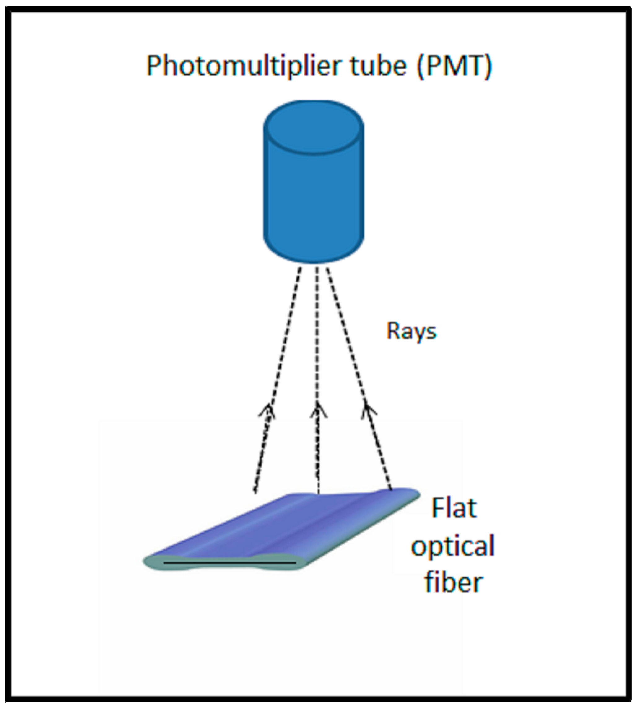
2.4. Samples Readout
3. Results and Discussion
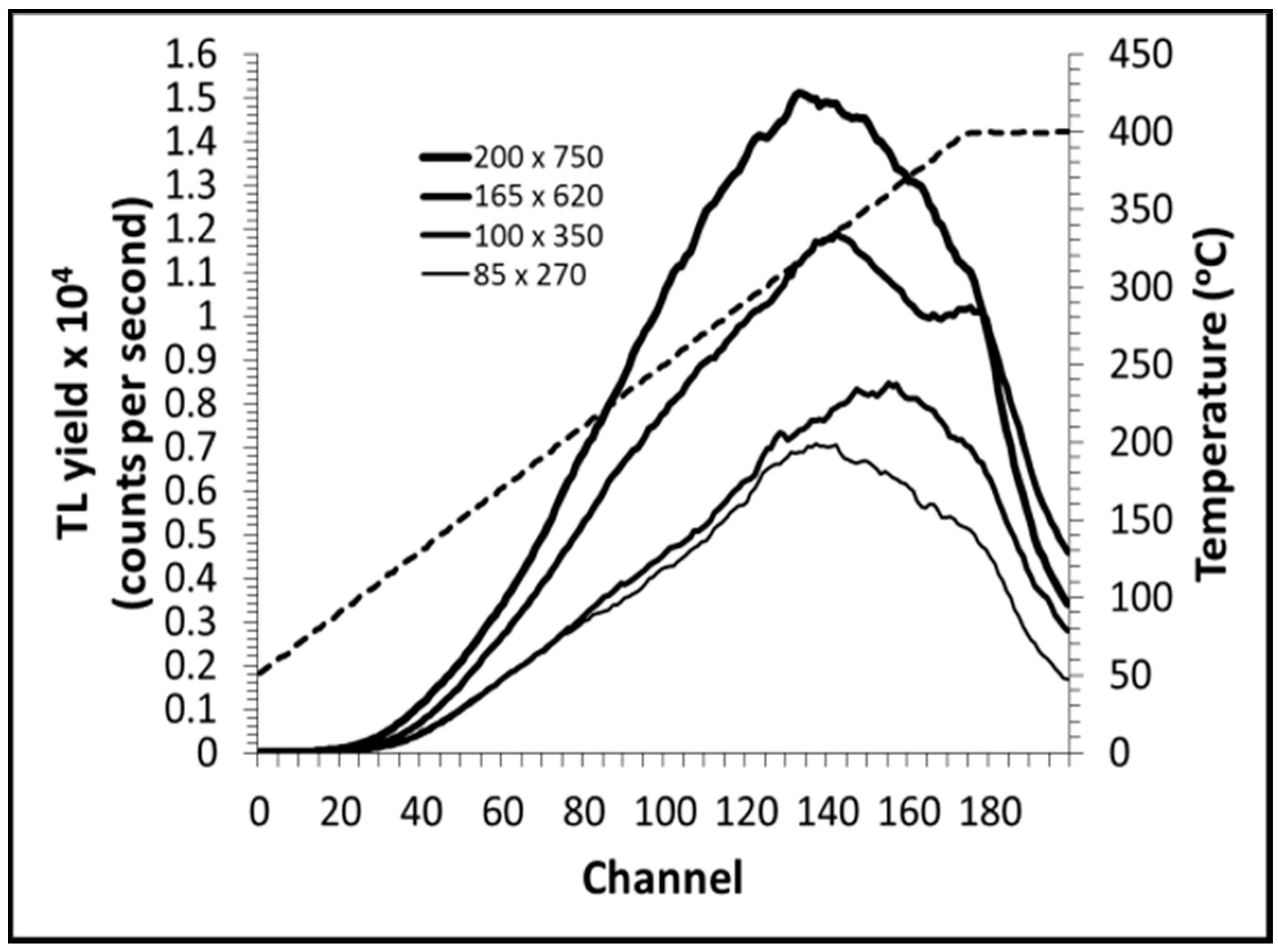
3.1. TL Glow Curve

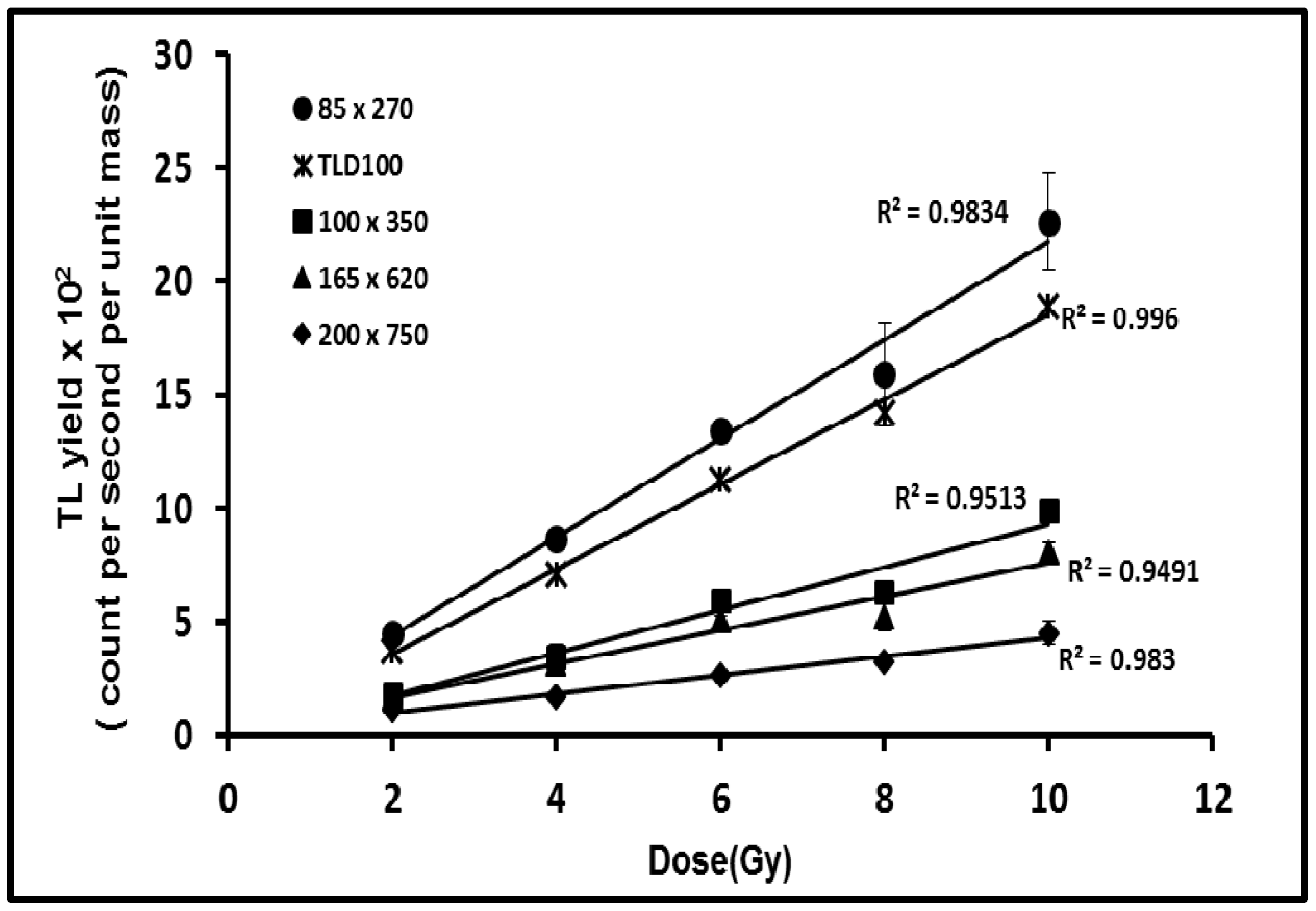
3.2. Dose Response


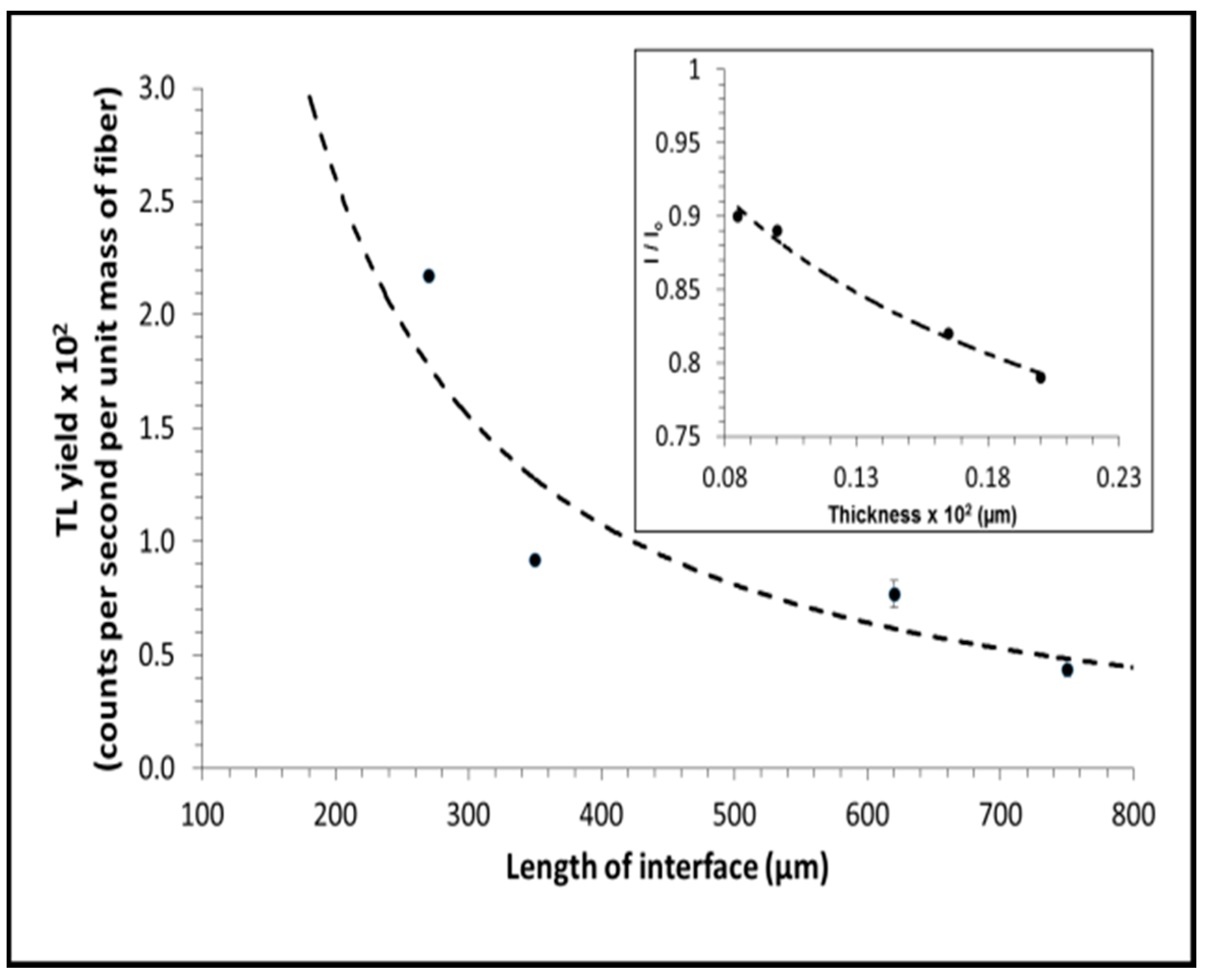
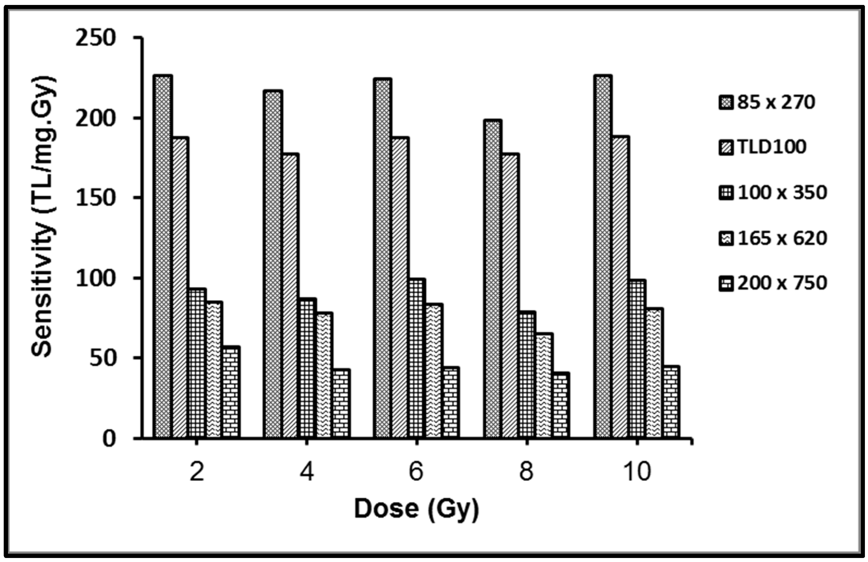
3.3. Sensitivity

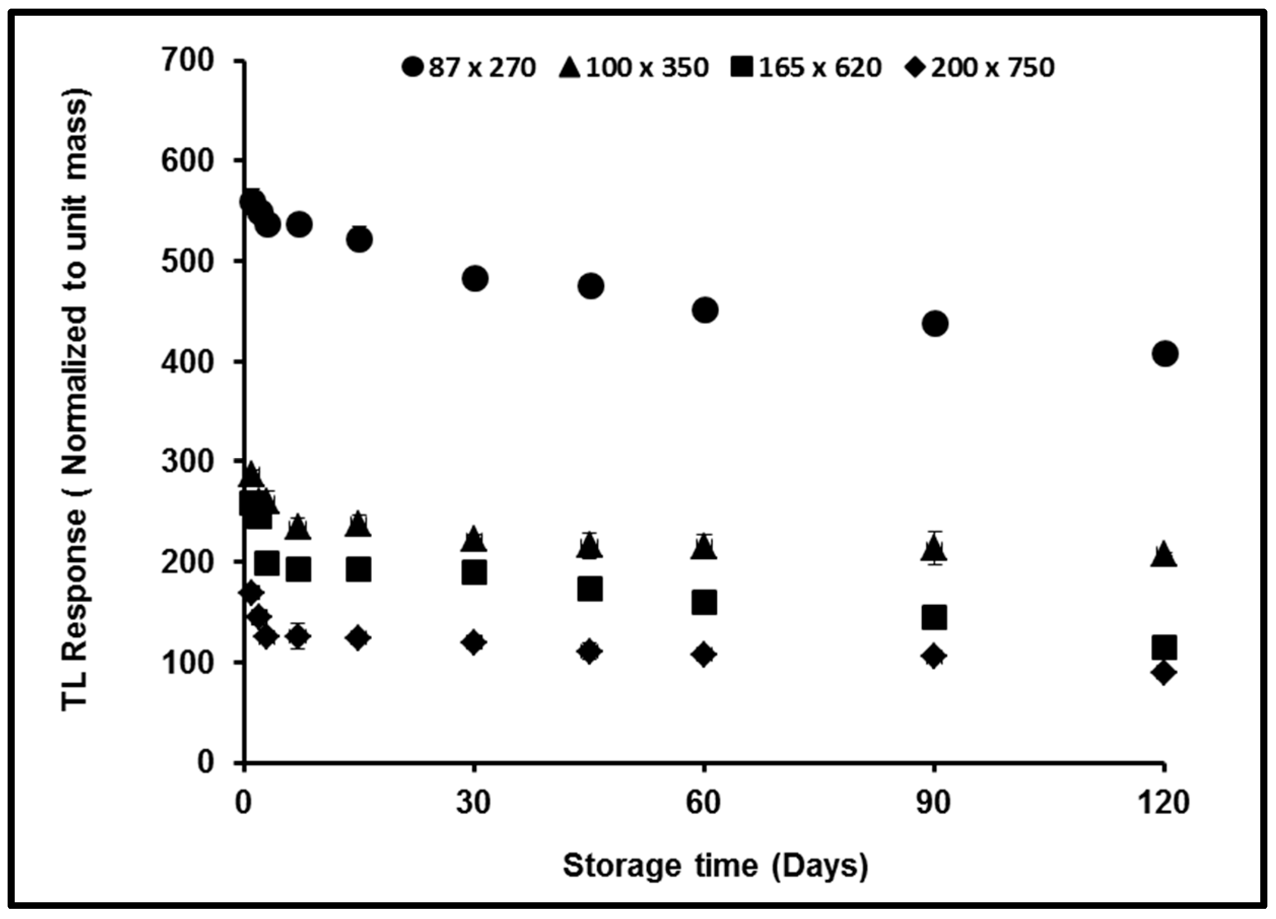
3.4. Fading
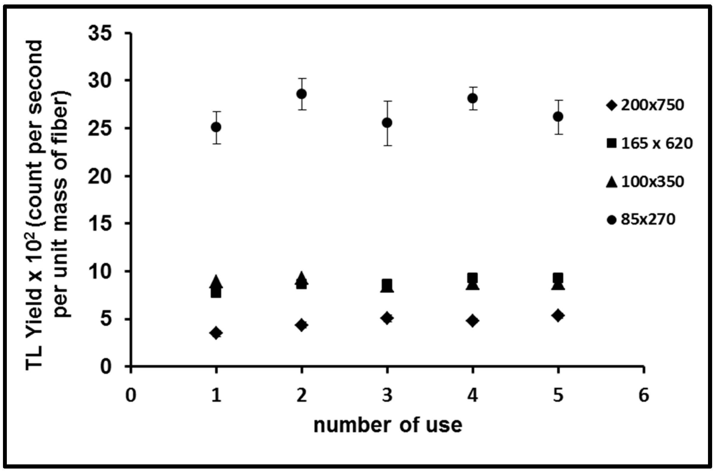
3.5. Reproducibility

4. Conclusions
Acknowledgments
Author Contributions
Conflicts of Interest
References
- Furetta, C. Handbook of Thermoluminescence; World Scientific Publishing: Singapore, 2003. [Google Scholar]
- Hashim, S.; Al-Ahbabi, S.; Bradley, D.A.; Webb, M.; Jeynes, C.; Ramli, A.T.; Wagiran, H. The thermoluminescence response of doped SiO2 optical fibers subjected to photon and electron irradiations. Appl. Radiat. Isot. 2009, 67, 423–427. [Google Scholar] [CrossRef] [PubMed]
- Yaakob, N.H.; Wagiran, H.; Hossain, M.I.; Ramli, A.T.; Bradley, D.A.; Ali, H. Low-dose photon irradiation response of Ge and Al-doped SiO2 optical fibers. Appl. Radiat. Isot. 2011, 69, 1189–1192. [Google Scholar] [CrossRef] [PubMed]
- Alawiah, A.; Intan, A.M.; Bauk, S.; Abdul-Rashid, H.A.; Yusoff, Z.; Mokhtar, M.R.; Wan Abdullah, W.S.; Mat Sharif, K.A.; Mahdiraji, G.A.; Mahamd Adikan, F.R.; et al. Thermoluminescence characteristics of flat optical fiber in radiation dosimetry under different electron irradiation conditions. Proc. SPIE 2013, 8775. [Google Scholar] [CrossRef]
- Abdulla, Y.A.; Amin, Y.M.; Bradley, D.A. The thermoluminescence response of Ge-doped optical fiber subjected to photon irradiation. Radiat. Phys. Chem. 2001, 61, 409–410. [Google Scholar] [CrossRef]
- Hashim, S.; Bradley, D.A.; Peng, N.; Ramli, A.T.; Wagiran, H. The thermoluminescence response of oxygen-doped optical fibers subjected to photon and electron irradiations. Nucl. Instrum. Methods Phys. Res. Sect. A Accel. Spectrom. Detect. Assoc. Equip. 2010, 619, 291–294. [Google Scholar] [CrossRef]
- Noor, N.M.; Hussein, M.; Bradley, D.A.; Nisbet, A. Investigation of the use of Ge-doped optical fiber for in vitro IMRT prostate dosimetry. Nucl. Instrum. Methods Phys. Res. Sect. A Accel. Spectrom. Detect. Assoc. Equip. 2011, 652, 819–823. [Google Scholar] [CrossRef]
- Fadzil, M.S.A.; Ramli, N.N.H.; Jusoh, M.A.; Kadni, T.; Bradley, D.A.; Ung, N.M.; Suhairul, H.; Noor, N.M. Dosimetric characteristics of fabricated silica fiber for postal radiotherapy dose audits. J. Phys. Conf. Ser. 2014, 546, 012010. [Google Scholar] [CrossRef]
- Sani, S.F.A.; Alalawi, A.I.; Abdul Rashid, H.A.; Mahdiraji, G.A.; Tamchek, N.; Nisbet, A.; Maah, M.J.; Bradley, D.A. High sensitivity flat SiO2 fibers for medical dosimetry. Radiat. Phys. Chem. 2014, 104, 134–138. [Google Scholar] [CrossRef]
- Bradley, D.A.; Mahdiraji, G.A.; Ghomeishi, M.; Dermosesian, E.; Adikan, F.R.M.; Abdul Rashid, H.A.; Maah, M.J. Enhancing the radiation dose detection sensitivity of optical fibers. Appl. Radiat. Isot. 2015, 100, 43–49. [Google Scholar] [CrossRef] [PubMed]
- Saeed, M.A.; Fauzia, N.A.; Hossain, I.; Ramli, A.T.; Tahir, B.A. Thermoluminescence response of germanium-doped optical fibers to X-ray irradiation. Chin. Phys. Lett. 2012, 29, 078701. [Google Scholar] [CrossRef]
- Shafiqah, A.S.S.; Amin, Y.M.; Nor, R.M.; Tamchek, N.; Bradley, D.A. Enhanced TL response due to radiation induced defects in Ge-doped silica preforms. Radiat. Phys. Chem. 2015, 111, 87–90. [Google Scholar] [CrossRef]
- Dambul, K.D.; Tamchek, N.; Sandoghchi, S.R.; Abu Hassan, M.R.; Tee, D.C.; Mahamd Adikan, F.R. Fabrication and Characterization of Flat Fibers. In Proceedings of the IEEE 2nd International Conference on Photonics (ICP), Kota Kinabalu, Malaysia, 17–19 October 2011; pp. 10–13.
- Hashim, S.; Omar, S.S.C.; Ibrahim, S.A.; Hassan, W.M.S.W.; Ung, N.M.; Mahdiraji, G.A.; Bradley, D.A.; Alzimami, K. Thermoluminescence response of flat optical fiber subjected to 9 MeV electron irradiations. Radiat. Phys. Chem. 2015, 106, 46–49. [Google Scholar] [CrossRef]
- Benabdesselam, M.; Mady, F.; Duchez, J.B.; Member, S.; Girard, S.; Member, S. The opposite effects of the heating rate on the TSL sensitivity of Ge- doped fiber and TLD500 dosimeters. IEEE Trans. Nucl. Sci. 2014, 61, 1–4. [Google Scholar] [CrossRef]
- Kristensen, M. Ultraviolet-light-induced processes in germanium-doped silica. Phys. Rev. B 2001, 64, 1–12. [Google Scholar] [CrossRef]
- Skuja, L. Optically active oxygen-deficiency-related centers in amorphous silicon dioxide. J. Non. Cryst. Solids 1998, 239, 16–48. [Google Scholar] [CrossRef]
- Hashim, S.; Ramli, A.T.; Bradley, D.A.; Wagiran, H. The Thermoluminesce Response of Ge-Doped Optical Fiber Subjected to Proton Irradiation. In Proceedings of the Fifth National Seminar on Medical Physics, Malaysia Medical Physics Association, Kuala Lumpur, Malaysia, 13–15 July 2006.
- Abdul Rahman, A.T.; Nisbet, A.; Bradley, D.A. Dose-rate and the reciprocity law: TL response of Ge-doped SiO2 optical fibers at therapeutic radiation doses. Nucl. Instrum. Methods Phys. Res. Sect. A 2011, 652, 891–895. [Google Scholar] [CrossRef]
© 2015 by the authors; licensee MDPI, Basel, Switzerland. This article is an open access article distributed under the terms and conditions of the Creative Commons Attribution license (http://creativecommons.org/licenses/by/4.0/).
Share and Cite
Nawi, S.N.B.M.; Wahib, N.F.B.; Zulkepely, N.N.B.; Amin, Y.B.M.; Min, U.N.; Bradley, D.A.; Nor, R.B.M.; Maah, M.J. The Thermoluminescence Response of Ge-Doped Flat Fibers to Gamma Radiation. Sensors 2015, 15, 20557-20569. https://doi.org/10.3390/s150820557
Nawi SNBM, Wahib NFB, Zulkepely NNB, Amin YBM, Min UN, Bradley DA, Nor RBM, Maah MJ. The Thermoluminescence Response of Ge-Doped Flat Fibers to Gamma Radiation. Sensors. 2015; 15(8):20557-20569. https://doi.org/10.3390/s150820557
Chicago/Turabian StyleNawi, Siti Nurasiah Binti Mat, Nor Fadira Binti Wahib, Nurul Najua Binti Zulkepely, Yusoff Bin Mohd Amin, Ung Ngie Min, David Andrew Bradley, Roslan Bin Md Nor, and Mohd Jamil Maah. 2015. "The Thermoluminescence Response of Ge-Doped Flat Fibers to Gamma Radiation" Sensors 15, no. 8: 20557-20569. https://doi.org/10.3390/s150820557