A Highly Sensitive and Selective Competition Assay for the Detection of Cysteine Using Mercury-Specific DNA, Hg2+ and Sybr Green I

Abstract
: We here report a rapid, sensitive, selective and label-free fluorescence detection method for cysteine (Cys). The conformation of mercury-specific DNA (MSD) changes from a random coil form to a hairpin structure in the presence of Hg2+ due to the formation of a thymine-Hg2+-thymine (T-Hg2+-T) complex. Cys can selectively coordinate with Hg2+ and extract it from the thymine-Hg2+-thymine complex. The hairpin structure dehybridizes and the fluorescence intensity of Sybr Green I (SG) decreases upon addition of Cys because SG efficiently discriminates mercury-specific DNA and mercury-specific DNA/Hg2+ complex. The detection can be finished within 5 min with high sensitivity and selectivity. In addition, we can obtain variable dynamic ranges for Cys by changing the concentration of MSD/Hg2+.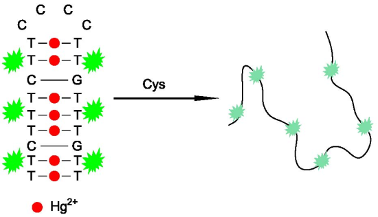
1. Introduction
Widely distributed in living cells, thiols such as cysteine (Cys) are involved in many biological functions. Their levels in biological fluids such as human plasma and urine are of great importance for clinical diagnostics of a variety of diseases [1–3]. Determination of these species can be achieved by effective separation/detection techniques, e.g., HPLC and capillary electrophoresis [4–6], spectrophotometry [7–9], electrochemical voltammetry [10–13], colorimetric methods [14–17], flow injection [18–20] and fluorescence analysis method [21–25]. However, some of them suffer from low sensitivity and/or selectivity or require cumbersome laboratory procedures. There is thus an intense demand for more sensitive, selective, convenient and low-cost methods to detect Cys.
Growing research interest has been focused on the development of sensitive, selective, and cost-effective biosensors based on target-responsive DNA structural switching [26–29]. The core technology of these sensors is a kind of special DNA molecules which change their conformations upon binding with the targets. For example, aptamers are in vitro selected functional oligonucleotides that can bind specifically to target molecules. In this work, we use a mercury-specific DNA (MSD) which presents a random coil form in the absence of Hg2+, and forms a hairpin structure in the presence of Hg2+ due to the formation of a thymine-Hg2+-thymine (T-Hg2+-T) complex. A fluorescent dye, Sybr Green I (SG), was applied to recognize the structural change due to the different interaction of SG with MSD and MSD/Hg2+ complex [30].
It is well-known that Cys can form a very stable complex with Hg2+ [31]. By using this property, Mirkin et al. [32] developed a highly sensitive and selective colorimetric detection method for Cys. However, their method needs an elevated temperature, which requires a long time. We recently developed a fluorescent turn-on “molecular beacon” probe for the detection of Cys, which is also based on the competitive ligation of Hg2+ ions by Cys and thymine-thymine (T-T) mismatches [33]. The method shows high sensitivity and selectivity, but still needs to be improved in terms of cost and convenience due to its requirement of a labelled “molecular beacon” and a solution heating process. Here we develop a simple, rapid, sensitive and selective method for detection of Cys by using a target-responsive DNA structural change. The MSD/Hg2+ complex is a hairpin structure, which dehybridizes when Hg2+ is extracted from the thymine-Hg2+-thymine complex by Cys due to the high formation constant of Hg2+-Cys complexes. The fluorescence intensity of SG then decreases upon addition of Cys due to the dehybridization of the MSD/Hg2+ complex.
2. Experimental Section
2.1. Chemicals and Apparatus
All chemicals used for these investigations were of analytical grade purity. L-Cysteine (Cys, minimum 98.5%) was purchased from Sigma Aldrich Chemical Company and used as received. MSD (5′-TTCTTTCTTCCCCTTGTTTGTT-3′) was synthesized and purified by HPLC (Takara Biotech. Co., Dalian). SG (10,000×) was purchased from Invitrogen Inc. A stock solution of 400× was prepared with DMSO/water (volume 1:1) before use. Milli-Q water (18.2 MΩ cm) was used in all procedures. The fluorescence measurements were recorded at room temperature (RT) on a Perkin Elmer LS-55 spectrophotometer equipped with a xenon lamp excitation source. Fluorescence spectra were measured at an excitation wavelength of 490 nm and the emission range from 500 to 650 nm with the excitation and emission slit widths set at 5 nm.
2.2. Performance of Cys Detection
10 nM MSD was first incubated with 70 nM Hg2+ solution in 2.0 mL of 10 mM MOPS (3-(N-morpholino)propanesulfonic acid) buffer containing 0.1 M NaNO3 (pH 7.50). 5 μL of 25 × SG was then added to the solution. After incubation for two minutes, different amounts of Cys were added to the solution. The mixture was then immediately used for the fluorescence measurements. For selectivity analysis, various kinds of amino acids with final concentration of 140 nM were used instead of Cys.
3. Results and Discussion
3.1. Sensor Operation Principle
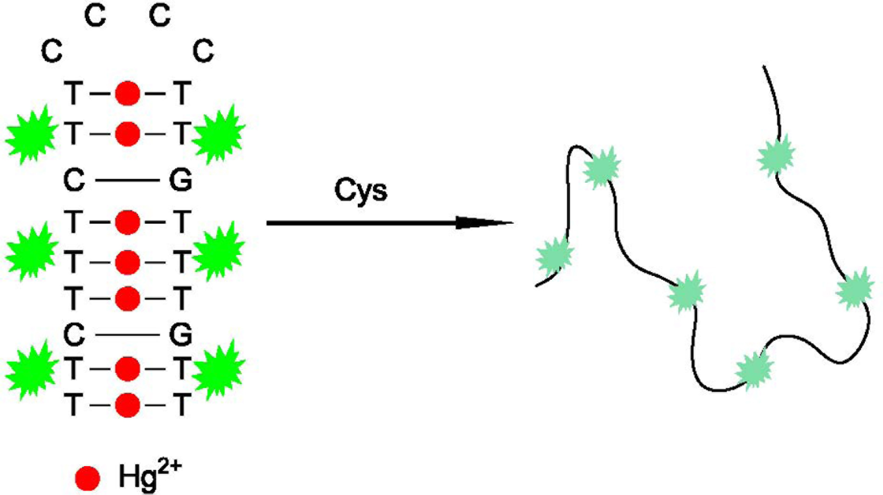
MSD, including seven T-T mismatches, could form a stable hairpin structure upon combination with Hg2+, which could be specifically stained by SG and produce a high fluorescence signal. However, in the presence of Cys, Hg2+ was extracted from the T-Hg2+-T structure because Cys can bind to Hg2+ with higher affinity (the formation constant for Cys to Hg2+ is ca. 1042) [31]. As a result, the hairpin structure dehybridizes and the fluorescence intensity of SG decreases. Scheme 1 depicts the designed fluorescence method for Cys detection. The detection can be completed in less than 5 min.
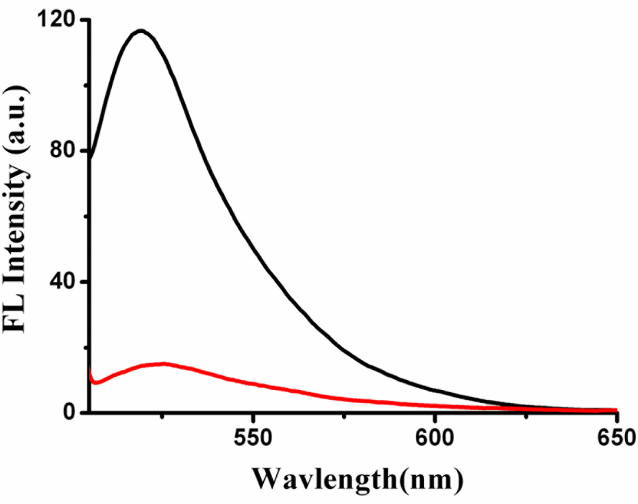
As shown in Figure 1, the fluorescence intensity of MSD/Hg2+/SG is 116.7 a.u., while MSD/Hg2+/SG/Cys is about 15 a.u. when the concentration of Cys is 140 nM.
3.2. Optimization of the Assay
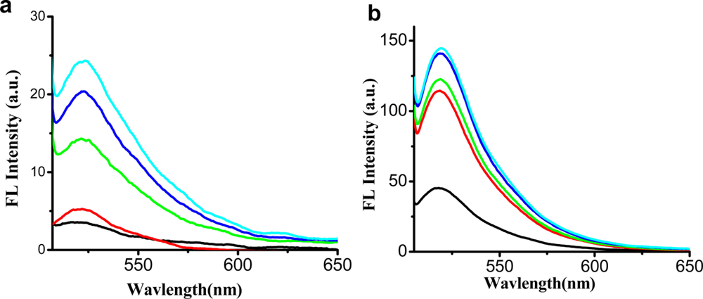
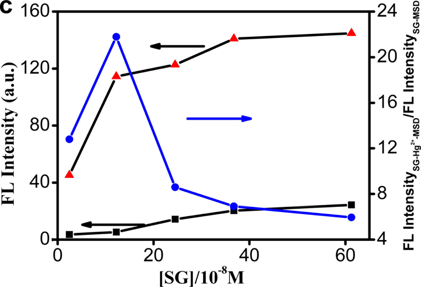
In previous study, Liu et al. [30] found that MSD kinetically accomplishes the hairpin structure at [Hg2+]/7[MSD] = 1. One equivalent of MSD thus requires seven equivalents of Hg2+ to completely form the hairpin structure due to the T-Hg2+-T chemistry. So we fixed the concentration of MSD to be 10 nM and Hg2+ 70 nM, and then the concentration of SG was optimized. Figure 2 shows the fluorescence spectra of MSD and MSD-Hg2+ upon addition of different concentration of SG.
We can see from the results that the fluorescence intensity of SG increases both in the absence of Hg2+ [Figure 2(a)] and in the presence of Hg2+ [Figure 2(b)]. The highest ratio of the fluorescence intensity of SG-Hg2+-MSD to that of SG-MSD is 21.8 [Figure 2(c)], at which the concentration of SG is 1.225 × 10⁻⁷ M, so we selected the concentration of SG to be 1.225 × 10⁻⁷ M when the concentration of MSD is 10 nM and Hg2+ 70 nM.
3.3. Cys Sensor Sensitivity
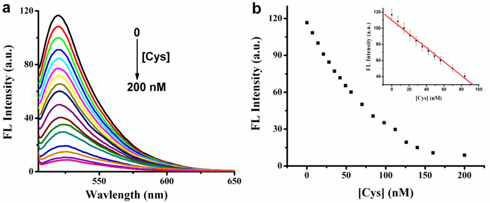
To evaluate the sensitivity of the assay, different concentrations of Cys were mixed with MSD/Hg2+/SG, and then the fluorescence intensity was immediately measured. As shown in Figure 3(a), the fluorescence intensity of SG decreases gradually upon adding increased concentrations of Cys.
When the concentration of Cys is two-fold higher than that of Hg2+, the fluorescence intensity decreases very slowly due to formation of a 2:1 Cys/Hg2+ adduct [31]. This implies that almost all the Hg2+ has been extracted from T-Hg2+-T complex when the concentration of Cys is two fold higher than that of Hg2+. When we increased the concentration of Cys further, we found that the fluorescence intensity decreased very slowly. Perhaps some Cys forms Hg(Cys)3 complexes. By measuring the fluorescence intensity at the emission maximum of SG-Hg2+-MSD-Cys, a linear response of fluorescence intensity vs. [Cys] was observed in the range 7–84 nM [Figure 3(b) inset]. The detection limit may be estimated from Equation (1):
What is more important, our method is really fast. In the first step of this assay, SG staining was finished in 2 min, which was proved to be fully adequate for the SG binding by a previous study [30]. In the second step, after the addition of Cys, the fluorescence spectra was measured immediately, and as anticipated, the fluorescence intensity remained stable for a long time (data not shown), because Cys can bind to Hg2+ very strongly [31].
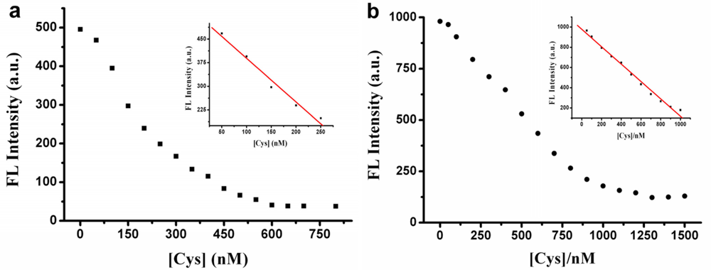
We further find that we can obtain different dynamic ranges by changing the concentration of MSD/Hg2+. For example, when we fixed the concentration of MSD to be 50 nM and that of Hg2+ to be 350 nM (MSD kinetically forms the hairpin structure at [Hg2+]/7[MSD] = 1 [30]), a linear dynamic range for Cys from 50 to 250 nM was obtained [Figure 4(a) inset]. When we changed the concentration of MSD and Hg2+ to be 100 nM and 700 nM, a very wide linear dynamic range for Cys from 50 to 1,000 nM was obtained [Figure 4(b) inset]. The concentration of SG was 6.125 × 10⁻⁷ M [Figure 4(a)] and 1.225 × 10⁻⁶ M [Figure 4(b)], respectively, which was determined by the same method mentioned above (Section 3.2). When the concentration of MSD/Hg2+ changed, different concentration of Cys was needed to extract Hg2+ from T-Hg2+-T structure due to the 2:1 Cys/Hg2+ adduct, which leads to the different dynamic ranges for Cys.
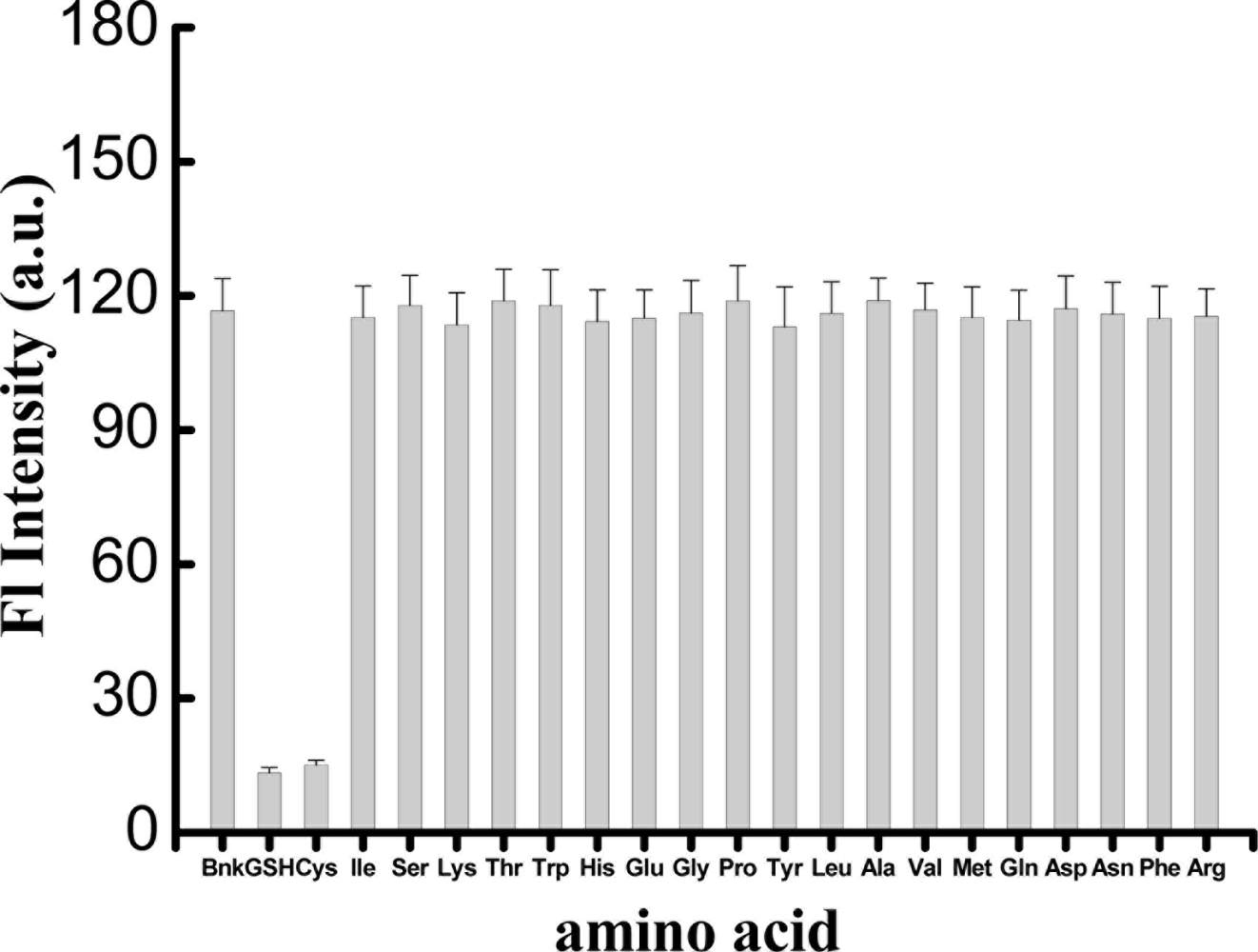
3.4. Cys Sensor Selectivity
To challenge the assay’s selectivity, other amino acids and the Cys-containing tripeptide glutathione (GSH) at a concentration of 140 nM were analyzed. The results are shown in Figure 5. It is clear that only Cys and GSH show significant fluorescence intensity changes. In contrast to the enormous fluorescence decrease observed for Cys and GSH, there is very little fluorescence change observed in the presence of other amino acids. A Cys-containing tripeptide GSH (γ-Glu-Cys-Gly) showed almost the same fluorescence decrease as Cys, indicating that Hg2+ can be extracted from the T-Hg2+-T structure upon adding GSH. This is because GSH can also form a very stable 2:1 GSH/Hg2+ complex with Hg2+ [34–36]. Hg2+ is well known to have an affinity to certain N-type ligands such as amino acids [37], but the amino acids, except for Cys, cannot extract Hg2+ from the thymine-Hg2+-thymine complex. The binding affinity of Hg2+ to T-T mismatch sites thus appears to be stronger enough than that of Hg2+ to all of the amino acids studied, except Cys. We also found from Figure 5 that a sulfur-containing amino acid like methionine did not result in a significant fluorescence decrease. The formation constant for Cys to Hg2+ (about 1042) is much higher than that of methionine to Hg2+ (about 1017.6) [31]. The method presented here therefore shows very high specificity to Cys.
4. Conclusions
This paper describes a rapid, highly selective and sensitive fluorescence assay for Cys using mercury-specific DNA and SG. This assay is based on the extraction of Hg2+ by Cys from a T-Hg2+-T complex. The whole procedure can be done within 5 min. In addition, the assay is label free, low-cost and can provide variable linear dynamic ranges.
Acknowledgments
The authors would like to thank the Promotional Foundation for the Excellent Middle-Aged and Young Scientists of Shandong Province of China (BS2009SW040), the National Natural Science Foundation (20902096, 21104030) and a Project of Shandong Province Higher Educational Science and Technology Program (J11LB03, J10LF02), Shanghai Postdoctoral Sustentation Fund, China (11R21420800), General Administration of Quality Supervision, Inspection and Quarantine of PRC (2009QK098, 2010QK294).
References
- Droge, W.; Holm, E. Role of cysteine and glutathione in HIV infection and other diseases associated with muscle wasting and immunological dysfunction. FASEB J 1997, 11, 1077–1089. [Google Scholar]
- Wang, X.F.; Cynader, M.S. Pyruvate released by astrocytes protects neurons from copper-catalyzed cysteine neurotoxicity. J. Neurosci 2001, 21, 3322–3331. [Google Scholar]
- Liu, J.; Yeo, H.C.; Overvik-Douki, E.; Hagen, T.; Doniger, S.J.; Chu, D.W.; Brook, G.A.; Ames, B.N. Chronically and acutely exercised rats: Biomarkers of oxidative stress and endogenous antioxidants. J. Appl. Physiol 2000, 89, 21–28. [Google Scholar]
- Lu, C.; Zu, Y.; Yam, V.W.W. Nonionic surfactant-capped gold nanoparticles as postcolumn reagents for high-performance liquid chromatography assay of low-molecular-mass biothiols. J. Chromatogr. A 2007, 1163, 328–332. [Google Scholar]
- Jin, W.R.; Wang, Y. Determination of cysteine by capillary zone electrophoresis with end-column amperometric detection at a gold/mercury amalgam microelectrode without deoxygenation. J. Chromatogr. A 1997, 769, 307–314. [Google Scholar]
- Nozal, M.J.; Bernal, J.L.; Toribio, L.; Marinero, P.; Moral, O.; Manzanas, L.; Rodriguez, E. Determination of glutathione, cysteine and N-acetylcysteine in rabbit eye tissues using high-performance liquid chromatography and post-column derivatization with 5,5′-dithiobis(2-nitrobenzoic acid). J. Chromatogr. A 1997, 778, 347–353. [Google Scholar]
- Abu Eid, M. Spectrophotometric determination of cysteine and N-acetylcysteine in pharmaceutical preparations. Microchim. Acta 1998, 129, 91–95. [Google Scholar]
- Wang, W.; Rusin, O.; Xu, X.; Kim, K.K.; Escobedo, J.O.; Fakayode, S.O.; Fletcher, K.A.; Lowry, M.; Schowalter, C.M.; Lawrence, C.M.; Fronczek, F.R.; Warner, I.M.; Strongin, R.M. Detection of homocysteine and cysteine. J. Am. Chem. Soc 2005, 127, 15949–15958. [Google Scholar]
- Lunar, M.L.; Rubio, S.; Pérez-Bendito, D.; Carreto, M.L.; McLeod, C.W. Hexadecylpyridinium chloride micelles for the simultaneous kinetic determination of cysteine and cystine by their induction of the iodine-azide reaction. Anal. Chim. Acta 1997, 337, 341–349. [Google Scholar]
- Deng, C.; Chen, J.; Chen, X.; Wang, M.; Nie, Z.; Yao, S. Electrochemical detection of l-cysteine using a boron-doped carbonnanotube-modified electrode. Electrochim. Acta 2009, 54, 3298–3302. [Google Scholar]
- Bai, Y.H.; Xu, J.J.; Chen, H.Y. Selective sensing of cysteine on manganese dioxide nanowires and chitosan modified glassy carbon electrodes. Biosens. Bioelectron 2009, 24, 2985–2990. [Google Scholar]
- Shahrokhian, S.; Karimi, M. Voltammetric studies of a Cobalt(II)-4-methylsalophen modified carbon-paste electrode and its application for the simultaneous determination of cysteine and ascorbic acid. Electrochim. Acta 2004, 50, 77–84. [Google Scholar]
- Amini, M.K.; Khorasani, J.H.; Khaloo, S.S.; Tangestaninejad, S. Cobalt(II) salophen-modified carbon-paste electrode for potentiometric and voltammetric determination of cysteine. Anal. Biochem 2003, 320, 32–38. [Google Scholar]
- Zhang, F.X.; Han, L.; Israel, L.B.; Daras, J.G.; Maye, M.M.; Ly, N.K.; Zhong, C.J. Colorimetric detection of thiol-containing amino acids using gold nanoparticles. Analyst 2002, 127, 462–465. [Google Scholar]
- Li, L.; Li, B. Sensitive and selective detection of cysteine using gold nanoparticles as colorimetric probes. Analyst 2009, 134, 1361–1365. [Google Scholar]
- Sudeep, P.K.; Joseph, S.T.S.; Thomas, K.G. Selective detection of cysteine and glutathione using gold nanorods. J. Am. Chem. Soc 2005, 127, 6516–6517. [Google Scholar]
- Jia, S.M.; Liu, X.F.; Li, P.; Kong, D.M.; Shen, H.X. G-quadruplex DNAzyme-based Hg2+ and cysteine sensors utilizing Hg2+-mediated oligonucleotide switching. Biosens. Bioelectron 2011, 27, 148–152. [Google Scholar]
- Waseem, A.; Yaqoob, M.; Nabi, A. Flow injection determination of cysteine in pharmaceuticals based on luminol-persulphate chemiluminescence detection. Luminescence 2008, 23, 144–149. [Google Scholar]
- Teshima, N.; Nobuta, T.; Sakai, T. Simultaneous flow injection determination of ascorbic acid and cysteine using double flow cell. Anal. Chim. Acta 2001, 438, 21–29. [Google Scholar]
- Zhao, C.; Zhang, J.C.; Song, J.F. Determination of l-Cysteine in amino acid mixture and human urine by flow-injection analysis with a biamperometric detector. Anal. Biochem 2001, 297, 170–176. [Google Scholar]
- Tanaka, F.; Mase, N.; Barbas, C.F., III. Determination of cysteine concentration by fluorescence increase: Reaction of cysteine with a fluorogenic aldehyde. Chem. Commun 2004, 1762–1763. [Google Scholar]
- Shang, L.; Qin, C.J.; Wang, T.; Wang, M.; Wang, L.X.; Dong, S.J. Fluorescent conjugated polymer-stabilized gold nanoparticles for sensitive and selective detection of cysteine. J. Phys. Chem. C 2007, 111, 13414–13417. [Google Scholar]
- Shang, L.; Dong, S.J. Sensitive detection of cysteine based on fluorescent silver clusters. Biosens. Bioelectron 2009, 24, 1569–1573. [Google Scholar]
- Huang, S.; Xiao, Q.; Li, R.; Guan, H.L.; Liu, J.; Liu, X.R.; He, Z.K.; Liu, Y. A simple and sensitive method for l-cysteine detection based on the fluorescence intensity increment of quantum dots. Anal. Chim. Acta 2009, 645, 73–78. [Google Scholar]
- Ruan, Y.B.; Li, A.F.; Zhao, J.S.; Shen, J.S.; Jiang, Y.B. Specific Hg2+-mediated perylene bisimide aggregation for highly sensitive detection of cysteine. Chem. Commun 2010, 46, 4938–4940. [Google Scholar]
- Li, D.; Song, S.P.; Fan, C.H. Target-responsive structural switching for nucleic acid-based sensors. Acc. Chem. Res 2010, 43, 631–641. [Google Scholar]
- Zhang, J.; Wang, L.H.; Zhang, H.; Boey, F.; Song, S.P.; Fan, C.H. Aptamer-based multicolor fluorescent gold nanoprobes for multiplex detection in homogeneous solution. Small 2010, 6, 201–204. [Google Scholar]
- Song, S.P.; Wang, L.H.; Li, J.; Zhao, J.; Fan, C.H. Aptamer-based biosensors. Trends Anal. Chem 2008, 27, 108–117. [Google Scholar]
- Zuo, X.L.; Song, S.P.; Zhang, J.; Pan, D.; Wang, L.H.; Fan, C.H. A target-responsive electrochemical aptamer switch (TREAS) for reagentless detection of nanomolar ATP. J. Am. Chem. Soc 2007, 129, 1042–1043. [Google Scholar]
- Wang, J.; Liu, B. Highly sensitive and selective detection of Hg2+ in aqueous solution with mercury-specific DNA and Sybr Green I. Chem. Commun 2008, 4759–4761. [Google Scholar]
- Berthon, G. The stability constants of metal complexes of amino acids with polar side chains. Pure App. Chem 1995, 67, 1117–1240. [Google Scholar]
- Lee, J.S.; Ulmann, P.A.; Han, M.S.; Mirkin, C.A. A DNA-gold nanoparticle-based colorimetric competition assay for the detection of cysteine. Nano Lett 2008, 8, 529–533. [Google Scholar]
- Xu, H.; Hepel, M. “Molecular beacon”-based fluorescent assay for selective detection of glutathione and cysteine. Anal. Chem 2011, 83, 813–819. [Google Scholar]
- Oram, P.D.; Fang, X.; Fernando, Q. The formation of constants of mercury (II)-glutathione complexes. Chem. Res. Toxicol 1996, 9, 709–712. [Google Scholar]
- Fuhr, B.J.; Rabenstein, D.L. Nuclear magnetic resonance studies of the solution chemistry of metal complexes. IX. The binding of cadmium, zinc, lead, and mercury by glutathione. J. Am. Chem. Soc 1973, 95, 6944–6950. [Google Scholar]
- Stricks, W.; Kolthoff, I.M. Reactions between mercuric mercury and cysteine and glutathione. Apparent dissociation constants, heats and entropies of formation of various forms of mercuric mercapto-cysteine and -glutathione. J. Am. Chem. Soc 1953, 75, 5673–5681. [Google Scholar]
- Corradi, A.B.; Cramarossa, M.R.; Vezzosi, I.M.; Giusti, J.G. trans-2-Styrylbenzothiazole complexes with mercury(II) halides. Synthesis, characterization and X-ray crystal structure of diiodobis(trans-2-styrylbenzothiazole)mercury(II). Polyhedron 1993, 12, 2235–2239. [Google Scholar]


| Detection method | Linear range (μmol L−1) | Detection limit (μmol L−1) | Reference |
|---|---|---|---|
| Spectrophotometry | 0.0082–0.12 | 0.0049 | [9] |
| Spectrophotometry | 0.17–50 | Not given | [7] |
| Fluorimetry | 100–5,000 | Not given | [21] |
| Fluorimetry | 0.05–4 | 0.025 | [22] |
| Fluorimetry | 0.025–6 | 0.02 | [23] |
| Fluorimetry | 0.01–0.8 | 0.0038 | [24] |
| Fluorimetry | 0.05–0.3 | 0.0096 | [25] |
| Flow injection | 0.001–0.5 | 0.0005 | [18] |
| Flow injection | 1–90 | 0.2 | [19] |
| Flow injection | 0.4–40 | 0.1 | [20] |
| Capillary zone electrophoresis | 0.1–100 | 0.058 | [5] |
| Voltammetry | 0.5–100 | 0.2 | [12] |
| Voltammetry | 2–10,000 | 1.0 | [13] |
| Voltammetry | 0.78–200 | 0.26 ± 0.01 | [10] |
| Voltammetry | 0.5–630 | 0.07 | [11] |
| Fluorimetry | 0.007–0.084 or 0.05–0.25 or 0.05–1 | 0.0034 | This method |
© 2011 by the authors; licensee MDPI, Basel, Switzerland. This article is an open access article distributed under the terms and conditions of the Creative Commons Attribution license (http://creativecommons.org/licenses/by/3.0/).
Share and Cite
Xu, H.; Gao, S.; Liu, Q.; Pan, D.; Wang, L.; Ren, S.; Ding, M.; Chen, J.; Liu, G. A Highly Sensitive and Selective Competition Assay for the Detection of Cysteine Using Mercury-Specific DNA, Hg2+ and Sybr Green I. Sensors 2011, 11, 10187-10196. https://doi.org/10.3390/s111110187
Xu H, Gao S, Liu Q, Pan D, Wang L, Ren S, Ding M, Chen J, Liu G. A Highly Sensitive and Selective Competition Assay for the Detection of Cysteine Using Mercury-Specific DNA, Hg2+ and Sybr Green I. Sensors. 2011; 11(11):10187-10196. https://doi.org/10.3390/s111110187
Chicago/Turabian StyleXu, Hui, Shuli Gao, Quanwen Liu, Dun Pan, Lihua Wang, Shuzhen Ren, Min Ding, Jingwen Chen, and Gang Liu. 2011. "A Highly Sensitive and Selective Competition Assay for the Detection of Cysteine Using Mercury-Specific DNA, Hg2+ and Sybr Green I" Sensors 11, no. 11: 10187-10196. https://doi.org/10.3390/s111110187