Communication

1,2,3,4-Tetra-O-Acetyl-β-d-Mannuronic Acid

Lennard-Jones Laboratory, School of Chemical and Physical Sciences, Keele University, Keele, Staffordshire ST5 5BG, UK
*
Author to whom correspondence should be addressed.
Molbank 2017, 2017(3), M947; https://doi.org/10.3390/M947
Submission received: 3 July 2017 / Revised: 10 July 2017 / Accepted: 11 July 2017 / Published: 14 July 2017
(This article belongs to the Section Organic Synthesis)

Abstract

:
1,2,3,4-Tetra-O-acetyl-β-d-mannuronic acid was synthesized in three steps from commercial d-mannose in 21% yield. Regioselective 6-O-tritylation followed by per-acetylation and 6-OTr removal using HBr/AcOH gave the required primary alcohol substrate, which was then oxidised to the target compound using TEMPO/BAIB. None of the synthetic steps required column chromatography and the product was fully characterized by 1H-NMR, 13C-NMR, 2D NMR, MS and IR.

Graphical Abstract

1. Introduction

d-Mannuronic acid is the constituent building block monosaccharide of the polysaccharide alginate: an important industrial biomaterial widely used across the textile, food and biomedical sectors [1,2] and a major exopolysaccharide secreted by the opportunistic human pathogen P. aeruginosa (PA) [3]. As part of our program developing sugar nucleotide mimetics to investigate the biosynthetic mechanisms underpinning alginate biosynthesis in mucoid PA [4], we required access to a suitably protected d-mannuronic acid building block.
d-Mannuronic acid is a uronic acid or sugar acid where C-6 is presented at the carboxylic acid oxidation level, as opposed to the C-6 alcohol oxidation level seen in common pyranoses. Uronic acids are thus often accessed synthetically via oxidation of the corresponding hexose [5,6]. As such, we required efficient and scalable access to the title compound from commercially available d-mannose and provide our record of its synthesis and characterization herein.

2. Results

d-Mannose (1) was regioselectively protected at the C-6 OH position using TrCl, followed by per-acetylation of the remaining hydroxyl groups (Scheme 1). This one-pot procedure delivered anomerically pure 1,2,3,4-tetra-O-acetyl-6-O-triphenylmethyl-β-d-mannopyranose (2) as a white solid on 40-g scale (46% yield, after recrystallization of the major β diastereoisomer). 1H and 13C-NMR data collected for (2) agreed with data already published [7,8], however, upon inspecting the literature more closely we found additional examples of published data for (2) [9] that were contrary to ours and others, recording the data as the α-anomer of (2), but utilizing the same reaction conditions. Given this inconsistency, we performed a NOESY experiment on crude (2), confirming a correlation from H1 to H2/H3/H5 and our assignment of (2) as the β-anomer. Additionally, we report the anomeric 1JH-C value of (2) as 159.4 Hz.
6-OTr derivative (2) was subjected to treatment with HBr/AcOH to remove the primary alcohol protecting group [7]. Almost immediately upon the addition of 33% HBr/AcOH solution to (1), a precipitate of TrBr formed and the reaction was halted to prevent acyl migration and delivered 1,2,3,4-tetra-O-acetyl-β-d-mannopyranose (3) as white solid (60%). To oxidise the resultant primary alcohol, we chose a TEMPO/BAIB reagent combination [5], an alternative to the earlier reported RuCl3/NaIO4 approach [6]. This furnished the title compound 1,2,3,4-tetra-O-acetyl-β-d-mannuronic acid (4) as white solid (75%). Analytical data collected for (4) supported the structural assignment and gave an indicative level of purity. Copies of NMR, MS and IR data are included in Supplementary Materials.

3. Materials and Methods

3.1. General

Analytical data for previously reported compounds matched those already described and are cited in the preceding text. 1H-NMR spectra were recorded on a Bruker Avance 300 (Coventry, UK), (300 MHz) instrument or Bruker Avance 400 (400 MHz) instrument using deuterated chloroform (or other indicated solvent) as reference. The chemical shift data for each signal are given as δ in units of parts per million (ppm) relative to tetramethylsilane (TMS) where δ = 0.00 ppm. The multiplicity of each signal is indicated by: s (singlet), bs (broad singlet), d (doublet), dd (doublet of doublets), t (triplet), appt (apparent triplet), m (multiplet). The number of protons (n) for a given resonance is indicated by nH. Coupling constants (J) are quoted in Hz and recorded to the nearest 0.1 Hz. 13C-NMR spectra were recorded on a Bruker Avance 300 (75 MHz) instrument or Bruker Avance 400 (100 MHz) instrument. The chemical shift data for each signal are given as δ in units of parts per million (ppm) relative to tetramethylsilane (TMS) where δ = 0.00 ppm. FT-IR spectra were recorded on a Thermo Scientific Nicolet iS10 instrument (Stafford, UK). Analytical thin layer chromatography (TLC) was carried out on pre-coated 0.25 mm Merck (Nottingham, UK) KGaA 60 F254 silica gel plates. Visualisation was by adsorption of UV light, or thermal development after dipping in a methanolic solution of sulfuric acid (5% v/v), vanillin or KMnO4. Chemicals were purchased from Acros/Fisher (Loughborough, UK), Aldrich (Gillingham, UK) or Alfa Aesar (Heysham, UK). All solvents and reagents were purified and dried where necessary. Where appropriate and if not stated otherwise, all non-aqueous reactions were performed under an inert atmosphere of nitrogen, using a vacuum manifold with nitrogen passed through molecular sieves. Brine refers to a saturated aqueous solution of sodium chloride. Hexane refers to the fraction from petroleum ether boiling between 40 and 60 °C. RT refers to room temperature.

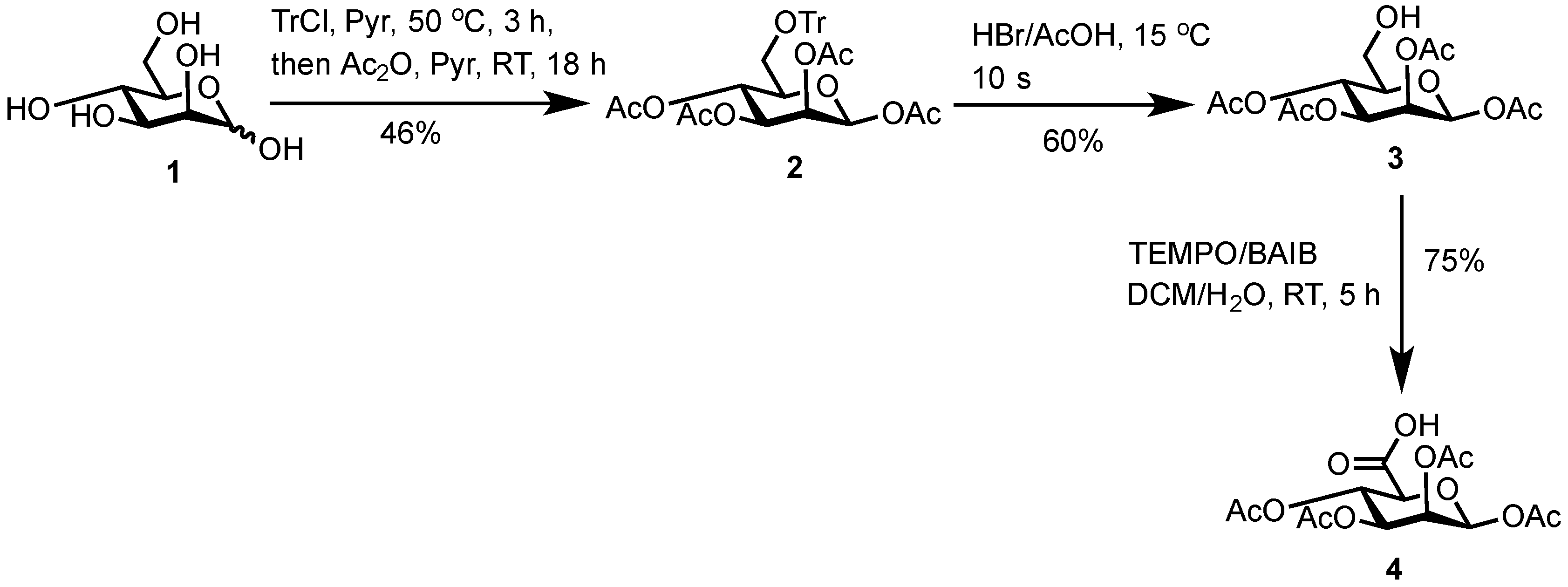
3.2. 1,2,3,4-Tetra-O-Acetyl-6-O-Triphenylmethyl-β-d-Mannopyranose (2)

d-Mannose (1) (25.8 g, 142 mmol, 1.00 equiv.) and trityl chloride (40.1 g, 143.0 mmol, 1.03 equiv.) were stirred in anhydrous pyridine (125 mL) under a N2 atmosphere. The reaction mixture was heated to 50 °C and stirred for 3 h. The solution was cooled to RT, placed in an ice bath and acetic anhydride (75 mL) added via a dropping funnel over 30 min. The solution was then stirred at RT for 18 h, whereupon TLC analysis (hexane/ethyl acetate, 3/1) showed complete conversion of starting material to a higher Rf value spot. The reaction mixture was poured onto ice (750 mL) and stirred vigorously for 1.5 h to afford a white solid (water was exchanged twice during this time to remove pyridine). The solid was collected by filtration, washed with ice-cold water (250 mL) and dried for 2.5 h under vacuum. The solid was recrystallized from the minimum amount of hot ethanol, collected by filtration and dried over P2O5 in a vacuum desiccator to afford 1,2,3,4-tetra-O-acetyl-6-O-triphenylmethyl-β-d-mannopyranose (2) as a white solid (39.0 g, 66.0 mmol, 46%). 1H-NMR, 13C-NMR and MS collected for (2) were in good agreement with those already reported [8].

3.3. 1,2,3,4-Tetra-O-Acetyl-β-d-Mannopyranose (3)

1,2,3,4-tetra-O-acetyl-6-O-triphenylmethyl-β-d-mannopyranose (2) (10.2 g, 17.3 mmol, 1.00 equiv.) was dissolved in acetic acid (120 mL) at 40 °C with stirring. The solution was cooled to 15 °C and HBr/acetic acid (33% w/w in acetic acid, 5.0 mL, 20.4 mmol, 1.20 equiv.) was added dropwise, forming a yellow precipitate almost immediately. After 10 s, the solution was filtered under vacuum through CeliteTM onto water (250 mL) and the solids washed with acetic acid (10 mL). The flitrate was quenched slowly with saturated aqueous NaHCO3 solution (250 mL), forming an opaque white suspension which was extracted with chloroform (2 × 200 mL). The organic layers were combined, washed with water (100 mL), saturated aqueous NaHCO3 solution (100 mL), dried (MgSO4), filtered and concentrated in vacuo to afford the crude product as an oil. TLC analysis (hexane/ethyl acetate, 3/1) showed complete conversion of starting material to a lower Rf value spot and a higher UV active Rf value spot corresponding to trityl bromide. The crude product was dissolved in the minimum amount of hot chloroform and induced to crystallise by addition of diethyl ether. The crystals formed were collected by filtration and dried in vacuo, affording 1,2,3,4-tetra-O-acetyl-β-d-mannopyranose (3) as a white solid (3.6 g, 10.4 mmol, 60%). 1H-NMR, 13C-NMR and MS collected for (3) were in good agreement with those already reported [8].

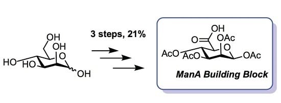
3.4. 1,2,3,4-Tetra-O-Acetyl-β-d-Mannuronic Acid (4)

To a vigorously stirred solution of 1,2,3,4-tetra-O-acetyl-β-d-mannopyranose (3) (1.5 g, 4.30 mmol, 1.0 equiv.) in dichloromethane (20 mL) and water (10 mL) was added (2,2,6,6-tetramethylpiperidin-1-yl)oxyl radical (TEMPO) (336 mg, 2.2 mmol, 0.50 equiv.) and [bis(acetoxy)iodo]benzene (BAIB) (6.9 g, 21.5 mmol, 5.00 equiv.). The solution was stirred at RT for 5 h, whereupon TLC analysis (hexane/ethyl acetate, 1/1) showed complete conversion of starting material to a baseline Rf value spot. The reaction was quenched with aqueous Na2S2O3 solution (10% w/v, 30 mL), the organic layer removed and the aqueous layer acidified to pH 3 using 1M HCl, followed by extraction with ethyl acetate (2 × 30 mL). The organic extracts were combined, dried (MgSO4), filtered and concentrated in vacuo to afford 1,2,3,4-tetra-O-acetyl-β-d-mannuronic acid (4) as a white solid (1.2 g, 3.3 mmol, 75%). Rf = 0.81 (dichloromethane/methanol, 9/1); 1H-NMR (300 MHz, CDCl3) δH 5.96 (1H, d, J = 1.5 Hz, H1), 5.50 (1H, dd, J = 3.2, 1.5 Hz, H2), 5.48 (1H, appt, J = 9.1 Hz, H4), 5.21 (1H, dd, J = 9.1, 3.2 Hz, H3), 4.2 (1H, d, J = 9.1 Hz, H5), 2.21 (3H, s, C(O)CH3), 2.13 (3H, s, C(O)CH3), 2.10 (3H, s, C(O)CH3), 2.04 (3H, s, C(O)CH3). 13C-NMR (100 MHz, CDCl3) δC 169.9 (C=O), 169.7 (C=O), 169.5 (C=O), 169.4 (C=O), 168.2 (C=O), 89.4 (C1), 72.3 (C5), 69.3 (C3), 66.9 (C2), 65.9 (C4), 20.4 (C(O)CH3), 20.4 (C(O)CH3), 20.3 (C(O)CH3), 20.2 (C(O)CH3); FT-HRMS NSI (ES) m/z Found: (M−H) 361.0767, C14H17O11 requires 361.0776; FT-IR vmax/cm−1 2986 (O–H), 1751 (C=O).

Supplementary Materials

NOESY/HSQC NMR data for (2) and NMR, MS and IR data for (4) are available online at https://www.mdpi.com/1422-8599/2017/3/M947.
Supplementary File 1Supplementary File 2Supplementary File 3Supplementary File 4

Acknowledgments

Keele University are thanked for a Faculty of Natural Sciences studentship to L.B. and the EPSRC UK National Mass Spectrometry Facility (NMSF) at Swansea University are thanked for MS analyses.

Author Contributions

L.B. and G.J.M conceived and designed the experiments; L.B. performed the experiments and analyzed the data; L.B. and G.J.M. wrote the manuscript.

Conflicts of Interest

The authors declare no conflict of interest.

References

  1. Rehm, B.H.A. Bacterial polymers: biosynthesis, modifications and applications. Nat. Rev. Microbiol. 2010, 8, 578–592. [Google Scholar] [CrossRef] [PubMed]
  2. Hay, I.D.; Rehman, Z.U.; Moradali, M.F.; Wang, Y.; Rehm, B.H.A. Microbial alginate production, modification and its applications. Microb. Biotechnol. 2013, 6, 637–650. [Google Scholar] [CrossRef] [PubMed]
  3. Franklin, M.J.; Nivens, D.E.; Weadge, J.T.; Howell, P.L. Biosynthesis of the Pseudomonas aeruginosa Extracellular Polysaccharides, Alginate, Pel, and Psl. Front. Microbiol. 2011, 2, 167. [Google Scholar] [CrossRef] [PubMed]
  4. Tatnell, P.J.; Russell, N.J.; Gacesa, P. A metabolic study of the activity of GDP-mannose dehydrogenase and concentrations of activated intermediates of alginate biosynthesis in Pseudomonas aeruginosa. Microbiology 1993, 139, 119–127. [Google Scholar] [CrossRef] [PubMed]
  5. De Mico, A.; Margarita, R.; Parlanti, L. A versatile and highly selective hypervalent iodine (III)/2, 2, 6, 6-tetramethyl-1-piperidinyloxyl-mediated oxidation of alcohols to carbonyl compounds. J. Org. Chem. 1997, 62, 6974–6977. [Google Scholar] [CrossRef]
  6. Knapp, S.; Nandan, S.R. Synthesis of capuramycin. J. Org. Chem. 1994, 59, 281–283. [Google Scholar] [CrossRef]
  7. Reynolds, D.; Lloyd, W. The synthesis of certain oligosaccharide acetates in the mannose series. J. Am. Chem. Soc. 1940, 62, 66–69. [Google Scholar] [CrossRef]
  8. Lin, K.; Kasko, A. Effect of branching density on avidity of hyperbranched glycomimetics for mannose binding lectin. Biomacromolecules 2013, 14, 350–357. [Google Scholar] [CrossRef] [PubMed]
  9. Lopez, M.; Trajokovic, J.; Bornaghi, L.F.; Innocenti, A.; Vullo, D.; Supuran, C.T.; Poulsen, S. Design, synthesis, and biological evaluation of novel carbohydrate-based sulfamates as carbonic anhydrase inhibitors. J. Med. Chem. 2011, 54, 1481–1489. [Google Scholar] [CrossRef] [PubMed]
Scheme 1. Synthesis of 1,2,3,4-tetra-O-acetyl-β-d-mannuronic acid (4) from d-mannose (1).
Molbank 2017 m947 sch001

