1. Introduction
Colorectal cancer (CRC) is the third most common cancer globally. However, only about 20% of CRC cases have been associated with heritable genetic changes, and the largest fraction has been linked to environmental factors. On the basis of our present knowledge of risk factors, inflammation bowel disease is closely related to CRC. For this reason, the inhibition of precancerous lesions is a considerable strategy for cancer prevention [
1]. The Azoxymethane/Dextran sodium sulphate (AOM/DSS) mouse model is an outstanding platform for colitis-related carcinogenesis and cancer-preventive intervention study. For analogizing human carcinogenesis, the AOM/DSS model is based on a single carcinogenic hit of azoxymethane (AOM) followed by one to three exposition cycles to dextran sodium sulphate (DSS). The model develops through two stages: the initial stage (the acute inflammation stage or the early stage), and the promotion stage [
2]. In the early stage (from week 1–7), normal crypts are initiated to form foci of aberrant crypts that proliferate by crypt fission to form micro-adenoma. This initial stage is marked by genetic changes that progress with morphological alternation, involving the Wnt/Apc/β-catenin pathway, K-Ras, c-Myc global hypomethylation, iNOS, and COX-2 [
2,
3,
4,
5,
6]. Following the resolution of acute inflammation, these micro-adenomas will grow and aggregate to form adenomas, adenomatous polyps, and finally adenocarcinomas in the subsequent weeks (the promotion stage). As described by Tanaka et al. [
7], the AOM/DSS model completely reproduced the multi-step process of carcinogenesis characterized by the canonical phases of initiation, promotion, and progression.Different preclinical studies support the use of the AOM/DSS model as aninflammation/oxidative stress-related carcinogenic model to perform the intervention studies of synthetic agents or natural plant compounds. For these reasons, the AOM/DSS model was chosen for our study to evaluate the effect of
Angelica sinensis root (ASR) extract on cancer prevention.
Traditional Chinese medicine (TCM) employs the dry roots of
Angelica sinensis (Oliv.) Diels.As a popular herb from umbelliferae, it has been used for medicinal and edible purposes for thousands of years in Asia. Traditionally,
Angelica sinensis, also known as “female ginseng”, is widely compatible with other herbs for its blood enriching, circulation promoting, and laxative activities [
8]. Some scientific reports have indicated the effectiveness of ASR for menopausal symptoms, migraine, anemia, dysmenorrhea, and ulcers [
9]. Chemical constituents in ASR show a variety of biological activities including anti-oxidative, anti-inflammatory gastrointestinal protective, and anti-cancer activities [
10]. There is also proven evidence of the possible oral and topical safety of ASR for adults [
11]. Thus,
Angelica sinensis has been of interest to researchers recently as a candidate source of agents resistant to oxidative stress-related disease.
Various dietary antioxidant supplements have also shown considerable promise as effective agents for cancer prevention by reducing oxidative stress. Previously, we reported on the cancer preventive potential of ASR extract by protecting cells from oxidative stress in HepG2-C8 and RAW 264.7 cells [
12]. However, some contradictory evidence has challenged the above statement. For instance, Sayin et al. [
13] reported that the antioxidant agent vitamin E markedly increased tumor progression and reduced survival in the mouse model of B-Raf- and K-Ras-induced lung cancer by disrupting the ROS-P53 axis. As is well-known, evading growth suppressors is one of the hallmarks of carcinogenesis. The circumvention of cellular programed death makes it possible for transformed cells to obtain sustaining proliferation [
14]. P53 as an intercellular abnormality sensor that monitors genome damage degree and levels of nucleotide pools, glucose, and oxygenation. If the damage to such cellular systems is irreparable or alarm signals indicate an overwhelming of such systems, P53 can trigger apoptosis. Notably, the activation of P53 is a complex and highly context-dependent progression, which depends on the severity and persistence of the conditions of cell stress and genomic damage [
15]. As the occurrence of cancer is a multi-step progression with various contexts of lesion, we suspect that the antioxidants for cancer prevention are stage-dependent. In other words, at specific stages of carcinogenesis, antioxidants protect cells from oxidative stress, and at other stages play an unpredictable role. To verify this assumption, ASR extract was administrated at different stages of carcinogenesis, and its cancer-preventive effect was investigated.
3. Discussion
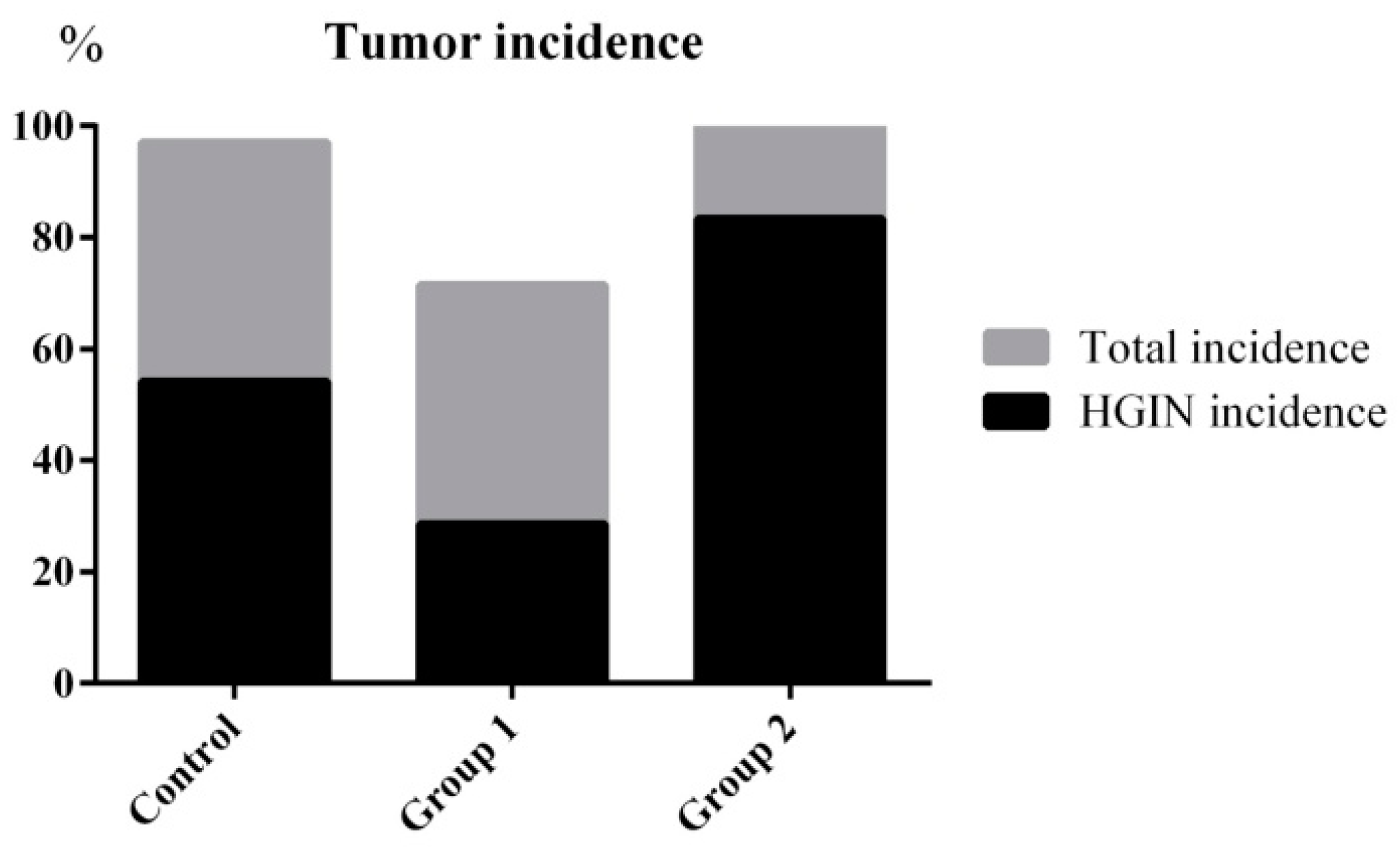
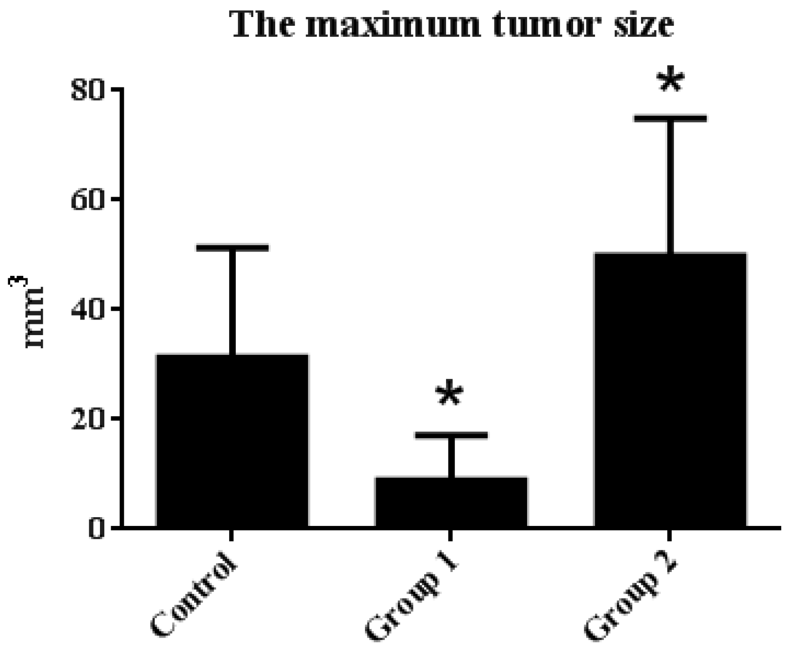
This study demonstrated that ASR extract administration either in the initial or promotion stages of the AOM/DSS model reduced amounts of 8-oxoguanine and γ-H2AX in tumors, which indicated that cells were protected from DNA damage. However, these administrations led to opposite effects on carcinogenesis through P53-mediated apoptosis. These results infer that the cancer-preventive effect of ASR extract was stage-dependent, as the antioxidant supplement exerted specific roles at different stages of carcinogenesis.
Recent studies have shown that ASR extracts have an antioxidant potential. Wu et al. [
16] reported that
Angelica sinensis extract inhibited lipid peroxidation in rat liver homogenate in vitro, and exhibited a superoxide anion scavenging activity in a concentration-dependent manner. Cao et al. [
17] proved the hepatoprotective and antioxidant effects of
Angelica sinensis extract (1.0%) against CCl
4-induced hepatotoxicity in Jian carp. Dietz et al. [
18] suggested that
Angelica sinensis dietary supplements had potential as chemopreventive agents through the induction of Detoxification Enzyme NAD(P)H: Quinone Oxidoreductase 1 by alkylating Keap1 in HepG2-ARE-C8 cells.
Many components from ASRhave also been reported as antioxidant or free radical scavengers. For instance, Kampa et al. [
19] reported that ferulic acid and caffeic acid showed a time-dependent and dose-dependent inhibitory effect on T27D human breast cancer cell growth with direct interaction with aryl hydrocarbon receptors and nitric oxide synthase inhibition.Cheng et al. [
20] reported that ferulic acid reduces cerebral infarct through its anti-oxidative and anti-inflammatory effects following transient focal cerebral ischemia in rats. Chou et al. [
21] reported that vanillic acid inhibited cellular reactive oxygen species in H
2O
2-treated BNLCL2 cells.
Previously, our group studied the composition of the ASR extract prepared by our group. FeCl
3-K
3(Fe(CN)
6) UV-VIS spectrophotometric results showed that the phenolic acid content reached 61.4 ± 3.51%, and the HPLC-UV test result showed that ferulic acid content reached 16.14 ± 0.21% (
m/
m). Twelve kinds of organic acids were identified using UPLC-MS/MS, including ferulic acid, sassinic acid, chlorogenic acid, camphoric acid, and so on [
22].
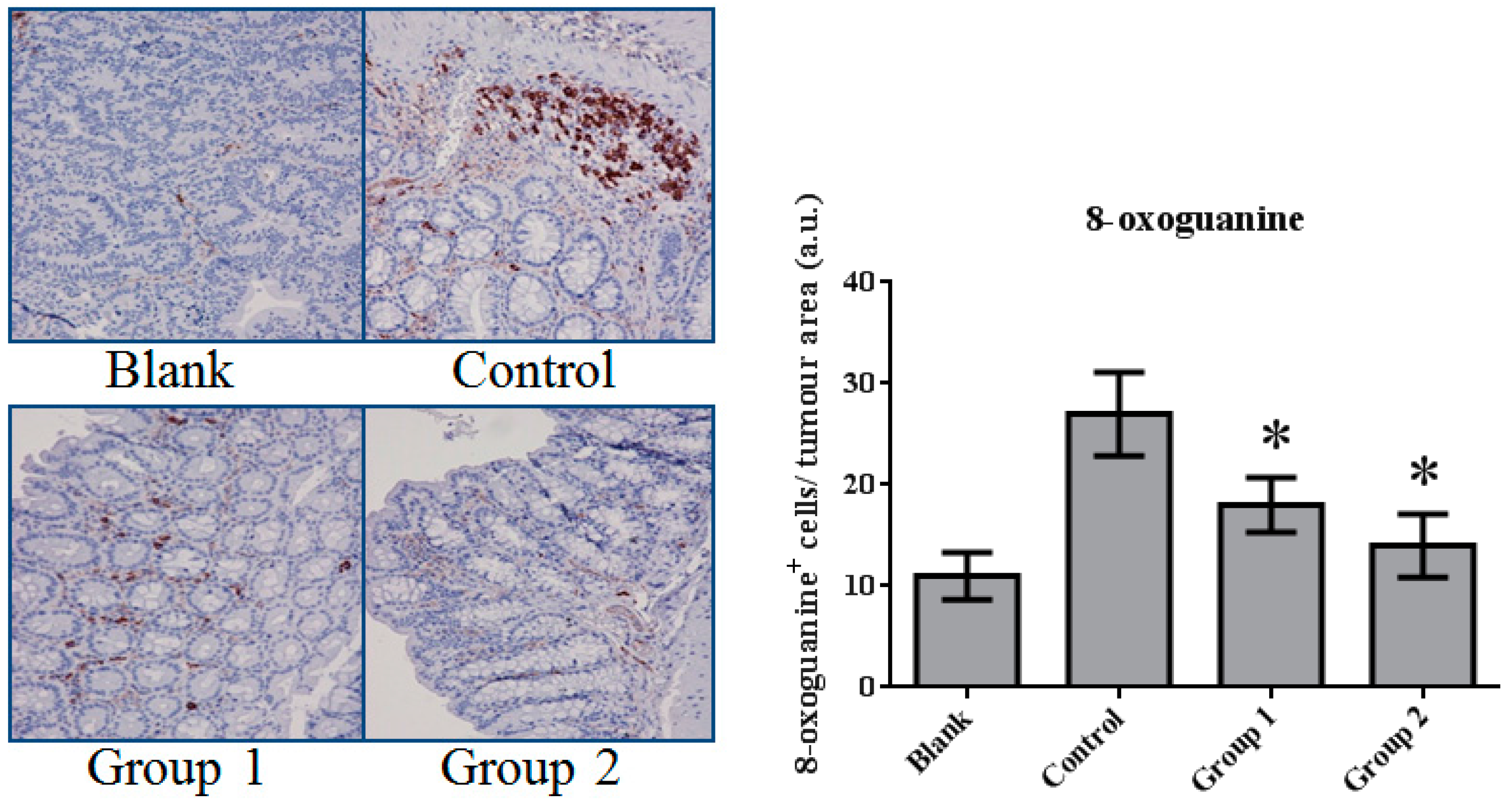
In this paper, mismatched pairing and DNA double-strand break were reflected by 8-oxoguanine and γ-H2AX levels. These DNA lesions mainly result from reactive oxygen species. We noticed the marked decrease of 8-oxoguanine and γ-H2AX levels after ASR extract administration. These results indirectly show the antioxidative properties of ASR extract.
Oxidative stress mainly results from excessive reactive oxygen species (ROS) [
23]. Normally, ROS homeostasis is balanced by the respiratory chain and Nrf2-Keap1 pathway. Pathologically increased levels of ROS can result in the hyperactivation of inflammatory responses and damage to lipids, proteins, and DNA [
24]. It is interesting that oxidative stress can also lead to the restriction of the proliferation of damaged cells and the enhancement of DNA repair, or it can induce apoptosis [
25].
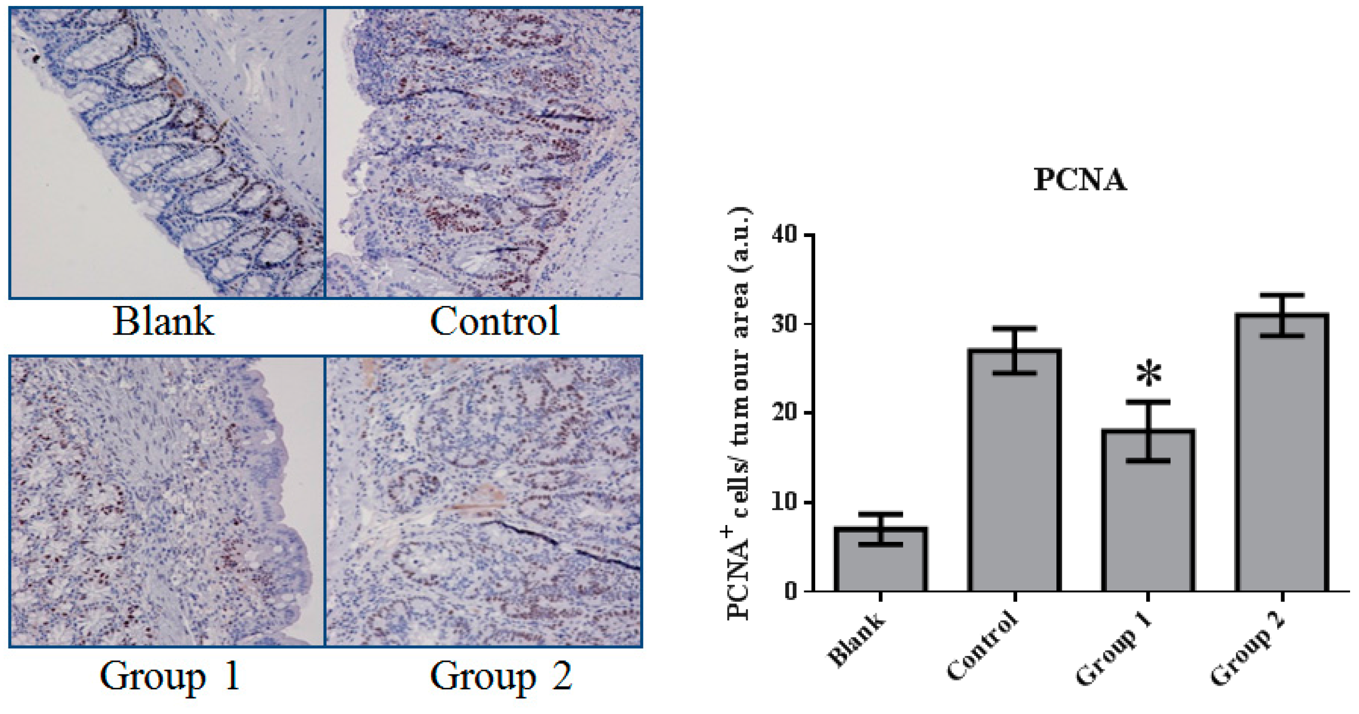
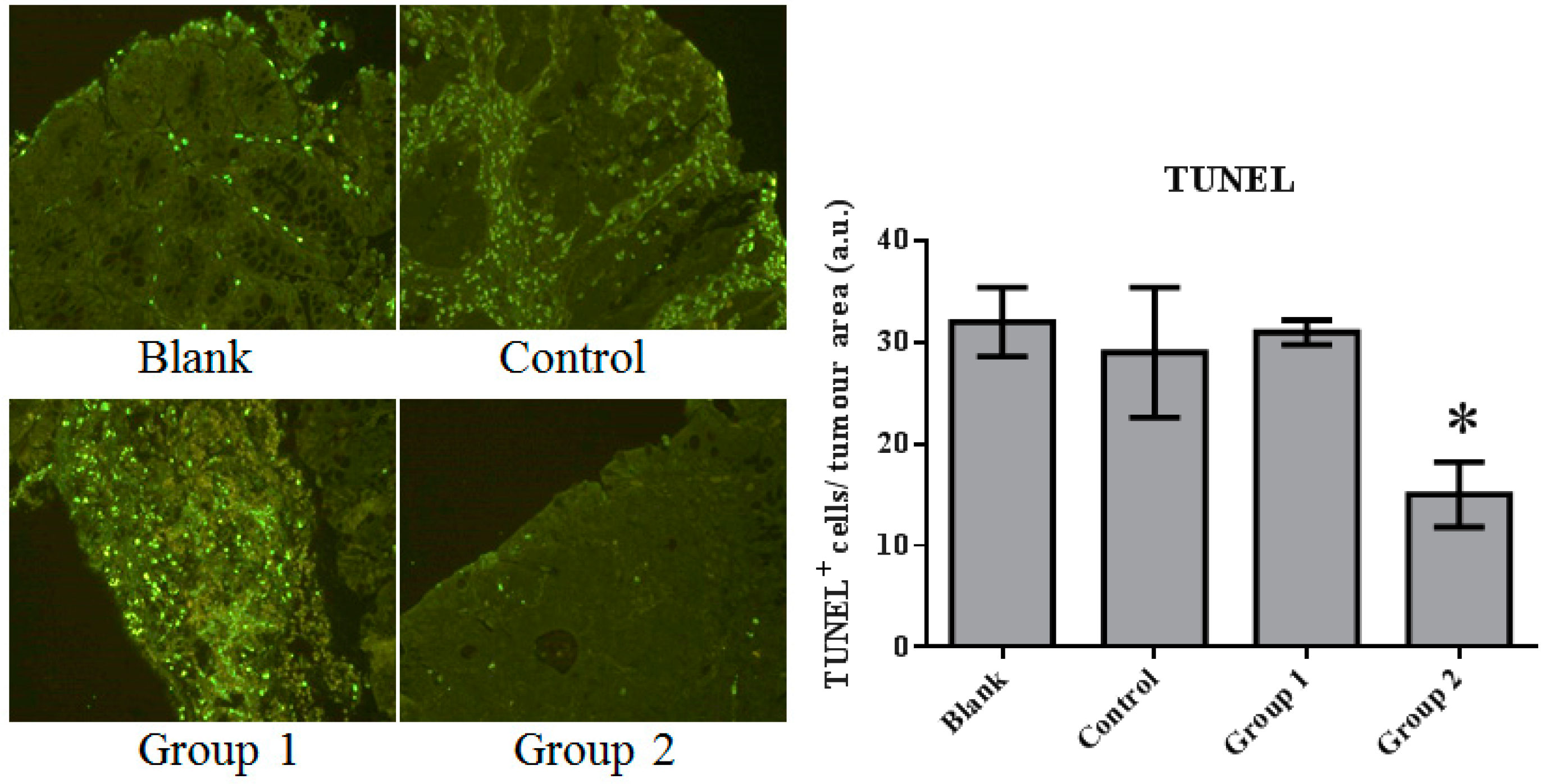
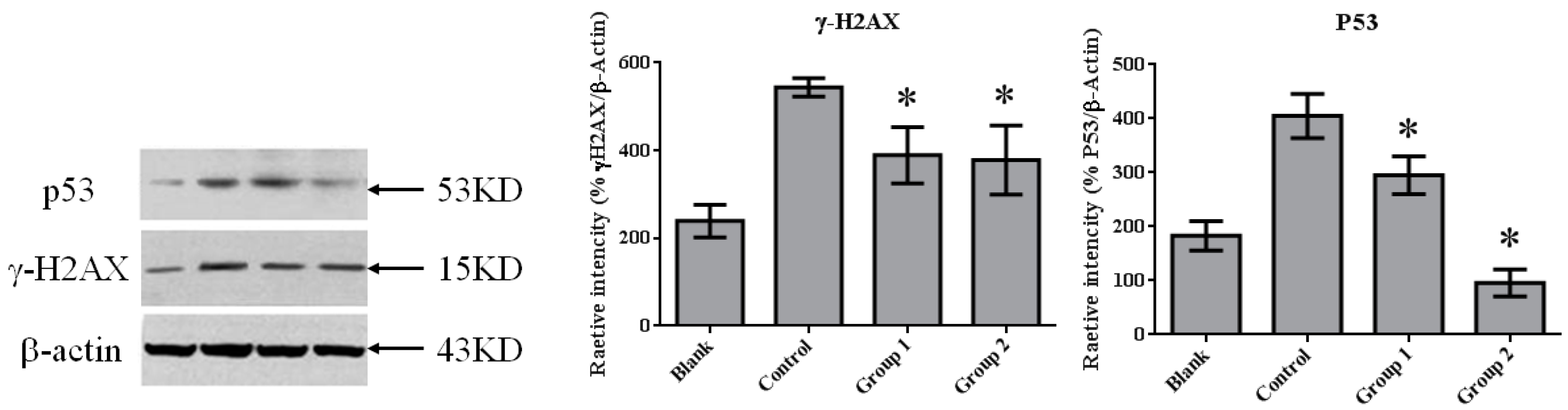
In our research, cell proliferation activity and apoptosis were characterized by PCNA immunohistochemistry and TUNEL assays. We found that DNA synthesis in Group 1 was inhibited by relatively high apoptotic activity. In contrast, in Group 2, apoptotic activity was significantly suppressed. This variation could be interpreted as the reason for the different effects of ASR extract administration in Groups 1 and 2. Based on the relationship of oxidative stress and apoptosis mediated by P53, Western blot assays were performed. Consistent with the results of the TUNEL assays, P53 expression was significantly suppressed by ASR extract administration that was even lower than the blank group. Studies on P53 revealed that DNA damage was detected by ATM (ataxia telangiectasia mutated) and ATR (ataxia telangiectasia and Rad3 related proteins), which signal downstream to CHK1 and CHK2 (checkpoint kinase) and P53. P53 exerts three crucial functions: regulation and stimulation of DNA repair; signaling cell-cycle checkpoints; and signaling apoptosis. In addition, a certain concentration of ROS acts as both an upstream signal triggering P53 activation and a downstream factor mediating apoptosis [
26]. Thus, combined with the component analysis and literature reports, we suggest that ASR extract played an antioxidant role and depleted ROS in epithelial tissue. As a result, ASR extract administration in the promotion stage disrupted the ROS-P53 axis. ASR extract administration in the initial stage reduced the accumulation of DNA damage without the intervention of ROS concentration in the promotion stage, and effectively reduced high-grade pathological changes.
There are studies that have questioned the use of antioxidants, including epidemiological and laboratory studies. Based on our study, we found that antioxidant administration in the initial stage of carcinogenesis could play a positive role, while it promoted carcinogenesis when administrated during the promotion stage. Hence, we hypothesized that effective cancer prevention is based on a clear judgment of precancerous lesions, and antioxidant treatment at an appropriate time is a reasonable choice for inflammation-related carcinogenesis prevention.
In conclusion, it was demonstrated that ASR extract administration in the early stages of the AOM/DSS model prevented DNA damage and reduced tumor incidence, but disrupted P53 activation in later stages, leading to the promotion of carcinogenesis.
In follow-up studies, the composition ofASRextract and the definition of the antioxidant compound, as well as its DNA protectionmechanism, should be further clarified. Furthermore, a clear dose-effect relationship should be studied with a determined ROS clearance timing and degree.
4. Materials and Methods
4.1. Chemicals and Reagents
Azoxymethane (AOM) was purchased from Sigma-Aldrich (St. Louis, MO, USA) and dextran sodium sulfate (DSS) was purchased from MP Biomedicals (Santa Ana, CA, USA). Antibodies of anti-P53, anti-gamma H2AX (phospho S139), anti-OXoguanine8, and anti-PCNA were purchased from Abcam (Cambridge, UK). Goat anti-mouse IgG (H+L) and goat anti-rabbit IgG (H+L) were purchased from Jackson Immuno-Research (West Grove, PA, USA). The Beta actin antibody was obtained from ZSGB-BIO (Beijing, China). In situ cell death detection kits, POD, were obtained from Roche Ltd. (Laval, QC, Canada). Phosphate buffer saline was obtained from Amresco (Solon, OH, USA). Liquid paraffin and neutral formalin were purchased from Beijing Chemical Works (Beijing, China).
4.2. Plant Material and Preparation of ASR Extract
Dry roots of
Angelica sinensis were purchased from TRT Chinese Materia Medica Company (Beijing, China). Plant identification was undertaken by Professor Liu Chunshen (Faculty of School of Chinese Material Medica, Beijing University of Chinese Medicine, Beijing, China) as per the identification standard of Pharmacopoeia of the People’s Republic of China. The sliced roots of
Angelic sinensis were extracted with 70% ethanol. The extract was concentrated and centrifuged at 3000×
g for 30 min to obtain the supernatant. The supernatant was loaded onto an NKA-9 macroporous resin column. To desorb impurities in the extract, the column was eluted by distilled water and 30% ethanol successively. The 45% ethanol eluate was collected and concentrated under vacuum at 60 °C into powder. The content of total organic acids and ferulic acid was determined by the FeCl
3-K
3(Fe(CN)
6) colorimetric method, and the HPLC-UV method reached 61.4 ± 3.51% and 16.14 ± 0.21% (m/m), respectively. The composition of the ASR extract, detected by UPLC-MS/MS, is shown in
Table 1,
Figure S1, and
Table S1.
4.3. Azoxymethane/Dextran Sodium Sulphate Colitis-Associated Carcinoma Mouse Model
The animals were maintained in the Beijing University of Traditional Chinese Medicine animal experimental center. Principles of laboratory animal care were followed and all experiments were carried out in accordance with the “Regulation for the Administration of Affairs Concerning Experimental Animals” (State Council of China, 1988). And the Animal Care and Use Committee is the School of Chinese Pharmacy, Beijing University of Chinese Medicine (Date: 7 May 2015; No.: 201505-03). Male 6-week-old Balb/c mice (18–20 g) were purchased from SPF Experimental Animal Technology Co., Ltd. (Beijing, China). Animals were randomly divided into four groups (n = 10). Colitis-associated carcinoma was induced by a single-dose intraperitoneally injection of azoxymethane (AOM) (10 mg/kg) in the first week. Dextran sodium sulfate (DSS) (2%) was dissolved in drinking water and administrated in the first, third, and fifth week. Animals were housed under controlled conditions of humidity (50 ± 10%), lighting (12 h light/dark cycle), and temperature (25 ± 2 °C) with pure water and a freely accessible pelleted basal diet. The same procedure was performed with intraperitoneal normal saline and drinking distilled water instead of the AOM/DSS treatment in the blank group.
4.4. Experimental Procedures
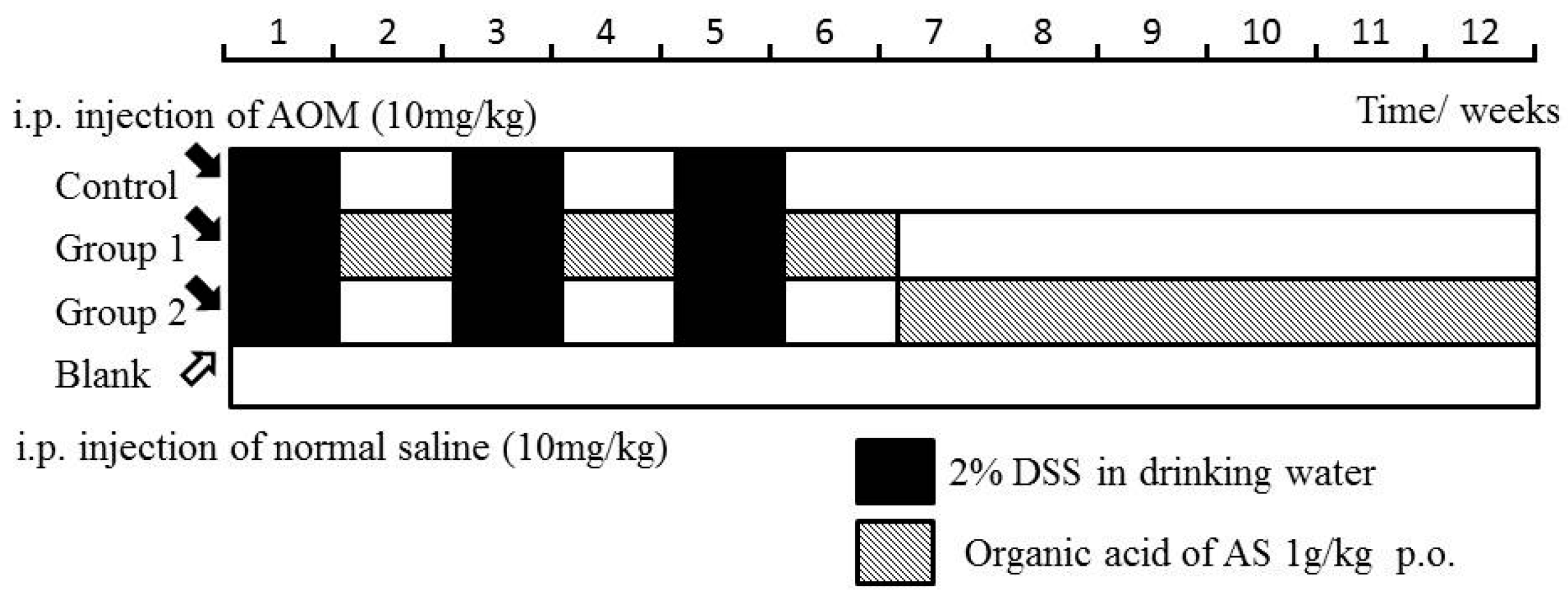
The mice in the control group were not given further treatment. Mice in Groups 1 and 2 were administered by ASR extract orally at different stages of carcinogenesis. The specific method is illustrated in
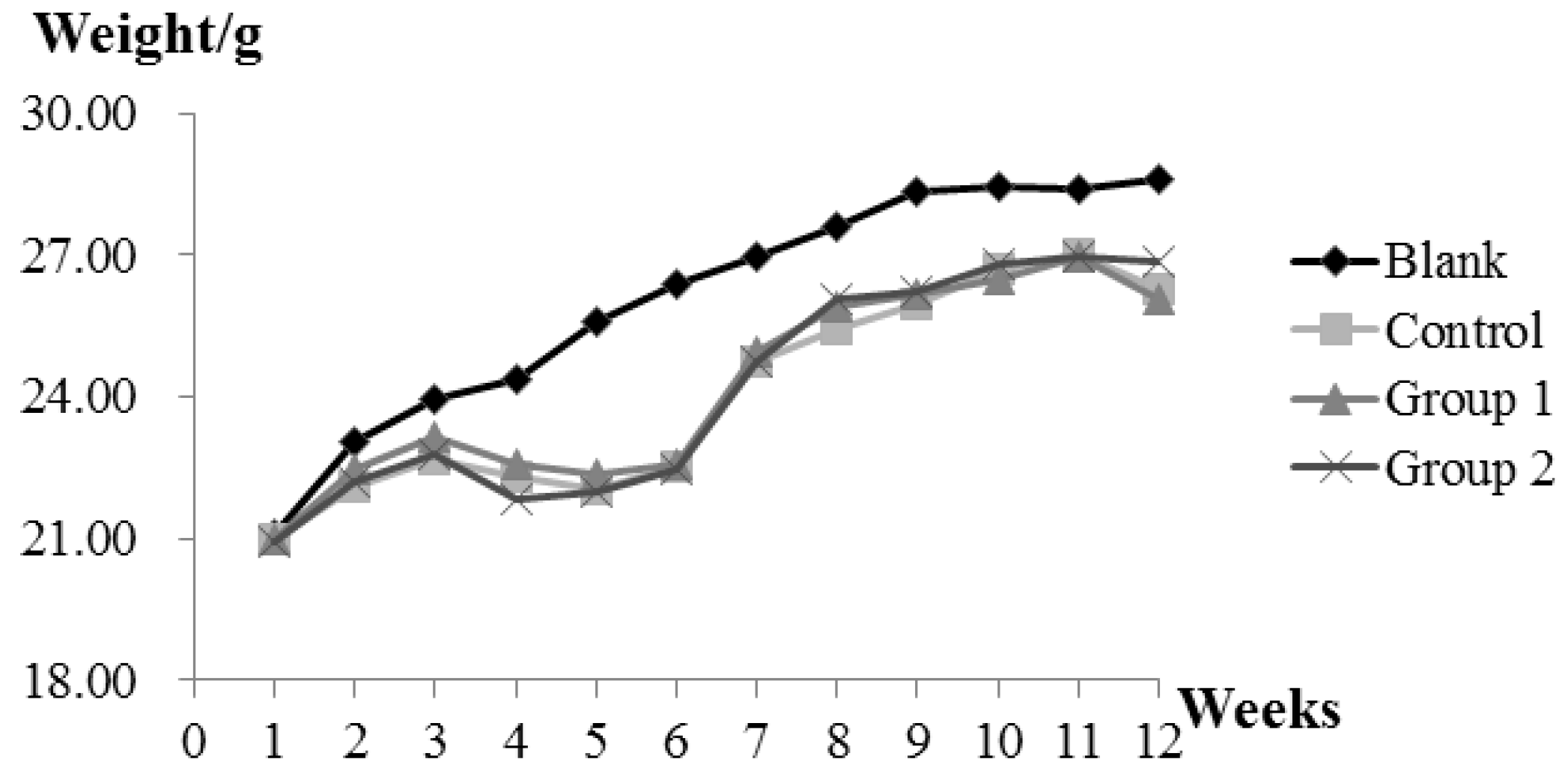
Figure 9. The bodyweight and health condition of the mice were recorded every week. Animals were sacrificed in the 12th week, and large bowels were excised for evaluating histological evidence. All handlings and procedures were carried out as per the protocol approved by the Institutional Animal Care and Usage Committee at Beijing University of Traditional Chinese Medicine.
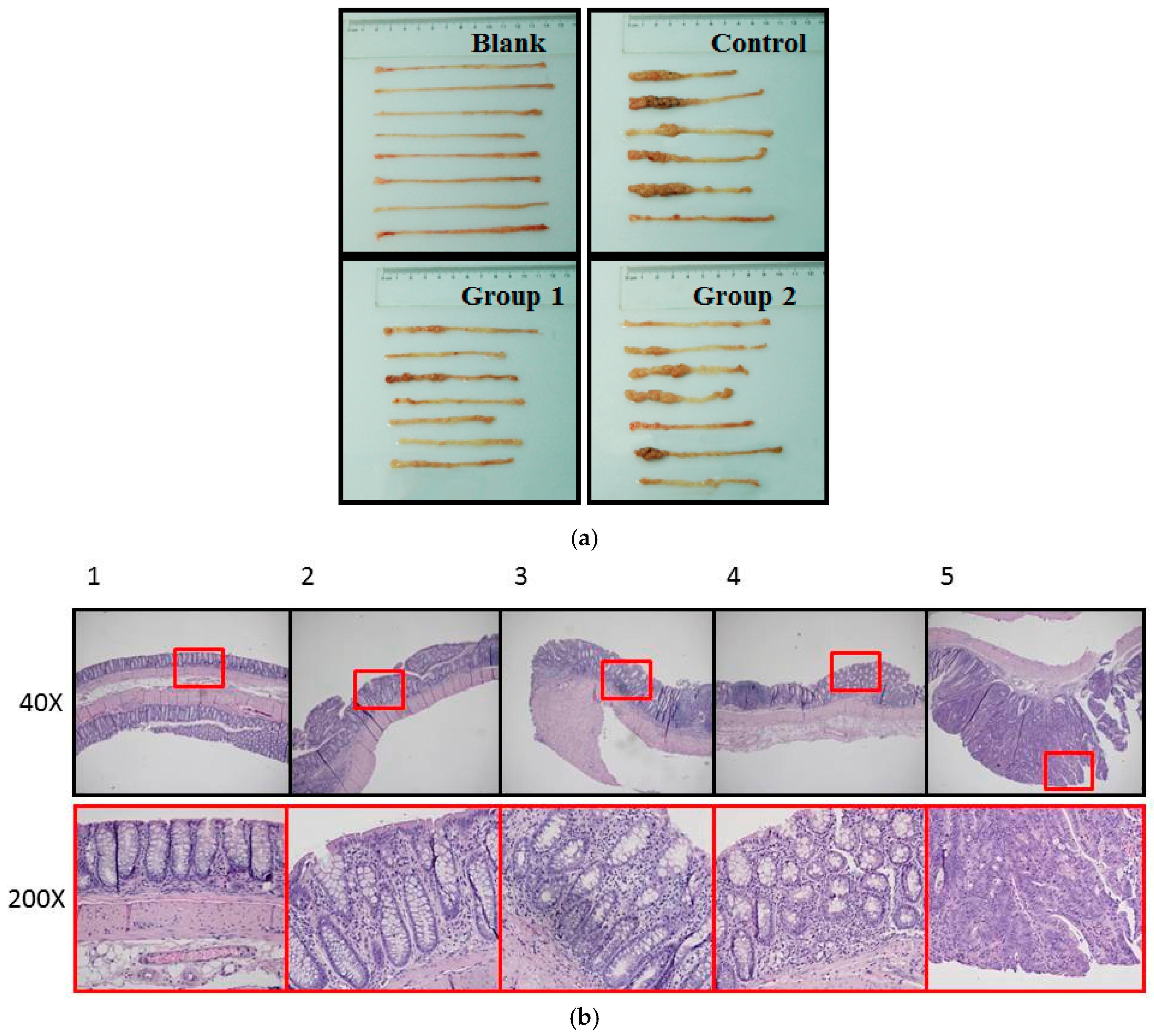
4.5. Hematoxylin-Eosin (HE) Staining and Histological Analysis
The formalin-fixed colon tissues were embedded in paraffin blocks. Sliced sections (4 μm) were deparaffinized and rehydrated by a xylene-ethanol-water gradient system. Hematoxylin and eosin (HE) staining was performed followed by a dehydrating process. Histopathological examination was performed under a light microscope by Olympus (Waltham, MA, USA). Neoplasms and inflammations were analyzed and diagnosed as normal gland, mild inflammation, moderate inflammation, severe inflammation (ulcerative colitis), hyperplasia, aberrant crypt foci (ACF), gastrointestinal intraepithelial neoplasia (GIN), adenoma, and adenocarcinoma as per the established criteria. Histopathological evaluation was determined by two pathologists from the pathology department of Wang-jing Hospital (Chao-yang, Beijing, China) who were not aware of the experimental protocols.
4.6. Immunohistochemistry Assay
For immunohistochemical analysis, 4-μm thick sections of colon-rectum tissue were dried and deparaffinized. After antigen retrieving and endogenous peroxidase activity inhibition, the sections were incubated with diluted primary antibodies of 8-oxoguanine (1:10,000) and PCNA (1:5000), and a secondary antibody. Sections were then stained withhaematoxylin slightly for three minutes and dehydrated for observation and photographing under a light microscope.
4.7. TUNEL Assay
An in situ cell death detection kit, POD, was applied tothe TUNEL (terminal deoxynucleotidyltransferase-mediated deoxyuridine triphosphate nick end labeling) assay to detect apoptotic cells as per the manufacturer’s instructions. Paraffinized sections were then embedded with the incubation of the TUNEL reaction mixture containingTdT and fluorescein-dUTP. The label incorporated at the damaged sites of the DNA was marked by an anti-fluorescein antibody conjugated with the reporter enzyme POD. Stained sections were examined and photographed under a Nikon Eclipse-Ti Fluorescence microscope system (Tokyo, Japan).
4.8. Western Blot Analysis
A 20-mg sample of frozen colon-rectal tissue was homogenized with a 200-μL protease inhibition cocktail and 100 μL lysis buffer for the extraction of total protein. The content of protein was determined by BCA assay, and the protein concentration was adjusted by radio immune-precipitation assay buffer to 5 mg/mL. Equal amounts of protein from tissue lysates were resolved using 10% DSS gel electrophoresis and immunoblotted with primary antibodies (P53 and gamma-H2AX) followed by HRP-conjugated secondary antibodies. After washing the membranes three times for five minutes in TBS buffer containing 0.05% tween-20, the targeted signals were detected on a Li-Cor Odyssey Imager.
4.9. Statistical Analysis
Values are presented as means ± SD. Statistical analyses were done by one-way ANOVA test and Fisher’s least significant difference t-test. p value of less than 0.05 was considered to be statistically significant.


