Article

Binding of Sulpiride to Seric Albumins

by
Viviane Muniz Da Silva Fragoso
1,*,
Carla Patrícia De Morais Coura
2,†,
Luanda Yanaan Hoppe
1,†,
Marília Amável Gomes Soares
3,†,
Dilson Silva
2,3,† and
Celia Martins Cortez
2,3,†
1
Laboratory of Innovations in Therapies, Education and Bioproducts, Oswaldo Cruz Institute/FIOCRUZ, Av. Brasil 4365, Rio de Janeiro 21045-900, Brazil
2
Postgraduation in Medical Sciences, Rio de Janeiro State University, Av. Manoel de Abreu, 444, Rio de Janeiro 20550-171, Brazil
3
Applied Mathematics, Rio de Janeiro State University, Rua São Francisco Xavier, 524, Rio de Janeiro 20559-900, Brazil
*
Author to whom correspondence should be addressed.
These authors contributed equally to this work.
Int. J. Mol. Sci. 2016, 17(1), 59; https://doi.org/10.3390/ijms17010059
Submission received: 14 September 2015 / Revised: 23 November 2015 / Accepted: 30 November 2015 / Published: 4 January 2016
(This article belongs to the Section Physical Chemistry, Theoretical and Computational Chemistry)

Abstract

:
The aim of this work was to study the interaction of sulpiride with human serum albumin (HSA) and bovine serum albumin (BSA) through the fluorescence quenching technique. As sulpiride molecules emit fluorescence, we have developed a simple mathematical model to discriminate the quencher fluorescence from the albumin fluorescence in the solution where they interact. Sulpiride is an antipsychotic used in the treatment of several psychiatric disorders. We selectively excited the fluorescence of tryptophan residues with 290 nm wavelength and observed the quenching by titrating HSA and BSA solutions with sulpiride. Stern-Volmer graphs were plotted and quenching constants were estimated. Results showed that sulpiride form complexes with both albumins. Estimated association constants for the interaction sulpiride–HSA were 2.20 (±0.08) × 104 M−1, at 37 °C, and 5.46 (±0.20) × 104 M−1, at 25 °C. Those for the interaction sulpiride-BSA are 0.44 (±0.01) × 104 M−1, at 37 °C and 2.17 (±0.04) × 104 M−1, at 25 °C. The quenching intensity of BSA, which contains two tryptophan residues in the peptide chain, was found to be higher than that of HSA, what suggests that the primary binding site for sulpiride in albumin should be located next to the sub domain IB of the protein structure.

Graphical Abstract

1. Introduction

Sulpiride is an antipsychotic drug of the benzamine class, which presents strong chemical and clinical similarity with amisulpride, and it is used in the treatment of a large range of psychotic disorders [1,2]. About 1% of the general population is affected by psychotic diseases with high prevalence (16%) among people with familial history of schizophrenia [3,4].
Antipsychotic drugs have been traditionally classified into typical and atypical, where the so called typical antipsychotics were the first developed drugs. Typicals have great efficacy on positive symptoms but present low response in the case of negative symptoms. Atypical antipsychotics, such as risperidone, are the second-generation antipsychotic drugs which are efficient to reduce both positive and negative symptoms, without important prejudice of physiological effects of dopamine via nigro-striatal. Atypicals present an important safety advantage over typicals because of their low potential to generate extrapyramidal effect and tardive dyskinesia [5].
Sulpiride is a selective post-synaptic dopamine D2 antagonist [2], and there are suggestions that it could be regarded as an atypical antipsychotic [6,7,8] because of its clinical benefits to minimize negative defective symptoms, as well as its partial activity against positive symptoms [9,10]. In addition, sulpirides have fewer extrapyramidal effects than other typical antipsychotics such as haloperidol [1]. Lai et al. [11] have shown, using pragmatic outcome measures, that the effectiveness of sulpiride was better than haloperidol and risperidone. The most troubling side effects of sulpiride were weight gain and endocrine-related symptoms, which may also affect patient persistence [4].
A meta-analysis study has concluded that atypical anti-psychotics comprise a heterogeneous group of drugs presenting different pharmacological properties, such as mechanisms of action, therapeutic efficacy and side effects besides cost-effective profile [12]. In such a context, we regard as questionable whether sulpiride is properly classified as an atypical antipsychotic.
It is known that the ratio of the binding of sulpiride to plasmatic proteins is about 40% [13]. The drug-protein binding is a very important pharmacokinetic parameter, since the balance between free and combined drug portions in plasma is a determinant factor for the intensity and the duration of their clinical effects, distribution and elimination [14,15].
In an earlier study [16], we used the spectrofluorimetric binding analysis [17] to explore the interaction of risperidone with albumins. Results showed that this drug quenches the fluorescence of albumin and forms complexes with both human serum albumin (HSA) and bovine serum albumin (BSA).
The present paper reports results produced by the spectrofluorimetric study of the interaction of sulpiride with HSA and BSA, and comparing data obtained for the two albumins, we enhanced possibilities to better identify and characterize the binding region, presenting a mathematical model to estimate the quenching of albumins by fluorescent drugs.

2. Results and Discussion

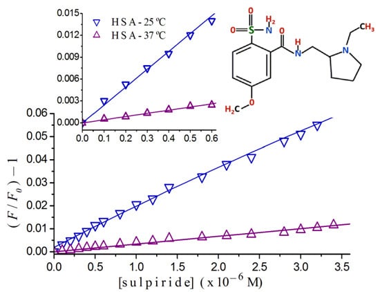
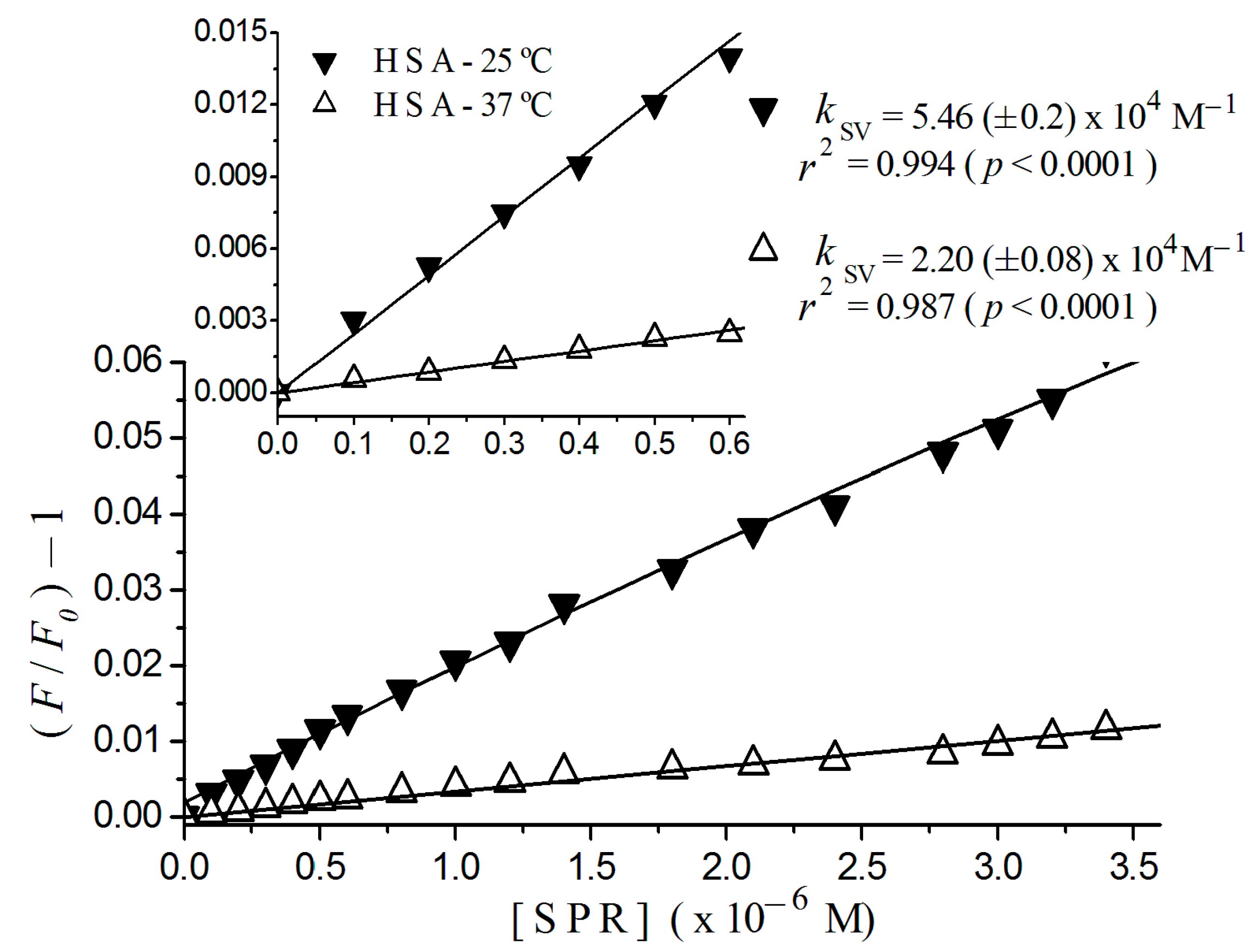
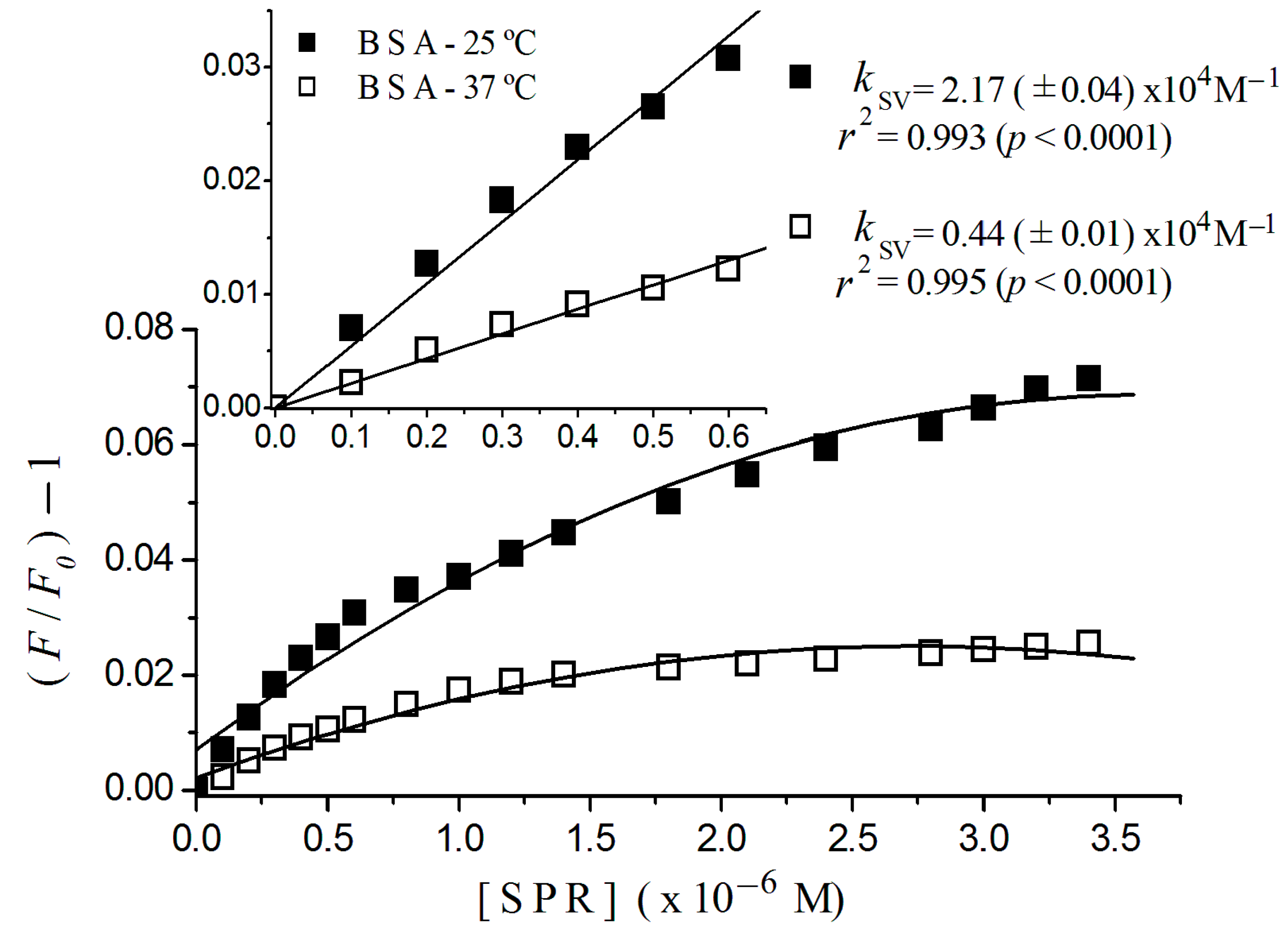
2.1. Fluorescence Quenching

HSA and BSA solutions were titrated by sulpiride at 25 and 37 °C and plots of fluorescence quenching, Stern-Volmer constants, binding constants and binding sites were shown. Figure 1 shows the normalized fluorescence quenching plots for HSA and BSA titrated by sulpiride related to the sulpiride concentration, at 37 °C, Figure 1a and at 25 °C, Figure 1b. Each point in the plot represents the average of three experiments, within standard deviations lower than 10%, ever considering four points around the maximum emission, λmax = 340 nm for BSA, and λmax = 338 nm for HSA. The values of F/F0 were corrected by Equation (3). For the molar ratio sulpiride/HSA and sulpiride/BSA of 1:10 at 37 °C, the ligand quenches 0.08 (±0.03)% and 0.51 (±0.10)% of HSA and BSA fluorescence, respectively. Otherwise, for the molar ratio 1:1 at the same conditions, the quenching increased to 0.74 (±0.10)% and 2.30 (±0.20)% for HSA and BSA, respectively.
Figure 1. Normalized fluorescence quenching plots of human serum albumin (HSA) and bovine serum albumin (BSA) titrated with sulpiride at 25 and 37 °C. Excitation λ = 290 nm; [albumin] = 2 × 10⁻⁶ M; 10−3 M phosphate buffer, pH 7.4. [SPR] = sulpiride concentration.
Ijms 17 00059 g001
The study of the interaction of sulpiride with albumin has great relevance once this protein represents 60% of the total plasmatic proteins and plays a crucial role in transport hydrophobic ligands [14,18,19].
Despite the exclusive use of sulpiride in humans, we also estimated spectrofluorimetric data for its interaction with BSA, once such comparative analysis allows us to verify the approximate localization of the primary binding site for HSA and BSA in albumins. The HSA molecule only has a single tryptophan residue located in sub domain IIA (tryptophan 214), while BSA contains another residue, in sub domain IB (tryptophan 134), in addition to that tryptophan residue 212 in subdomain IIA [20,21,22,23].
Sulpiride has quenched the fluorescence of both albumins just after the first additions (Figure 1), although the intensity of the fluorescence emission of BSA is stronger than the fluorescence emitted by HSA, due to its two tryptophan residues. The quenching of the BSA fluorescence by sulpiride was more intense than that of HSA, especially at 37 °C and even at low concentrations (<0.6 × 10⁻⁶ M) of the drug. That suggests that sulpiride suppresses more intensively the emission of the tryptophan residue 134 than the emission of tryptophan residue 212, indicating that the affinity sites to sulpiride in albumins are closer to sub domain IB (or within this) than the sub domain IIA.

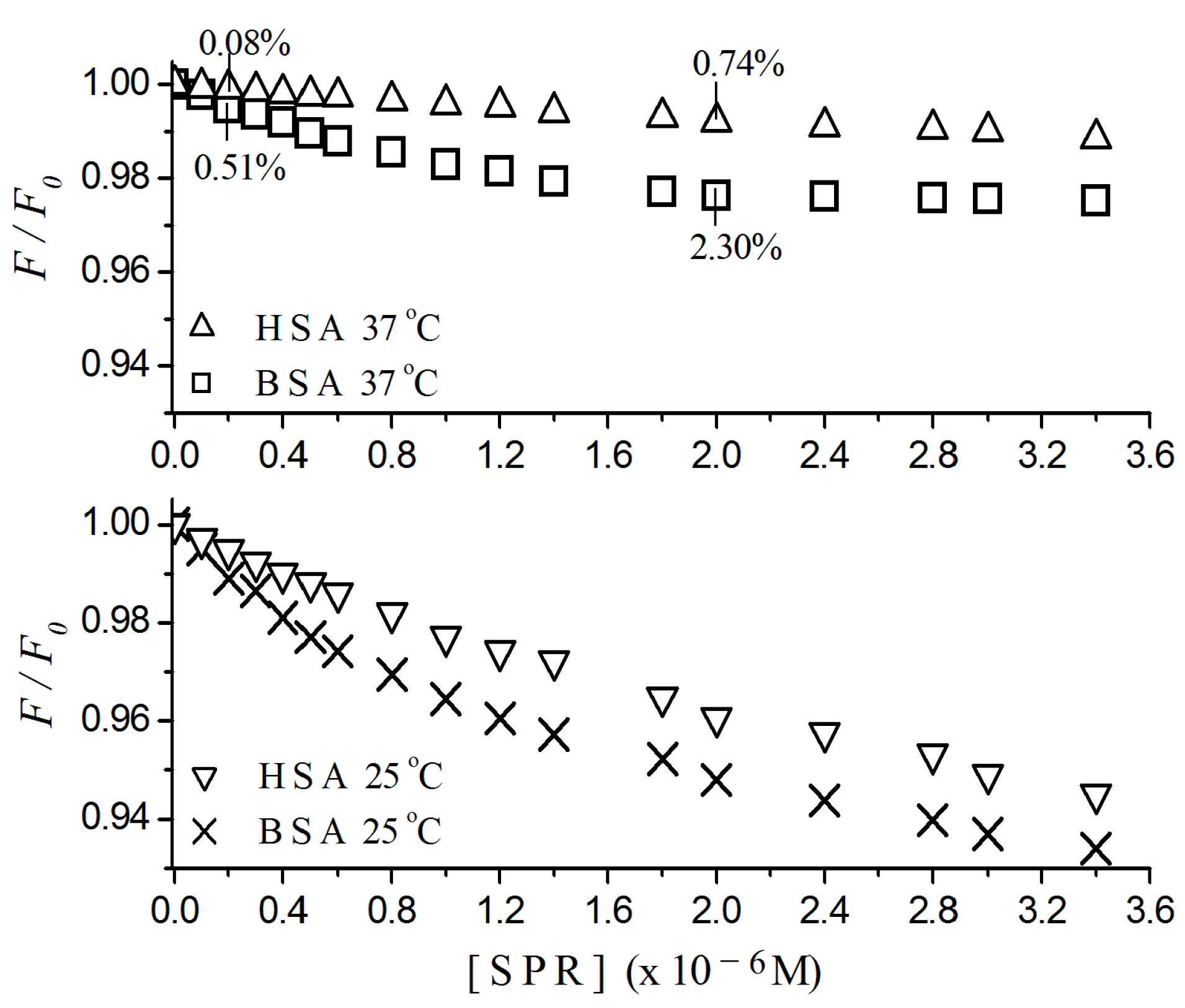
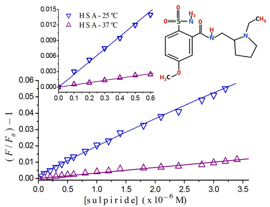
2.2. Stern-Volmer Graphs of Albumins Titrated with Sulpiride

In Figure 2, it is possible to observe the linear tendency of Stern-Volmer plots of HSA at 25 and 37 °C. The detail of this figure shows plots for (sulpiride) <0.6 × 10⁻⁶ M with respective constants, 2.20 (±0.08) × 104 M−1, at 37 °C, and 5.46 (±0.20) × 104 M−1, at 25 °C. It also presents regression coefficients, r2 = 0.984 at 37 °C and r2 = 0.994 at 25 °C for p < 0.0001.
Figure 2. Stern-Volmer plots of HSA titrated with sulpiride at 25 and 37 °C. Excitation λ= 290 nm; (albumin) = 2 × 10⁻⁶ M; 10−3 M phosphate buffer, pH 7.4, [SPR] = sulpiride concentration. Detail shows plot region for low sulpiride concentration region with regression coefficient and Stern-Volmer constant estimated for this region.
Ijms 17 00059 g002
Figure 3 shows plots for BSA, where it was possible to fit a linear adjust to sulpiride concentrations lower than 0.6 × 10⁻⁶ M, with regression coefficients of 0.995 at 37 °C, and 0.993 at 25 °C for p < 0.0001. From an overall view, we can see a downward concave curvature toward the x-axis for sulpiride for concentrations higher than 0.6 × 10⁻⁶ M. In the figure inset, we can see Stern-Volmer constants of 0.44 (±0.01) × 104 M−1, at 37 °C and 2.17 (±0.04) × 104 M−1, at 25 °C.
In sequence, to calculate constants and to define the quenching mechanism, we have plotted the Stern-Volmer graphs, as shown in Figure 2. The linear tendency (r2 > 0.996, p < 0.0001) of the plot of sulpiride–HSA for increasing concentrations, is compatible to the existence of one single binding site to sulpiride in the proximity of the residue of tryptophan, combined to the occurrence of a single type of quenching [17].
Otherwise, the nonlinearity of the Stern-Volmer plot for BSA shown in Figure 3 confirms that the two tryptophan-residues in BSA are not equally accessible to sulpiride. The concave curvature toward the x-axis in addition suggests that other binding sites could have been exposed as the drug concentration increased [17].
Figure 3. Stern-Volmer plots of BSA titrated with sulpiride at 25 and 37 °C. Excitation λ = 290 nm; (albumin) = 2 × 10⁻⁶ M; 10−3 M phosphate buffer, pH 7.4, [SPR] = sulpiride concentration. Detail shows plot region for low sulpiride concentration region, with regression coefficient and Stern-Volmer constant estimated for this region.
Ijms 17 00059 g003
Examining the influence of the temperature on the Stern-Volmer plot for HSA (Figure 2) and BSA (Figure 3), we can observe that an increase of 12 °C in the temperature causes a quenching decreasing, pointing to the occurrence of static quenching, which means the formation of a complex sulpiride-albumin. In that case, the Stern-Volmer constants estimated here can be named association constants [17].
In previous work [16], we found the presence of a high affinity binding site for risperidone in albumins next to or in the neighboring of sub domain IB, where the ligand binds forming the complex risperidone-albumin. The formation of a complex drug-HSA has been also found to occur with other antipsychotic drugs, such as clozapine [24], and phenotiazines: tioridazine, triflupromazine, and levomepromazine [25,26]. On the other hand, Silva et al. showed that the phenotiazine clorpromazine interacts with HSA without forming a complex [27].
HSA has two hydrophobic cavities located in subdomains II and III, which are considered primary sites for high affinity binding to the majority of drugs. Anionic heterocyclic compounds of large molecular size bind especially in the site Sudlow I, located in subdomain IIA. The site II (in subdomain IIIA) presents higher affinity to aromatic carboxylic drugs in therapeutic concentration, being pointed out as one of the principal binding sites for these kinds of compounds. However, low affinity and selectivity sites are also involved, for higher concentrations [28,29,30].

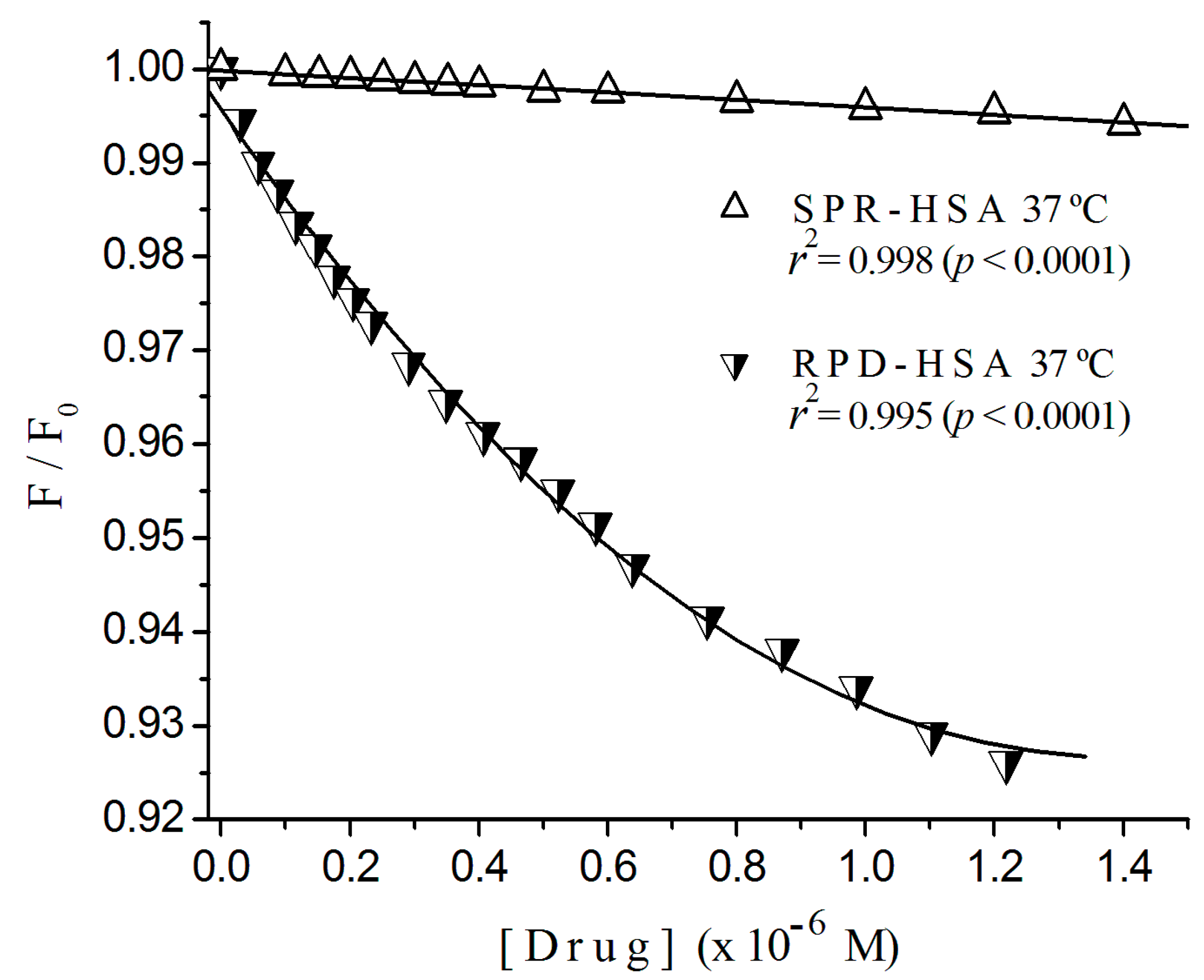
2.3. Fluorescence Quenching of HSA Titrated with Antipsychotics

Figure 4 illustrates the normalized fluorescence quenching plots of HSA by risperidone [16] and sulpiride at 37 °C, where we can see the great difference in the quenching rate for both ligands. In Table 1, we resume and compare results for HSA interacting with both ligands at 25 and 37 °C. It shows the ratio of Stern-Volmer constants and the type of the quenching, found to be static. The constant ratios risperidone/sulpiride at 37 and 25 °C were 6.5 and 4.6, respectively.
Comparing Ksv values from Table 1, we can see that Ksv for the interaction risperidone-HSA at 25 °C (2.56 (±0.09) × 105 M−1) to that of sulpiride, 5.46 (±0.20) × 104 M−1 we found a ratio of about 4.6-fold. By the other hand, at 37 °C, the ratio of constants, 1.43 (±0.05) × 105 M−1 and 2.20 (±0.08) × 104 M−1 was about 6.5-fold. These comparisons point to the occurrence of conformational changes in albumin chains which expose fluorophore groups and/or open new sites.
Figure 4. Normalized fluorescence quenching plots of HSA titrated with sulpiride and risperidone at 37 °C. Excitation λ = 290 nm; (albumin) = 2 × 10⁻⁶ M; 10−3 M phosphate buffer, pH 7.4.
Ijms 17 00059 g004
Table 1. Stern-Volmer Constant for risperidone and sulpiride with human serum albumin (HSA).
DrugQuenchingStern-Volmer Constant—KSV (M−1)
37 °C25 °C
SulpirideStatic2.20 (±0.08) × 1045.46 (±0.20) × 104
Risperidone *Static1.43 (±0.05) × 1052.56 (±0.09) × 105
Risperidone/Sulpiride-6.54.6
* Fragoso et al. [17].

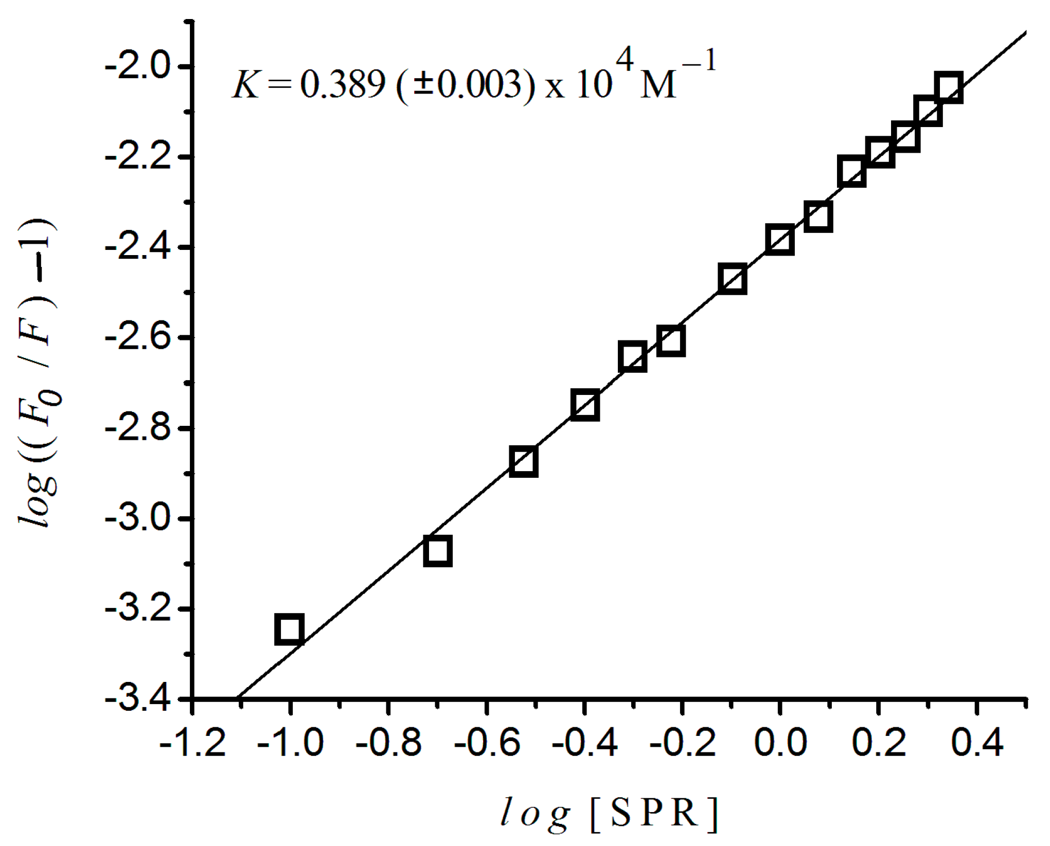
2.4. Binding Constant and the Number of Binding Sites

Figure 5 presents the variation of l o g ( F 0 F 1 ) against the log([sulpiride]) for HSA at 37 °C, estimated by means of Equation 1. It shows a linear plot (r2 = 0.998, p < 0.0001) with binding constant of 0.389 (±0.003) × 104 M−1, revealing one single binding site. They were estimated from the interception and the inclination of this plot, respectively, and these values are presented in Table 2, where, for comparison purposes, we included results of risperidone obtained from [16]. The n values shown in this table point to the existence of only one independent binding site for sulpiride in HSA, at 37 °C.
Figure 5. Variation of log((F0/F) − 1) versus log((sulpiride)) for HSA at 37 °C. Excitation λ = 290 nm; (albumin) = 2 × 10⁻⁶ M; 10−3 M phosphate buffer, pH 7.4.
Ijms 17 00059 g005
In addition, Table 2 provides evidence that binding constant, K, for risperidone-HSA (7.46 (±0.01) × 104 M−1) [16] was many times higher than K for sulpiride–HSA (3.89 (±0.03) × 103 M−1), Figure 5. Observing the K values, we can evaluate the distribution of a drug by the plasma. A small K value corresponds to a weak binding of a drug to albumin, and it means a short life time or poor distribution, while a high K value means a strong binding. In such cases, the life time is longer and the free drug concentration in plasma is lower [31,32].
Table 2. Binding constant K and number of binding sites n for interactions risperidone–HSA and sulpiride–HSA.
DrugK (M−1)n
Sulpiride3.890 (±0.003) × 103≈1
Risperidone7.460 (±0.010) × 104≈1
The above-mentioned strong binding of risperidone could be a consequence of its hydrophobicity, with its metabolizing path by the liver, where it is converted into a hydrophilic metabolite and then eliminated by the kidneys. Otherwise, since 1978, Denisoff and Molle [33] have shown that sultopride, a benzamide derivative like sulpiride, presents reduced tendency to bind to albumins. Sulpiride is a hydrophilic compound, with lower constant values, which presents reduced capacity to entry in the central nervous system and appears to not undergo hepatic action, once no sulpiride metabolite has been identified in plasma, reinforced by the fact that 100% of sulpiride parenteral administration, and 15% to 25% of oral administration are excreted unchanged by the kidneys [13,34,35].
However, despite a different hydrophobicity degree, primary sites for both antipsychotics, sulpiride and risperidone, seem to be located in the same albumin region, within or next to the sub domain IB, where tryptophan residue 134 of BSA is. This subdomain is more exposed to the hydrophilic environment than subdomain IIA, where the tryptophan residue 212 of BSA is. The hydrophobic cavity containing tryptophan residue 134 is less hydrophobic than the subdomain IIA cavity that contains residue 212, which is buried in an internal region of the protein. Sub domain IIA, namely Sudlow I, which normally drives strong hydrophobic interactions involving hydrogen bridges and histidine (residue 242) [36,37].
Thus, results suggest the existence of only one primary binging site for this sulpiride in HSA probably located within or next to the sub domain IB, where the primary site for risperidone also seems to be located. However, the comparison between K-values for two antipsychotics provides evidence that the interaction of albumin to risperidone is very higher than the interaction to sulpiride.

3. Material and Methods

3.1. Materials

This study was performed in vitro, using fluorescence measurements from a Hitachi-F3010 spectrofluorometer (Tokyo, Japan). Albumins (codes A-0281 and A-8763) and sulpiride (S2191000) were purchased from Sigma-Aldrich Co., St. Louis, MO, USA.

3.2. Binding of Albumin to Sulpiride—Fluorescence Quenching of Albumin by Spectrofluorimetry

Following the Fragoso et al. [16] experimental design, for each quencher addition (from 35.0 to 1191.7 ng/mL increments), we used the wavelength of 290 nm to selectively excite the fluorescence of intrinsic tryptophan residues in albumin. The emission and excitation bandwidths were to 3 nm, and the emission spectra registered from 300 to 400 nm. Final concentrations of sulpiride were within the therapeutic window for psychiatric patients, i.e., from 70.1 to 1121.2 ng/mL, according to Tokunaga et al. [38].
For each quenching measurement, sulpiride was added from concentrated stock solution in 2 mL solutions of albumin 2 × 10⁻⁶ M (in 10 mM phosphate at pH 7.4), at two different temperatures: 25 and 37 °C.
Stern-Volmer graphs were plotted according to the equation   F 0 = F ( 1 + K S V Q ) , where F0 and F are the relative fluorescence intensities of albumins in the absence and in the presence of sulpiride respectively (F0 is taken to be 100% always). KSV is the Stern-Volmer quenching constant, which is related to the bimolecular collisional process, and Q is the quencher concentration [17].
The presence of primary and secondary inner filter effects was verified by means of absorbance measurements (Shimadzu UV-160A spectrophotometer, Columbia, Portland, OR, USA) of albumin solutions at emission and excitation wavelengths. The secondary inner filter effect was negligible, and the primary inner filter effect was corrected by Parker’s equation [39]. Comparing Stern-Volmer plots at two temperatures, 25 and 37 °C, we verified the dynamic or static nature of the quenching process but only for HSA.
Regression coefficients in Stern-Volmer analysis allow us to fit a linear adjust when r2 > 0.991 and p < 0.0001. In the case where r2 < 0.9980, the polynomial adjust was used [16].
The constant of binding (K) and the number of binding sites (n) of sulpiride to HSA were obtained through the equation [25]
l o g ( F 0 F F ) = l o g ( K ) + n   l o g ( [ Q ] )

3.3. Mathematical Modeling of Albumin Quenching by a Fluorescent Quencher

Due to the fluorescence of the sulpiride molecule, which difficult the direct determination of the quenching of the albumin, a mathematical model has been developed aiming to distinguish the albumin fluorescence from the sulpiride fluorescence, in the solution where they interact.
For a certain concentration of ligand i added in the solution, we can consider that the fluorescence measured for albumin, Fi, at a given temperature can be written as
F i = F A 0 + F L 0 F S T
where F A 0 is the fluorescence intensity of albumin before the addition of the quencher, F L 0 is the fluorescence intensity of the pure quencher solution, and F S T is the total fluorescence quenched due to the interaction of those molecules. F S T can be represented by the sum of the quenched fluorescence portion of albumin F A S and the quenched fluorescence portion of the quencher F L S , i.e., F S T = F A S + F L S .
Supposing that each chemical species contributes with half of the quenched fluorescence when interacting, we can write that F S T = 2 F A S . Substituting Equation (4) into Equation (2), the fluorescence intensity involved in the interaction is given by F A S = 1 2 ( F A 0 + F L 0 F i ) . Subtracting F A 0   from both sides of this equation and dividing them by F A 0 , we have
F A i F A 0 = 1 2 F L 0 2 F A 0 + F i 2 F A 0  
which refers to the recorded fluorescence portion emitted by albumin. This equation is valid for all fluorescence measures, from the beginning of the titration of albumin solution by a fluorescent quencher, such as sulpiride.

4. Conclusions

Results support the conclusion that sulpiride quenches HSA and BSA fluorescence by a process of static quenching, interacting with the protein by forming complexes. Association constants calculated for sulpiride/HSA <0.6 µM, by the Stern-Volmer equation, were 2.20 (±0.08) × 104 and 5.46 (±0.20) × 104 M−1, at 37 and 25 °C, respectively. For sulpiride–BSA interaction, association constants were 0.44 (±0.01) × 104 and 2.17 (±0.04) × 104 M−1 at 37 and 25 °C, respectively.
The apparent binding constant estimated for sulpiride–HSA at 37 °C was 3.89 (±0.03) × 103 M−1, which was lower than that found by Fragoso et al. [16] to risperidone-HSA at 37 °C.
The comparison between Ksv values allows us to conclude that the quenching potential of RPD on HSA at 37 °C is about six times higher than that for sulpiride. Despite different hydrophobicity degrees, primary sites for both antipsychotics appear to be located in the same albumin region, within or next to the sub domain IB, where tryptophan residue 134 of BSA is located, the less hydrophobic between both considered regions.
The biological meaning of this study lies in its contribution to the knowledge about the pharmacokinetics of sulpiride, and its comparison with risperidone. It is of great relevance in some clinical pharmacology and psychiatric questions to rationalize and personalize therapeutics. In such a sense, the simultaneous administration of drugs and therapeutics requires critical care due to their possible competition for the same site.
In addition, the present study contributes to the molecular biochemistry of sulpiride, establishing the number of binding sites, found to be only one, and its localization in the chain.

Acknowledgments

This study was financially supported by the Brazilian Government Agency CAPES. The authors thank Sonia W.R. Louro (Pontifical Catholic University, Rio de Janeiro, Brazil) and Jaime Cunha Bastos (Rio de Janeiro State University, Rio de Janeiro, Brazil) for permitting the use of all their laboratories facilities. The authors thank Tânia Cremonini de Araújo-Jorge, Head of the Laboratory of Innovations in Therapies, Education and Bioproducts of the Oswaldo Cruz Institute, Oswaldo Cruz Foundation–Brazil, for the logistical support to this research.

Author Contributions

Viviane Muniz da Silva Fragoso and Celia Martins Cortez contributed to the design of the experiments; Viviane Muniz da Silva Fragoso and Dilson Silva performed the experiments; Dilson Silva and Viviane Muniz da Silva Fragoso wrote the paper; Celia Martins Cortez, Carla Patrícia de Morais Coura, Luanda Yanaan Hoppe and Marília Amável Gomes Soares revised the manuscript; Celia Martins Cortez and Viviane Muniz da Silva Fragoso analyzed the data; Carla Patrícia de Morais Coura contributed reagents.

Conflicts of Interest

The authors declare no conflict of interest.

References

  1. Leucht, S.; Corves, C.; Arbter, D.; Engel, R.R.; Li, C.; Davis, J.M. Second-generation versus first-generation antipsychotic drugs for schizophrenia: A meta-analysis. Lancet 2009, 37, 31–41. [Google Scholar] [CrossRef]
  2. Seeman, P.; van Tol, H.H.M. Dopamine receptor pharmacology. Trends Pharmacol. Sci. 1994, 15, 264–270. [Google Scholar] [CrossRef]
  3. Bhugra, D. The global prevalence of schizophrenia. PLoS Med. 2005, 2, e151. [Google Scholar] [CrossRef] [PubMed] [Green Version]
  4. Souza, B.R.; Torres, K.C.L.; Miranda, D.M.; Motta, B.S.; Scotti-Muzzi, E.; Guimarães, M.M.; Carneiro, D.S.; Rosa, D.V.F.; Souza, R.P.; Reis, H.J.; et al. Lack of effects of typical and atypical antipsychotics in DARPP-32 and NCS-1 levels in PC12 cells overexpressing NCS-1. J. Negat. Results Biomed. 2010, 9, 4–7. [Google Scholar] [CrossRef] [PubMed]
  5. Peluso, M.J.; Lewis, S.W.; Barnes, T.R.; Jones, P.B. Extrapyramidal motor side-effects of first- and second-generation antipsychotic drugs. Br. J. Psychiatry 2012, 200, 387–392. [Google Scholar] [CrossRef] [PubMed]
  6. Lerner, V.; Libov, I.; Kotler, M.; Strous, R.D. Combination of “atypical” antipsychotic medication in the management of treatment-resistant schizophrenia and schizoaffective disorder. Prog. Neuropsychopharmacol. Biol. Psychiatry 2004, 28, 89–98. [Google Scholar] [CrossRef] [PubMed]
  7. Mauri, M.C.; Bravin, S.; Bitetto, A.; Rudelli, R.; Invernizzi, G. A risk-benefit assessment of sulpiride in the treatment of schizophrenia. Drug Saf. 1996, 14, 288–298. [Google Scholar] [CrossRef] [PubMed]
  8. Moore, S.; Kenyon, P. Atypical antipsychotics, clozapine and sulpiride do not antagonize amphetamine-induced stereotyped locomotion. Psychopharmacology 1994, 114, 123–130. [Google Scholar] [CrossRef] [PubMed]
  9. Azorin, J.M.; Dassa, D.; Jalfre, M. The atypical neuroleptic concept. Encephale 1992, 18, 453–457. [Google Scholar] [PubMed]
  10. Jones, P.B.; Barnes, T.R.; Davies, L.; Dunn, G.; Lloyd, H.; Hayhurst, K.P.; Murray, R.M.; Markwick, A.; Lewis, S.W. Randomized controlled trial of the effect on Quality of Life of second- vs. first-generation antipsychotic drugs in schizophrenia: Cost utility of the latest antipsychotic drugs in schizophrenia study (CUtLASS 1). Arch. Gen. Psychiatry 2006, 63, 1079–1087. [Google Scholar] [CrossRef] [PubMed]
  11. Lai, E.C.C.; Chia-Hsien, C.; Yang, Y.H.K.; Lin, S.J.; Lin, C.Y. Effectiveness of sulpiride in adult patients with schizophrenia. Schizophr. Bull. 2013, 39, 673–683. [Google Scholar] [CrossRef] [PubMed]
  12. Leucht, S.; Komossa, K.; Rummel-Kluge, C.; Corves, C.; Hunger, H.; Schmid, F.; Lobos, C.A.; Schwarz, S.; Davis, J.M. A meta-analysis of head-to-head comparisons of second-generation antipsychotics in the treatment of schizophrenia. Am. J. Psychiatry 2009, 166, 152–163. [Google Scholar] [CrossRef] [PubMed]
  13. Frota, L.H.; Bueno, J.R.; Silva Filho, J.F. Risperidona, amisulpirida, quetiapina e ziprasidona: Comentários finais ao protocolo do Ministério da Saúde para antipsicóticos atípicos de segunda geração. Braz. J. Psychiatry 2001, 50, 337–362. [Google Scholar]
  14. Fanali, G.; di Mais, A.; Trezza, V.; Marino, M.; Fasano, M.; Ascenzi, P. Human serum albumin: From bench to bedside. Mol. Asp. Med. 2012, 33, 209–290. [Google Scholar] [CrossRef] [PubMed]
  15. Fasano, M.; Curry, S.; Terreno, E.; Galliano, M.; Fanali, G.; Narciso, P.; Notari, S.; Ascenzi, P. The extraordinary ligand binding properties of human serum albumin. IUBMB Life 2005, 57, 787–796. [Google Scholar] [CrossRef] [PubMed]
  16. Fragoso, V.M.S.; Silva, D.; Cruz, F.A.O.; Cortez, C.M. Modeling the process of interaction of risperidone and serum albumin. Environ. Toxicol. Pharmacol. 2012, 33, 262–266. [Google Scholar] [CrossRef] [PubMed]
  17. Lakowicz, J.R. Principles of Fluorescence Spectroscopy, 3rd ed.; Springer Science: Philadelphia, PA, USA, 2006; pp. 278–327. [Google Scholar]
  18. Bal, W.; Sokołowska, M.; Kurowska, E.; Faller, P. Binding of transition metal ions to albumin: Sites, affinities and rates. Biochim. Biophys. Acta 2013, 1830, 5444–5455. [Google Scholar] [CrossRef] [PubMed]
  19. Silva, D.; Cortez, C.M.; Silva, C.M.; Missailidis, S. A fluorescent spectroscopy and modelling analysis of anti-heparanase aptamers–serum protein interactions. J. Photochem. Photobiol. B 2013, 127, 68–77. [Google Scholar] [CrossRef] [PubMed]
  20. Carter, D.C.; Ho, J.X. Structure of serum albumin. Adv. Protein Chem. 1994, 45, 153–203. [Google Scholar] [PubMed]
  21. Kragh-Hansen, U. Molecular aspects of ligand binding to serum albumin. Pharmacol. Rev. 1981, 33, 17–53. [Google Scholar] [PubMed]
  22. Simard, J.R.; Zunszain, P.A.; Ha, C.E.; Yang, J.S.; Bhagavan, N.V.; Petitpas, I.; Curry, S.; Hamilton, J.A. Locating high-affinity fatty acid-binding sites on albumin by X-ray crystallography and NMR spectroscopy. Proc. Natl. Acad. Sci. USA 2005, 102, 17958–17963. [Google Scholar] [CrossRef] [PubMed]
  23. Tavirani, M.; Moghaddamnia, S.; Ranjbar, B.; Amani, M.; Marashi, S. Conformational study of human serum albumin in pre-denaturation temperatures by differential scanning calorimetry, circular dichroism and UV spectroscopy. J. Biochem. Mol. Biol. 2006, 39, 530–536. [Google Scholar] [CrossRef]
  24. Wu, X.; Liu, J.; Wang, Q.; Xue, W.; Yao, X.; Zhang, Y.; Jin, J. Spectroscopic and molecular modeling evidence of clozapine binding to human serum albumin at subdomain IIA. Spectrochim. Acta A Mol. Biomol. Spectrosc. 2011, 79, 1202–1209. [Google Scholar] [CrossRef] [PubMed]
  25. Kandagal, P.B.; Shaikh, S.M.T.; Manjunatha, S.D.; Seetharamappa, J.; Nagaralli, S.J. Spectroscopic studies on the binding of bioactive phenothiazine compounds to human serum albumin. J. Photochem. Photobiol. A Chem. 2007, 189, 121–127. [Google Scholar] [CrossRef]
  26. Kandagal, P.B.; Kalanur, S.S.; Manjunatha, D.H.; Seetharamappa, J. Mechanism of interaction between human serum albumin and N-alkyl phenothiazines studied using spectroscopic methods. J. Pharm. Biomed. Anal. 2008, 47, 260–267. [Google Scholar] [CrossRef] [PubMed]
  27. Silva, D.; Cortez, C.M.; Louro, S.R.W. Chlorpromazine Interactions to Sera Albumins. A study by the quenching of fluorescence. Spectrochim. Acta A Mol. Biomol. Spectrosc. 2004, 60, 1215–1223. [Google Scholar] [CrossRef] [PubMed]
  28. Fanali, G.; Pariani, G.; Ascenzi, P.; Fasano, M. Allosteric and binding properties of Asp1–Glu382 truncated recombinant human serum albumin—An optical and NMR spectroscopic investigation. FEBS J. 2009, 276, 2241–2250. [Google Scholar] [CrossRef] [PubMed]
  29. Kitamura, K.; Kume, M.; Yamamoto, M.; Takegami, S.; Kitade, T. 19F NMR spectroscopic study on the binding of triflupromazine to bovine and human serum albumins. J. Pharm. Biomed. Anal. 2004, 36, 411–414. [Google Scholar] [CrossRef] [PubMed]
  30. Watanabe, H.; Kragh-Hansen, U.; Tanase, S.; Nakajou, K.; Mitarai, M.; Iwao, Y.; Maruyama, T.; Otagiri, M. Conformational stability and warfarin-binding properties of human serum albumin studied by recombinant mutants. Biochem. J. 2001, 357, 269–274. [Google Scholar] [CrossRef] [PubMed]
  31. Yamasaki, K.; Chuang, V.T.; Maruyama, T.; Otagiri, M. Albumin–drug interaction and its clinical implication. Biochim. Biophys. Acta 2013, 1830, 5435–5443. [Google Scholar] [CrossRef] [PubMed]
  32. Yang, F.; Zhang, Y.; Liang, H. Interactive Association of drugs binding to human serum albumin. Int. J. Mol. Sci. 2014, 15, 3580–3595. [Google Scholar] [CrossRef] [PubMed]
  33. Denisoff, O.; Molle, L. Binding study of benzamides to serum albumins by fluorescence probe technique. Arzneimittelforschung 1978, 28, 2156–2157. [Google Scholar] [PubMed]
  34. Bressolle, F.; Bres, J.; Blanchin, M.D.; Gomeni, R. Sulpiride pharmacokinetics in humans after intramuscular administration at three dose levels. J. Pharm. Sci. 1984, 73, 1128–1136. [Google Scholar] [CrossRef] [PubMed]
  35. Nyberg, S.; Eriksson, B.; Oxenstierna, G.; Halldin, C.; Farde, L. Suggested minimal effective dose of risperidone based on PET-measured D2 and 5-HT2A receptor occupancy in schizophrenic patients. Am. J. Psychiatry 1999, 156, 869–875. [Google Scholar] [CrossRef] [PubMed]
  36. Wang, T.; Xiang, B.; Wang, Y.; Chen, C.; Dong, Y.; Fang, H.; Wang, M. Spectroscopic investigation on the binding of bioactive pyridazinone derivative to human serumalbumin and molecular modeling. Colloids Surf. B Biointerfaces 2008, 65, 113–119. [Google Scholar] [CrossRef] [PubMed]
  37. Hongwei, Z.; Min, G.; Zhaoxia, Z.; Wenfeng, W.; Guozhong, W. Spectroscopic studies on the interaction between riboflavin and albumins. Spectrochim. Acta A Mol. Biomol. Spectrosc. 2006, 65, 811–817. [Google Scholar]
  38. Tokunaga, H.; Kudo, K.; Jitsufuchi, N.; Ohtsuka, Y.; Imamura, T. Sensitive determination of sulpiride in human plasma by high-performance liquid chromatography. J. Chromatogr. B Biomed. Sci. Appl. 1997, 691, 203–207. [Google Scholar] [CrossRef]
  39. Puchalski, M.M.; Morra, M.J.; von Wandruszka, R. Assessment of inner filter effect corrections in fluorimetry. Fresenius J. Anal. Chem. 1991, 340, 341–344. [Google Scholar] [CrossRef]

