Article

Identification of the Avian Pasteurella multocida phoP Gene and Evaluation of the Effects of phoP Deletion on Virulence and Immunogenicity

1
Institute of Preventive Veterinary Medicine, College of Veterinary Medicine, Sichuan Agricultural University, Chengdu 611130, China
2
Department of Bioengineering, College of Veterinary Medicine, Sichuan Agricultural University, No. 211 Huimin Road, Wenjiang District, Chengdu 611130, China
3
Avian Disease Research Center, College of Veterinary Medicine, Sichuan Agricultural University, Wenjiang, Chengdu 611130, China
4
Key Laboratory of Animal Disease and Human Health of Sichuan Province, Sichuan Agricultural University, Wenjiang, Chengdu 611130, China
*
Authors to whom correspondence should be addressed.
Int. J. Mol. Sci. 2016, 17(1), 12; https://doi.org/10.3390/ijms17010012
Submission received: 27 October 2015 / Revised: 14 December 2015 / Accepted: 15 December 2015 / Published: 23 December 2015
(This article belongs to the Section Biochemistry)

Abstract

:
Pasteurella multocida (P. multocida) is an animal pathogen of worldwide economic significance that causes fowl cholera in poultry and wild birds. Global gene regulators, including PhoP are important in regulating bacterial virulence and are good targets for developing attenuated vaccines against many pathogenic bacteria. However, the biological significance of phoP gene has not been identified in P. multocida. Here, we identified the phoP gene in P. multocida, and we evaluated the roles of phoP in P. multocida by deleting the phoP gene. The P. multocida phoP mutant exhibited similar growth curves and lipopolysaccharide and outer membrane protein profiles but displayed defective polymyxin resistance in vitro compared with the parent strain. Additionally, the phoP deletion resulted in decreased virulence. The LD50 of the ΔphoP mutant was 32- and 154-fold higher than the parent strain via the oral and intranasal routes, respectively. Transcriptome sequencing analysis showed that 161 genes were up-regulated and 173 genes were down-regulated in the absence of the phoP gene. Finally, the immunogenicity and protective efficacy of the ΔphoP mutant were evaluated. Immunized ducks produced significantly higher levels of serum IgY and bile IgA compared to the control ducks, and immunization with the ΔphoP mutant conferred 54.5% protection efficiency against challenge with the virulent P. multocida. This work provides a platform to dissect the function of phoP and develop a new vaccine against P. multocida.

Graphical Abstract

1. Introduction

Pasteurella multocida (P. multocida) is a Gram-negative encapsulated bacterium that is the causative agent of a range of animal pasteurellosis diseases, including fowl cholera in poultry and wild birds, hemorrhagic septicemia in cattle and buffalo, atrophic rhinitis in swine, and snuffles in rabbits [1]. P. multocida can also infect human via small-mammal bites. Fowl cholera is a severe systemic disease that occurs in domestic poultry and wild birds and results in significant economic losses to poultry industries worldwide [2]. The use of antibiotics is a successful method of controlling P. multocida infection, but the emergence of drug-resistant strains poses a serious challenge for antibiotic use [3]. Vaccination of fowl would provide protection against P. multocida infection [4]. Current vaccines against fowl cholera include bacterins and live attenuated vaccines: the former provides only limited protection against homologous serotypes, and the latter were developed empirically and were observed to revert to the virulent strain [4]. Therefore, there is a pressing need to develop new vaccines, especially well-defined live vaccines for fowl cholera control.
Live attenuated vaccines were successfully developed by the introduction of mutations into global regulators including two-component systems (TCS) in many pathogenic bacteria [5,6]. TCS are highly conserved prokaryotic signal transduction pathways that consist of a histidine kinase as the sensor and a response regulator as the effector. Intracellular pathogens usually use TCS to respond to host defenses and are often essential for virulence; the Salmonella phoP is the most widely studied example [6]. phoP was first identified and suggested to perform a regulatory function in Salmonella because a strain with a phoP mutation lost acid phosphatase activity [7]. phoP and phoQ constitute a vital TCS involved in bacterial invasion and survival in the host and can positively and negatively regulate a network of genes in many Gram-negative pathogens, including Mycobacterium tuberculosis [8], Yersinia pestis [9], Escherichia coli (E. coli) [10], and Salmonella enterica [11]. In response to a number of environmental signals, including low magnesium [12], low pH [13], and sub-lethal cationic antimicrobial peptides [14], PhoQ phosphorylates PhoP, which regulates the expression of genes containing an 18-bp PhoP box sequence within their promoters [15]. Activation of this operon leads to modifications of lipopolysaccharides (LPS) or outer membrane proteins (OMPs) to cope with environmental extremes and enhance bacterial survival [16]. Deletion of phoP significantly decreases the virulence of some bacteria [17], and increases the sensitivity to antimicrobial cationic peptides such as polymyxin B [18] and the phoP mutant strain also confers protective immunity against challenge with pathogenic bacteria [19,20,21], indicating that the phoP gene would be an ideal target for developing an attenuated live vaccine.
During infection, P. multocida encounters a wide-range of adverse environments including dramatic shifts in nutrient acquisition, pH, iron and innate immune molecules such as reactive oxygen species, antimicrobial peptides and those found in the immune cells [22]. Overcoming these adverse limitations are essential for establishing a P. multocida infection in the host. P. multocida may exploit a similar TCS mechanism as Salmonella to sense the exterior signals. The sequence and functions of the phoP gene in P. multocida are not known. In this study, the phoP gene was first cloned from the highly virulent P. multocida 0818 strain isolated from ducks in southwest China. A non-polar ΔphoP mutant of P. multocida 0818 was then constructed and was systemically investigated for several phenotypes, including bacterial growth, LPS and OMP profiles, resistance to polymyxin B, and virulence in ducks. The phoP-regulated genes were also identified by transcriptome sequencing. Moreover, the immunogenicity and protective efficacy of the ΔphoP mutant were determined.

2. Results and Discussion

2.1. Results

2.1.1. Cloning and Characterization of the phoP Gene of P. multocida

Based on the bioinformatics analysis, two potential phoP genes, named phoP1 (PM0432, NCBI Gene ID: 1243779) and phoP2 (PM0214, NCBI Gene ID: 1243561), were cloned from P. multocida. The proteins encoded by the phoP1 and phoP2 gene shared 27.23% and 36.53% identity with the PhoP of Salmonella enterica serovar Typhimurium (S. Typhimurium), respectively. To study the cloned genes, the recombinant Asd+ (aspartate semialdehyde dehydrogenase) plasmids pQK167 (phoP1) and pQK168 (phoP2) were transformed into S412 (S. Typhimurium Δasd ΔphoP). Phosphatase activity and the resistance to polymyxin B were determined in S184 (S. Typhimurium Δasd) harboring the empty plasmid pQK664, S412 harboring pQK664, S412 harboring pQK167 and S412 harboring pQK168. Asd is required for the synthesis of diaminopimelic acid (DAP), an essential component for biosynthesis of cell walls in Salmonella. It has been demonstrated that S. Typhimurium is incapable of survival in the absence of DAP ex vivo. A balanced-lethal host-vector system based on the essential bacterial gene for Asd has been used to specify recombinant proteins from Asd+ plasmids that are retained in Salmonella strains with the asd gene deleted [23,24]. Here, the aim of selecting Δasd strains for identifying the potential phoP gene was to stably maintain the exogenous Asd+ plasmids in the bacteria without antibiotic pressure. As shown in Figure 1A, the S412 strain displayed a white colony morphology and S184 showed blue colony morphology on X-P plates, indicating that phoP deletion leads to suppressed phosphatase activity [14]. Meanwhile, S412 harboring pQK168 (phoP2) rather than pQK167 (phoP1) displayed a similar phenotype (blue clones) as strain S184 (Figure 1A). The survival ratio of S184 after treatment with polymyxin B was significantly higher than that of S412, which lacked the phoP gene (Figure 1B). The plasmid pQK168 (phoP2), but not pQK167 (phoP1), had the ability to restore resistance of S412 to polymyxin B (Figure 1B), indicating that the phoP2 gene of P. multocida could complement the phoP gene mutation in S. Typhimurium. Both the phosphatase assay and the polymyxin B resistance assay demonstrated that phoP2 was the functional phoP gene in P. multocida.
Figure 1. Identification of the phoP gene of P. multocida. (A) Detection of phosphatase activity. Four S. Typhimurium strains, S184 (Δasd) harboring empty plasmid pQK664, S412 (Δasd ΔphoP) harboring pQK664, S412 harboring pQK167 and S412 harboring pQK168, were cultured on X-P plates supplemented with 50 μg/mL diaminopimelic acid (DAP), and the colony colors were observed; (B) The resistance to polymyxin B. The four strains described in A were cultured in Luria-Bertani (LB) broth with or without 0.1 μg/mL polymyxin B for 1 h, and then the survival ratio of each strain was calculated. 1, 2, 3 and 4 refer to S184 (pQK664), S412 (pQK664), S412 (pQK167) and S412 (pQK168), respectively. The data in B are expressed as the mean value ± standard deviation (SD) and were analyzed with the significance level set at 0.05 (*) compared to the S184 strain.
Ijms 17 00012 g001
Figure 2. Construction of the ΔphoP mutant in P. multocida strain 0818. (A) Schematic strategy for deletion of the target phoP gene (PM0214) by single-crossover recombination. The targeted phoP gene of P. multocida 0818 was replaced with a kanR cassette through homologous recombination. The primers termed 1, 2, 3, 4, 5 and 6 were designed for selection and characterization of the mutant clones; (B) Characterization of the constructed ΔphoP mutant. The parent strain and the ΔphoP mutant were identified with three primer pairs: 1&2, 3&4 and 5&6. The theoretical sizes of the PCR products from primer pairs of 1&2, 3&4, 5&6 and 16sRNA-F/R were 672, 1168, 1282 and 501 bp, respectively. M refers to the DNA marker; 16S RNA denotes amplification of the positive-control gene in both strains.
Ijms 17 00012 g002

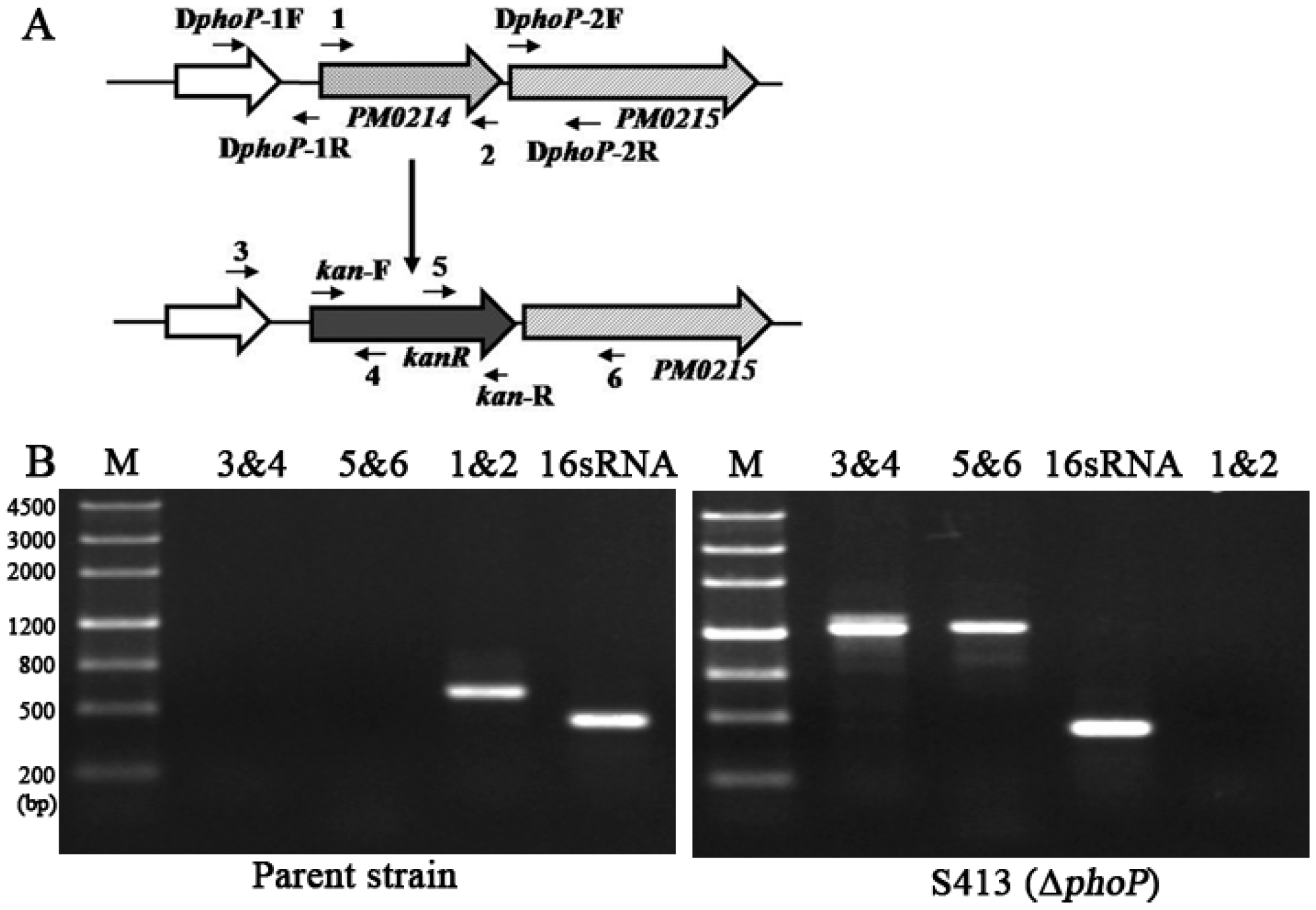
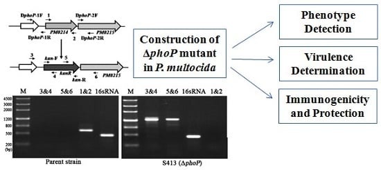
2.1.2. Construction of the Non-Polar ΔphoP Mutant in P. multocida 0818

To determine the roles of the phoP gene in P. multocida 0818, a P. multocida ΔphoP mutant named S413 was constructed through suicide plasmid-mediated homologous recombination. The target phoP (PM0214) gene was replaced with the kanR (kanamycin resistance) cassette by single-crossover recombination (Figure 2A). The mutant was selected and purified by kanamycin pressure and was confirmed by PCR amplification with three primer pairs (1&2, 3&4 and 5&6) (Figure 2A). As shown in Figure 2B, the fragment containing phoP upstream sequence and partial kanR cassette sequence, or phoP downstream sequence and partial kanR cassette sequence, was amplified from strain S413 but not the parent strain (P. multocida 0818), and the complete phoP gene was only amplified from the parent strain. The positive-control 16S rRNA gene was present in both strains. Thus, the phoP gene was successfully deleted in S413.

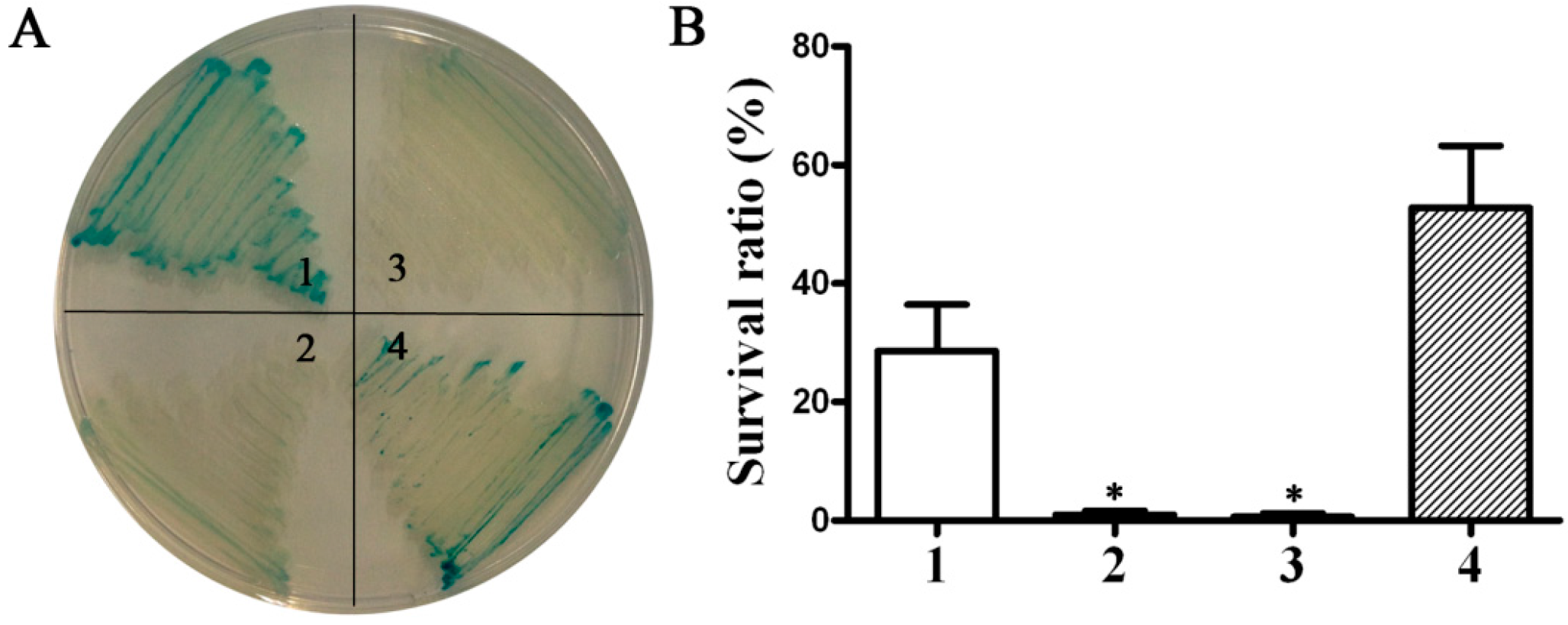
2.1.3. Phenotype of the ΔphoP Mutant Strain

To evaluate effects of the phoP mutation on the P. multocida phenotypes, the growth curve, the OMPs and LPS profiles and the resistance to polymyxin B were measured. Strain S413 (P. multocida ΔphoP) and the parent strain showed similar growth curves with a short lag phase (0–2 h) and a log phase where major bacterial growth occurred (2–10 h) followed by a stationary phase (10–14 h) (Figure 3A). The OMP and LPS bands were similar in both strains and were mainly composed of proteins larger than 25 kDa and short-length LPS, respectively (Figure 3B,C); however, subtle differences in OMPs or LPS might be present that can not detected by the Coomassie blue or silver staining. In contrast, the polymyxin B resistance significantly decreased in the absence of phoP. The survival ratio of strain S413 after treatment with polymyxin B was significantly lower than that of the parent strain and the complement strain, S413 with pQK180 carrying the phoP gene of P. multocida (Figure 3D). The successful complementation by pQK180 also demonstrated the non-polar phoP mutation.
Figure 3. Phenotype characterization of the P. multocida ΔphoP mutant. (A) The growth curve of the parent strain and the ΔphoP mutant. P. multocida were cultured in brain heart infusion (BHI) broth supplemented with or without kanamycin, and then the OD600 value of each strain was measured every 2 h over a period of 14 h; (B,C) The OMP and LPS profiles of P. multocida. OMPs or LPS were extracted from the parent strain or the ΔphoP mutant and were then subjected to sodium dodecyl sulfate polyacrylamide gel electrophoresis (SDS-PAGE). Next, Coomassie blue staining and silver staining were applied to visualize the OMPs (B) and LPS (C), respectively. M refers to the protein Marker; (D) The resistance to polymyxin B. The parent strain, S413 and the complement stain S413 (pQK180) were cultured in BHI broth with or without 0.5 μg/mL polymyxin B for 1 h, and the survival ratio of each strain was calculated as the mean CFU of the polymyxin B-treated group divided by the mean CFU of the untreated group. The data in D are expressed as the mean ± SD and were analyzed at the significance level of 0.05 (*).
Ijms 17 00012 g003

2.1.4. Virulence of Wild-Type P. multocida and the ΔphoP Mutant

To determine the effect of the phoP mutation on bacterial virulence, the 50% lethal dose (LD50) of the parent strain and S413 (P. multocida ΔphoP) were measured in a duck animal model. Ducklings were inoculated with gradual doses of P. multocida via the intranasal and oral routes. The LD50 of S413 was 1.33 × 10⁷ CFU via the intranasal route, which was approximately 154-fold higher than the parent strain LD50 of 8.66 × 10⁴ (Table 1). The LD50 of S413 was 1.56 × 10⁸ CFU via the oral route, which was approximately 32-fold higher than the parent strain LD50 of 4.87 × 10⁶ (Table 1). These results indicate that the phoP deletion decreased the virulence of P. multocida 0818.
Table 1. Determination of the LD50 of P. multocida 0818 and the ΔphoP mutant.
RouteStrainsInfection Dose (CFU) and SurvivalLD50
103104105106107108109
IntranasalP. multocida 08185/57/88/161/160/160/80/88.66 × 104
S413 (ΔphoP)8/88/87/812/169/165/160/81.33 × 107
OralP. multocida 08188/88/812/165/160/130/84.87 × 106
S413 (ΔphoP)8/88/88/811/137/131/131.56 × 108
–, not detected.

2.1.5. Identification of Genes Regulated by phoP in P. multocida

Transcriptome sequencing was applied to search for genes regulated by phoP in P. multocida. Compared to the parent strain, 334 genes were differentially expressed in the S413 (ΔphoP) strain during the exponential growth stage, of which 161 genes were up-regulated and 173 genes were down-regulated (Supplementary Material, Table S1). The genes with a greater than four-fold difference in transcription between the parent strain and the ΔphoP mutant are listed in Table 2. Down-regulated genes with more than a four-fold difference were classified into several groups, including metabolism genes (aspA, deaD, PM0751, rnc, resA, trmD and eptA), membrane protein genes (PM0803, PM1164 and tonB), infB (translation), nusA (transcription), dnaK (RNA degradation) and PM1295 (unknown function). The genes that were up-regulated more than four-fold included comF (competence gene), PM0213 (membrane protein), stress reaction genes (cspA and lexA), PM1891 (mineral absorption), def (peptide deformylase), impA (type VI secretion protein), rraA (regulator of ribonuclease activity), acpP (acyl carrier protein, involved in fatty acid biosynthetic process) and PM1316 (unknown function). KEGG enrichment analysis showed that the regulated genes were significantly involved in five pathways, including bacterial secretion, RNA degradation, protein export, nitrogen metabolism and ribosome (Supplementary Material, Figure S1).
Table 2. Partial list of differentially expressed genes between the parent strain and the ΔphoP mutant.
GeneID aLocusDescriptionFold Change (log2)
Genes up-regulated in strain S413 (ΔphoP)
1244903comFcompetence protein ComF18.25
1243560PM0213membrane protein11.79
1244002cspAcold-shock protein5.98
1244528lexArepressor LexA5.73
1245238PM1891hypothetical protein5.39
1244663PM1316hypothetical protein4.66
1244906defpeptide deformylase, partial4.66
1244239impAtype VI secretion protein ImpA4.29
1244515rraAribonuclease activity regulator protein RraA4.06
1245264acpPMULTISPECIES: acyl carrier protein4.00
Genes down-regulated in strain S413 (ΔphoP)
1244450aspAaspartate ammonia-lyase9.92
1244459deaDRNA helicase7.62
1244098PM0751DNA glycosylase7.16
1244150PM0803TonB-dependent receptor5.28
1243408rncribonuclease III5.24
1243794resAPasteurella ResA protein5.03
1244644trmDtRNA (guanine-N1)-methyltransferase4.96
1244642PM1295hypothetical protein4.66
1244511PM1164membrane protein4.66
1244107nusApeptidase M544.53
1244535tonBcell envelope protein TonB4.47
1244106infBtranslation initiation factor IF-24.29
1244083dnaKmolecular chaperone DnaK4.14
1244389eptAsulfatase4.11
a NCBI accession number of the identified gene.

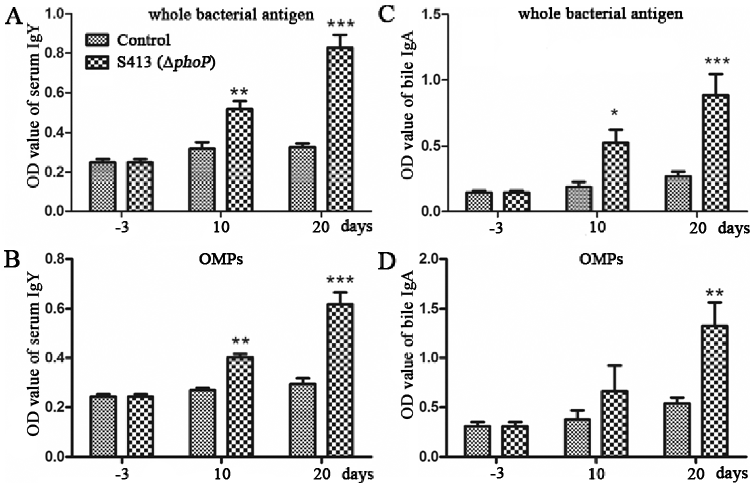
2.1.6. Evaluation of the Immune Responses and Protection Rate Conferred by ΔphoP Mutant

To evaluate protective efficacy of strain S413 (ΔphoP), one-week-old ducks were immunized with 1 × 10⁵ CFU of S413 (ΔphoP) orally twice at an interval of 10 days. Then, ducks were challenged with approximately 200 × LD50 of the wild-type P. multocida 0818 at Day 10 after the second immunization (Table 3). The serum IgY and bile IgA responses specific to whole-bacterial antigen and OMPs were measured 3 days before immunization and 10 and 20 days post-immunization by indirect ELISA. No serum IgY or bile IgA responses specific to P. multocida antigens were present before immunization (Figure 4). In contrast, immunized ducks produced strong serum IgY and bile IgA antibodies against whole-bacterial antigen, both of which were significantly higher than that of the control ducks (Figure 4A,B). The serum IgY and bile IgA responses specific to P. multocida OMPs were significantly higher in the vaccinated group than in the control group (Figure 4C,D). In the immunized group, 54.5% of the ducks survived after challenge with 200 × LD50 of the wild-type P. multocida 0818, and all control ducks were dead within one week (Table 3), indicating that the ΔphoP mutant is a putative vaccine candidate that could provide moderate protection against P. multocida infection.
Figure 4. The antibody responses induced by the ΔphoP mutant in ducks. Ducks were immunized with 1 × 10⁵ CFU of ΔphoP mutant orally twice at an interval of 10 days. Then, the serum IgY responses specific to whole-bacterial antigen (A) and OMPs (B) and the bile IgA responses specific to whole-bacterial antigen (C) and OMPs (D) were measured by indirect ELISA. The data in B are expressed as the mean value ± SD and were analyzed with the significance levels set at 0.05 (*), 0.01 (**) and 0.001 (***).
Ijms 17 00012 g004
Table 3. Protection rate conferred by the ΔphoP mutant.
GroupImmunizationChallengeSurvivalProtection
Immune group105 CFU of S413 (ΔphoP)109 CFU of P. multocida 08186/1154.5%
ControlPBS109 CFU of P. multocida 08180/80

2.2. Discussion

P. multocida is associated with a wide range of diseases in many species of animals. Determination of the roles of its global regulators will promote the understanding of bacterial survival and evasion in the host and will contribute to the development of vaccines. Recently, several global regulators have been identified in Pasteurella, such as Fis (nucleoid-associated protein) [25], Fur (ferric uptake regulation) [26,27] and FnrP [28,29], all of which are associated with bacteria virulence. PhoP and PhoQ constitute a two-component system that senses exterior signals and plays vital roles in bacterial virulence [30]. In this study, we found that the PM0214 gene (phoP2) of P. multocida performs phoP functions. Three main lines of evidence support this conclusion: (1) The P. multocida PM0214 gene has the ability to restore the resistance of a S. Typhimurium ΔphoP mutant to polymyxin B (Figure 1B); (2) The P. multocida PM0214 gene exhibited alkaline phosphatase activity (Figure 1A); (3) The PM0214 and PM0215 (NCBI Gene ID: 1243562) genes constitute an operon sharing the same promoter (Figure 2), and PM0215 (NCBI protein ID, WP_010906530.1) is an inner membrane protein sharing 45% similarity with PhoQ of Salmonella, which is the cognate sensor of PhoP. Therefore, the PM0214 gene of P. multocida is a phoP gene, which was interchangeable with phoP of Salmonella. In addition, the protein encoded by PM0432 (phoP1) shared only 27.23% identity to Salmonella PhoP and did not exhibit any alkaline phosphatase activity, which indicates of PhoP activity [31]. However, the protein shared approximately 60% identity to E. coli PhoB, which responds to phosphate starvation in E. coli. Hence, phoP1 might be the phoB gene of P. multocida. Considering the relationship between phoP mutation and virulence, we focused this study on the phoP gene of P. multocida.
The virulence of the S413 strain (ΔphoP) decreased 32- and 154-fold after oral and intranasal inoculation, respectively (Table 1). The reduction level of the P. multocida ΔphoP mutant was much lower than that of the Salmonella ΔphoP mutant, whose virulence was reduced by five orders of magnitude [32]. Similar results have been observed for the phoP mutation in Yersinia pseudotuberculosis [33], indicating that PhoP does not always have significant impacts on virulence. These results suggest that there are species-specific virulence aspects of phoP or that other genes in P. multocida may compensate for the adverse effects of a phoP deletion. The LD50 of P. multocida inoculated via the oral route was higher than that in the intranasal route, which was consistent with a previous study [34].
Global regulators are capable of regulating hundreds of genes essential for the virulence [30]. In this study, the phoP gene influenced the transcription of 334 genes, including 161 up-regulated genes and 173 down-regulated genes in P. multocida. Notably, three iron uptake genes, tonB, exbD and PM0803 (TonB-dependent receptor) were down-regulated in the absence of the phoP gene (Table S1). A previous report has demonstrated that tonB and exbD are necessary for the virulence of P. multocida cells and contribute equally to the infectious process, indicating that tonB and exbD down-regulation might partially contribute to the decreased virulence of the ΔphoP. The phoP gene was also involved in the down-regulation of some global regulators, such as csrA and fis, and the up-regulation of regulators such as mraZ (Table S1). CsrA, a post-transcriptional regulator, directly activates genes necessary for pedestal formation in enteropathogenic E. coli and for the type III secretion system in P. aeruginosa [35]. Fis is involved in the transcriptional regulation of genes encoding certain virulence factors in P. multocida [25], Erwinia chrysanthemi [36], E. coli [37] and Salmonella [38]. Overproduction of E. coli MraZ inhibits cell division and is lethal in rich medium [39]. Therefore, down-regulated CsrA and Fis and up-regulated MraZ in the P. multocida ΔphoP mutant may also partially account for its decreased virulence. Besides, phoP deletion down-regulated five membrane protein genes (Table S1). Salmonella PhoP/Q promotes the bacteria resistance to antimicrobial peptides by activating the synthesis of unique outer membrane proteins [40], indicating that these genes may be essential for P. multocida resistance to polymyxin B. In addtion, fourteen membrane protein genes, such as PM0213, an outer membrane protein with an 11.79-fold increased transcription level, were up-regulated in the ΔphoP mutant, which prompted us to investigate the function and immunogenicity of the fourteen proteins.
The P. multocida ΔphoP mutant stimulated strong serum IgY and bile IgA responses in ducks, indicating that ΔphoP mutant was a good vaccine candidate. Because the P. multocida ΔphoP mutant was not fully attenuated, low doses of this mutant were used to immunize ducks. Therefore, this mutant only provided approximately 55% protection against a challenge with 200 × LD50 of the P. multocida virulent strain (Table 3), which was a lower protective effect than was observed for the Salmonella phoP mutant [41,42] or the Mycobacterium tuberculosis phoP mutant [43]. Other mutations need to be introduced into the phoP mutant to achieve full attenuation for vaccine development. Our study provides a foundation to explore the mechanism of phoP-regulated genes and a platform for P. multocida attenuated vaccine development.

3. Experimental Section

3.1. Materials and Methods

3.1.1. Bacterial Strains, Plasmids, Media and Growth Conditions

The bacterial strains and plasmids used in this study are listed in Table 4. The P. multocida strains were grown in BHI broth (BD Bioscience, San Jose, CA, USA). S. Typhimurium and E. coli were grown in LB broth. DAP was added to the LB broth at a final concentration of 50 μg/mL for growth of the E. coli and S. Typhimurium asd mutant strains. Solid media were prepared by the addition of 1.5% agar. When required, the media were supplemented with kanamycin (50 μg/mL) or chloramphenicol (25 μg/mL for S. Typhimurium and E. coli, 2.5 μg/mL for P. multocida). The plasmids were transformed into the bacterial strains by electroporation. Notably, Salmonella Δasd strains and Asd+ plasmids were selected to identify the putative phoP gene of P. multocida. The asd gene encoding Asd is required for synthesis of DAP, an essential component for biosynthesis of cell walls of Gram bacteria. The S. Typhimurium Δasd strain will undergo lysis in the absence of DAP unless exogenous Asd+ plasmid is retained in bacteria or DAP is provided [23,24]. Thus, the Asd+ plasmid carrying phoP gene can be stably maintained in S. Typhimurium Δasd strains without antibiotic pressure post-transformation.
Table 4. Bacterial strains and plasmids used in the study.
Strain or PlasmidDescriptionSource
Strains
S184S. Typhimurium Δasd66Lab collection
S412S. Typhimurium Δasd66 ΔphoP85Lab collection
P. multocida 0818Wild type P. multocida serotype A strain. Capsulated and virulentLab collection
S413P. multocida 0818 ΔphoP::kanRThis work
χ7232E. coli K-12, endA1 hsdR17 (rK-, mK+) glnV44 thi-1 recA1 gyrA relA1 Δ(lacZYA-argF)U169 λpir deoR80dlac Δ(lacZ)M15)[39]
χ7213E. coli K-12, thi-1 thr-1 leuB6 glnV44 fhuA21 lacY1 recA1 RP4-2-Tc::Mu λpirasdA4zhf-2::Tn10[39]
Plasmids
pQK664Asd+ Ptrc pSC101 origin replicationp15a, asd+, spec rDerived from pYA3337 [36]
pQK167Insertion of phoP1 into pQK664This work
pQK168Insertion of phoP2 into pQK664This work
pYA4278pRE112 derivative, sacB mobRP4 R6K ori Cm+[38]
pQK171pYA4278-ΔphoPThis work
pQK173pYA4278-ΔphoP::kanR, for deletion of phoP in P. multocidaThis work
pMC-ExpressA broad host-range shuttle vector derived from pMIDG100, chloramphenicol r[40]
pQK180Insertion of phoP into pMC-ExpressThis work
r antibiotic resistance.

3.1.2. Molecular and Genetic Procedures

DNA was amplified using the PrimeSTAR Max DNA polymerase (Takara Bio Inc., Shiga, Japan) or Taq DNA polymerase (Tiangen Biotech Co., Ltd., Beijing, China). The primers used in this study are listed in Table 5. When required, PCR products were purified using the Universal DNA Purification Kit (Tiangen Biotech) and commercially sequenced by BGI Tech (BGI Tech Solutions Co., Ltd., Shenzhen, China). Plasmids were restriction-digested and ligated according to the manufacturers’ instructions using enzymes obtained from NEB and TAKARA, respectively, and the plasmids were prepared using the TIANprep Mini Plasmid Kit (Tiangen Biotech).
Table 5. Primers used in the study.
PrimersSequence 5’-3’
CphoP1-FGCATGCCATGGAGGTACAAATGACCAAAAT
CphoP1-RCGCGGATCCATACTTAATTGAAAAGGTTC
CphoP2-FGCATGCCATGGCGGTCAAAAAAATGCGAAT
CphoP2-RCGCGGATCCACGTGAAAATTAAACGAACC
CphoP-FcgcGGATCCatgcgaattttattaatagaag
CphoP-RataagaatGCGGCCGCccgtaaacttgtttgcttaagc
DphoP-1FTGCCAGTTTTCAATGGTGTC
DphoP-1RCCTGCAGGGATGCGGCCGCTTTTTTGACCGCACTTTTTTC
DphoP-2FGCGGCCGCATCCCTGCAGGGCTAGGAAAAAATGATGAAATG
DphoP-2RATTTTCCTTGATTGACTGGC
Kan-FATAAGAATGCGGCCGCTCAGTGGAACGAAAACTC
Kan-RCCTGCAGGTTAGAAAAACTCATCGAGCATC
Primer 1TTAATTGGCAATGGCTTACAG
Primer 2TACCCTACACCATGCACAGT
Primer 3TATTACACACGTTATAACCCG
Primer 4CTGATTCAGGTGAAAATATTG
Primer 5CATTTGTGCAACTTCAGTTTG
Primer 6CAATATTTTCACCTGAATCAG
16sRNA-FTAATACCGCGTATTCTCTGAGG
16sRNA-RCCCTCCCTAAAGTACTCTAGAC

3.1.3. Plasmid and Mutant Strain Construction

To clarify the function of the phoP gene of P. multocida, two pairs of primers, CphoP1-F/CphoP1-R and CphoP2-F/CphoP2-R, were used to amplify phoP1 and phoP2 from the P. multocida 0818 genome, respectively. The amplified DNA fragments and the Asd+ plasmid pQK664 derived from pYA3337 [44] were digested with NcoI and BamHI, and then the digested phoP1 and phoP2 PCR products were inserted into pQK664 to generate pQK167 and pQK168, respectively.
A modification of the previously described single-crossover insertion mutagenesis method [45], which utilizes the λ pir-dependent plasmid pUA826, was applied for phoP deletion in P. multocida. To generate a non-polar mutation, a different suicide plasmid, T-vector pYA4278 [46] was used. In brief, the upstream and downstream fragments (approximately 450 bp) of the phoP gene were amplified from P. multocida 0818 genome with two pairs of primers, DphoP-1F/DphoP-1R and DphoP-2F/DphoP-2R (Figure 2A), respectively. The two fragments were then joined by PCR using the primers DphoP-1F and DphoP-2R. The terminal A was added at both ends to the resulting PCR product by using the Tailing-A Reaction Kit (Tiangen Biotech). The suicide plasmid T-vector pYA4278 was digested with AhdI to generate a linear T-vector, and the PCR product was ligated to this T-vector to generate the plasmid pQK171. Next, the kanR cassette amplified with the primers kan-F/kan-R was inserted into pQK171 between the NotI and SbfI sites to generate the plasmid pQK173. This plasmid was then mobilized from E. coli χ7213 [47] into P. multocida 0818 strain by conjugation, and the ΔphoP mutant termed S413 was selected on BHI agar containing 50 μg/mL kanamycin. Finally, the selected mutant was confirmed by PCR using primers 1, 2, 3, 4, 5 and 6, which flanked the genomic sequence and the kanR cassette. As a positive control, the 16S ribosomal RNA gene was amplified with the primers 16sRNA-F/16sRNA-R, which were designed according to the NCBI sequence JX869945.1.
To complement the phoP mutant in P. multocida, the phoP gene sequence was amplified from P. multocida 0818 genomic DNA using the primers CphoP-F/CphoP-R containing NotI and BamHI sites. The amplified fragment was digested and cloned into NotI-BamHI-digested pMC-Express (kindly provided by Paul R Langford from Imperial College London) [48], generating pQK180. The plasmids pQK167 and pQK168 were separately transformed into S412 (S. Typhimurium Δasd ΔphoP), and pQK180 was transformed into S413 (P. multocida ΔphoP), generating the corresponding complementary strains S412 (pQK167), S412 (pQK168) and S413 (pQK180).

3.1.4. Phosphatase Activity and Polymyxin B Resistance Assays

To detect phosphatase activity, X-P plates were obtained by addition of 5-bromo-4-chloro-3-indolyl-phosphate (BCIP) (Sigma-Aldrich, St. Louis, MO, USA) to LB agar at a final concentration of 50 μg/mL [31]. Four S. Typhimurium strains, S184 (Δasd) harboring the control plasmid pQK664, S412 (Δasd ΔphoP) harboring pQK664, S412 harboring pQK167 and S412 harboring pQK168, were grow at 37 °C on X-P plates for 18 h, and the colony color was observed.
To detect the resistance to polymyxin B, bacteria were grown to an OD600 of 0.8 in LB broth (S. Typhimurium) or BHI broth (P. multocida), harvested, and washed in PBS. The cells were diluted to 1 × 10⁶ CFU and were cultured at 37 °C in culture media supplemented with or without polymyxin B (0.1 μg/mL for S. Typhimurium and 0.5 μg/mL for P. multocida) for 1 h [49]. Then, the cells were 10-fold serially diluted and 100 μL of the diluted suspension was spread on LB agar or BHI agar to determine the number of bacteria. Percent survival was calculated as the mean CFU of the polymyxin B-treated group divided by the mean CFU of the untreated group.

3.1.5. Phenotype Determinations

The P. multocida 0818 and S413 (P. multocida ΔphoP) stains were grown in BHI broth, and the OD600 values were recorded every 2 h over a period of 14 h to generate the growth curves.
LPS of P. multocida was prepared as previously described [50]. P. multocida was grown in BHI broth overnight, harvested and washed three times using distilled water. The cells were re-suspended in 150 µL of sample buffer (0.5 M Tris–HCl pH 6.8, 2% SDS, 0.05% ethyl alcohol, 10% glycerol, 5% β-mercaptoethanol), boiled for 10 min, and centrifuged at 10,000× g for 10 min. Ten microliters of the supernatant was mixed with 90 µL of loading buffer and 1 µL of proteinase K (20 mg/mL, Sigma-Aldrich), followed by incubation for 1 h at room temperature. Next, 15 µL of the mixture was subjected to 12.5% SDS-PAGE, and the SDS-PAGE gels were visualized using a standard silver-staining protocol [51].
The OMPs of P. multocida were extracted as previously described [52]. P. multocida was harvested after growth to an OD600 of 0.8, and the pellet was re-suspended in 2 mL of HEPES buffer (10 mM, pH 7.4) on ice. The cells were disrupted by sonication (six bursts, 10 s each), and the unbroken cells were removed by centrifugation (15,600× g, 2 min, 4 °C). The supernatant containing the OMPs was transferred into a new tube and centrifuged again (15,600× g, 30 min, 4 °C). The membrane pellet was re-suspended in 0.2 mL of HEPES buffer. To solubilize the cytoplasmic membrane, 0.2 mL of 2% sarcosyl was added and incubated at room temperature for 30 min with constant shaking. After centrifugation (15,600× g, 30 min, 4 °C), the pellet containing the OMPs was washed once with 0.5 mL of HEPES buffer and re-suspended in 50 µL of HEPES buffer. The OMP concentration was measured using a BCA Protein Assay Kit (Thermo Scientific, Rockford, IL, USA). Next, the protein samples were prepared in sample buffer (50 mM Tris, 5% β-mercaptoethanol, 20% glycerol, 0.005% bromophenol blue, 4% SDS) and boiled at 95 °C for 5 min. The samples were analyzed by 12.5% SDS-PAGE and were subjected to staining with Coomassie Brilliant Blue R-250 (Sigma-Aldrich) for protein band detection.

3.1.6. Determination of LD50 in Ducks

All experiments with animals in this study were performed in strict accordance with the recommendations in the Guide for the Care and Use of Laboratory Animals of the Ministry of Science and Technology of China. All animal procedures were approved by the Animal Ethics Committee of the Sichuan Agricultural University (Approval No. 2015-015, Approval date: January 2015).
One-day-old Sheldrake ducks were purchased from a hatchery and acclimated for 7 days after arrival before starting the experiments. For determination of the 50% lethal dose (LD50), the P. multocida strains were grown statically overnight at 37 °C in BHI broth, diluted 1:100 in fresh medium and grown with aeration (180 rpm) at 37 °C. When the cultures reached an OD600 of 0.8–0.9, they were harvested at room temperature at 4000 rpm, washed in PBS, and suspended in PBS. Groups of 1-week-old ducks (5–16 ducks per group) were infected orally with 500 μL of PBS containing various doses of P. multocida 0818 or S413 (ΔphoP) ranging from 1 × 10⁴ to 1 × 10⁹ CFU. Meanwhile, groups of 2-week-old ducks were infected intranasally with 100 μL of PBS containing various doses of P. multocida 0818 or S413 (ΔphoP) strains, ranging from 1 × 10³ to 1 × 10⁹ CFU. The animals were observed for 2 weeks post-infection, and deaths were recorded daily. The LD50 was calculated with the method of Reed and Muench.

3.1.7. Transcriptome Sequencing

RNA sequencing was performed on an Illumina HiSeq2500/MiSeq instrument (Illumina, San Diego, CA, USA) by Shanghai Majorbio Bio-pharm Biotechnology Co., Ltd. (Shanghai, China). For preparation of the samples, the P. multocida 0818 and S413 (ΔphoP) strains were cultured in BHI broth to an OD600 of 0.8, and the total RNA was isolated from each sample with Trizol Reagent (Invitrogen, Carlsbad, CA, USA). The ribosomal RNA was removed from RNA samples before construction of the cDNA library and Illumina deep sequencing.
For sequence analysis, we chose the published sequence of P. multocida PM70 as the reference genome sequence [53], and the clean reads from each sample were mapped to isogenes using Bowtie 2 software [54]. The number of mapped reads per kilobase of exon per million mapped reads (RPKM) was used as the transcription activity of each gene. The differentially expressed genes were analyzed using RSEM and edgeR software [55,56]. The RPKM of genes showing a ≥2-fold intensity change between P. multocida 0818 and S413 with a false discovery rate (FDR) of <0.5 were considered differentially expressed.

3.1.8. Immunization and Challenge

Seventeen 1-week-old commercial Sheldrake ducks were inoculated with 500 μL of PBS containing 1 × 10⁵ CFU of S413 orally on Day 0 and were boosted on Day 10 with the same dose of the same strain. Another 14 ducks were inoculated with 500 μL of PBS as unimmunized controls. Blood and bile were collected from three randomly selected ducks in both groups on Day –3, Day 10, and Day 20. Blood was allowed to coagulate at 37 °C for 2 h. Following centrifugation, the serum was removed and stored at −80 °C. For the challenge experiment, the remaining immunized (11 ducks) and control ducks (8 ducks) were challenged orally with 500 μL of PBS containing 1 × 10⁹ of P. multocida strain 0818. The animals were observed for 2 weeks post-challenge, and deaths were recorded daily. Necropsy and bacterial isolation were carried out routinely after death.

3.1.9. Enzyme-Linked Immunosorbent Assay (ELISA)

The serum IgY and bile IgA responses were measured by indirect ELISA as previously described [34]. P. multocida strain 0818 was inactivated by heating at 80 °C for 10 min in a water bath, and the nonviable bacteria were washed in saline and suspended to approximately 1 × 10¹¹ CFU/mL (as determined by viable counts prior to boiling). A 1:100 dilution of the whole-cell antigen or 0.25 μg/mL purified OMPs were prepared in carbonate-bicarbonate buffer (pH 9.6), and 100 μL of antigen was added to each well of a 96-well ELISA microtiter plate (Nunc-immuno MaxiSorp plate, Nunc, Roskilde, Denmark). The plate was incubated at 4 °C overnight. After washing and blocking, the plate was incubated with 1:160 diluted serum or 1:40 diluted bile for 1 h at 37 °C. Then, 100 μL of 1:5000 diluted alkaline phosphatase (AP)-labeled mouse anti-duck IgY or IgA (AbD Serotec, Puchheim, Germany) was added to the wells, and the plate was incubated at 37 °C for 1 h followed by four washes. AP solution (Sigma-Aldrich) was added to the wells for 20 min. The reaction was stopped by the addition of 100 μL of 0.2 M NaOH. The plate was read at 415 nm by an ELISA reader (Bio-Rad Laboratories, Richmond, CA, USA).

3.1.10. Statistical Analyses

Statistical analysis was performed using the GraphPad Prism 5 software package (Graph Software, San Diego, CA, USA). Data are expressed as the means ± SD and were analyzed using Student’s t-test at significance levels of 0.05, 0.01 and 0.001. The animal experiments were repeated at least twice, and the in vitro experiments were performed independently three times in triplicate. The representative results are presented in the study.

4. Conclusions

In this study, we identified the phoP gene in P. multocida and constructed and characterized a P. multocida ΔphoP mutant. Compared to the parent strain, this mutant is more sensitive to polymyxin B, exhibits lower virulence in ducks, and provides moderate immune protection against challenge with virulent P. multocida strains.

Supplementary Materials

Supplementary materials can be found at https://www.mdpi.com/1422-0067/17/1/12/s1.

Acknowledgments

We are thankful to Paul R Langford for providing the pMC-Express plasmid, and Dekang Zhu for providing the wild-type P. multocida 0818, and Anchun Cheng, Mingshu Wang and Kunfeng Sun for providing animal housing. This research was supported by the Foundation of Educational Commission of Sichuan Province of China (15ZA0018), the Special Foundation for Young Scientists of Sichuan Province (2014JQ0006) and the Applied Basic Research Programs of Science and Technology Department of Sichuan Province (2015JY0244).

Authors Contributions

Kangpeng Xiao carried out most of the experimental work, and Xinxin Zhao and Kangpeng Xiao drafted the manuscript. Qing Liu, Xueyan Liu and Yunlong Hu assisted in the animal experiments and assays. Xinxin Zhao and Qingke Kong secured funding and analyzed and interpreted the data. Qingke Kong designed this study and revised the manuscript. All authors read and approved the final manuscript.

Conflicts of Interest

The authors declare that they have no competing interests.

References

  1. Harper, M.; Boyce, J.D.; Adler, B. Pasteurella multocida pathogenesis: 125 years after pasteur. FEMS Microbiol. Lett. 2006, 265, 1–10. [Google Scholar] [CrossRef] [PubMed]
  2. Chrzastek, K.; Kuczkowski, M.; Wieliczko, A.K.; Bednarek, K.J.; Wieliczko, A. Molecular epidemiologic investigation of polish avian pasteurella multocida strains isolated from fowl cholera outbreaks showing restricted geographical and host-specific distribution. Avian Dis. 2012, 56, 529–536. [Google Scholar] [CrossRef] [PubMed]
  3. San Millan, A.; Escudero, J.A.; Gutierrez, B.; Hidalgo, L.; Garcia, N.; Llagostera, M.; Dominguez, L.; Gonzalez-Zorn, B. Multiresistance in pasteurella multocida is mediated by coexistence of small plasmids. Antimicrob. Agents Chemother. 2009, 53, 3399–3404. [Google Scholar] [CrossRef] [PubMed]
  4. Ahmad, T.A.; Rammah, S.S.; Sheweita, S.A.; Haroun, M.; El-Sayed, L.H. Development of immunization trials against pasteurella multocida. Vaccine 2014, 32, 909–917. [Google Scholar] [CrossRef] [PubMed]
  5. Desin, T.S.; Koster, W.; Potter, A.A. Salmonella vaccines in poultry: Past, present and future. Expert Rev. Vaccines 2013, 12, 87–96. [Google Scholar] [CrossRef] [PubMed]
  6. Broset, E.; Martin, C.; Gonzalo-Asensio, J. Evolutionary landscape of the mycobacterium tuberculosis complex from the viewpoint of phopr: Implications for virulence regulation and application to vaccine development. MBio 2015. [Google Scholar] [CrossRef] [PubMed]
  7. Kier, L.D.; Weppelman, R.M.; Ames, B.N. Regulation of nonspecific acid phosphatase in salmonella: Phon and phop genes. J. Bacteriol. 1979, 138, 155–161. [Google Scholar] [PubMed]
  8. Gonzalo Asensio, J.; Maia, C.; Ferrer, N.L.; Barilone, N.; Laval, F.; Soto, C.Y.; Winter, N.; Daffe, M.; Gicquel, B.; Martin, C.; et al. The virulence-associated two-component PhoP-PhoR system controls the biosynthesis of polyketide-derived lipids in mycobacterium tuberculosis. J. Biol. Chem. 2006, 281, 1313–1316. [Google Scholar] [CrossRef] [PubMed]
  9. Perez, J.C.; Groisman, E.A. Transcription factor function and promoter architecture govern the evolution of bacterial regulons. Proc. Natl Acad. Sci. USA 2009, 106, 4319–4324. [Google Scholar] [CrossRef] [PubMed]
  10. Coornaert, A.; Chiaruttini, C.; Springer, M.; Guillier, M. Post-transcriptional control of the escherichia coli PhoQ-PhoP two-component system by multiple sRNAs involves a novel pairing region of GcvB. PLoS Genet. 2013, 9, e1003156. [Google Scholar] [CrossRef] [PubMed]
  11. Zwir, I.; Latifi, T.; Perez, J.C.; Huang, H.; Groisman, E.A. The promoter architectural landscape of the salmonella PhoP regulon. Mol. Microbiol. 2012, 84, 463–485. [Google Scholar] [CrossRef] [PubMed]
  12. Garcia Vescovi, E.; Soncini, F.C.; Groisman, E.A. Mg2+ as an extracellular signal: Environmental regulation of salmonella virulence. Cell 1996, 84, 165–174. [Google Scholar] [CrossRef]
  13. Prost, L.R.; Daley, M.E.; le Sage, V.; Bader, M.W.; le Moual, H.; Klevit, R.E.; Miller, S.I. Activation of the bacterial sensor kinase PhoQ by acidic pH. Mol. Cell 2007, 26, 165–174. [Google Scholar] [CrossRef] [PubMed]
  14. Bader, M.W.; Sanowar, S.; Daley, M.E.; Schneider, A.R.; Cho, U.; Xu, W.; Klevit, R.E.; le Moual, H.; Miller, S.I. Recognition of antimicrobial peptides by a bacterial sensor kinase. Cell 2005, 122, 461–472. [Google Scholar] [CrossRef] [PubMed]
  15. Lejona, S.; Aguirre, A.; Cabeza, M.L.; Garcia Vescovi, E.; Soncini, F.C. Molecular characterization of the Mg2+-responsive PhoP-PhoQ regulon in salmonella enterica. J. Bacteriol. 2003, 185, 6287–6294. [Google Scholar] [CrossRef] [PubMed]
  16. Dalebroux, Z.D.; Miller, S.I. Salmonellae phopq regulation of the outer membrane to resist innate immunity. Curr. Opin. Microbiol. 2014, 17, 106–113. [Google Scholar] [CrossRef] [PubMed]
  17. Lee, J.S.; Krause, R.; Schreiber, J.; Mollenkopf, H.J.; Kowall, J.; Stein, R.; Jeon, B.Y.; Kwak, J.Y.; Song, M.K.; Patron, J.P.; et al. Mutation in the transcriptional regulator PhoP contributes to avirulence of mycobacterium tuberculosis h37ra strain. Cell Host Microbe 2008, 3, 97–103. [Google Scholar] [CrossRef] [PubMed]
  18. Prost, L.R.; Miller, S.I. The salmonellae PhoQ sensor: Mechanisms of detection of phagosome signals. Cell. Microbiol. 2008, 10, 576–582. [Google Scholar] [CrossRef] [PubMed]
  19. Martin, C.; Williams, A.; Hernandez-Pando, R.; Cardona, P.J.; Gormley, E.; Bordat, Y.; Soto, C.Y.; Clark, S.O.; Hatch, G.J.; Aguilar, D.; et al. The live mycobacterium tuberculosis PhoP mutant strain is more attenuated than bcg and confers protective immunity against tuberculosis in mice and guinea pigs. Vaccine 2006, 24, 3408–3419. [Google Scholar] [CrossRef] [PubMed]
  20. Nambiar, J.K.; Pinto, R.; Aguilo, J.I.; Takatsu, K.; Martin, C.; Britton, W.J.; Triccas, J.A. Protective immunity afforded by attenuated, PhoP-deficient mycobacterium tuberculosis is associated with sustained generation of Cd4+ T-cell memory. Eur. J. Immunol. 2012, 42, 385–392. [Google Scholar] [CrossRef] [PubMed]
  21. Lee, H.Y.; Cho, S.A.; Lee, I.S.; Park, J.H.; Seok, S.H.; Baek, M.W.; Kim, D.J.; Lee, S.H.; Hur, S.J.; Ban, S.J.; et al. Evaluation of PhoP and Rpos mutants of salmonella enterica serovar typhi as attenuated typhoid vaccine candidates: Virulence and protective immune responses in intranasally immunized mice. FEMS Immunol. Med. Microbiol. 2007, 51, 310–318. [Google Scholar] [CrossRef] [PubMed]
  22. Wilson, B.A.; Ho, M. Pasteurella multocida: From zoonosis to cellular microbiology. Clin. Microbiol. Rev. 2013, 26, 631–655. [Google Scholar] [CrossRef] [PubMed]
  23. Galan, J.E.; Nakayama, K.; Curtiss, R., 3rd. Cloning and characterization of the asd gene of salmonella typhimurium: Use in stable maintenance of recombinant plasmids in Salmonella vaccine strains. Gene 1990, 94, 29–35. [Google Scholar] [CrossRef]
  24. Tacket, C.O.; Kelly, S.M.; Schodel, F.; Losonsky, G.; Nataro, J.P.; Edelman, R.; Levine, M.M.; Curtiss, R., 3rd. Safety and immunogenicity in humans of an attenuated Salmonella typhi vaccine vector strain expressing plasmid-encoded hepatitis B antigens stabilized by the asd-balanced lethal vector system. Infect. Immun. 1997, 65, 3381–3385. [Google Scholar] [PubMed]
  25. Steen, J.A.; Steen, J.A.; Harrison, P.; Seemann, T.; Wilkie, I.; Harper, M.; Adler, B.; Boyce, J.D. Fis is essential for capsule production in Pasteurella multocida and regulates expression of other important virulence factors. PLoS Pathog. 2010, 6, e1000750. [Google Scholar] [CrossRef] [PubMed]
  26. Paustian, M.L.; May, B.J.; Cao, D.; Boley, D.; Kapur, V. Transcriptional response of Pasteurella multocida to defined iron sources. J. Bacteriol. 2002, 184, 6714–6720. [Google Scholar] [CrossRef] [PubMed]
  27. Bosch, M.; Tarrago, R.; Garrido, M.E.; Campoy, S.; Fernandez de Henestrosa, A.R.; Perez de Rozas, A.M.; Badiola, I.; Barbe, J. Expression of the Pasteurella multocida ompH gene is negatively regulated by the Fur protein. FEMS Microbiol. Lett. 2001, 203, 35–40. [Google Scholar] [CrossRef] [PubMed]
  28. Uhlich, G.A.; McNamara, P.J.; Iandolo, J.J.; Mosier, D.A. Cloning and characterization of the gene encoding Pasteurella haemolytica fnrp, a regulator of the Escherichia coli silent Hemolysin shea. J. Bacteriol. 1999, 181, 3845–3848. [Google Scholar] [PubMed]
  29. Uhlich, G.A.; McNamara, P.J.; Iandolo, J.J.; Mosier, D.A. FnrP interactions with the Pasteurella haemolytica leukotoxin promoter. FEMS Microbiol. Lett. 2000, 186, 73–77. [Google Scholar] [CrossRef] [PubMed]
  30. Ryndak, M.; Wang, S.; Smith, I. Phop, a key player in mycobacterium tuberculosis virulence. Trends Microbiol. 2008, 16, 528–534. [Google Scholar] [CrossRef] [PubMed]
  31. Curtiss, R., 3rd; Wanda, S.Y.; Gunn, B.M.; Zhang, X.; Tinge, S.A.; Ananthnarayan, V.; Mo, H.; Wang, S.; Kong, W. Salmonella enterica serovar typhimurium strains with regulated delayed attenuation in vivo. Infect. Immun. 2009, 77, 1071–1082. [Google Scholar] [CrossRef] [PubMed]
  32. Garcia Vescovi, E.; Soncini, F.C.; Groisman, E.A. The role of the PhoP/PhoQ regulon in Salmonella virulence. Res. Microbiol. 1994, 145, 473–480. [Google Scholar] [CrossRef]
  33. Pisano, F.; Heine, W.; Rosenheinrich, M.; Schweer, J.; Nuss, A.M.; Dersch, P. Influence of PhoP and intra-species variations on virulence of yersinia pseudotuberculosis during the natural oral infection route. PLoS ONE 2014, 9, e103541. [Google Scholar] [CrossRef] [PubMed]
  34. Wilkie, I.W.; Grimes, S.E.; O’Boyle, D.; Frost, A.J. The virulence and protective efficacy for chickens of Pasteurella multocida administered by different routes. Vet. Microbiol. 2000, 72, 57–68. [Google Scholar] [CrossRef]
  35. Timmermans, J.; Van Melderen, L. Post-transcriptional global regulation by CsrA in bacteria. Cell. Mol. Life Sci. 2010, 67, 2897–2908. [Google Scholar] [CrossRef] [PubMed]
  36. Lautier, T.; Nasser, W. The DNA nucleoid-associated protein Fis co-ordinates the expression of the main virulence genes in the phytopathogenic bacterium Erwinia chrysanthemi. Mol. Microbiol. 2007, 66, 1474–1490. [Google Scholar] [CrossRef] [PubMed]
  37. Saldana, Z.; Xicohtencatl-Cortes, J.; Avelino, F.; Phillips, A.D.; Kaper, J.B.; Puente, J.L.; Giron, J.A. Synergistic role of curli and cellulose in cell adherence and biofilm formation of attaching and effacing Escherichia coli and identification of Fis as a negative regulator of curli. Environ. Microbiol. 2009, 11, 992–1006. [Google Scholar] [CrossRef] [PubMed]
  38. Kelly, A.; Goldberg, M.D.; Carroll, R.K.; Danino, V.; Hinton, J.C.; Dorman, C.J. A global role for Fis in the transcriptional control of metabolism and type III secretion in Salmonella enterica serovar typhimurium. Microbiology 2004, 150, 2037–2053. [Google Scholar] [CrossRef] [PubMed]
  39. Eraso, J.M.; Markillie, L.M.; Mitchell, H.D.; Taylor, R.C.; Orr, G.; Margolin, W. The highly conserved MraZ protein is a transcriptional regulator in Escherichia coli. J. Bacteriol. 2014, 196, 2053–2066. [Google Scholar] [CrossRef] [PubMed]
  40. Guina, T.; Yi, E.C.; Wang, H.; Hackett, M.; Miller, S.I. A PhoP-regulated outer membrane protease of Salmonella enterica serovar typhimurium promotes resistance to α-helical antimicrobial peptides. J. Bacteriol. 2000, 182, 4077–4086. [Google Scholar] [CrossRef] [PubMed]
  41. Hohmann, E.L.; Oletta, C.A.; Killeen, K.P.; Miller, S.I. PhoP/PhoQ-deleted Salmonella typhi (ty800) is a safe and immunogenic single-dose typhoid fever vaccine in volunteers. J. Infect. Dis. 1996, 173, 1408–1414. [Google Scholar] [CrossRef] [PubMed]
  42. Methner, U.; Barrow, P.A.; Berndt, A.; Rychlik, I. Salmonella enteritidis with double deletion in phopflic—A potential live salmonella vaccine candidate with novel characteristics for use in chickens. Vaccine 2011, 29, 3248–3253. [Google Scholar] [CrossRef] [PubMed]
  43. Aguilar, D.; Infante, E.; Martin, C.; Gormley, E.; Gicquel, B.; Hernandez Pando, R. Immunological responses and protective immunity against tuberculosis conferred by vaccination of Balb/C mice with the attenuated mycobacterium tuberculosis (Phop) so2 strain. Clin. Exp. Immunol. 2007, 147, 330–338. [Google Scholar] [CrossRef] [PubMed]
  44. Torres-Escobar, A.; Juarez-Rodriguez, M.D.; Curtiss, R., 3rd. Biogenesis of yersinia pestis psaa in recombinant attenuated Salmonella typhimurium vaccine (RASV) strain. FEMS Microbiol. Lett. 2010, 302, 106–113. [Google Scholar] [CrossRef] [PubMed]
  45. Cardenas, M.; Fernandez de Henestrosa, A.R.; Campoy, S.; Perez de Rozas, A.M.; Barbe, J.; Badiola, I.; Llagostera, M.; Molecular Microbiology, G. Virulence of Pasteurella multocidarecA mutants. Vet. Microbiol. 2001, 80, 53–61. [Google Scholar] [CrossRef]
  46. Kong, Q.; Yang, J.; Liu, Q.; Alamuri, P.; Roland, K.L.; Curtiss, R., 3rd. Effect of deletion of genes involved in lipopolysaccharide core and O-antigen synthesis on virulence and immunogenicity of Salmonella enterica serovar Typhimurium. Infect. Immun. 2011, 79, 4227–4239. [Google Scholar] [CrossRef] [PubMed]
  47. Roland, K.; Curtiss, R., 3rd; Sizemore, D. Construction and evaluation of a Δcya Δcrp salmonella Typhimurium strain expressing aviasn pathogenic Escherichia coli O78 LPS as a vaccine to prevent airsacculitis in chickens. Avian Dis. 1999, 43, 429–441. [Google Scholar] [CrossRef] [PubMed]
  48. Bosse, J.T.; Durham, A.L.; Rycroft, A.N.; Kroll, J.S.; Langford, P.R. New plasmid tools for genetic analysis of Actinobacillus pleuropneumoniae and other Pasteurellaceae. Appl. Environ. Microbiol. 2009, 75, 6124–6131. [Google Scholar] [CrossRef] [PubMed]
  49. Kawasaki, K.; China, K.; Nishijima, M. Release of the lipopolysaccharide deacylase PagL from latency compensates for a lack of lipopolysaccharide aminoarabinose modification-dependent resistance to the antimicrobial peptide polymyxin B in salmonella enterica. J. Bacteriol. 2007, 189, 4911–4919. [Google Scholar] [CrossRef] [PubMed]
  50. Hitchcock, P.J.; Brown, T.M. Morphological heterogeneity among salmonella lipopolysaccharide chemotypes in silver-stained polyacrylamide gels. J. Bacteriol. 1983, 154, 269–277. [Google Scholar] [PubMed]
  51. Fomsgaard, A.; Freudenberg, M.A.; Galanos, C. Modification of the silver staining technique to detect lipopolysaccharide in polyacrylamide gels. J. Clin. Microbiol. 1990, 28, 2627–2631. [Google Scholar] [PubMed]
  52. Roier, S.; Leitner, D.R.; Iwashkiw, J.; Schild-Prufert, K.; Feldman, M.F.; Krohne, G.; Reidl, J.; Schild, S. Intranasal immunization with nontypeable Haemophilus influenzae outer membrane vesicles induces cross-protective immunity in mice. PLoS ONE 2012, 7, e42664. [Google Scholar] [CrossRef] [PubMed]
  53. May, B.J.; Zhang, Q.; Li, L.L.; Paustian, M.L.; Whittam, T.S.; Kapur, V. Complete genomic sequence of Pasteurella multocida, Pm70. Proc. Natl Acad. Sci. USA 2001, 98, 3460–3465. [Google Scholar] [CrossRef] [PubMed]
  54. Langmead, B.; Salzberg, S.L. Fast gapped-read alignment with bowtie 2. Nat. Method 2012, 9, 357–359. [Google Scholar] [CrossRef] [PubMed]
  55. Li, B.; Dewey, C.N. Rsem: Accurate transcript quantification from RNA-Seq data with or without a reference genome. BMC Bioinform. 2011, 12, 323. [Google Scholar] [CrossRef] [PubMed]
  56. Robinson, M.D.; McCarthy, D.J.; Smyth, G.K. Edger: A bioconductor package for differential expression analysis of digital gene expression data. Bioinformatics 2010, 26, 139–140. [Google Scholar] [CrossRef] [PubMed]

