Review

Characteristics of Antisense Transcript Promoters and the Regulation of Their Activity

1
Department of Animal Genetics, Breeding and Reproduction, College of Animal Science, South China Agricultural University, Guangzhou 510642, China
2
Guangdong Provincial Key Lab of Agro-Animal Genomics and Molecular Breeding, Guangzhou 510642, China
3
Key Lab of Chicken Genetics, Breeding and Reproduction, Ministry of Agriculture, Guangzhou 510642, China
4
Agricultural College, Guangdong Ocean University, Zhanjiang 524088, China
*
Author to whom correspondence should be addressed.
Int. J. Mol. Sci. 2016, 17(1), 9; https://doi.org/10.3390/ijms17010009
Submission received: 19 October 2015 / Revised: 23 November 2015 / Accepted: 16 December 2015 / Published: 23 December 2015
(This article belongs to the Section Biochemistry)

Abstract

:
Recently, an increasing number of studies on natural antisense transcripts have been reported, especially regarding their classification, temporal and spatial expression patterns, regulatory functions and mechanisms. It is well established that natural antisense transcripts are produced from the strand opposite to the strand encoding a protein. Despite the pivotal roles of natural antisense transcripts in regulating the expression of target genes, the transcriptional mechanisms initiated by antisense promoters (ASPs) remain unknown. To date, nearly all of the studies conducted on this topic have focused on the ASP of a single gene of interest, whereas no study has systematically analyzed the locations of ASPs in the genome, ASP activity, or factors influencing this activity. This review focuses on elaborating on and summarizing the characteristics of ASPs to extend our knowledge about the mechanisms of antisense transcript initiation.

1. Introduction

Natural antisense transcripts comprising both coding and non-coding regulatory RNAs are widespread in the mammalian transcriptome. Indeed, it is estimated that at least 22%–40% of genes have an antisense partner [1,2,3]. These antisense transcripts contain sequences that are partially or completely complementary to sense transcripts and can interact with their sense RNAs through these complementary regions [4,5].
Previous studies have revealed many characteristics of natural antisense transcripts, including their classification [6,7,8,9], spatial and temporal expression profiles [10,11,12], regulatory functions and molecular mechanisms [10,13,14,15,16,17,18,19,20,21,22]. Natural antisense transcripts are involved in early embryonic developmental processes, such as cell proliferation [10,23], and cell migration and sprouting [10], as well as in healthy growth and disease. Through direct interaction with sense transcripts or indirect interaction with other targets, natural antisense transcripts have a widespread impact on conventional gene expression at the DNA and genome, transcriptional, post-transcriptional and translational levels [20]. The reported models of action include transcriptional collision [24], DNA methylation, histone modification [25], mRNA stability [10,26,27], endogenous siRNA [28], and mRNA splicing, editing and translation [4,29]. Chromatin modification as a result of ectopic expression of natural antisense transcripts has even been found to be dysregulated in human disease [5,13,23,25,30]. These previous studies indicate that antisense transcripts are tissue specific and may be regulated by promoters in the same manner as sense transcripts.
However, the transcription mechanism of antisense transcripts remains poorly understood. Similar to sense transcription driven by a promoter, antisense transcripts are controlled by antisense promoters (ASPs) that can be recognized by transcription factors (TFs) to form a transcription initiation complex [31]. Therefore, further antisense transcript studies may focus on identifying the regulation of target gene expression by new functional natural antisense transcripts in specific tissues or cells, simultaneously exploring the relationship between certain critical diseases and the regulation mechanisms of natural antisense transcripts themselves. For example, expression of the lymphoid enhancer factor 1 (LEF1) gene is attenuated by a natural antisense transcript that is regulated by the balance between its spliced and unspliced forms [32]. Greater insight through biomedical research into the regulation mechanisms of natural antisense transcripts may provide novel diagnostic labels and drug targets in clinical applications.

2. Location of Antisense Promoters (ASPs)

Identification of the positions of ASPs can help in clarifying their potential regulatory functions and analyzing the transcription start sites (TSSs) and transcriptional mechanisms of natural antisense transcripts [33]. ASPs are generally located on the strand opposite from coding genes [33,34]. The locations of ASPs can be divided into three types relative to the locations of the sense transcript promoters (SPs): in the same gene as the SP (Figure 1A), in genes upstream or downstream of the SP (Figure 1B), or in intergenic regions (Figure 1C). When an ASP is located in the same gene as the SP, it can be present in the 5′-UTR, exons, introns or 3′-UTR of the gene (Figure 1A).
Figure 1. Characteristics of the locations of antisense promoters (ASPs). When an ASP (red reverse dotted arrow) and the sense promoter (SP) (black solid arrow) are located in the same gene (A); the ASP can be located at any position in the 5′-UTR, exons and introns or 3′-UTR. ASPs can also be located in genes upstream or downstream (gene B or C, respectively) of the SP gene (B); or an ASP may be located between two genes, with SP in the same region (green rectangle) (C). In the figure, “/ /” indicates other exons and introns.
Ijms 17 00009 g001

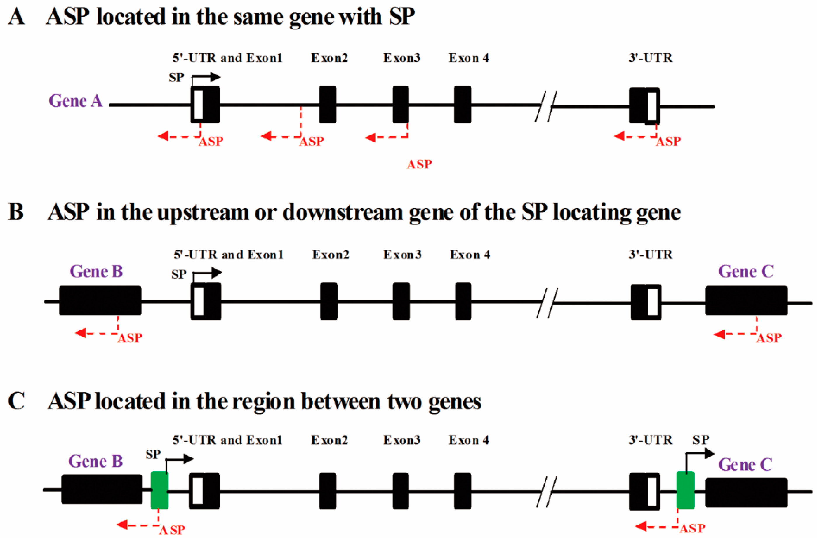
2.1. Both ASPs and Sense Promoters (SPs) Can Be Located in the Same Gene

2.1.1. ASPs and SPs Are Located in the 5′-UTR of the Same Gene

SPs are usually located in the 5′-UTR of a gene, though ASPs can be simultaneously present in the gene’s 5′-UTR. For example, the V1 promoter, the SP of the chicken growth hormone receptor (GHR) gene is located in the 5′-UTR of the gene [35]. Additionally, both the SP and ASP of human long interspersed elements (L1s) are located in the 5′-UTR [3,36,37], and the SP and ASP can compete, leading to six alternative splicing patterns of L1 5′-UTR transcripts (Figure 2) [36,38]. Recently, it has been reported that an L1 primate-specific open reading frame-0 (ORF0), can contribute to the generation of ORF0-proximal exon fusion proteins and enhance L1 mobility (Figure 2) [25]. In addition, the human tryptophan hydroxylase-2 (hTPH2) 5′-UTR harbors a bidirectional promoter; the ASP is much more active than the SP, but both are cell line dependent, with the highest activities observed in Human Embryonic Kidney 293T cell line containing the SV40 Large T-antigen (HEK-293T) and the lowest in a human neuroblastoma cell line (SK-N-MC) [39]. Bidirectional promoters can transcribe both sense transcripts and antisense transcripts, and their TSSs may be located more than 1 kb apart [7,40,41]. Thus, it is clear that an ASP and SP can exist simultaneously in a 5′-UTR.
Figure 2. Schematic representation of the position of L1 ASPs in humans and mice. (A) The L1 5′-UTR (human) harbors an ASP (red reverse arrow) and a SP (black arrow) [36,37]; the mouse L1 ASP (red arrow showed in (B) is within ORF1 [37]. The G-C base rich (GC-rich) region (0–485 bp, black box) plays a role in the activity of the L1 ASP. There are 24 TSSs (transcription start sites) in purple arrow mapped to the opposite strand of the L1 5′-UTR. Interestingly, the L1 ASP functions as an alternative promoter, generating not only antisense transcripts, but also chimeric mRNAs [36]. ORF0 (yellow rectangle), a primate-specific open reading frame-0 located in 236–452 bp, not only contributes to the generation of ORF0-proximal exon fusion proteins but also enhances L1 mobility and there are 24 TSSs of various tissues located in 386–503 bp (purple bracket) [25]. The wright arrows means the transcriptional direction of ORF0, and the black dotted line and connected black boxes represent its transcript alternative splicing; (B) The mouse L1 ASP is located in ORF1, which includes a TATAA sequence at 2689 bp (orange arrow) and the ASP region with the highest activity from 2125 to 2823 bp (green frame) [37]. It has been demonstrated that RNA polymerase II (orange circle) and Dicer (blue circle) participate in the L1 ASP transcriptional initiation complex, with the latter playing a modest role in limiting native L1 retrotransposition. There are also different TSSs located in the L1 ASP, such as brain tissue-specific TSSs located at 2240–2250 bp and 2430–2440 bp (black reverse arrow); a kidney-specific TSS is located at 2320–2330 bp (black reverse arrow). The difference between the L1 ASPs of humans and mice is due to evolutionary events [3].
Ijms 17 00009 g002
SPs and ASPs compete for a portion or all of the elements in the 5′-UTR region and generate typical head-to-head transcripts. Finocchiaro et al. [33] discovered 7072 sense/antisense transcript pairs in the human genome, and found that 76% exhibit a head-to-head formation, whereas the rest are intragenic pairs. Remarkably, 58% of antisense transcripts begin from a 500 bp region upstream of the TSS of protein-coding genes, indicating that both sense and antisense transcripts can be produced from the same promoter element. This phenomenon is characterized by a specialized transcriptional control mechanism that is directly coupled to relaxed bidirectional transcription [42]. Therefore, bidirectional transcription may involve many specific TF binding sites (TFBSs) controlling different directions of SPs and ASPs in an orderly manner.

2.1.2. ASPs Are Located in Exons

ASPs may be located in the exons of a gene when SPs are located in the 5′-UTR. For example, the Dictyostelium discoideum prespore gene EB4-PSV ASP is located in exon 3, driving an antisense transcript that terminates in the SP region [43]. Unlike the human L1 ASP located in the L1 5′-UTR, the rat L1-ASP is located in open reading frame-1 (ORF1) (Figure 2) [37], and the L1-ASPs of many human genes are located in different exons, such as exons 2 and 4, producing different antisense transcripts of neighboring genes [12]. The difference between the location of human and mouse L1-ASP may be a result of the evolution of these species [3]. L1 family members comprise numerous extracellular immunoglobulins and fibronectins, and the gene structure of human L1 family members differs from that of chicken and fruit fly, further revealing the different characteristics of ASPs among species.

2.1.3. ASPs Are Located in Introns

Recent studies have shown that ASPs can be located in different gene introns; however, ASPs do appear to be preferentially located in the first intron, as observed for “initiator” (InR, the ASP of the human eukaryotic initiation factor 2α (eIF-2α) gene) [44], the mouse insulin-like growth factor 2 gene (Igf2) ASP [45] and pIRAIN, which generates an antisense transcript of insulin-like growth factor type I receptor (IGF1R) [46].
Some ASPs can also be located in the second intron, such as those of human BCL-2-related ovarian killer (BOK) [47], a human natural killer cell (NK) I type MHC receptor gene (killer cell immunoglobulin-like receptor surface, KIR) [48] and the murine c-myc gene [49].
ASPs can also be located in the third intron of a gene. For example, the ASP of the PU.1 TF gene is located in intron 3 [50]. Of course, ASPs can be located in other introns. For instance, the ASP of the imprinted Kcnq1 gene is located in intron 11 [51], and the ASP of the L1-COL11A1 (collagen type XI α1, COL11A1) transcript is located in the 46 intron. Previous studies have demonstrated that L1 ASPs can be distributed in various introns in different genes [12,38].
High-throughput sequencing has identified a great number of sense/antisense transcript pairs, yet only some of them exhibit a head-to-head formation, indicating that both directions of many genes fragments can generate transcripts [1,2,3]. Hence, we speculate that there are many ASPs located in the introns of other genes, though they remain to be identified and further studied.

2.1.4. ASPs Are Located in the 3′-UTR

To date, only rare ASPs have been found in the 3′-UTR of genes. The galanin (GAL) gene cluster, including GAL1, GAL7 and GAL10, is a highly regulated galactose-induced genetic unit. GAL7 and GAL10 are tandem genes, and GAL1 and GAL10 are divergent genes; these three genes share a bidirectional promoter, and the GAL10 antisense transcript is initiated within the GAL10 3′-UTR [31]. The antisense transcripts initiated by the ASP in the 3′-UTR influence the transcription initiation of sense transcripts by forming a tail-to-tail pair complement with them. Head-to-head sense/antisense transcript pairs are less common than those showing a tail-to-tail formation, likely because most antisense transcripts (especially those with a tail-to-tail formation) often play a role in post transcriptional regulation [52]. However, most ASPs are either located in the 5′-UTR, or in introns and exons (Figure 3) [33].
Although, the antisense transcripts of different human cell types correspond to exon, intron, promoter and terminator positions [53]. These antisense transcripts more frequently correspond to promoters and exons than they correspond to other positions of a gene. This finding coincides with the results of Finocchiaro et al. [33] who found that antisense transcript start regions (ATSRs) are more likely to be located in intron 1, followed by exon 1, intron 2 and the last intron of protein coding genes. Compared with other exons and introns, the TSSs of antisense transcripts demonstrate a strong tendency to be located in the first exon. Overall, more than half of ATSRs are located in exon 1, intron 1 and intron 2, indicating that ATSRs are distributed at the 5′-end of genes (Figure 3) [33].
Figure 3. The distribution of antisense transcript start regions (ATSRs). Here, we hypothesized that one or more ASPs are located in ATSRs. The pie chart shows the ATSR distribution per genomic element. The distribution was evaluated for RefSeq genes with at least three exons for which it was possible to unambiguously distinguish the first and last introns. Globally, the distribution among 2671 RefSeq genes indicated that 94.3% of 2830 RefSeq genes contain an ATSR [33]. More than 50% of the ASPs are located in exon 1, intron 1 and intron 2.
Ijms 17 00009 g003

2.2. ASPs Are Located in Adjacent Genes

According to the reported classification, natural antisense transcripts originating from an adjacent gene are considered trans-natural antisense transcripts [20,54]. Mammalian Kcnq1to1 derives from intron 11 of the Kcnq1 gene in the antisense direction and is only expressed paternally. In early embryonic development, Kcnq1to1 can silence three upstream genes, including cyclin-dependent kinase inhibitor 1C (Cdkn1c), solute carrier family 22 member 18 (Slc22a18) and Pleckstrin homology-like domain family A member 2 (Phlda2) [51], and the ASPs of these three genes are located in adjacent genes. Moreover, a number of L1 ASPs are located in the L1 5′-UTR (400–600 bp), and various antisense transcripts of adjacent genes originate from L1 ASPs if they are activated [12,36,37]. For example, in both tumor cells and normal tissues or cells, an L1-ASP with transcriptional activity has been shown to be responsible for the transcription of multiple contiguous genes [36,37].

2.3. ASPs Are Located in Intergenic Regions

ASPs located in intergenic regions may serve as alternative promoters or bidirectional promoters that share or compete for upstream regulatory elements (UREs) in the opposite direction [55]. It is worth noting that the promoters of the human immune deficiency virus type 1 (HIV-1) and human T-cell leukemia virus (HTLV-1) genes are bidirectional promoters [56,57], whereas the L1-ASP is an alternative promoter [36,37]. Mammalian L1-ASPs are tissue specific, and when this type of ASP is activated, a variety of antisense transcripts of different adjacent genes are initiated [12,36,37]. Unexpectedly, a bidirectional promoter can play a crucial role in the expression of related genes. For example, KIR3DL1, the bidirectional promoter of the human KIR gene, can regulate the frequent expression of KIR [48].
Regardless, the mechanism underlying the transcriptional control of bidirectional genes by this type of promoter remains largely unknown.

3. Characteristics of ASPs

Promoters can be recognized by RNA polymerase either directly or indirectly. The core promoter (CP), which is approximately 100 bp and situated near the TSS, is identified by and combined with other cis elements, such as enhancers and silencers, for transcriptional regulation [58]. CP elements identified in eukaryotic protein-coding genes include the TATAAA sequence (TATA box), upstream promoter element (UPE), TFII B recognition element (BRE), initiator and downstream promoter element (DPE), and the newly discovered motif ten element (MTE) near the DPE [58,59]. Bidirectional promoters contain a CCAAT box and CpG islands (CpGIs) (Table 1) [7].

3.1. ASPs Are RNA Pol II Promoters

Some of the identified ASPs are RNA polymerase (Pol) II promoters, which are recognized by and recruit RNA Pol II, including the ASPs of the murine c-myc gene [49,60], α1(I) collagen gene [61], and rat L1 [37]. A recent study confirmed that RNA Pol II participates in the initiation of antisense transcript transcription and explained how RNA Pol II associates with target genes and antisense transcripts. This study also showed that the GAL10 ASP depends on transcription factor II D (TFIID), which recruits the Reb1p activating factor and interacts with the ASP at the TSS to produce antisense transcripts [31].

3.2. A TATA Box Is Not a Necessary Component of ASPs

The RNA Pol II promoter mainly integrates four components, a TATA box, an initiator, a GC box, and a CCAAT box. Although the first two components are also known as the CP [7], not all promoters contain a TATA box. For example, Trinklein et al. [62] reported that many bidirectional promoters lack a TATA box, but include a GC-rich box. In the human genome, only 10%–20% of promoters contain a TATA box, and GC boxes (6–8 nucleotides) are mainly concentrated in the TSS + 100 bp region [63]. However, the frequency of GC boxes in the chicken genome is only approximately 10% of that observed in humans; in the former, a GC content of approximately 0–70% occurs in the region from −600 bp to the TSS but is as high as 70% in the area neighboring the TSS. However, the CG content declines markedly due to the presence of TATA boxes from −25 to −30 bp [64].
Although the frequency of TATA boxes in eukaryotes is less than 20%, many promoter regions are TATA-less. For example, the c-myc gene ASP lacks a TATA box, has a low GC content, and contains ATCCAAAT sequences in the upstream TSS and a similar ATGCAAAT octamer binding site. Octamer binding sites also exist in the ASP of the N-myc (v-myc avian myelocytomatosis viral oncogene neuroblastoma derived homolog, Mycn) gene [49]. The bidirectional transcriptional promoters of the HIV-1 and HTLV-1 genes also lack TATA boxes but contain a GC-box, which is easily identified by and binds with other TFs [55,56].
A comparative analysis of sequence motifs showed that unidirectional promoters lack TATA boxes [43], and the frequency of TATA-less ASPs is higher than for SPs [7] (Table 1). It is noteworthy that some promoters without TATA boxes contain multiple TSSs [35]. For example, the rat L1 ASP lacks a TATA box, and different tissues, including the testicles, kidney and cerebellum, possess different TSSs [37]. However, some ASPs do harbor a TATA box, such as that of the N-myc gene [50,61] and the Igf2r (Igf2 receptor) ASP Air [65]. These findings indicated that TATA boxes are not necessary components of ASPs. A comprehensive prediction combined with experimentation would be beneficial for elucidating many types of promoters and their functions.
Table 1. Information of different transcription factors (TFs) in uni- and bi-directional promoters.
TATA-Less or TATA-Containing PromotersTF NameLocationSequence (5′–3′)Bidirectional PromoterUnidirectional PromoterMechanism
TATA-box lacking promotersINR−3–+5YYANWYY25.30% [7,59]30.80% [7,59]It can function independently, and together with the TATA box or DPE [7,44,45,59].
BRE−37–−32SSRCGCC16.50% [7,59]11.10% [7,59]TFIIB–BRE interaction may play a dominant role in the assembly of the pre-initiation complex and transcription initiation [7,58,59].
CCAAT−75–−80GGCCAATCT12.9% [7,59]6.90% [7,59]Enhances the transcriptional rate [7].
DPE+28–+32DSWYVY(T)46.65% [7,59]50.60% [7,59]It can be recognized by TBP-associated factors (TAFs), such as TAF6 and TAF9 [58,59].
TATA-box containing promotersSp1Ubiquitously located near the TSS [66]GGGGCGG//Sp1 acts through its zinc finger domain at the carboxyl end to interact with GC rich sequences of downstream target genes [56,57,67]. It binds to the GC-rich box and recruits TFIID to regulate the bidirectional promoter HIV-1 LTR [66,68]. Recognizes the core sequence 5-GGGGCGGG-3 of the target promoter.
NF-κB////It binds to the DNA sequence: 5-GGGRNYYY-3 [68,69,70].
TATA−25–−30TATAAA9% [59]29% [59]TATA box and INR interact synergistically when they are separated by 25–30 bp [7,44,59,64].
CpGI5′-regions of housekeeping genes and many tissues-specific genes [66]size: 0.5–2 kb in length77% [7,71], enriched binding sites of many transcription factors: Sp1, GABPA, MYC, E2F1, E2F4, Nrf1, YY1, NF-Y [42,72,73]; 86.37% [72].56% [43]; 38%, compared with a bidirectional promoter, more lack GC-pairs [7,71]; 28.48% [72].Hypermethylation of a CpGI in the promoter region usually suppresses gene expression [74,75,76], and the promoters of some tumor suppressor genes are hypermethylated in cancer [7,71,77]. Bi-directional genes tend to relate to housekeeping functions in metabolism pathways and nuclear processes [66,72].
Information derived from Orekhova and Rubtsov (2013) [7], Lepoivre et al. (2013) [42], Hildebrandt et al. (1992) [43], Silverman et al. (1992) [44], Duart-Garcia and Braunschweig (2014) [45], Gazon et al. (2012) [56], Yoshida et al. (2008) [57], Xie et al. (2007) [58], Yang and Elnitski (2008) [59], Abe and Gemmell (2014) [64], Wierstra (2008) [66], Zanotto et al. (2008) [67], Arpin-André et al. (2014) [68], Bentley et al. (2004) [69], Kobayashi-Ishihara et al. (2012) [70], Tufarelli et al. (2013) [71], Liu et al. (2011) [72], Lin et al. (2007) [73], Li et al. (2011) [74], Weber et al. (2010) [75], Rao et al. (2013) [76] and Shu et al. (2006) [77]. (DNA fragment no less than 500 bp with a GC-content ≥55% and Obs/Exp value ≥0.60). R, N, and Y denote for any purine, any pyrimidine, and any nucleotide, respectively.

3.3. ASPs Contain Specific TF Binding Sites (TFBSs)

ASPs contain many specific TFBSs that have been shown to respond in vitro to extracellular “stress”. For instance, bidirectional promoters in the human and rat genomes contain NF-Y TFBSs, which must combine with a CCAAT box to initiate gene transcription [67]. The PU.1 TF is a member of the hematopoietic cell line-specific ETS family and is necessary in normal cells [78]. Furthermore, the expression level of PU.1 plays a key role in the fate of certain cells, and a slight decrease in its expression can lead to leukemia and lymphoma [79,80]. Previous studies have shown that the expression level of the PU.1 gene is regulated through its proximal promoter [81] and a URE [80,82]. It has also been reported that PU.1 can be regulated by antisense transcripts at the translational level and that UREs are essential for the generation of both antisense and sense transcripts [50]. UREs can be selected and shared by SPs and ASPs. KIR3DL1, the promoter of the human KIR gene, is a bidirectional promoter with a myeloid zinc finger 1 (MZF-1) binding site in the CP region [48].
In mammalian genomes, many sense/antisense transcript pairs are driven by bidirectional promoters. A comparative analysis showed that the composition of CP elements between unidirectional and bidirectional promoters differs, including the four main components. In addition, protein-coding genes share RNA Pol II, whereas non-protein encoding genes share RNA Pol III [7]. The different types of TFs found in different promoters and their ratios are listed in Table 1.
Many studies have shown that certain ASPs belong to TF-dependent promoters. For example, the GAL10 ASP is TFIID and Reb1p dependent [31]. Lyle et al. [67] reported that Air promoter regions also harbor specific TFBSs, such as AP2, MseI and Sp1. Additionally, more than one-third of c-myc promoters must recruit the TF P-TEFb to activate transcriptional activity in mouse embryonic stem cells (mESCs) [60,83]. The different L1 splicing forms [36,37] mentioned above are related to L1 ASP regulation by TFs [43]; Lepoivre et al. [42] identified binding sites for GATA3, ETS1 and RUNX1 TFs, which contribute to cell differentiation and development, in the L1 ASP. Bidirectional promoters are more frequently combined with other TF binding sites, such as NRF-1, CCAAT, YY1 and ACTACAnnTCCC; however, few TFs show a preference for participating in unidirectional transcriptional promoters [72,73], including MYC, ELK1, NF-Y, SP1, ATF, GABPA, SREB-1, NF-E2 and SOX, STAT5A, and NF-1-9. We suggest that antisense transcripts could be transcribed in a temporally regulated, tissue-specific expression pattern, as these specific TFBSs within ASPs result in specific temporal and spatial activity patterns.

3.4. ASPs Are Selected by Evolution and Possess Lower Activity than SPs

Previous studies have indicated that approximately 20% of the human genome can produce nature antisense transcripts [1,53]; in contrast, more than 70% of the mouse genome produces antisense transcripts [3]. In vertebrates, many new functional transcripts and at least some coding functional transcripts in the opposite direction have arisen during evolution through functional selection. It was shown that the emergence of bidirectional promoters is conducive to maintaining the specificity of different species [41,84]. A case in point is the location of the mouse L1 ASP, which unlike that in humans (Figure 2), is the result of evolutionary selection [3].
Additionally, a large number of experimental investigations have shown that numerous pairs of sense/antisense transcripts exist in the human genome, even though the expression level of antisense transcripts is lower than that of sense transcripts [7,33]. We speculate that this phenomenon is due to the following reasons. First, ASPs have a higher CG content, resulting in an elevated level of ASP methylation and, thus, inhibition of their transcriptional activity; Second, ASPs exhibit higher activity in the embryonic period than in later stages, as most antisense transcripts play an important role during early growth and development; Third, many factors regulate the activity of ASPs; Fourth, an antisense transcript may be less stable compared with sense RNA, exhibiting a shorter half-life; Fifth, in the sense direction, a more active U1 snRNP protects the sense transcripts of protein-coding genes from premature cleavage and polyadenylation in promoter-proximal regions, thus reinforcing the transcriptional directionality of genes [85]; Sixth, in some cases, antisense transcripts can directly or indirectly bind to the SP region of their target genes, allowing for an open chromatin conformation and positively regulating the expression of sense transcripts. As the expression of a large number of antisense transcripts is very low and displays a temporally and spatially specific pattern, the transcriptional activity of the corresponding ASPs should also present a temporal and spatial expression pattern under the influence of different TFs. There are, however, exceptions. For example, for the hTPH2 5′-UTR bidirectional transcriptional promoter, ASP activity is much stronger than that of the SP [40].

4. Factors Affecting the Activity of ASPs

As ASPs are distributed throughout the genome, they can be activated by many types of “signals” to generate antisense transcripts. DNA unwinding requires the binding of one or more TFs to corresponding sites in ASPs to simultaneously interact with the template chain and form a transcription initiation complex; the ASP is then activated to drive antisense transcription [31].

4.1. The Activity of ASPs Is Closely Related to the Functions of Different Genes

The activity of ASPs is strongly related to the functions of different genes. Genes expressed during embryonic development are induced and transcribed quickly when cells are proliferating. When the initiation of antisense transcription is complete, the activity of ASPs declines rapidly or is even silenced, though it can be activated again at a later time. Thus, there are ASPs with temporal specificity or spatial regularity, such as the c-myc [50] and N-myc genes [61]. In 1992, Silverman et al. [44] found that the expression of eIF-2α increased rapidly in G0 phase, whereas both the eIF-2α sense transcript and the InR antisense transcript showed very low levels in G1 phase, demonstrating that eIF-2α/InR exhibits time-dependent expression. In another study, approximately 60% of long noncoding RNAs (lncRNAs) in ESCs were shown to be located near the TSS of coding genes. When the L1 ASP is responsible for adjacent gene transcription, natural antisense transcripts regulate cell differentiation and development by combining with matching mRNAs to form dsRNAs [43]. Multiple such genes are present in cells, guaranteeing the production of sufficient antisense transcripts together with sense transcripts for the regulation of an individual’s early (embryonic) cell differentiation and development [11].
For oncogenes such as the L1 ASP, specific natural antisense transcripts of cancer genes can be produced [37,71]. Additionally, approximately 25% of promoters in cancer cells are bidirectional transcription promoters with CpGIs and are highly methylated [77]. When cancer cells proliferate, the activity of ASPs is relatively strengthened [71].
It was shown that the expression of tissue-specific promoter-associated ncRNAs (pancRNAs) is positively correlated with the expression of mRNAs and that pancRNAs can activate facultative genes when constitutively expressed genes lack pancRNAs [86]. That is to say that the ASPs of facultative genes show strong activity and the ASPs of constitutively expressed genes show no or weak transcriptional activity.

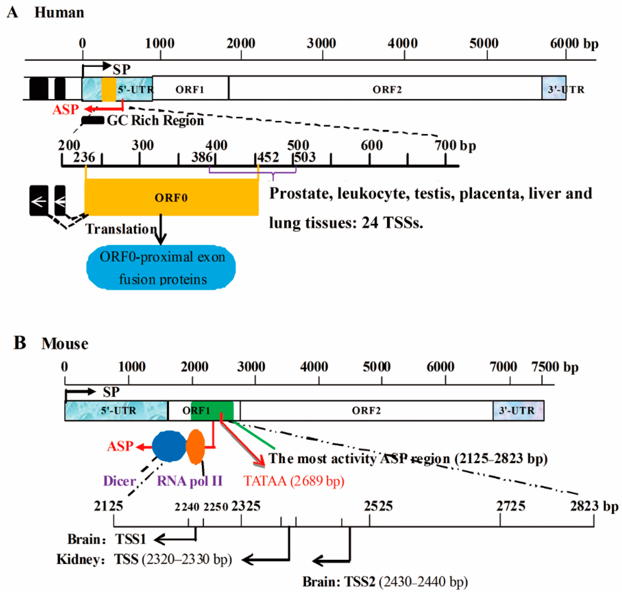
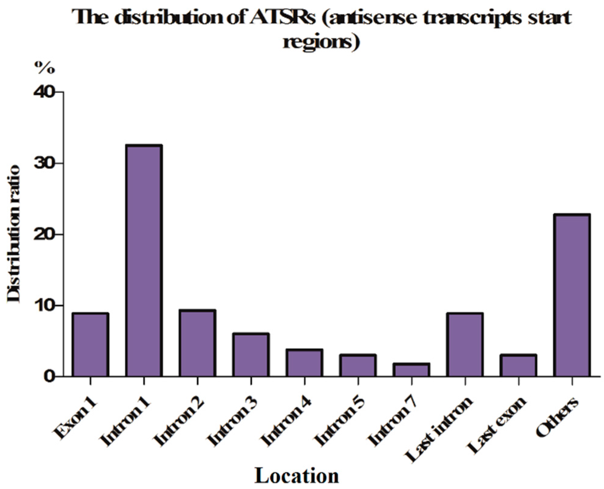
4.2. ASP Activity Is Associated with the Distribution Density of TSSs

The methylation signals of the 5′-end of intron 1 and the promoter region in most human genes are very low. However, TSS signals of antisense transcripts are gradually reduced from the 5′-end to the 3′-end of intron 1 but are markedly higher than those of the other introns, by 1.5–5 times, and the antisense transcript TSSs are normally distributed on both sides of the junction between exon 1 and intron 1 [33]. In other words, antisense transcript TSSs are mainly distributed at the 3′-end of exon 1 and the 5′-end of intron 1. He et al. [53] reported the interesting finding that higher ASP activity is found with a greater distribution of antisense transcript TSSs.
Cap analysis of gene expression (CAGE) in the human genome showed that a transcriptional unit possesses 16.3 sense transcript TSSs and 5.8 antisense transcript TSSs [87,88]. Conley et al. [87] found that partial sense transcript TSSs originate from the same alternative promoter as the mRNA-TSSs previously reported by Carninci et al. [88]. Interestingly, Osato et al. [89] argued that sense transcript TSSs are the result of transcriptional collision between antisense and sense transcripts.

4.3. ASP Activity Is Associated with the Degree of Methylation of CpGIs

Promoter regions are demethylated in vivo, decreasing their methylation signals compared to those of other areas [67,90]. In the vertebrate genome, an interesting observation is that genes are expressed in multiple tissues if their promoters contain CpGIs but are only active in a particular pattern when ASPs lack CpGIs [63]. This finding suggests that the temporal and spatial expression of antisense transcripts in particular tissues might be induced by the extent of CpGI methylation in their ASPs.
In fact, the lower the CpGI methylation signals in an ASP, the denser is the distribution of the antisense transcript TSS signal, and the greater is the activity of the ASP. More than 90% of bidirectional promoters contain significant CpGIs and CCG and CGG repeat sequences, which are enriched in the upstream and downstream regions of the TSSs of bidirectional promoters [77,91]. In contrast, only 56% of unidirectional transcription promoters contain CpGIs [43]. Bidirectional promoters in mESCs harbor many CpGIs, with an asymmetric distribution in the upstream and downstream regions of TSSs, which are inhibited by polycomb complexes [43].
The methylation level of CpGIs is negatively related to gene expression levels, with an inhibitory effect on gene transcription [74,75]. Weber et al. [75] revealed that the L1-cMet ASP is highly methylated in most human cells: when the ASP is demethylated, L1 antisense transcript expression is remarkably increased, whereas cMet expression and methylation signals are reduced.
In the chicken genome, most CpGIs are maintained in a state of demethylation [74], and the GC content of the 5′-UTR is positively related to the gene expression level, the breadth of expression and maximum expression, demonstrating that the GC content plays a key role in chicken gene regulation networks [76], though further analysis is necessary to elucidate the molecular mechanism.

4.4. ASP Promoter Activity Is Strongly Regulated by TFs

In the complex genome, a promoter is a characteristic sequence showing a transcriptional function and cis regulation, located upstream of a TSS, and contains a variety of sequence motifs involved in gene regulation [63]. Eukaryotic promoter regions contain motifs responsible for the transcription of the gene, and core elements often coexist and form composite motifs, including TFBSs, short tandem repeats, G-quadruplexes, and CpGIs.
Previous studies have shown that TFs play key roles in regulating the activity of ASPs. For example, Sp1 binding sites play an important role in regulating the activity of ASPs, such as by participating in HTLV-1 ASP transcription [56,57], combining with the GC box and recruiting the TF TFIID to activate the transcription of antisense transcripts [66]. Various studies have shown that NF-κB is also involved in ASP regulation [69,70]. Moreover, the HIV-1 long terminal repeat (LTR) acts as a bidirectional transcriptional promoter, the activity of which is regulated by NF-κB- and Sp1 binding sites in both orientations [68]. Of course, ASPs are regulated by specific TFs determined by the TFBSs present; these TFs can be independent or play a role in complex formation with RNA II to activate the ASP.
It is worth noting that one TF can interact with different gene promoters and regulate the expression of different genes. The human double homeobox 4 (DUX4) gene encodes the TF DUX4, which is expressed in testicular germ cells but is epigenetically silenced in ovarian tissue. When this TF binds to the ASPs of the MLT1C, THE1C and DDX10 genes, lncRNAs and natural antisense transcripts can be produced [92].

4.5. The U1 Small Nuclear Ribonucleoprotein (U1 snRNP) Affects ASP Promoter Activity

The mammalian U1 snRNP exhibits a weak recognition capability for natural antisense transcripts but strong recognition of sense transcripts [85]. Correlation analysis of a mathematical model of the relationship between sense transcripts or antisense transcripts and U1 showed that with an increase in the age of genes, the U1 site in downstream sense regions at the 5′-end (the first 1 kb) of protein-coding genes is significantly positively correlated with sense transcripts and negatively correlated with antisense transcripts. Interestingly, the U1 site in upstream antisense regions at the 5′-end (the first 1 kb) of protein-coding genes shows a significant negative correlation with sense transcripts and a positive correlation with antisense transcripts, suggesting that at least a subset of uaRNAs may be functionally important for antisense transcripts to become more extensively transcribed. Sense U1 sites are intensively distributed within the TSS + 200 bp region [85]. These observations indirectly demonstrate that the U1 snRNP restrains the activity of ASPs but enhances the activity of SPs.

4.6. Polyadenylation Site Signals Are Implicated in ASP Activity

It has been shown that chicken GHR mRNA transcripts are initiated from at least two SPs (V1 and V6), with a polymerase phosphorylation signal (PAS) in intron 6 [36]. Are ASPs also affected by PASs? Almada et al. [85] discovered that the percentage of mammalian PASs in the upstream region of TSSs was twice as high as in downstream regions. In addition, correlation analysis of a mathematical model of the relationship between sense/antisense transcripts and PASs revealed that the expression of both sense and natural antisense transcripts is significantly negatively correlated with both upstream and downstream PASs [85]. When ASPs are subject to the inhibitory effect of the U1 snRNP and PASs, the expression of natural antisense transcripts is much lower than that of sense transcripts. Compared with the transcription of mRNA, the extension of antisense transcripts is subject to relatively more interference from PASs, and transcription is forced to terminate more quickly [85]. PASs clearly also affect the activity of ASPs. Most genes exhibit more than one antisense transcript, and different antisense transcripts may be transcribed by one or more ASP. The formation of different transcripts is also highly dependent on PASs.
Some other enzymes may influence the activity of ASPs. Early in 1998, Vanhée-Brossoll and Vaquero reported a negative correlation between α1 (I) collagen gene mRNA and natural antisense transcripts [93]. In addition, the activity of the eIF-2α ASP may be regulated by an InR-associated binding protein through the control of RNA Pol II to interact with the InR-ASP. When entering the cell cycle, the expression of InR is low because its association with Inr-associated binding protein is restrained [45]. Moreover, topology isomerase I interacts with TF sites within the ASP, playing an indispensable role in stimulating and regulating the activity of this promoter [45]. The structure of bidirectional promoters is associated with lncRNAs, which are induced or inhibited in a rapid retinoic acid-dependent manner. The rat L1-ASP is Dicer enzyme dependent [37]. This process is similar to the generation of miRNAs, as it is subject to dsRNA formation between sense and antisense transcripts and then produces small interfering RNAs (siRNAs) or miRNAs through Dicer cleavage [94,95]. However, it remains to be determined whether certain enzymes in tissues or cells control the activity of ASPs. If the genes encoding these enzymes show tissue specificity, the regulation of the ASP will also be tissue specific, ultimately resulting in natural antisense transcripts only being expressed in some tissues.

4.7. Stimulation by Material Outside of Cells Influences the Activity of ASPs

The cell environment is particularly important for cell survival and reproduction, and many complex molecular biological processes, such as DNA replication, RNA transcription and protein translation, must be carried out in this environment. However, extracellular stressors may interfere with the transfer of genetic information. In studying the impact of stimuli on the cell environment, most researchers are currently focusing on extracellular inhibitors.
The expression of the α2 (I) collagen gene in cartilage cells and its corresponding promoter inactivates and decreases the expression of α2 (I) collagen to one-third of initial levels after BrdU treatment [62]. Through bioinformatics analysis, Nigumann et al. [39] revealed that L1-ASP could initiate the expression of natural antisense transcripts with high expression in expressed sequence tag databases. Recent studies have shown that various cytosine nucleosides, such as azacytidine, cytidine analogues, and DNMT inhibitors, can activate illegitimate transcription from the L1-ASP [44]. Weber et al. [75] performed an in-depth analysis of the regulation of L1-cMet ASPs and found that most human L1-cMet ASPs are hypermethylated, significantly increasing the expression of L1 antisense transcripts. Additionally, L1-cMet transcript expression levels and methylation signals are reduced. The same result can be obtained using micromolar doses of decitabine in cancer cell cultures.

4.8. ASP Activity Is Influenced by Chromatin and Histone Modifications

Epigenetic regulatory pathways influence gene transcription by modulating chromatin structure without altering primary DNA sequences [96]. The mechanisms that regulate chromatin structure include ATP-dependent chromatin remodeling, replacement of major histones with variants, cytosine methylation, the small RNA pathway, and covalent posttranslational modifications of histones [97]. Histone H3 trimethylated at Lys-4 and Lys-36 (H3K4met3, H3K36met3) can be used as a marker of chromatin activity, whereas histone H3 trimethylated at Lys-9 and Lys-27 (H3K9met3, H3K27met3) is a marker of transcriptionally silent chromatin. Histone acetylation, such as H3 acetylated at Lys-14 (H3K14ac), can also alter chromatin conformation, allowing transcriptional co-activating factors access to the promoter region and consequently favoring transcription. These markers associated with antisense promoters are distinct from the features associated with sense transcription. In general, high levels of antisense transcription result in pronounced differences in a broad range of chromatin features, both for the sense promoter and within the gene body. Features associated with newly deposited dynamic chromatin-histone H3 acetylation, turnover, chromatin remodeling enzymes and histone chaperones are typically increased, whereas features associated with established chromatin-H3K79me3, H3K36me3 and H2B123ub are reduced. These features are distinct from those associated with sense transcription [98]. Additionally, ASPs can form an R-loop structure which can be disfavored in vitro and in vivo by ribonuclease H1 overexpression, thereby resulting in gene down-regulation [34]. Overall, ASPs can regulate themselves at a low-affinity chromatin structure, and some antisense transcript can recruit other TFs to bind to the SP region to increase chromatin accessibility, followed by a high level of sense transcript expression [26].
Regulation of the activity of ASPs is complicated, as there are many factors affecting ASPs’ activity, and the same ASP may be influenced by many factors. This situation can be exemplified by the fact that the activity of different mESC ASPs is P-TEFb dependent, in addition to being affected by CpGIs [54].

5. Conclusions

Briefly, ASPs can be located in any position within a gene or intergenic region. The position of ASPs determines the initiation of the antisense transcript, and the dsRNAs formed by antisense transcripts match their target sense transcripts, ultimately determining the role of antisense transcripts in regulating gene expression. Therefore, it is necessary to conduct further analyses of the locations of interesting ASP in target genes, which will provide beneficial clues for understanding the initiation of antisense transcripts.
The initiation of antisense transcription is an extremely complex process. Many transcriptional elements can combine with ASPs, with different elements playing different roles, and interactions among components may even exist. Subsequent efforts should focus on the interactions between these transcriptional elements to elucidate the molecular mechanisms of ASPs and their antisense transcripts.

Acknowledgments

This work was supported by the Chinese Postdoctoral Science Foundation (2012M521611, 2013T60808), the National Natural Science Foundation (31301958), the Guangdong Provincial Natural Science Foundation (S2013010013382) and the China Agriculture Research System (CARS-42-G05). The funders played no role in the study design, the decision to publish, or in the preparation of the manuscript.

Author Contributions

The manuscript was written through contributions of all authors. Shudai Lin substantially contributed to the conception and design and drafted the article. Li Zhang revised it critically for important intellectual content. Wen Luo provided many important opinions in writing the manuscript. Xiquan Zhang gave the final approval of the article to be published.

Conflicts of Interest

The authors declare no conflict of interest.

References

  1. Chen, J.; Sun, M.; Kent, W.J.; Huang, X.; Xie, H.; Wang, W.; Zhou, G.; Shi, R.Z.; Rowley, J.D. Over 20% of human transcripts might form sense-antisense pairs. Nucleic Acids Res. 2004, 32, 4812–4820. [Google Scholar] [CrossRef] [PubMed]
  2. Engstrom, P.G.; Suzuki, H.; Ninomiya, N.; Akalin, A.; Sessa, L.; Lavorgna, G.; Brozzi, A.; Luzi, L.; Tan, S.L.; Yang, L.; et al. Complex Loci in human and mouse genomes. PLoS Genet. 2006, 2, e47. [Google Scholar] [CrossRef] [PubMed]
  3. Katayama, S.; Tomaru, Y.; Kasukawa, T.; Waki, K.; Nakanishi, M.; Nakamura, M.; Nishida, H.; Yap, C.C.; Suzuki, M.; Kawai, J.; et al. Revisiting the evolution of mouse LINE-1 in the genomic era. Mob. DNA 2013, 4, 3. [Google Scholar] [CrossRef]
  4. Mahmoudi, S.; Henriksson, S.; Corcoran, M.; Méndez-Vidal, C.; Wiman, K.G.; Farnebo, M. Wrap53, a natural p53 antisense transcript required for p53 induction upon DNA damage. Mol. Cell 2009, 33, 462–471. [Google Scholar] [CrossRef] [PubMed]
  5. Yu, W.; Gius, D.; Onyango, P.; Muldoon-Jacobs, K.; Karp, J.; Feinberg, A.P.; Cui, H. Epigenetic silencing of tumour suppressor gene p15 by its antisense RNA. Nature 2008, 451, 202–206. [Google Scholar] [CrossRef] [PubMed]
  6. Suzuki, K.; Ahlenstiel, C.; Marks, K.; Kelleher, A.D. Promoter targeting RNAs: Unexpected contributors to the control of HIV-1 transcription. Mol. Ther. Nucl. Acids 2015, 4, e222. [Google Scholar] [CrossRef] [PubMed]
  7. Orekhova, A.S.; Rubtsov, P.M. Bidirectional promoters in the transcription of mammalian genomes. Biochemistry 2013, 78, 335–341. [Google Scholar] [CrossRef] [PubMed]
  8. Kaikkonen, M.U.; Lam, M.T.; Glass, C.K. Non-coding RNAs as regulators of gene expression and epigenetics. Cardiovasc. Res. 2011, 90, 430–440. [Google Scholar] [CrossRef] [PubMed]
  9. Rezaeian, A.H.; Nishibori, M.; Hiraiwa, N.; Yoshizawa, M.; Yasue, H. Expression profile and localization of mouse calcitonin (CT) sense/antisense transcripts in pre- and postnatal tissue development. J. Vet. Med. Sci. 2009, 71, 561–568. [Google Scholar] [CrossRef] [PubMed]
  10. Li, K.; Chowdhury, T.; Vakeel, P.; Koceja, C.; Sampath, V.; Ramchandran, R. δ-Like 4 mRNA is regulated by adjacent natural antisense transcripts. Vasc. Cell 2015, 7, 3. [Google Scholar] [CrossRef] [PubMed]
  11. Chiba, M. Differential expression of natural antisense transcripts during liver development in embryonic mice. Biomed. Rep. 2014, 2, 918–922. [Google Scholar] [CrossRef] [PubMed]
  12. Mätlik, K.; Redik, K.; Speek, M. L1 antisense promoter drives tissue-specific transcription of human genes. J. Biomed. Biotechnol. 2006, 2006, 1–16. [Google Scholar] [CrossRef] [PubMed]
  13. Huang, M.D.; Chen, W.M.; Qi, F.Z.; Xia, R.; Sun, M.; Xu, T.P.; Yin, L.; Zhang, E.B.; De, W.; Shu, Y.Q. Long non-coding RNA ANRIL is upregulated in hepatocellular carcinoma and regulates cell apoptosis by epigenetic silencing of KLF2. J. Hematol. Oncol. 2015, 8, 50. [Google Scholar] [CrossRef] [PubMed]
  14. Xi, Q.; Gao, N.; Zhang, X.; Zhang, B.; Ye, W.; Wu, J.; Zhang, X. A natural antisense transcript regulates acetylcholinesterase gene expression via epigenetic modification in Hepatocellular Carcinoma. Int. J. Biochem. Cell Biol. 2014, 55, 242–251. [Google Scholar] [CrossRef] [PubMed]
  15. Wight, M.; Werner, A. The functions of natural antisense transcripts. Essays Biochem. 2013, 54, 91–101. [Google Scholar] [CrossRef] [PubMed]
  16. Rinn, J.L.; Chang, H.Y. Genome regulation by long noncoding RNAs. Annu. Rev. Biochem. 2012, 81, 145–166. [Google Scholar] [CrossRef] [PubMed]
  17. Wery, M.; Kwapisz, M.; Morillon, A. Noncoding RNAs in gene regulation. Rev. Syst. Biol. Med. 2011, 3, 728–738. [Google Scholar] [CrossRef] [PubMed]
  18. Au, P.C.K.; Zhu, Q.H.; Dennis, E.S.; Wang, M.B. Long non-coding RNA-mediated mechanisms independent of the RNAi pathway in animals and plants. RNA Biol. 2010, 8, 404–414. [Google Scholar] [CrossRef]
  19. Aartsma-Rus, A.; van-Ommen, G.J. Progress in therapeutic antisense applications for neuromuscular disorders. Eur. J. Hum. Genet. 2010, 18, 146–153. [Google Scholar] [CrossRef] [PubMed]
  20. Su, W.; Xiong, H.; Fang, J. Natural antisense transcripts regulate gene expression in an epigenetic manner. Biochem. Biophys. Res. Commun. 2010, 396, 177–181. [Google Scholar] [CrossRef] [PubMed]
  21. Faghihi, M.A.; Wahlestedt, C. Regulatory roles of natural antisense transcripts. Nat. Rev. Mol. Cell Biol. 2009, 10, 637–643. [Google Scholar] [CrossRef] [PubMed]
  22. Grigoriadis, A.; Oliver, G.R.; Tanney, A.; Kendrick, H.; Smalley, M.J.; Jat, P.; Neville, A.M. Identification of differentially expressed sense and antisense transcript pairs in breast epithelial tissues. BMC Genom. 2009, 10, 324. [Google Scholar] [CrossRef] [PubMed]
  23. Beckedorff, F.C.; Ayupe, A.C.; Crocci-Souza, R.; Amaral, M.S.; Nakaya, H.I.; Soltys, D.T.; Menck, C.F.; Reis, E.M.; Verjovski-Almeida, S. The intronic long noncoding RNA ANRASSF1 recruits PRC2 to the RASSF1A promoter, reducing the expression of RASSF1A and increasing cell proliferation. PLoS Genet. 2013, 9, e1003705. [Google Scholar] [CrossRef] [PubMed] [Green Version]
  24. Chatterjee, A.; Drews, L.; Mehra, S.; Takano, E.; Kaznessis, Y.N.; Hu, W.S. Convergent transcription in the butyrolactone regulon in Streptomyces coelicolor confers a bistable genetic switch for antibiotic biosynthesis. PLoS ONE 2011, 6, e21974. [Google Scholar] [CrossRef] [PubMed]
  25. Denli, A.M.; Narvaiza, I.; Kerman, B.E.; Pena, M.; Benner, C.; Marchetto, M.C.; Diedrich, J.K.; Aslanian, A.; Ma, J.; Moresco, J.J.; et al. Primate-specific ORF0 contributes to retrotransposon-mediated diversity. Cell 2015, 163, 583–593. [Google Scholar] [CrossRef] [PubMed]
  26. Saldaña-Meyer, R.; González-Buendía, E.; Guerrero, G.; Narendra, V.; Bonasio, R.; Recillas-Targa, F.; Reinberg, D. CTCF regulates the human p53 gene through direct interaction with its natural antisense transcript, Wrap53. Genes Dev. 2014, 28, 723–734. [Google Scholar] [CrossRef] [PubMed]
  27. Mahmoudi, S.; Henriksson, S.; Weibrecht, I.; Smith, S.; Söderberg, O.; Strömblad, S.; Wiman, K.G.; Farnebo, M. WRAP53 is essential for Cajal body formation and for targeting the survival of motor neuron complex to Cajal bodies. PLoS Biol. 2010, 8, e1000521. [Google Scholar] [CrossRef] [PubMed]
  28. Werner, A.; Cockell, S.; Falconer, J.; Carlile, M.; Alnumeir, S.; Robinson, J. Contribution of natural antisense transcription to an endogenous siRNA signature in human cells. BMC Genom. 2014, 15, 19. [Google Scholar] [CrossRef] [PubMed]
  29. Peters, N.T.; Rohrbach, J.A.; Zalewski, B.A.; Byrkett, C.M.; Vaughn, J.C. RNA editing and regulation of Drosophila 4f-rnp expression by sas-10 antisense readthrough mRNA transcripts. RNA 2003, 9, 698–710. [Google Scholar] [CrossRef] [PubMed]
  30. Rong, J.; Yin, J.; Su, Z. Natural antisense RNAs are involved in the regulation of CD45 expression in autoimmune diseases. Lupus 2015, 24, 235–239. [Google Scholar] [CrossRef] [PubMed]
  31. Malik, S.; Durairaj, G.; Bhaumik, S.R. Mechanisms of antisense transcription initiation from the 3′-end of the GAL10 coding sequence in vivo. Mol. Cell. Biol. 2013, 33, 3549–3567. [Google Scholar] [CrossRef] [PubMed]
  32. Beltran, M.; Aparicio-Prat, E.; Mazzolini, R.; Millanes-Romero, A.; Massó, P.; Jenner, R.G.; Díaz, V.M.; Peiró, S.; de Herreros, A.G. Splicing of a non-coding antisense transcript controls LEF1 gene expression. Nucleic Acids Res. 2015, 43, 5785–5797. [Google Scholar] [CrossRef] [PubMed]
  33. Finocchiaro, G.; Carro, M.S.; Francois, S.; Parise, P.; DiNinni, V.; Muller, H. Localizing hotspots of antisense transcription. Nucleic Acids Res. 2007, 35, 1488–1500. [Google Scholar] [CrossRef] [PubMed]
  34. Boque-Sastre, R.; Soler, M.; Oliveira-Mateos, C.; Portela, A.; Moutinho, C.; Sayols, S.; Villanueva, A.; Esteller, M.; Guil, S. Head-to-head antisense transcription and R-loop formation promotes transcriptional activation. Proc. Natl. Acad. Sci. USA 2015, 112, 5785–5790. [Google Scholar] [CrossRef] [PubMed]
  35. Lau, S.L. Molecular Characterization of the Chicken Growth Hormone Receptor Gene. Ph.D. Thesis, University of Hong Kong, Hong Kong, China, September 2005. [Google Scholar]
  36. Cruickshanks, H.A.; Tufarelli, C. Isolation of cancer-specific chimeric transcripts induced by hypomethylation of the LINE-1 antisense promoter. Genomics 2009, 94, 397–406. [Google Scholar] [CrossRef] [PubMed]
  37. Li, J.; Kannan, M.; Trivett, A.L.; Liao, H.; Wu, X.; Akagi, K.; Symer, D.E. An antisense promoter in mouse L1 retrotransposon open reading frame-1 initiates expression of diverse fusion transcripts and limits retrotransposition. Nucleic Acids Res. 2014, 42, 4546–4562. [Google Scholar] [CrossRef] [PubMed]
  38. Nigumann, P.; Redik, K.; Mätlik, K.; Speek, M. Many human genes are transcribed from the antisense promoter of L1 retrotransposon. Genomics 2002, 79, 628–634. [Google Scholar] [CrossRef] [PubMed]
  39. Chen, G.L.; Miller, G.M. 5′-Untranslated region of the tryptophan hydroxylase-2 gene harbors an asymmetric bidirectional promoter but not internal ribosome entry site in vitro. Gene 2009, 435, 53–62. [Google Scholar] [CrossRef] [PubMed]
  40. Gotea, V.; Petrykowska, H.M.; Elnitski, L. Bidirectional promoters as important drivers for the emergence of species-specific transcripts. PLoS ONE 2013, 8, e57323. [Google Scholar] [CrossRef] [PubMed]
  41. Batagov, A.O.; Yarmishyn, A.A.; Jenjaroenpun, P.; Tan, J.Z.; Nishida, Y.; Kurochkin, I.V. Role of genomic architecture in the expression dynamics of long noncoding RNAs during differentiation of human neuroblastoma cells. BMC Syst. Biol. 2013, 7, S11. [Google Scholar] [CrossRef] [PubMed]
  42. Lepoivre, C.; Belhocine, M.; Bergon, A.; Griffon, A.; Yammine, M.; Vanhille, L.; Zacarias-Cabeza, J.; Garibal, M.A.; Koch, F.; Maqbool, M.A.; et al. Divergent transcription is associated with promoters of transcriptional regulators. BMC Genom. 2013, 14, 914. [Google Scholar] [CrossRef] [PubMed] [Green Version]
  43. Hildebrandt, M.; Nellen, W. Differential antisense transcription from the dictyostelium EB4 gene locus: Implications on antisense mediated regulation of mRNA stability. Cell 1992, 69, 197–204. [Google Scholar] [CrossRef]
  44. Silverman, T.A.; Noguchi, M.; Safer, B. Role of sequences within the first intron in the regulation of expression of eukaryotic initiation factor 2α. J. Biol. Chem. 1992, 267, 9738–9742. [Google Scholar] [PubMed]
  45. Duart-Garcia, C.; Braunschweig, M.H. Functional expression study of Igf2 antisense transcript in mouse. Int. J. Genom. 2014, 2014, 1–7. [Google Scholar] [CrossRef] [PubMed]
  46. Sun, J.; Li, W.; Sun, Y.; Yu, D.; Wen, X.; Wang, H.; Cui, J.; Wang, G.; Hoffman, A.R.; Hu, J.F. A novel antisense long noncoding RNA within the IGF1R gene locus is imprinted in hematopoietic malignancies. Nucleic Acids Res. 2014, 42, 9588–9601. [Google Scholar] [CrossRef] [PubMed]
  47. Luo, D.; Caniggia, I.; Post, M. Hypoxia-inducible regulation of placental BOK expression. Biochem. J. 2014, 461, 391–402. [Google Scholar] [CrossRef] [PubMed]
  48. Wright, P.W.; Huehn, A.; Cichocki, F.; Li, H.; Sharma, N.; Dang, H.; Lenvik, T.R.; Woll, P.; Kaufman, D.; Miller, J.S.; et al. Identification of a KIR antisense lncRNA expressed by progenitor cells. Genes Immun. 2013, 14, 427–433. [Google Scholar] [CrossRef] [PubMed]
  49. Spicer, D.B.; Sonenshein, G.E. An antisense promoter of the murine c-myc gene is localized within intron 2. Mol. Cell. Biol. 1992, 12, 1324–1329. [Google Scholar] [CrossRef] [PubMed]
  50. Ebralidze, A.K.; GuibalF, C.; Steidl, U.; Zhang, P.; Lee, S.; Bartholdy, B.; JordaM, A.; Petkova, V.; Rosenbauer, F.; Huang, G.; et al. PU.1 expression is modulated by the balance of functional sense and antisense RNAs regulated by a shared cis-regulatory element. Genes Dev. 2008, 22, 2085–2092. [Google Scholar] [CrossRef] [PubMed]
  51. Schultz, B.M.; Gallicio, G.A.; Cesaroni, M.; Lupey, L.N.; Engel, N. Enhancers compete with a long non-coding RNA for regulation of the Kcnq1 domain. Nucleic Acids Res. 2015, 43, 745–759. [Google Scholar] [CrossRef] [PubMed]
  52. Barann, M.; Esser, D.; Klostermeier, U.C.; Lappalainen, T.; Luzius, A.; Kuiper, J.W.; Ammerpohl, O.; Vater, I.; Siebert, R.; Amstislavskiy, V.; et al. Janus—A comprehensive tool investigating the two faces of transcription. Bioinformatics 2013, 29, 1600–1606. [Google Scholar] [CrossRef] [PubMed]
  53. He, Y.; Vogelstein, B.; Velculescu, V.E.; Papadopoulos, N.; Kinzler, K.W. The antisense transcriptomes of human cells. Science 2008, 322, 1855–1857. [Google Scholar] [CrossRef] [PubMed]
  54. Lapidot, M.; Pilpel, Y. Genome-wide natural antisense transcription: Coupling its regulation to its different regulatory mechanisms. EMBO Rep. 2006, 7, 1216–1222. [Google Scholar] [CrossRef] [PubMed]
  55. Dreos, R.; Ambrosini, G.; Cavin-Périer, R.; Bucher, P. EPD and EPDnew, high-quality promoter resources in the next-generation sequencing era. Nucleic Acids Res. 2013, 41, D157–D164. [Google Scholar] [CrossRef] [PubMed]
  56. Gazon, H.; Lemasson, I.; Polakowski, N.; Césaire, R.; Matsuoka, M.; Barbeau, B.; Mesnard, J.M.; Peloponese, J.M. Human T-cell leukemia virus type 1 (HTLV-1) bZIP factor requires cellular transcription factor JunD to upregulate HTLV-1 antisense transcription from the 3′-long terminal repeat. J. Virol. 2012, 86, 9070–9078. [Google Scholar] [CrossRef] [PubMed]
  57. Yoshida, T.; Kawano, Y.; Sato, K.; Ando, Y.; Aoki, J.; Miura, Y.; Komano, J.; Tanaka, Y.; Koyanagi, Y. A CD63 mutant inhibits T-cell tropic human immunodeficiency virus type 1 entry by disrupting CXCR4 trafficking to the plasma membrane. Traffic 2008, 9, 540–558. [Google Scholar] [CrossRef] [PubMed]
  58. Xie, Z.C.; Pan, Q.H.; Yu, Y.C.; Sun, F.Y. Core promoter of eukaryotic genes. J. Med. Mol. Biol. 2007, 4, 132–135. [Google Scholar]
  59. Yang, M.Q.; Elnitski, L.L. Diversity of core promoter elements comprising human bidirectional promoters. BMC Genom. 2008, 9, S3. [Google Scholar] [CrossRef] [PubMed]
  60. Flynn, R.A.; Almada, A.E.; Zamudio, J.R.; Sharp, P.A. Antisense RNA polymerase II divergent transcripts are P-TEFb dependent and substrates for the RNA exosome. Proc. Natl. Acad. Sci. USA 2011, 108, 10460–10465. [Google Scholar] [CrossRef] [PubMed]
  61. Farrell, C.M.; Lukens, L.N. Naturally occurring antisense transcripts are present in chick embryo chondrocytes simultaneously with the down-regulation of the α1(I) collagen gene. J. Biol. Chem. 1995, 270, 3400–3408. [Google Scholar] [PubMed]
  62. Trinklein, N.D.; Aldred, S.F.; Hartman, S.J.; Schroeder, D.I.; Otillar, R.P.; Myers, R.M. An abundance of bidirectional promoters in the human genome. Genome Res. 2004, 14, 62–66. [Google Scholar] [CrossRef] [PubMed]
  63. Putta, P.; Mitra, C.K. Conserved short sequences in promoter regions of human genome. J. Biomol. Struct. Dyn. 2010, 27, 599–610. [Google Scholar] [CrossRef] [PubMed]
  64. Abe, H.; Gemmell, N.J. Abundance, arrangement, and function of sequence motifs in the chicken promoters. BMC Genom. 2014, 15, 900. [Google Scholar] [CrossRef] [PubMed]
  65. Lyle, R.; Watanabe, D.; Te Vruchte, D.; Lerchner, W.; Smrzka, O.W.; Wutz, A.; Schageman, J.; Hahner, L.; Davies, C.; Barlow, D.P. The imprinted antisense RNA at the Igf2r locus overlaps but does not imprint Mas1. Nat. Genet. 2000, 25, 19–21. [Google Scholar] [CrossRef] [PubMed]
  66. Wierstra, I. Sp1: Emerging roles—Beyond constitutive activation of TATA-less housekeeping genes. Biochem. Biophys. Res. Commun. 2008, 372, 1–13. [Google Scholar] [CrossRef] [PubMed]
  67. Zanotto, E.; Lehtonen, V.; Jacobs, H.T. Modulation of Mrps12/Sarsm promoter activity in response to mitochondrial stress. Biochim. Biophys. Acta 2008, 1783, 2352–2362. [Google Scholar] [CrossRef] [PubMed]
  68. Arpin-André, C.; Laverdure, S.; Barbeau, B.; Gross, A.; Mesnard, J.M. Construction of a reporter vector for analysis of bidirectional transcriptional activity of retrovirus LTR. Plasmid 2014, 74, 45–51. [Google Scholar] [CrossRef] [PubMed]
  69. Bentley, K.; Deacon, N.; Sonza, S.; Zeichner, S.; Churchill, M. Mutational analysis of the HIV-1 LTR as a promoter of negative sense transcription. Arch. Virol. 2004, 149, 2277–2294. [Google Scholar] [CrossRef] [PubMed]
  70. Kobayashi-Ishihara, M.; Yamagishi, M.; Hara, T.; Matsuda, Y.; Takahashi, R.; Miyake, A.; Nakano, K.; Yamochi, T.; Ishida, T.; Watanabe, T. HIV-1-encoded antisense RNA suppresses viral replication for a prolonged period. Retrovirology 2012, 9, 38. [Google Scholar] [CrossRef] [PubMed]
  71. Tufarelli, C.; Cruickshanks, H.A.; Meehan, R.R. LINE-1 activation and epigenetic silencing of suppressor genes in cancer: Causally related events? Mob. Genet. Elements 2013, 3, e26832. [Google Scholar] [CrossRef] [PubMed]
  72. Liu, B.; Chen, J.; Shen, B. Genome-wide analysis of the transcription factor binding preference of human bi-directional promoters and functional annotation of related gene pairs. BMC Syst. Biol. 2011, 5, S2. [Google Scholar] [CrossRef] [PubMed]
  73. Lin, J.M.; Collins, P.J.; Trinklein, N.D.; Fu, Y.; Xi, H.; Myers, R.M.; Weng, Z. Transcription factor binding and modified histones in human bidirectional promoters. Genome Res. 2007, 17, 818–827. [Google Scholar] [PubMed]
  74. Li, Q.; Li, N.; Hu, X.; Li, J.; Du, Z.; Chen, L.; Yin, G.; Duan, J.; Zhang, H.; Zhao, Y.; et al. Genome-wide mapping of DNA methylation in chicken. PLoS ONE 2011, 6, e19428. [Google Scholar] [CrossRef] [PubMed]
  75. Weber, B.; Kimhi, S.; Howard, G.; Eden, A.; Lyko, F. Demethylation of a LINE-1 antisense promoter in the cMet locus impairs Met signalling through induction of illegitimate transcription. Oncogene 2010, 29, 5775–5784. [Google Scholar] [CrossRef] [PubMed]
  76. Rao, Y.S.; Chai, X.W.; Wang, Z.F.; Nie, Q.H.; Zhang, X.Q. Impact of GC content on gene expression pattern in chicken. Genet. Sel. Evol. 2013, 45, 9. [Google Scholar] [CrossRef] [PubMed]
  77. Shu, J.; Jelinek, J.; Chang, H.; Shen, L.; Qin, T.; Chung, W.; Oki, Y.; Issa, J.P. Silencing of bidirectional promoters by DNA methylation in tumorigenesis. Cancer Res. 2006, 66, 5077–5084. [Google Scholar] [CrossRef] [PubMed]
  78. Mueller, B.U.; Pabst, T.; Osato, M.; Asou, N.; Johansen, L.M.; Minden, M.D.; Behre, G.; Hiddemann, W.; Ito, Y.; Tenen, D.G. Heterozygous PU.1 mutations are associated with acute myeloid leukemia. Blood 2003, 101, 2074. [Google Scholar] [CrossRef] [PubMed]
  79. Rosenbauer, F.; Owens, B.M.; Yu, L.; Tumang, J.R.; Steidl, U.; Kutok, J.L.; Clayton, L.K.; Wagner, K.; Scheller, M.; Iwasaki, H.; et al. Lymphoid cell growth and transformation are suppressed by a key regulatory element of the gene encoding PU.1. Nat. Genet. 2006, 38, 27–37. [Google Scholar] [CrossRef] [PubMed]
  80. Huang, G.; Zhang, P.; Hirai, H.; Elf, S.; Yan, X.; Chen, Z.; Koschmieder, S.; Okuno, Y.; Dayaram, T.; Growney, J.D. PU.1 is a major downstream target of AML1 (RUNX1) in adult mouse hematopoiesis. Nat. Genet. 2008, 40, 51–60. [Google Scholar] [CrossRef] [PubMed]
  81. Chen, H.; Ray-Gallet, D.; Zhang, P.; Hetherington, C.J.; Gonzalez, D.A.; Zhang, D.E.; Moreau-Gachelin, F.; Tenen, D.G. PU.1 (Spi-1) autoregulates its expression in myeloid cells. Oncogene 1995, 11, 1549–1560. [Google Scholar] [PubMed]
  82. Okuno, Y.; Huang, G.; Rosenbauer, F.; Evans, E.K.; Radomska, H.S.; Iwasaki, H.; Akashi, K.; Moreau-Gachelin, F.; Li, Y.; Zhang, P.; et al. Potential autoregulation of transcription factor PU.1 by an upstream regulatory element. Mol. Cell. Biol. 2005, 25, 2832–2845. [Google Scholar] [CrossRef] [PubMed]
  83. Rahl, P.B.; Lin, C.Y.; Seila, A.C.; Flynn, R.A.; McCuine, S.; Burge, C.B.; Sharp, P.A.; Young, R.A. c-Myc regulates transcriptional pause release. Cell 2010, 141, 432–445. [Google Scholar] [CrossRef] [PubMed] [Green Version]
  84. Xu, C.; Chen, J.; Shen, B. The preservation of bidirectional promoter architecture in eukaryotes: What is the driving force? BMC Syst. Biol. 2012, 6, S21. [Google Scholar] [CrossRef] [PubMed]
  85. Almada, A.E.; Wu, X.B.; Kriz, A.J.; Burge, C.B.; Sharp, P.A. Promoter directionality is controlled by U1 snRNP and polyadenylation signals. Nature 2013, 499, 360–363. [Google Scholar] [CrossRef] [PubMed]
  86. Uesaka, M.; Nishimura, O.; Go, Y.; Nakashima, K.; Agata, K.; Imamura, T. Bidirectional promoters are the major source of gene activation-associated non-coding RNAs in mammals. BMC Genom. 2014, 15, 35. [Google Scholar] [CrossRef] [PubMed] [Green Version]
  87. Conley, A.B.; Miller, W.J.; Jordan, I.K. Human cis natural antisense transcripts initiated by transposable elements. Trends Genet. 2008, 24, 53–56. [Google Scholar] [CrossRef] [PubMed]
  88. Carninci, P.; Sandelin, A.; Lenhard, B.; Katayama, S.; Shimokawa, K.; Ponjavic, J.; Semple, C.A.; Taylor, M.S.; Engström, P.G.; Frith, M.C.; et al. Genome-wide analysis of mammalian promoter architecture and evolution. Nat. Genet. 2006, 38, 626–635. [Google Scholar] [CrossRef] [PubMed]
  89. Osato, N.; Suzuki, Y.; Ikeo, K.; Gojobori, T. Transcriptional interferences in cis natural antisense transcripts of humans and mice. Genetics 2007, 176, 1299–1306. [Google Scholar] [CrossRef] [PubMed]
  90. Klose, R.J.; Bird, A.P. Genomic DNA methylation: The mark and its mediators. Trends Biochem. Sci. 2006, 31, 89–97. [Google Scholar] [CrossRef] [PubMed]
  91. Bibikova, M.; Lin, Z.; Zhou, L.; Chudin, E.; Garcia, E.W.; Wu, B.; Doucet, D.; Thomas, N.J.; Wang, Y.; Vollmer, E.; et al. High-throughput DNA methylation profiling using universal bead arrays. Genome Res. 2006, 16, 383–393. [Google Scholar] [CrossRef] [PubMed]
  92. Young, J.M.; Whiddon, J.L.; Yao, Z.; Kasinathan, B.; Snider, L.; Geng, L.N.; Balog, J.; Tawil, R.; vander-Maarel, S.M.; Tapscott, S.J. DUX4 binding to retroelements creates promoters that are active in FSHD muscle and testis. PLoS Genet. 2013, 9, e1003947. [Google Scholar] [CrossRef] [PubMed]
  93. Vanhée-Brossollet, C.; Vaquero, C. Do natural antisense transcripts make sense in eukaryotes? Gene 1998, 211, 1–9. [Google Scholar] [CrossRef]
  94. Burns, K.H.; Boeke, J.D. Human transposon tectonics. Cell 2012, 149, 740–752. [Google Scholar] [CrossRef] [PubMed]
  95. Martin, S.L.; Cruceanu, M.; Branciforte, D.; Wai-Lun Li, P.; Kwok, S.C.; Hodges, R.S.; Williams, M.C. LINE-1 retrotransposition requires the nucleic acid chaperone activity of the ORF1 protein. J. Mol. Biol. 2005, 348, 549–561. [Google Scholar] [CrossRef] [PubMed]
  96. Li, B.; Carey, M.; Workman, J.L. The role of chromatin during transcription. Cell 2007, 128, 707–719. [Google Scholar] [CrossRef] [PubMed]
  97. Goldberg, A.D.; Allis, C.D.; Bernstein, E. Epigenetics: A landscape takes shape. Cell 2007, 128, 635–638. [Google Scholar] [CrossRef] [PubMed]
  98. Murray, S.C.; Haenni, S.; Howe, F.S.; Fischl, H.; Chocian, K.; Nair, A.; Mellor, J. Sense and antisense transcription are associated with distinct chromatin architectures across genes. Nucleic Acids Res. 2015, 43, 7823–7837. [Google Scholar] [CrossRef] [PubMed]

