2.1. Antiradical Activity of p-Hydroxybenzoic (HB) and p-Hydroxycinnamic (HC)
In the present work,
p-hydroxybenzoic (HB) and
p-hydroxycinnamic (HC) acids with various substituents (
Figure 1,
Table 1) were chosen as the objects of study.
p-Hydroxybenzoic (
p-HBA), vanillic (VA) and syringic acid (SyrA) are structural analogs of HC acids:
p-coumaric (CA), ferulic (FA) and sinapic (SinA) ones.
Antioxidant capacity (AOC) of HB and HC acids was determined against the ABTS radical cation (TEAC assay) and peroxyl radical (ORAC assay) at pH 7.4 (
Table 2).
The AOC of the studied HB and HC acids is, on the average, three times higher than that of
d,
l-α-tocopherol, and 3.0 and 5.7 times higher than AOC of
l-ascorbic acid against the ABTS radical cation and peroxyl radical, respectively (
Table 2).
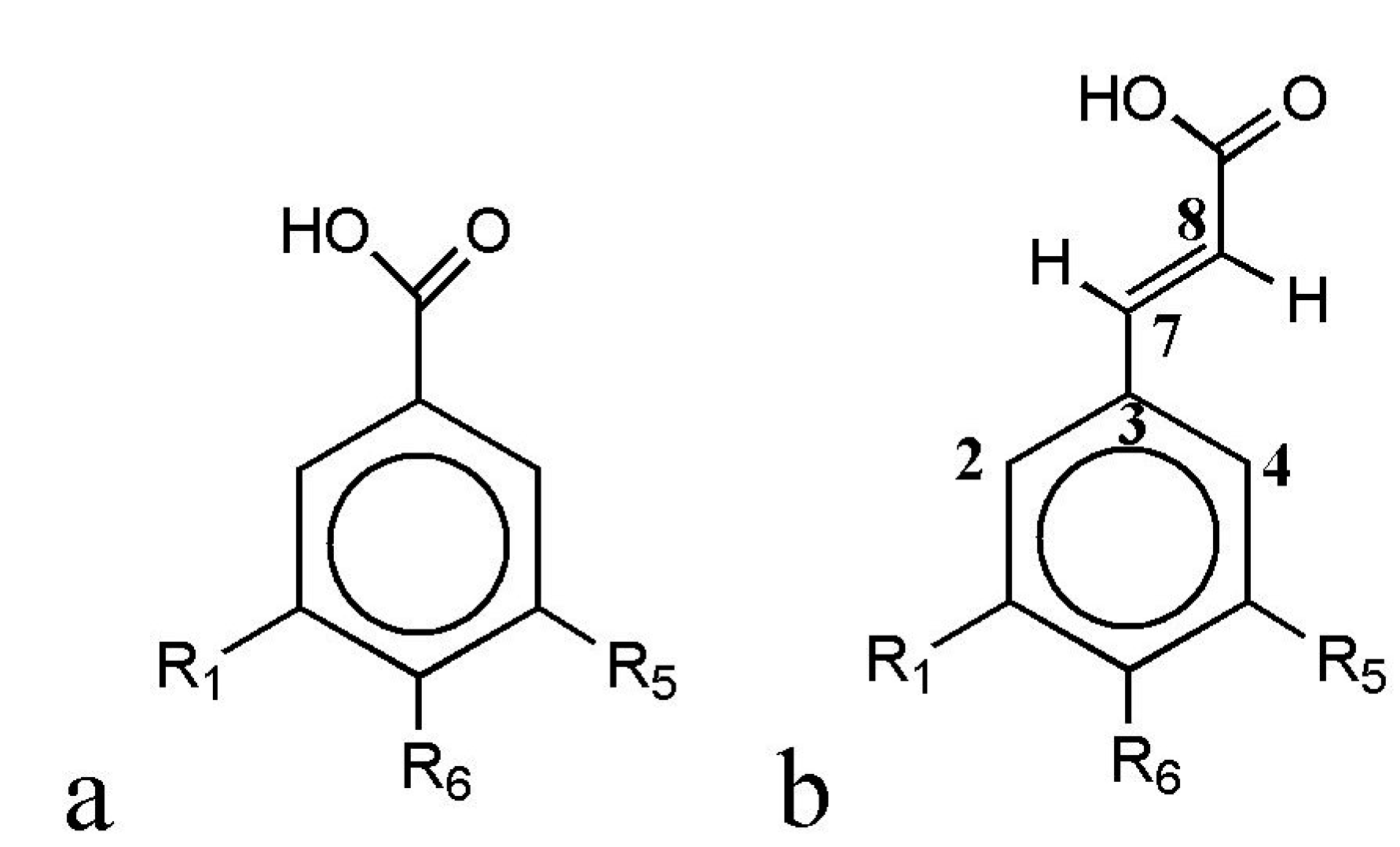
Figure 1.
General structures of p-hydroxybenzoic (HB, a) and p-hydroxycinnamic (HC, b) acids.
Figure 1.
General structures of p-hydroxybenzoic (HB, a) and p-hydroxycinnamic (HC, b) acids.
Table 1.
Substituents in the HB and HC acid structures.
Table 1.
Substituents in the HB and HC acid structures.
| Phenolic Acids | R1 | R5 | R6 |
|---|
| p-Hydroxybenzoic (HB) acids: |
| p-Hydroxybenzoic (HBA) | H | H | OH |
| Vanillic (VA) | H | OCH3 | OH |
| Syringic (SyrA) | OCH3 | OCH3 | OH |
| Gallic acid (GalA) | OH | OH | OH |
| p-Hydroxycinnamic (HC) acids: |
| p-Coumaric (CA) | H | H | OH |
| Ferulic (FA) | H | OCH3 | OH |
| Sinapic (SinA) | OCH3 | OCH3 | OH |
HC acids are characterized by 1.2–2.5 times higher AOC values against both types of radicals compared to the corresponding HB acids (
Table 2). Such high values are explained by the stabilization of phenoxyl radicals of HC acids due to the delocalization of the unpaired electron in case of benzene ring conjugation with C3 chain, and less pronounced influence of the carboxyl group, notable for its negative inductive and mesomeric effects, on the distribution of electron density in the benzene ring due to the presence of ethenyl bridge.
Table 2.
Antioxidant capacity of HB and HC acids against the 2,2'-azino-bis-(3-ethylbenzothiazoline-6-sulphonic acid) diammonium salt (ABTS) radical cation (Trolox Equivalent Antioxidant Capacity—TEAC, 50 µM PBS, pH 7.4) and peroxyl radical (Oxygen Radical Absorbance Capacity—ORAC, 75 µM Na-phosphate buffer, pH 7.4).
| Phenolic Acids | TEAC (μmol TE/μmol) | ORAC (μmol TE/μmol) |
|---|
| p-Hydroxybenzoic (HBA) | 2.64 ± 0.07 | 2.17 ± 0.18 |
| Vanillic (VA) | 2.63 ± 0.08 | 3.44 ± 0.19 |
| Syringic (SyrA) | 1.46 ± 0.09 | 1.52 ± 0.11 |
| Gallic acid (GalA) | 5.76 ± 0.21 | 1.08 ± 0.05 |
| p-Coumaric (CA) | 3.04 ± 0.18 | 5.02 ± 0.26 |
| Ferulic (FA) | 3.90 ± 0.21 | 4.59 ± 0.21 |
| Sinapic (SinA) | 3.66 ± 0.15 | 2.94 ± 0.19 |
| l-Ascorbic acid | 1.01 ± 0.02 | 0.52 ± 0.04 |
| d,l-α-Tocoferol a | 1.00 ± 0.02 | 1.01 ± 0.06 |
Experimentally determined the AOC values of HB and HC acids against peroxyl radical are in a good agreement with literature data [
39,
40]. Specifically, experimental and earlier published values of AOC against peroxyl radical for VA were 3.44 ± 0.19 and 3.21 ± 0.14 µmol TE/µmol [
41]; for CA—5.02 ± 0.26 and 4.47 ± 0.21 µmol TE/µmol [
42]; for GA—1.08 ± 0.05 and 1.05–1.64 µmol TE/µmol [
43,
44]; for FA—4.59 ± 0.21 and 3.88–4.47 µmol TE/µmol [
41,
42], respectively. In contrast to the above phenolic acids, the obtained AOC values for SinA are 1.8 times higher than those described in the work of Nenadis
et al. [
43]. For
p-HBA and SyrA the AOC values against peroxyl radical determined by ORAC method with use of fluorescein as an oxidation substrate were originally defined in the present work. It should be noted that the
in vitro antiradical behavior of the studied compounds can be different from that explicated
in vivo. Either synergic or infra-additive effects can occur when the antioxidant compounds are studied in living systems. The adding of propolis, for instance, substantially alters the antiradical properties of essential oil, although propolis itself exhibits almost zero antioxidant activity in FRAP (Ferric Reducing Antioxidant Power) and TEAC assays [
45].
The AOC values of HC acids against peroxyl radical (
Table 2) decreases in the row CA > FA > SinA, which is consistent with the AOC analysis for HB and HC acids by ORAC method with use of β-phycoerythrin protein as a fluorescent oxidation substrate [
46]. Thus, the introduction of a methoxy substituent in the
O-position to the 4-hydroxy group leads to a decrease in AOC of HC acids against peroxyl radical, which can be explained by a decrease of electron density in the benzene ring and a reduced interaction efficiency of HC acids with electrophilic particles.
In contrast to HC acids, the introduction of one methoxy group in the O-position to the p-hydroxy group increases the AOC of HB acids, but the introduction of the second methoxy substituent reduces the AOC. The increase of AOC of HB acids having a methoxy group in O-position to the phenolic hydroxyl is caused by a more effective stabilization of the formed phenoxyl radical.
The AOC values of HB acids against peroxyl radical decrease in a row VA >
p-HBA > SyrA > GalA (
Table 2), which conflicts with the data of Yen
et al. (GalA >
p-HBA > SyrA > VA) [
46]. It may be caused by using different fluorescent markers in ORAC analysis. This dissimilarity confirms that the structure-functional assay for HB and HC acids should only be carried out in an invariable experimental model. This can be illustrated by literature data on AOC of phenolic acids against the ABTS radical cation. The AOC values of phenolic acids vary considerably depending on the ABTS radical cation generation system (enzymatic/non-enzymatic), time of analysis and solvent system. The overall trend is an increase in AOC of phenolic acids upon incubation time prolongation and increase of the polarity and pH of reaction medium [
47,
48].
One of the limitations of the TEAC method is that AOC is determined at a fixed duration of reaction of antioxidants with the ABTS radical cation (usually 1–10 min). As a result, the stationary phase in kinetics of interaction of phenolic antioxidants with the ABTS radical cation is often not achieved, which naturally results in underestimated AOC values. Today, two stages are differentiated in the kinetics of interaction of the ABTS cation–radical with phenolic antioxidants: the fast stage and the slow one [
49,
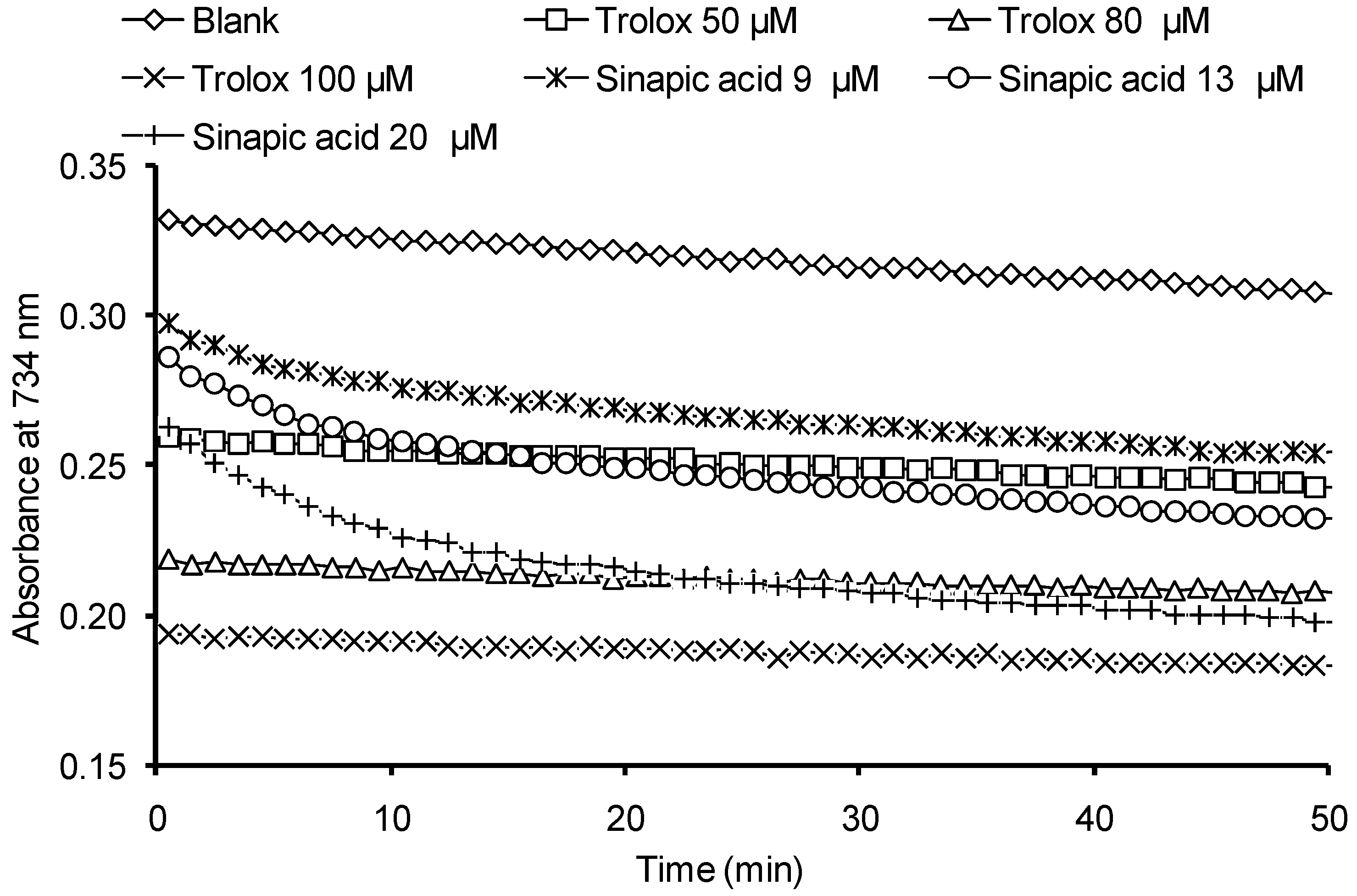
50]. The duration of the fast stage is 0.1 s, while the duration of the slow one is determined by the structure of an antioxidant and can be up to several hours. Thus, to properly quantify the AOC of HB and HC acids against the ABTS radical cation it appears necessary to determine the optimal reaction monitoring time range, in which the kinetics of the reaction for all the compounds involved would achieve the stationary phase. Examples of kinetic curves are shown in
Figure 2 describing the optical density decrease at interaction of Trolox and SinA with the ABTS cation–radical. For the rest of the investigated compounds the dependences obtained were similar to the SinA.
Figure 2.
Kinetic curves describing the optical density decrease at interaction of the ABTS radical cation solution with various antioxidants.
Figure 2.
Kinetic curves describing the optical density decrease at interaction of the ABTS radical cation solution with various antioxidants.
Trolox is characterized by mono-phase kinetics of interaction with the ABTS radical cation, whereas for the studied HB and HC acids the two-phase kinetics is typical, the stationary phase being achieved after 30–40 min of the reaction (
Figure 2). Thus, the AOC of phenolic acids against the ABTS radical cation is determined at 40th min of reaction.
The studied hydroxybenzoic and hydroxycinnamic acids have been previously found in saffron [
51], coffee [
41], olive oil [
52] and other antioxidant natural sources. The obtained AOC values of HB and HC acids against the ABTS radical action were compared to the earlier published data (
Table 3). For all investigated compounds with the exception of SirA, the AOC values defined in this paper were 1.2–1.6 times higher than those in literature, with the largest differences observed for sinapic acid due to the increase in reaction time.
Table 3.
Comparison of the experimental AOC of phenyl carbonic acids against the ABTS radical cation with literature data.
Table 3.
Comparison of the experimental AOC of phenyl carbonic acids against the ABTS radical cation with literature data.
| Phenolic Acid | TEAC (μmol TE/μmol) |
|---|
| p-HBA | 2.64 ± 0.07 | 2.64 ± 0.07 | – | – | – |
| VA | 2.63 ± 0.08 | 1.8 | – | 1.52 ± 0.01 | 1.42 ± 0.3 |
| SyrA | 1.46 ± 0.09 | 1.6 | – | – | 1.40 ± 0.03 |
| GalA | 5.76 ± 0.21 | 4.3 | – | – | 2.15 ± 0.01 |
| CA | 3.04 ± 0.18 | 2.3 | 2.39 ± 0.09 | – | 2.27 ± 0.01 |
| FA | 3.90 ± 0.21 | 3.2 | 1.97 ± 0.02 | 2.32 ± 0.09 | 1.87 ± 0.06 |
| SinA | 3.66 ± 0.15 | 2.2 | 2.09 ± 0.11 | – | – |
| Conditions of analysis | PBS, pH 7.40, 40th min | PBS, pH 7.40, 1st min | PBS, pH 7.40, 6th min | PBS, pH 7.40, 10th min | ethanol, 1st min |
| Data from | Experiment | [46] | [53] | [41] | [54] |
2.2. Influence of pH on Antioxidant Capacity (AOC)
It is known that the antioxidant action efficiency of HB and HC acids depends on the ionization degree of carboxyl group and phenolic hydroxyl. The ionization degree affects the distribution of electronic density in aromatic system. In particular, the increase of AOC against the ABTS radical cation–radical with increasing pH has been shown for a number of flavonoids and fluorinated
p-hydroxybenzoic acids [
22,
47].
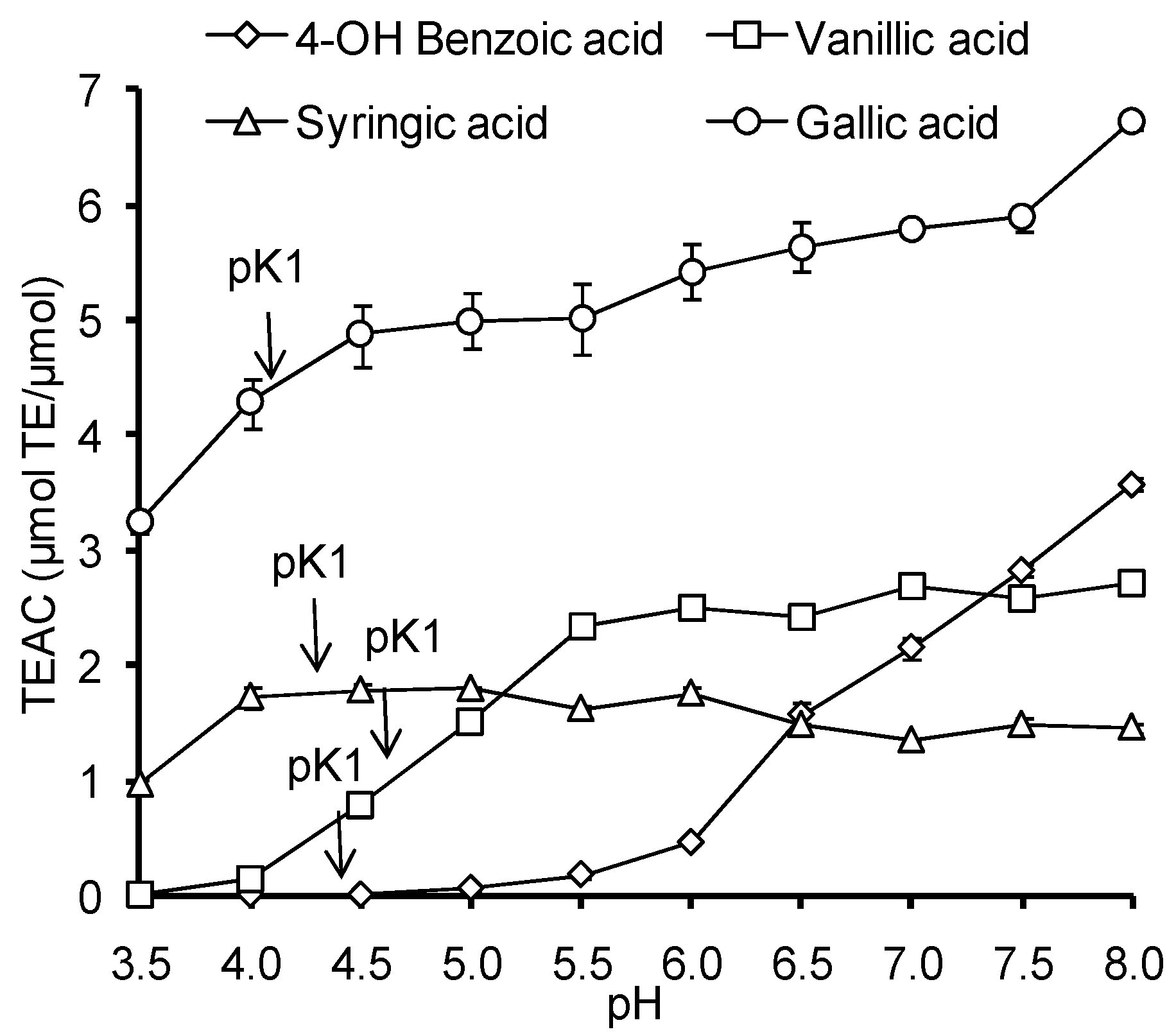
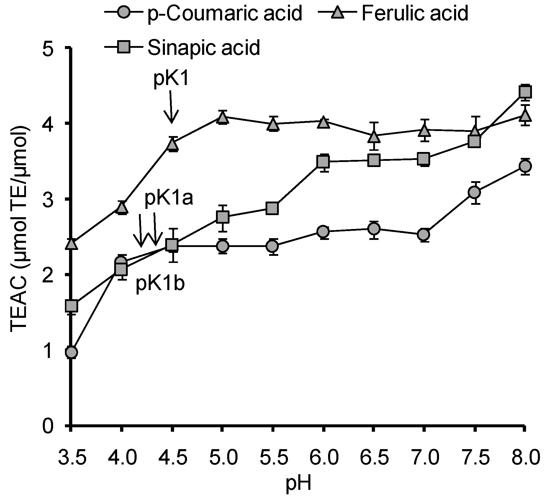
The influence of pH in the range of 3.5–8.0 on the AOC against the ABTS radical cation was studied for HB and HC acids (
Figure 3). In this pH range the ionization of carboxyl groups of HB and HC acids takes place, (pKa 4.10–4.58), and the ionization of phenolic hydroxyl begins in the case of GalA and SinA (p
Kphenol 8.90–8.94).
The AOC values of standard antioxidant—Trolox—in the investigated range of pH did not change despite the ionization of carboxyl group in a chromane ring (p
K = 3.89), due to the spatial remoteness of carboxyl and phenolic groups, lack of conjugation between benzene and chromane rings and high p
K value of trolox phenolic hydroxyl (p
K = 11.80) [
55].
Unlike Trolox, in all studied HB and HC acids the values of AOC increased with increasing pH, however, the dynamics and the intensity of that growth were individual for each compound. It was observed that
p-HBA and VA do not react with the ABTS radical cation at low pH, and exhibit the most pronounced growth of the AOC values with increasing pH (
Figure 3). For most acids (with the exception of
p-HBA), a pronounced growth of AOC value was observed in the range of carboxyl group ionization (4.0–5.5). It should be noted that the features of pH dependence of gallic acid AOC against the ABTS radical cation are in good agreement with the data of Lemanska
et al [
22] for pH range of 5.5 to 8.0.
The analysis of pH-dependencies of AOC for all investigated HB and HC acids, with the exception of
p-HBA, indicates that the carboxyl group deprotonation results in 1.7–2.4 times increase of their AOC against the ABTS radical cation (
Figure 3 and
Figure 4). At pH > 7.5 the deprotonation of phenolic hydroxyl begins, accompanied by a change of the antioxidant mechanism of phenolic acids the corresponding phenolate ions provide direct single electron transfer to the ABTS radical cation, which leads to an increase of AOC at pH 8.0 for GalA (p
Kphenol = 8.90), sinapic acid (p
Kphenol = 8.94), CA (p
Kphenol = 9.21) and, to a lesser extent, for FA (p
Kphenol = 9.21). For SirA and VA the p
K values representing the phenolic hydroxyl dissociation shifted to a more alkaline region (9.49 and 9.39 respectively), so the increase of AOC at pH 8.0 was not observed. Thus, the dissociation processes of ionogenic groups in HB and HC acids have a significant impact on their antioxidant properties. That is why for the further research of molecular and electronic descriptors, as well as for discrimination of antioxidant mechanisms of HB and HC acids, the quantum-chemical calculations were carried out for different states of ionization.
Figure 3.
pH-Dependence of AOC against the ABTS radical cation for HB and HC acids.
Figure 3.
pH-Dependence of AOC against the ABTS radical cation for HB and HC acids.
Figure 4.
pH-Dependence of AOC against the ABTS radical cation for HC acids (pK1a is sinapic acid related, pK1b is p-coumaric acid related).
Figure 4.
pH-Dependence of AOC against the ABTS radical cation for HC acids (pK1a is sinapic acid related, pK1b is p-coumaric acid related).
2.4. Spin Density and Electrophilic Attack of Radicals
The calculated Mulliken charges on the carbon atoms in the benzene ring, as well as oxygen and hydrogen atoms in phenolic hydroxyl and metoxy groups are presented in
Table 4. The atoms with the highest electronic density values are known to be sensitive to free-radical attacks [
8]. According to the
Table 4a,c, in the studied compounds the carbon atoms most preferable for electrophilic agent attacks are those located in
m-positions (C
2, C
4) to the phenolic hydroxyl. For VA, SyrA,
cis-CA,
trans-CA,
trans-FA or the corresponding hydroxybenzoates/hydroxycinnamates such preferred position is C
2.
Table 4.
(
a) Mulliken charges at carbon atoms of aromatic systems of HB acids, their radicals and cation–radicals in various states of ionization; (
b) Mulliken charges at oxygen and some hydrogen atoms of HB acids, their radicals and cation–radicals in various states of ionization; (
c) Mulliken charges at carbon atoms of aromatic systems of HC acids, their radicals and cation–radicals in various states of ioni
zation; (
d) Mulliken charges at oxygen and some hydrogen atoms of HB acids, their radicals and cation–radicals in various states of ionization (numbering of atoms according to
Figure 1).
Table 4.
(a) Mulliken charges at carbon atoms of aromatic systems of HB acids, their radicals and cation–radicals in various states of ionization; (b) Mulliken charges at oxygen and some hydrogen atoms of HB acids, their radicals and cation–radicals in various states of ionization; (c) Mulliken charges at carbon atoms of aromatic systems of HC acids, their radicals and cation–radicals in various states of ionization; (d) Mulliken charges at oxygen and some hydrogen atoms of HB acids, their radicals and cation–radicals in various states of ionization (numbering of atoms according to Figure 1).
| (a) |
| Phenolic Acid | State of Ionization | C3 | C4 | C5 | C6 | C1 | C2 |
| p-OH Benzoic | z
= 0 | 0.489 | −0.133 | −0.440 | −0.119 | −0.003 | −0.434 |
| z = −1 | 1.099 | −0.838 | 0.393 | −.0.890 | 0.468 | −0.885 |
| z = −2 | 0.932 | −0.619 | 0.314 | −1.108 | 0.314 | −0.619 |
| radical, z = 0 | 1.037 | −0.832 | 0.455 | −0.797 | 0.515 | −0.915 |
| radical, z = −1 | 1.151 | −0.938 | 0.535 | −0.838 | 0.535 | −0.938 |
| radical cation, z = −1 | 1.091 | −0.827 | 0.535 | −0.774 | 0.610 | −1.118 |
| radical cation, z = 0 | 1.301 | −0.909 | 0.520 | −0.809 | 0.446 | −0.190 |
| Vanillic | z = 0 | 0.569 | 0.518 | −0.455 | −0.633 | 0.194 | −0.775 |
| z = −1 | 0.590 | 0.379 | −0.585 | −0.410 | −0.104 | −0.371 |
| z = −2 | 0.763 | −0.176 | −0.375 | −0.513 | 0.164 | −0.597 |
| radical, z = 0 | 0.853 | 0.155 | −0.325 | −0.664 | 0.409 | −0.872 |
| radical, z = −1 | 0.931 | −0.078 | −0.287 | −0.622 | 0.501 | −0.858 |
| radical cation, z = −1 | 0.704 | −0.302 | −0.398 | −0.301 | −0.112 | −0.268 |
| radical cation, z = 0 | 0.782 | 0.335 | −0.497 | −0.324 | −0.062 | −0.446 |
| Syringic | z = 0 | 1.481 | −0.149 | −0.456 | −0.504 | 0.0552 | −0.328 |
| z = −1 | −0.240 | 0.278 | 0.120 | −0.038 | 0.142 | −0.278 |
| z = −2 | 0.547 | −0.040 | −0.794 | 0.516 | −.0793 | −0.042 |
| radical, z = 0 | 1.243 | −0.201 | −0.478 | −0.386 | −0.380 | −0.303 |
| radical, z = −1 | 1.249 | −0.071 | −0.459 | −0.498 | −0.459 | −0.071 |
| radical cation, z = −1 | 1.513 | −0.099 | −0.421 | −0.475 | −0.534 | −0.286 |
| radical cation z = 0 | 0.412 | 0.045 | 0.050 | 0.001 | 0.252 | −0.035 |
| Gallic | z = 0 | 0.859 | −0.376 | −0.371 | −0.166 | −0.219 | −0.152 |
| z = −1 | 0.910 | −0.262 | −0.425 | −0.127 | −0.225 | −0.237 |
| radical cation, z = −1 | 0.867 | −0.034 | 0.204 | −0.107 | −0.309 | −0.426 |
| radical cation, z = 0 | 1.113 | −0.256 | −0.168 | −0.138 | −0.391 | −0.234 |
| radical C1, z = 0 | 0.843 | −0.113 | −0.446 | 0.233 | 0.556 | 0.154 |
| radical C6, z = 0 | 0.788 | −0.147 | −0.354 | −0.079 | −0.350 | −0.274 |
| radical C5, z = 0 | 0.900 | −0.185 | −0.450 | 0.250 | −0.619 | −0.182 |
| radical C1, z = 0 | 0.796 | −0.111 | −0.502 | 0.284 | −0.637 | −0.069 |
| radical C6, z = −1 | 0.863 | −0.184 | −0.334 | −0.139 | −0.334 | −0.184 |
| radical C5, z = −1 | 0.900 | −0.114 | −0.506 | 0.301 | −0.696 | −0.108 |
| z = −2, C1 | 0.758 | −0.192 | −0.642 | 0.343 | −0.735 | −0.090 |
| z = −2, C6 | 0.695 | 0.040 | −0.486 | −0.048 | −0.655 | −0.145 |
| z = −2, C5 | 0.758 | −0.09 | −0.735 | 0.343 | −0.641 | −0.192 |
| z = −3, C1, C5 | 0.549 | 0.006 | −1.006 | 0.487 | −0.863 | 0.072 |
| z = −3, C1, C6 | 1.599 | −3.920 | 2.661 | 1.152 | −0.608 | −0.329 |
| z = −3, C6, C5 | 1.598 | −0.329 | −0.609 | 1.150 | 3.664 | −3.924 |
| (b) |
| Phenolic Acid | State of Ionization | O(C6) | O(C5) | O(C1) | H [O(C6)] | H [O(C5)] | H [O(C1)] |
| p-OH Benzoic | z = 0 | −0.223 | – | – | 0.273 | – | – |
| z = −1 | −0.269 | – | – | 0.242 | – | – |
| z = −2 | −0.559 | – | – | – | – | – |
| radical, z = 0 | −0.233 | – | – | – | – | – |
| radical, z = −1 | −0.350 | – | – | – | – | – |
| radical cation, z = −1 | −0.046 | – | – | 0.321 | – | – |
| radical cation, z = 0 | −0.209 | – | – | 0.268 | – | – |
| Vanillic | z
= 0 | −0.239 | −0.262 | – | 0.295 | – | – |
| z = −1 | −0.296 | −0.281 | – | 0.276 | – | – |
| z = −2 | −0.555 | −0.194 | – | – | – | – |
| radical, z = 0 | −0.250 | −0.106 | – | – | – | – |
| radical, z = −1 | −0.341 | −0.134 | – | – | – | – |
| radical cation, z = −1 | −0.094 | −0.180 | – | 0.332 | – | – |
| radical cation, z = 0 | −0.233 | −0.258 | – | 0.298 | – | – |
| Syringic | z
= 0 | −0.253 | −0.267 | −0.172 | 0.304 | – | – |
| z = −1 | −0.568 | −0.461 | −0.375 | 0.404 | – | – |
| z = −2 | −0.579 | −0.185 | −0.185 | – | – | – |
| radical, z = 0 | −0.253 | −0.122 | −0.121 | – | – | – |
| radical, z = −1 | −0.329 | −0.149 | −0.149 | – | – | – |
| radical cation, z = −1 | −0.099 | −0.242 | −0.096 | 0.347 | – | – |
| radical cation z = 0 | −0.500 | −0.447 | −0.353 | 0.424 | – | – |
| Gallic | z
= 0 | −0.240 | −0.232 | −0.306 | 0.290 | 0.270 | 0.288 |
| z = −1 | −0.297 | −0.273 | −0.337 | 0.270 | 0.259 | 0.274 |
| radical cation, z = −1 | −0.078 | −0.258 | −0.125 | 0.341 | 0.329 | 0.318 |
| radical cation, z = 0 | −0.232 | −0.301 | −0.227 | 0.293 | 0.289 | 0.274 |
| radical C1, z = 0 | −0.181 | −0.300 | −0.239 | 0.294 | 0.286 | – |
| radical C6, z = 0 | −0.247 | −0.176 | −0.175 | – | 0.271 | 0.268 |
| radical C5, z = 0 | −0.177 | −0.306 | −0.230 | 0.295 | – | 0.273 |
| radical C1, z = −1 | −0.237 | −0.330 | −0.306 | 0.273 | 0.271 | – |
| radical C6, z = −1 | −0.332 | −0.220 | −0.220 | – | 0.254 | 0.254 |
| radical C5, z = −1 | −0.235 | −0.374 | −0.271 | 0.274 | – | 0.257 |
| z = −2, C1 | −0.352 | −0.311 | −0.592 | 0.242 | 0.222 | – |
| z = −2, C6 | −0.613 | −0.320 | −0.295 | – | 0.235 | 0.211 |
| z = −2, C5 | −0.352 | −0.592 | −0.311 | 0.242 | – | 0.222 |
| z = −3, C1, C5 | −0.382 | −0.660 | −0.613 | 0.191 | – | – |
| z = −3, C1, C6 | −0.515 | −0.486 | −0.515 | – | −1.179 | – |
| z = −3, C6, C5 | −0.515 | −0.515 | −0.486 | – | – | −1.179 |
| (c) |
| Phenolic Acid | State of Ionization | C3 | C4 | C5 | C6 | C1 | C2 |
| cis-Caffeic | z = 0 | 0.809 | 0.029 | −0.410 | −0.296 | 0.027 | −1.045 |
| z = −1 | 1.331 | 0.245 | −0.386 | −0.328 | −0.009 | −1.293 |
| z = −2 | 1.767 | 0.231 | −0.163 | −0.860 | −0.008 | −1.315 |
| radical, z = 0 | 1.352 | 0.164 | −0.140 | −0.400 | 0.000 | −1.195 |
| radical, z = −1 | 1.706 | −0.885 | 0.312 | −0.729 | 0.308 | −0.884 |
| radical cation, z = −1 | 1.545 | −1.342 | 0.081 | −0.303 | −0.385 | 0.277 |
| radical cation, z = 0 | 1.537 | −1.409 | 0.093 | −0.422 | 0.379 | 0.230 |
| trans-Caffeic | z
= 0 | 1.316 | 0.342 | −0.319 | −0.481 | −0.099 | −1.262 |
| z = −1 | 1.193 | 0.320 | −0.327 | −0.497 | −0.071 | −1.258 |
| z = −2 | 1.549 | −0.520 | 0.393 | −1.076 | 0.247 | −1.356 |
| radical, z = 0 | 1.272 | 0.230 | −0.141 | −0.487 | −0.257 | −1.042 |
| radical, z = −1 | 1.314 | 0.012 | 0.047 | −0.648 | −0.070 | −1.297 |
| radical cation, z = −1 | 1.420 | 0.260 | −0.101 | −0.382 | −0.410 | −1.053 |
| radical cation, z = 0 | 1.272 | −1.287 | −0.102 | −0.484 | −0.320 | −0.355 |
| cis-Ferulic | z = 0 | 1.352 | −1.094 | 0.018 | 0.088 | −0.517 | 0.061 |
| z = −1 | 1.626 | −1.456 | −0.041 | 0.123 | −0.644 | 0.138 |
| z = −2 | 1.625 | −1.188 | −0.182 | −0.258 | −0.326 | −0.012 |
| radical, z = 0 | 1.481 | 0.054 | −0.329 | −0.006 | 0.152 | −1.425 |
| radical, z = −1 | 1.231 | −1.162 | −0.238 | 0.264 | −0.083 | −0.093 |
| radical cation, z = −1 | 1.589 | −1.724 | 0.350 | −0.431 | −0.668 | 0.570 |
| radical cation z = 0 | 1.896 | −1.503 | 0.002 | 0.159 | −0.599 | −0.015 |
| trans-Ferulic | z = 0 | 1.710 | 0.107 | −0.285 | −0.453 | −0.401 | −0.948 |
| z = −1 | 1.488 | 0.173 | −0.706 | −0.046 | 0.018 | −1.381 |
| z = −2 | 1.424 | −0.170 | −0.125 | −0.298 | −0.200 | −0.116 |
| radical, z = 0 | 1.136 | −0.652 | −0.640 | −0.396 | 0.109 | −0.181 |
| radical, z = −1 | 1.542 | 0.042 | −0.−23 | −0.297 | −0.169 | −1.250 |
| radical cation, z = +1 | 1.377 | −0.639 | −0.519 | −0.551 | −0.014 | −0.107 |
| radical cation z = 0 | 1.683 | 0.083 | −0.254 | −0.439 | −0.426 | −0.930 |
| cis-Sinapic | z = 0 | 1.564 | −0.739 | −0.477 | −0.093 | −0.368 | 0.042 |
| z = −1 | 2.014 | −0.856 | −0.739 | 0.027 | −0.226 | −0.337 |
| z = −2 | 1.411 | −1.014 | −0.219 | 0.011 | −0.289 | −0.044 |
| radical, z = 0 | 1.454 | −0.262 | −0.592 | −0.353 | 0.152 | −0.334 |
| radical, z = −1 | 1.740 | −0.754 | −0.883 | −0.112 | 0.162 | −0.146 |
| radical cation, z = −1 | 1.475 | −0.495 | −0.094 | −0.707 | −0.185 | −0.083 |
| radical cation, z = 0 | 1.662 | 0.164 | −0.490 | −0.63 | −0.537 | −0.630 |
| trans-Sinapic | z = 0 | 1.384 | −0.670 | −0.384 | −0.636 | −0.352 | 0.230 |
| z = −1 | 1.339 | −0.809 | −0.407 | −0.645 | −0.375 | 0.229 |
| z = −2 | 1.295 | −0.230 | −0.038 | 0.391 | −0.483 | −1.216 |
| radical, z = 0 | 1.648 | −0.180 | −0.357 | −0.373 | −0.564 | −0.742 |
| radical, z = −1 | 1.543 | −0.218 | 0.277 | −0.212 | −0.715 | −0.898 |
| radical cation, z = −1 | 1.761 | −0.092 | −0.139 | −0.666 | −0.843 | −0.425 |
| radical cation, z = 0 | 1.368 | −0.579 | −0.407 | −0.619 | −0.375 | −0.253 |
| (d) |
| Phenolic Acid | State of Ionization | O(C6) | O(C5) | O(C1) | H [O(C6)] | H [O(C5)] | H [O(C1)] |
| cis-Caffeic | z = 0 | | – | – | 0.268 | – | – |
| z = −1 | | – | – | 0.253 | – | – |
| z = −2 | | – | – | – | – | – |
| radical, z = 0 | | – | – | – | – | – |
| radical, z = −1 | | – | – | – | – | – |
| radical cation, z = −1 | | – | – | 0.315 | – | – |
| radical cation, z = 0 | | – | – | 0.271 | – | – |
| trans-Caffeic | z = 0 | | – | – | 0.271 | – | – |
| z = −1 | | – | – | 0.254 | – | – |
| z = −2 | | – | – | – | – | – |
| radical, z = 0 | | – | – | – | – | – |
| radical, z = −1 | | – | – | – | – | – |
| radical cation, z = −1 | | – | – | 0.310 | – | – |
| radical cation, z = 0 | | – | – | 0.273 | – | – |
| cis-Ferulic | z = 0 | | – | −0.162 | 0.276 | – | – |
| z = −1 | | – | −0.168 | 0.262 | – | – |
| z = −2 | | – | −0.200 | – | – | – |
| radical, z = 0 | | – | −0.175 | – | – | – |
| radical, z = −1 | | – | −0.367 | – | – | – |
| radical cation, z = −1 | | −1.724 | −0.086 | 0.315 | – | – |
| radical cation z = 0 | | −1.503 | 0.154 | 0.280 | – | – |
| trans-Ferulic | z = 0 | | 0.107 | – | 0.296 | – | – |
| z = −1 | | 0.173 | – | 0.263 | – | – |
| z = −2 | | −0.170 | – | – | – | – |
| radical,z = 0 | | −0.652 | – | – | – | – |
| radical, z = −1 | | 0.042 | – | – | – | – |
| radical cation, z = −1 | | −0.639 | – | 0.335 | – | – |
| radical cation z = 0 | | 0.083 | – | 0.298 | – | – |
| cis-Sinapic | z = 0 | | −0.739 | −0.163 | 0.308 | – | – |
| z = −1 | | −0.856 | −0.170 | 0.296 | – | – |
| z = −2 | | −1.014 | −0.195 | – | – | – |
| radical, z = 0 | | −0.262 | −0.199 | – | – | – |
| radical, z = −1 | | −0.754 | −0.180 | – | – | – |
| radical cation, z = −1 | | −0.495 | −0.123 | 0.342 | – | – |
| radical cation, z = 0 | | 0.164 | −0.152 | 0.311 | – | – |
| trans-Sinapic | z = 0 | | −0.670 | −0.174 | 0.305 | – | – |
| z = −1 | | −0.809 | −0.187 | 0.291 | – | – |
| z = −2 | | −0.230 | −0.177 | – | – | – |
| radical, z = 0 | | −0.180 | −0.270 | – | – | – |
| radical, z = −1 | | −0.218 | −0.145 | – | – | – |
| radical cation, z = −1 | | −0.092 | −0.124 | 0.344 | – | – |
| radical cation, z = 0 | | −0.579 | −0.171 | 0.308 | – | – |
For GalA,
cis-SinA, and
trans-SinA in both states of ionization the preferred position for electrophilic attack is C
4. For GalA and
p-HBA with ionized carboxyl groups electrophilic attacks could equally well be aimed at both positions C
2 and C
4. In the case of
cis- and
trans-isomers of HC acids and
p-HBA the deprotonation of carboxyl group leads to 1.1–1.4 times increase of the electron density on the carbon atom preferred for electrophilic attacks (
Table 4a,c). On the contrary to the HC acids, for GalA, SyrA and VA the deprotonation of carboxyl group was noted to reduce the spin density at C
4 (GalA) and C
2 (SyrA, VA) positions.
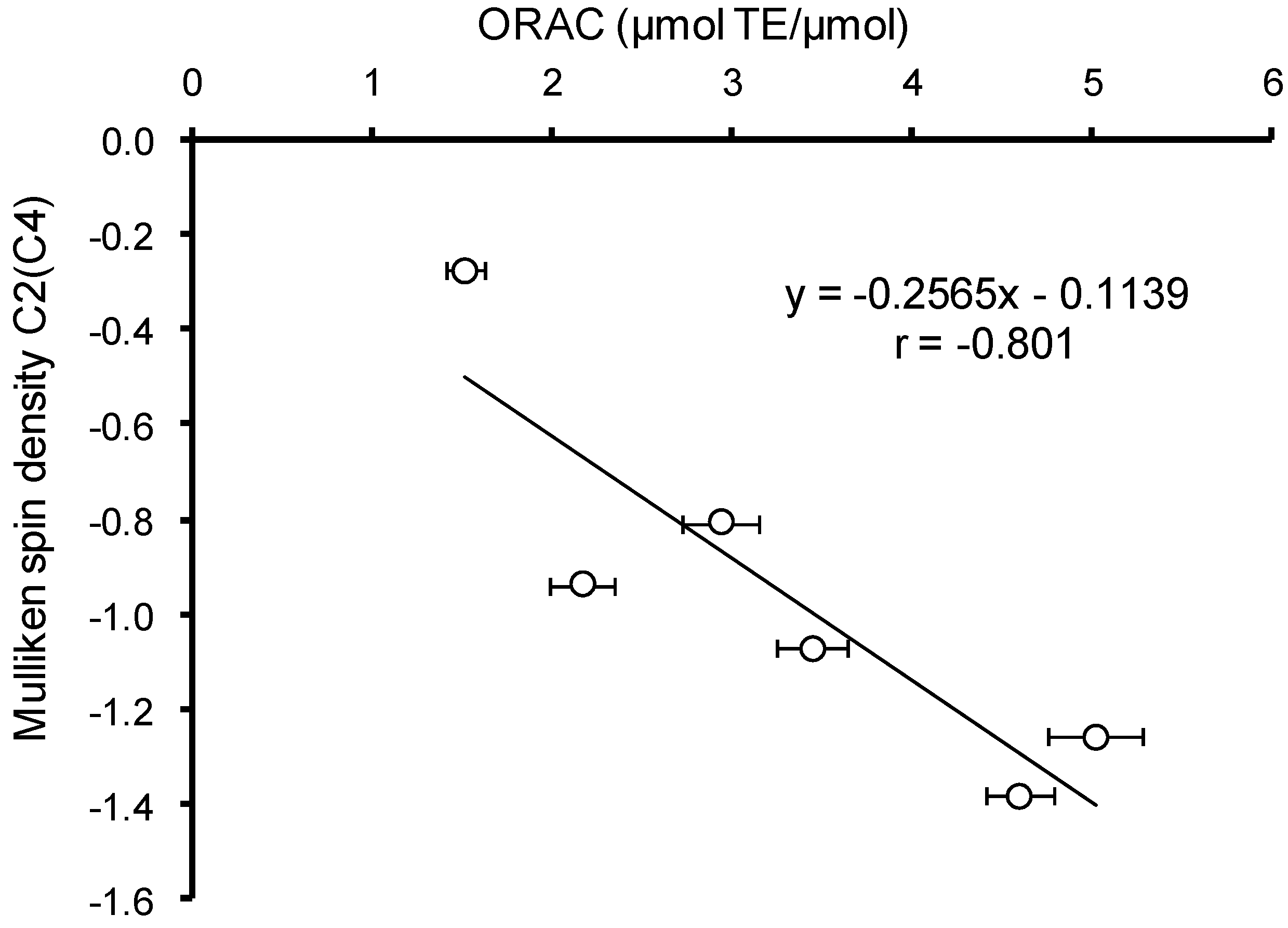
The increase of the electron density was expected to contribute to the efficiency of the primary interaction of hydroxyaromatic acids with free radicals and other electrophilic particles. To confirm this assumption was performed the correlation analysis between Mulliken charge values on the carbon atom preferable for electrophilic compound attacks and AOC values of HB and HC acids against the ABTS radical cation and peroxyl radical. In the case of ABTS no significant correlation was revealed for various states of ionization, due to the uncompetitive nature of this analysis method and the existence of a large number of secondary reactions, contributing to the AOC value. In the case of peroxyl radical, the experiments were carried out at pH 7.40, thus, the correlation analysis was carried out only for monoanion forms of HB and HC acids. A significant (
p < 0.05) inverse correlation (
r = −0.801) between the Mulliken charge and AOC against peroxyl radical was observed (
Figure 5). The exception of VA from the analysis arrays increases the value of the Spearman correlation coefficient to 0.925 without change of correlation significance.
Figure 5.
Correlation analysis between AOC of hydroxyaromatic acids against peroxyl radical and Mulliken charge at carbon atom preferred for electrophilic agent attacks.
Figure 5.
Correlation analysis between AOC of hydroxyaromatic acids against peroxyl radical and Mulliken charge at carbon atom preferred for electrophilic agent attacks.
HB acids are characterized by higher values of Mulliken charge on the carbon atom in
meta-position to the phenolic hydroxyl compared to their structural analogues among HC acids, which correlates with the previously identified differences in the values of their AOC against peroxyl radical (
Table 2).
In addition to the changes in the electron density at C
2/C
4 position in the benzene ring, the carboxyl groups, deprotonation results in reduction of O–H bond polarity and facilitates the hydrogen atom transfer. Thus, the sum of Mulliken charges for oxygen and hydrogen atoms of phenolic hydroxyl in non-ionized form of HB and HC acids varies within 0.050–0.056 and 0.032–0.056, respectively (
Table 4b,d). For the relevant benzoates and cinnamates the sum of Mulliken charges for oxygen and hydrogen atoms of phenolic hydroxyl reduced to (−0.164)–(−0.020) and (−0.024)–(−0.001).
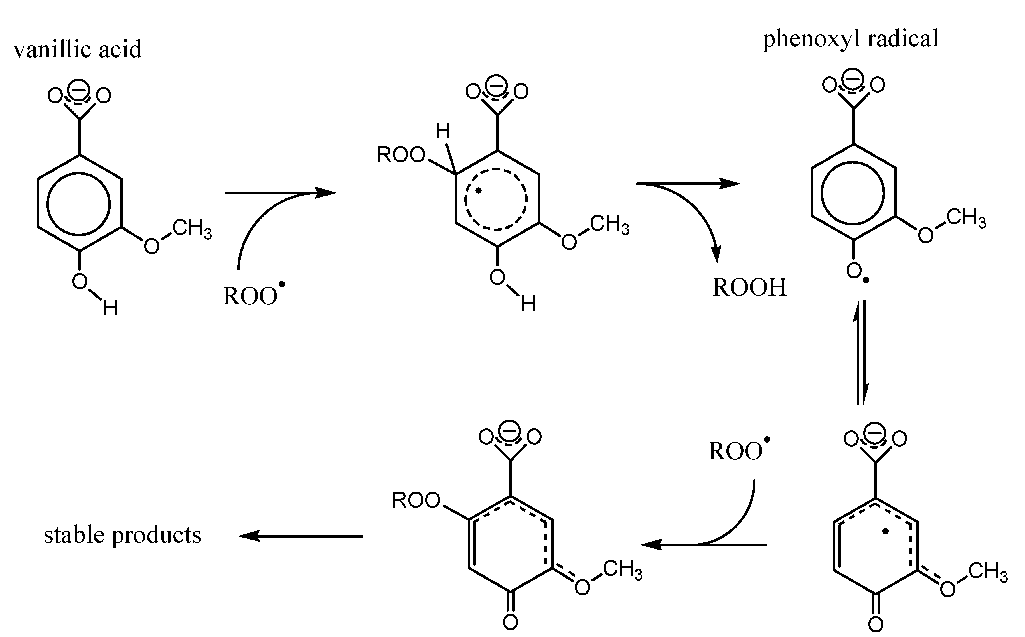
Based on the data obtained, the scheme was suggested that describes the mechanism of interaction of
p-HB and
p-HC acids (VA as an example) with peroxyl radical (
Figure 6). The primary attack by the peroxyl radical aims the aromatic system at a carbon atom with the largest electronic density (lowest Mulliken charge), which is C
2 in the case of VA, with further hydrogen atom abstraction and formation of hydroperoxide ROOH and phenoxyl radical. Further the delocalization of the unpaired electron leads to the formation of the most thermodynamically stable form of phenoxyl radical. In case of vanillic acid, maximum electron density is localized on C
2 carbon atom, where the secondary peroxyl radical attack aims, followed by further chemical transformations yielding the stable products.
As shown in
Figure 6, the presence of the substituents in
ortho-positions to the phenolic hydroxyl can stabilize the resulting phenoxyl radicals, reducing the probability of recombination and increasing the efficiency of secondary reactions with peroxyl radical. This correlates with the increase of the AOC of phenolic antioxidants.
Figure 6.
Mechanism of peroxyl radical interaction with p-HB and p-HC acids (Vanillic (VA) as an example).
Figure 6.
Mechanism of peroxyl radical interaction with p-HB and p-HC acids (Vanillic (VA) as an example).
As follows from the data in
Table 4a–d, the stabilization effect was ascertained for radicals and radical cations of VA, SyrA,
cis-/
trans-FA and
cis-/
trans-SinA with non-ionised carboxyl group. However, the introduction of substituents with negative inductive effect in the benzene ring creates a steric barrier for interaction with bulk radicals, and also variants the electron density in the benzene ring.
Thus, AOC of HB and HC acids depends on the nature and positions of substituents, namely:
- (1)
The influence of substituents on Mulliken charge on carbon atoms in C2/C4 positions.
- (2)
The stabilization effects due to delocalization of the unpaired electron in phenoxyl radicals.
- (3)
The steric effects of the substituents.
A following trend is observed when comparing the trans-CA and trans-SinA: the introduction of two metoxy groups leads to a decrease of the electron density in C4 position and creates steric barriers, which in combination results in 1.7 times lower trans-SinA AOC against peroxyl radical compared to trans-CA. Introduction of 1 metoxy group in the benzene ring (trans-FA) leads only to a minor (1.1 times) reduction of AOC against peroxyl radical, correlating with an insignificant increase of the spin density at C2 carbon atom of the benzene ring.
2.5. Structural Descriptors and Stabilizzation of the Phenoxyl Radical
The stabilization mechanism in HC acids can involve, besides metoxy groups, the C
3 chain, its conjugation degree with the benzene ring being dependent on their relative positions. Such stabilization effects are considered to cause higher values of AOC in HC acids compared to their structural analogues among HB acids [
54], which is confirmed by results of the quantum-chemical calculations (
Table 2). It was observed that the side chain participates in delocalization of unpaired electrons in
cis-isomers of FA and SinA during the formation of a radical with a deprotonated carboxyl group. The side chain of
trans-isomers of HC acids participate in delocalization of unpaired electrons in case of radicals and radical cations corresponding to the uncharged acid form, and also a radical of monoanion form.
The maximum degree of conjugation between the benzene ring and the side chain in HC acids will be achieved when the carbon atoms in the benzene ring and the double bond in C
3 chain share one plane. To estimate the possibility of π–π conjugation of the carbon atoms in the benzene ring with C3 side chain for different states of ionization of
cis- and
trans-isomers of HC acids, the values of dihedral angles between 4, 3, 7, 8 and 2, 3, 7, 8 carbon atoms were calculated (
Table 5). The analysis of the data in
Table 5 shows that the delocalization of the unpaired electron in the phenoxyl radicals of HC acids is possible when the angle between the planes of the benzene ring and C3 side chain is no more than ±10°. Thus, such delocalization is impossible in cation–radicals of
cis-isomers of HC acids and uncharged radicals of HC acids. At the same time, in the case of
trans-isomers there is a possibility of electron delocalization in phenoxyl radicals and radical cations in all states of ionization of HC acids, which increases their stability. Apparently, it is the greater stability of HC acid phenoxyl radicals that explains the occurrence of
trans-HC acid and aldehyde links in the chains of natural lignins.
Table 5.
Geometry parameters of HC acid molecules and corresponding radicals and radical cations in various states of ionization. Atom numbering according to
Figure 1.
Table 5.
Geometry parameters of HC acid molecules and corresponding radicals and radical cations in various states of ionization. Atom numbering according to Figure 1.
| Phenolic Acid | State of Ionization | D (°) | Length of the Bond (Å) |
|---|
| (4,3,7,8) | (2,3,7,8) | O–H | C6–O | C1–O | C5–O |
|---|
| cis-p-Coumaric | z = 0 | 159.35 | 24.14 | 0.96 | 1.36 | – | – |
| z = −1 | −167.98 | 13.06 | 0.96 | 1.39 | – | – |
| z = −2 | 180.00 | 0.01 | – | 1.28 | – | – |
| radical, z = 0 | −161.99 | 20.29 | – | 1.24 | – | – |
| radical, z = −1 | −96.46 | 94.99 | – | 1.26 | – | – |
| radical cation, z = − 1 | −11.80 | 169.21 | 0.97 | 1.32 | – | – |
| radical cation, z = 0 | −8.21 | 172.39 | 0.96 | 1.36 | – | – |
| trans-p-Coumaric | z = 0 | 179.99 | −0.01 | 0.96 | 1.36 | – | – |
| z = −1 | 176.18 | −3.89 | 0.96 | 1.38 | – | – |
| z = −2 | −180.01 | −0.01 | – | 1.28 | – | – |
| radical, z = 0 | −180.02 | −0.01 | – | 1.24 | – | – |
| radical, z = −1 | 178.09 | −1.60 | – | 1.25 | – | – |
| radical cation, z = − 1 | −179.99 | 0.01 | 0.97 | 1.32 | – | – |
| radical cation, z = 0 | 0.00 | −180.00 | 0.96 | 1.36 | – | – |
| cis-Ferulic | z = 0 | −38.48 | 144.81 | 0.96 | 1.36 | 1.37 | – |
| z = −1 | −12.28 | 168.97 | 0.97 | 1.38 | 1.39 | – |
| z = −2 | 0.71 | −179.02 | – | 1.28 | 1.40 | – |
| radical, z = 0 | −153.78 | 28.62 | – | 1.24 | 1.33 | – |
| radical cation, z = 0 | −7.05 | 174.42 | – | 1.26 | 1.37 | – |
| radical cation, z = − 1 | −13.65 | 167.82 | 0.97 | 1.32 | 1.32 | – |
| trans-Ferulic | z = 0 | −180.03 | −0.02 | 0.97 | 1.36 | – | 1.37 |
| z = −1 | 179.66 | −0.06 | 0.97 | 1.37 | – | 1.38 |
| z = −2 | 179.69 | 0.08 | – | 1.28 | – | 1.40 |
| radical, z = 0 | −0.01 | 179.99 | – | 1.23 | – | 1.34 |
| radical cation, z = 0 | 180.00 | −0.00 | 0.97 | 1.35 | – | 1.37 |
| cis-Sinapic | z = 0 | −33.83 | 148.72 | 0.97 | 1.36 | 1.37 | 1.37 |
| z = −1 | −11.67 | 169.50 | 0.97 | 1.38 | 1.39 | 1.38 |
| z = −2 | 2.01 | −177.59 | – | 1.28 | 1.40 | 1.40 |
| radical, z = 0 | −24.99 | 156.92 | – | 1.24 | 1.34 | 1.34 |
| radical, z = −1 | −9.20 | 172,50 | – | 1.25 | 1.37 | 1.35 |
| radical cation, z = −1 | −13.75 | 167.28 | 0.98 | 1.31 | 1.32 | 1.34 |
| radical cation, z = 0 | −140.16 | 42.29 | 0.97 | 1.36 | 1.36 | 1.37 |
| trans-Sinapic | z = 0 | 0.01 | 180.01 | 0.97 | 1.35 | 1.36 | 1.37 |
| z = −1 | −7.10 | 172.93 | 0.97 | 1.37 | 1.37 | 1.38 |
| z = −2 | 178.44 | −0.53 | – | 1.28 | 1.40 | 1.40 |
| radical, z = −1 | 176.02 | −2.91 | – | 1.25 | 1.36 | 1.37 |
| radical cation, z = −1 | −180.00 | −0.01 | 0.98 | 1.31 | 1.32 | 1.34 |
| radical cation, z = 0 | −4.02 | 175.93 | 0.97 | 1.35 | 1.36 | 1.37 |
2.7. Energy Descriptors and Antioxidant Capacity
For further analysis of the antioxidant action mechanisms, the corresponding sets of parameters were calculated for HB and HC acids in different states of ionization:
- (1)
The mechanism of direct hydrogen atom transfer (HAT): bond dissociation enthalpy (BDE) for O–H of the phenolic hydroxyl.
- (2)
The mechanism of single electron transfer followed by proton transfer (SET–PT): ionization potential (IP) and the proton dissociation enthalpy (PDE).
- (3)
The mechanism of sequential proton loss and electron transfer (SPLET): proton affinity (PA) and electron transfer enthalpy (ETE).
The IP values were calculated on the basis of the data on molecular orbital energies (IPo) and the total energy of the molecules (IPe), according to Equations (6) and (7). The values of ETE, PA, and PDE were calculated for vacuum at a temperature of 298K with subtraction of proton (H
+) and electron formation enthalpy. The obtained values are not the absolute values of the thermodynamic parameters considered, but they can be properly used to compare different HB and HC acids and reveal the correlation relationships in the context of the present work. A similar approach was used when characterizing the antioxidant properties of flavonoids quercetin and myricetin [
20]. The calculated thermodynamic and energy parameters for HB and HC acid molecules are presented in
Table 6a,b.
where: E
HOMO—energy of the highest occupied molecular orbital; (
εo +
Etot)—total energy for the molecule and cation radical of the antioxidant, respectively.
For non-ionised form of HB and HC acids the IPe values range from 7.27 eV (
cis-SinA) to 8.62 eV (
p-HBA). The typical IP values in vacuum for the phenolic compounds in non-ionised form are ≤9 eV [
56]. For monoanion forms of HB and HC acids the calculated IPe values range from 3.33 to 3.85 eV (
Table 6a). The obtained IPe values for monoanion forms of HB and HC acids are 1.2–1.3 times lower than those found in literature for
p-HBA (4.49 eV), GalA (4.44 eV),
trans-CA (4.32 eV),
trans-FA (4.89 eV) and
trans-SinA (4.51 eV) [
47,
53]. These differences emerge due to the features of semiempirical calculation methods in the framework of the density functional theory [
57].
The data in
Table 6b shows that the PDE of phenolic hydroxyl O-H in non-ionised forms of HB and HC acids decreases in the row:
p-HBA (86.55 kcal/mol) > VA (85.76 kcal/mol) >
trans-CA (82.54 kcal/mol) > GalA (82.35 kcal/mol) >
trans-FA (82.14 kcal/mol) > SyrA (81.65 kcal/mol) >
trans-SinA (78.57 kcal/mol), which agrees with the data in other works [
58,
59]. It should also be noted that the calculated O–H bond BDE in the gas phase for non-ionized forms of HB and HC acids fits within the range of values of the same thermodynamic parameter (75.78–81.41 kcal/mol) for widely known natural antioxidants—tocopherols [
60].
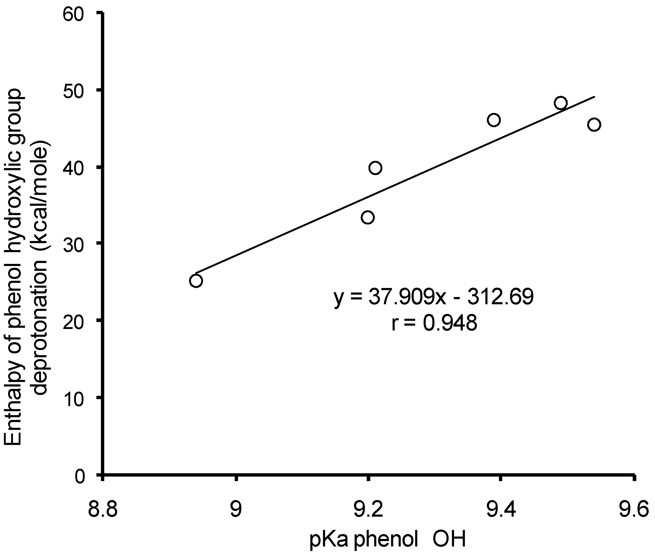
Based on the values of enthalpy of formation for monoanion and dianion forms of HB and HC acids, the enthalpies of phenyl hydroxyl deprotonation have been calculated. The enthalpy of H
+ formation was 1530.17 kJ/mol [
20]. The comparison of calculated values for enthalpy of phenyl hydroxyl deprotonation to the experimental pKa values for mono-HB and mono-HC acids in aqueous solutions [
61] revealed a significant (
p < 0.05) correlation (
r = 0.948) (
Figure 7).
Table 6.
(a,b) Thermodynamic and energy parameters of HB and HC acids in various states of ionization in the gas phase (298 K).
Table 6.
(a,b) Thermodynamic and energy parameters of HB and HC acids in various states of ionization in the gas phase (298 K).
| (a) |
| Phenolic Acid | BDE, kcal/mol | IPe, eV | IPo, eV | EHOMO, eV | ELUMO, eV | χ, eV |
| p-OH Benzoic | | | | | | |
| z = 0 | 86.55 | 8.62 | 6.86 | −6.866 | −1.576 | 4.221 |
| z = −1 | 72.95 | 3.42 | 1.63 | −1.627 | 1.884 | −0.129 |
| z = −2 | – | −1.11 | −2.76 | 3.757 | 5.421 | −4.089 |
| Vanillic | | | | | | |
| z = 0 | 85.76 | 8.04 | 6.41 | −6.413 | −1.527 | 3.970 |
| z = −1 | 74.83 | 3.43 | 1.68 | −1.679 | 1.873 | −0.097 |
| z = −2 | – | −1.05 | −2.55 | 2.554 | 4.587 | −3.570 |
| Siringic | | | | | | |
| z = 0 | 81.65 | 7.66 | 6.12 | −6.121 | −1.485 | 3.803 |
| z = −1 | 73.21 | 3.40 | 1.69 | −1.691 | 1.906 | −0.107 |
| z = −2 | – | −1.01 | −2.36 | 2.363 | 4.478 | −3.421 |
| Gallic | | | | | | |
| z = 0, C1 | 82.35 | 8.06 | 6.41 | −6.405 | −1.645 | 4.025 |
| z = 0, C6 | 82.86 | 8.06 | 6.41 | −6.405 | −1.645 | 4.025 |
| z = 0, C5 | 77.97 | 8.06 | 6.41 | −6.405 | −1.645 | 4.025 |
| z = −1, C1 | 75.70 | 3.57 | 1.83 | −1.827 | 1.933 | −0.053 |
| z = −1, C6 | 73.26 | 3.57 | 1.83 | −1.827 | 1.933 | −0.053 |
| z = −1, C5 | 69.97 | 3.57 | 1.83 | −1.827 | 1.933 | −0.053 |
| z = −2, C1 | – | −0.68 | −2.56 | 2.580 | 4.923 | −3.571 |
| z = −2, C6 | – | −0.74 | −2.65 | 2.651 | 5.064 | −3.858 |
| z = −2, C5 | – | −0.93 | −2.58 | 2.580 | 4.293 | 3.751 |
| cis-Caffeic | | | | | | |
| z = 0 | 87.06 | 8.12 | 6.41 | −6.413 | −2.119 | 4.266 |
| z = −1 | 77.73 | 3.44 | 1.74 | −1.736 | 1.831 | −0.047 |
| z = −2 | – | −0.44 | −2.18 | 2.175 | 4.775 | −3.475 |
| trans-Caffeic | | | | | | |
| z = 0 | 82.54 | 8.00 | 6.43 | −6.435 | −2.150 | 4.292 |
| z = −1 | 67.80 | 3.33 | 1.58 | −1.581 | 1.429 | 0.076 |
| z = −2 | – | −0.52 | −1.97 | 1.968 | 4.873 | −3.420 |
| cis-Ferulic | | | | | | |
| z = 0 | 77.46 | 7.53 | 6.65 | −6.646 | −2.105 | 4.376 |
| z = −1 | 76.03 | 3.44 | 1.79 | −1.787 | 1.673 | 0.051 |
| z = −2 | – | −0.54 | −2.02 | 2.022 | 4.210 | −3.117 |
| trans-Ferulic | | | | | | |
| z = 0 | 82.14 | 7.66 | 6.14 | −6.144 | −2.100 | 4.122 |
| z = −1 | 82.64 | 3.85 | 1.54 | −1.537 | 1.615 | −0.038 |
| z = −2 | – | −0.44 | −1.82 | 1.822 | 4.172 | −2.997 |
| cis-Sinapic | | | | | | |
| z = 0 | 78.92 | 7.27 | 6.35 | −6.347 | −2.089 | 4.218 |
| z = −1 | 70.20 | 3.61 | 1.89 | −1.890 | −1.816 | 0.037 |
| z = −2 | – | −0.80 | −1.88 | 1.885 | 4.144 | −3.014 |
| trans-Sinapic | | | | | | |
| z = 0 | 78.57 | 7.38 | 5.94 | −5.940 | −2.082 | 4.011 |
| z = −1 | 69.25 | 3.58 | 1.64 | −1.635 | 1.541 | 0.047 |
| z = −2 | – | −0.38 | −1.68 | – | 4.085 | −2.882 |
| (b) |
| Phenolic Acid | η, eV | ω × 103, eV | EHOMO–ELUMO, eV | ETE, kJ/mol | PA, kJ/mol | PDE, kJ/mol |
| p-OH Benzoic | | | | | | |
| z = 0 | 2.645 | 842 | 5.290 | – | – | 838.5 |
| z = −1 | 1.755 | 1.179 | 3.511 | −106.87 | 1720.5 | 1283.3 |
| z = −2 | 1.131 | 1569 | 2.664 | – | – | – |
| Vanillic | | | | | | |
| z = 0 | 2.443 | 806 | 4.887 | – | – | 891.5 |
| z = −1 | 1.776 | 0.658 | 3.552 | −101.69 | 1723.1 | 1290.9 |
| z = −2 | 1.017 | 1567 | 2.033 | – | – | – |
| Siringic | | | | | | |
| z = 0 | 2.318 | 780 | 4.636 | – | – | 910.0 |
| z = −1 | 1.798 | 0.800 | 3.597 | −97.85 | 1260.4 | 834.9 |
| z = −2 | 1.058 | 1383 | 2.116 | – | – | – |
| Gallic | | | | | | |
| z = 0, C1 | 2.380 | 851 | 4.760 | – | – | 875.3 |
| z = 0, C6 | 2.380 | 851 | 4.760 | – | – | 877.4 |
| z = 0, C5 | 2.380 | 851 | 4.760 | – | – | 856.9 |
| z = −1, C1 | 1.880 | 0.188 | 3.760 | −65.99 | 1691.1 | 1281.0 |
| z = −1, C6 | 1.880 | 0.188 | 3.760 | −71.05 | 1686.0 | 1270.8 |
| z = −1, C5 | 1.880 | 0.188 | 3.760 | −89.97 | 1691.1 | 1257.0 |
| z = −2, C1 | 1.172 | 1501 | 2.343 | – | – | – |
| z = −2, C6 | 1.207 | 1541 | 2.414 | – | – | – |
| z = −2, C5 | 1.171 | 1501 | 2.343 | – | – | – |
| cis-Caffeic | | | | | | |
| z = 0 | 2.147 | 1060 | 4.294 | – | – | – |
| z = −1 | 1.783 | 0.156 | 3.567 | −42.30 | 1675.9 | 889.4 |
| z = −2 | 1.300 | 1161 | 2.600 | – | – | 1301.3 |
| trans-Caffeic | | | | | | |
| z = 0 | 2.143 | 10.75 | 4.285 | – | – | – |
| z = −1 | 1.505 | 0.481 | 3.010 | −49.82 | 1641.8 | 881.7 |
| z = −2 | 1.452 | 1007 | 2.905 | – | – | 1270.6 |
| cis-Ferulic | | | | | | |
| z = 0 | 2.270 | 1054 | 4.541 | – | – | – |
| z = −1 | 1.730 | 0.232 | 3.460 | −42.27 | 1656.2 | 906.1 |
| z = −2 | 1.094 | 1109 | 2.188 | – | – | 1141.3 |
| trans-Ferulic | | | | | | |
| z = 0 | 2.022 | 1050 | 4.044 | – | – | – |
| z = −1 | 1.576 | 0.119 | 3.152 | −42.45 | 1696.6 | 913.4 |
| z = −2 | 1.175 | 955 | 2.350 | – | – | 1283.0 |
| cis-Sinapic | | | | | | |
| z = 0 | 2.129 | 1044 | 4.258 | – | – | – |
| z = −1 | 1.853 | 0.091 | 3.706 | −77.64 | 1679.7 | 936.9 |
| z = −2 | 1.130 | 1006 | 2.259 | – | – | 1253.5 |
| trans-Sinapic | | | | | | |
| z = 0 | 1.929 | 1043 | 3.858 | – | – | 925.2 |
| z = −1 | 1.588 | 0.071 | 3.176 | −37.04 | 1635.2 | 1253.1 |
| z = −2 | 1.203 | 863 | 2.405 | – | – | – |
Figure 7.
Correlation analysis of deprotonation enthalpy versus pKa values of phenyl hydroxyls of mono-HB and mono-HC acids.
Figure 7.
Correlation analysis of deprotonation enthalpy versus pKa values of phenyl hydroxyls of mono-HB and mono-HC acids.
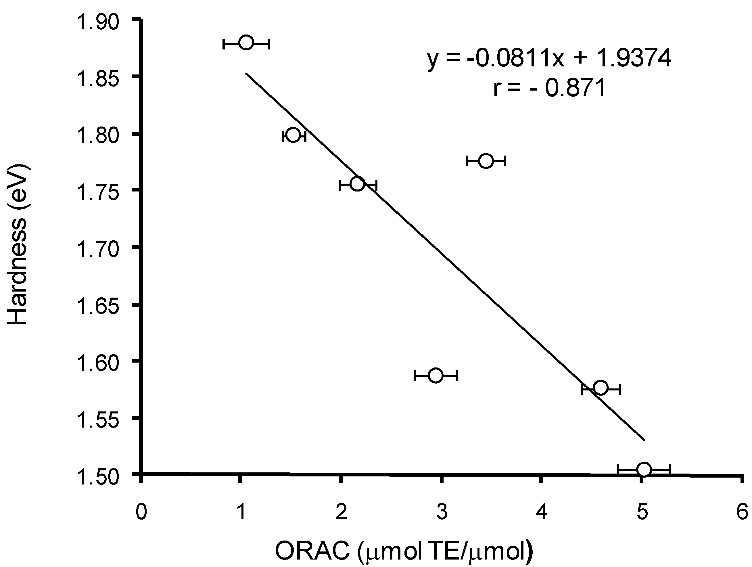
The comparative analysis of the AOC of HB and HC acids against peroxyl radical (
Table 2)
versus calculated quantum-chemical descriptors of the hydroxy-aromatic acids (
Table 6a,b) has revealed a significant (
p < 0.05) correlation between the values of AOC, energy of HOMO (
r = 0.824), and rigidity (
r = −0.871) for monoanion forms of these compounds (
Figure 8 and
Figure 9). Thus, the lower values of AOC against peroxyl radical for HB acids compared to HC acids can be explained by their higher HOMO energy and rigidity.
Figure 8.
Correlation analysis of AOC against peroxyl radical for HB and HC acids versus HOMO energy values.
Figure 8.
Correlation analysis of AOC against peroxyl radical for HB and HC acids versus HOMO energy values.
Figure 9.
Correlation analysis of AOC against peroxyl radical for HB and HC acids versus rigidity (η).
Figure 9.
Correlation analysis of AOC against peroxyl radical for HB and HC acids versus rigidity (η).
Since AOC of HB and HC acids is largely dependent on pH of reaction medium (
Figure 2), the understanding of mechanisms for their antioxidant action required to analyse how thermodynamic and energy parameters of HB and HC acids depend on their state of ionization. The analysis of the data in
Table 6a,b indicates that carboxyl group deprotonation in GB and GC acids leads to the 1.1 times reduction of O–H bond BDE, the 2.2 times IPe reduction, and also the 1.3 times reduction of rigidity and difference between LUMO and HOMO energies. In addition, the carboxyl group deprotonation in HB and HC acids increases the HOMO energy and reduces the electronegativity of the molecules. Altogether, the above changes in energy and thermodynamic parameters indicate a significantly lower stability of the monoanion form of HB and HC acids compared to the non-ionised form, therefore, the carboxyl group deprotonation should result in greater reactivity of HB and HC acids. The last assumption is fully confirmed experimentally by the significant AOC increase of HB and HC acids in the pH range corresponding to the ionization change of carboxyl group (
Figure 2).
Further phenolic hydroxyl deprotonation and phenolate-anion formation leads to a more severe destabilization of HB and HC acid molecules—the HOMO energy reaches positive values whereas the IPe values drop significantly (
Table 6a). The rigidity of the molecules and the difference between LUMO and HOMO energies reduce 2.2 times compared to the non-ionized form of HB and HC acids. The negative IPe values of the phenolate anions of HB and HC acids indicate their high electron-donating abilities. Therefore, even the smallest quantities of HB and HC acids present in the solution in phenolate form will very quickly eliminate due to the interaction with oxidizing agents, with a result of continuous shift of chemical equilibrium in the reaction of dissociation of phenolic hydroxyl in HB and HC acids. Thus, the most likely mechanism of interaction between HB/HC acids and ABTS radical cation at pH > 4.5 appears to be sequential proton loss and electron transfer (SPLET).
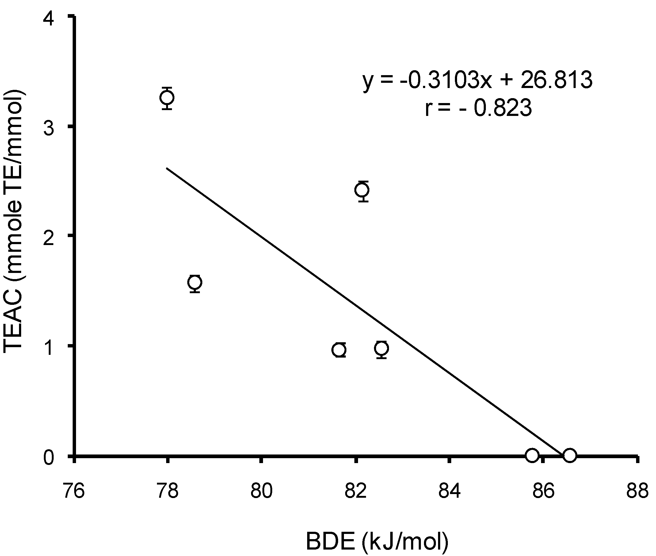
To identify the mechanism of interaction between HB/HC acids and ABTS radical cation, the correlation analysis of AOC with the thermodynamic parameters of uncharged and monoanion forms of HB and HC acids was carried out at different pH. For the uncharged forms, among all parameters a significant (
p < 0.05) correlation (
r = −0.832) was established between BDE values and AOC of HB and HC acids against the ABTS cation–radical at pH 3.5 (
Figure 10). This means that the dominating mechanism of interaction of HB and HC acids with ABTS radical cation at pH ≤ 3.5 is hydrogen atom transfer (HAT). In the range of pH 4.0–7.5 the only value to significantly (
p < 0.05) correlate with AOC of mono-HB and mono-HC acids against the ABTS radical cation is ETE (
r = 0.901) (
Figure 11). Consequently, when the pH of the reaction medium is 4.0–7.5, the main mechanism of implementation of the antioxidant properties of HB and HC acids against the ABTS cation–radical becomes sequential proton loss and electron transfer (SPLET). Thus, the mechanism of antioxidant action of HB and HC acids appears to depend on pH.
Figure 10.
The correlation analysis of HB/HC acid AOC against the ABTS cation–radical at pH 3.5 versus values of bond dissociation enthalpy (BDE) in phenolic hydroxyl.
Figure 10.
The correlation analysis of HB/HC acid AOC against the ABTS cation–radical at pH 3.5 versus values of bond dissociation enthalpy (BDE) in phenolic hydroxyl.
Figure 11.
Correlation analysis of HB/HC acid AOC against the ABTS cation radical at pH 7.4 versus values of electron transfer enthalpy (ETE) from phenolate ion.
Figure 11.
Correlation analysis of HB/HC acid AOC against the ABTS cation radical at pH 7.4 versus values of electron transfer enthalpy (ETE) from phenolate ion.
To resolve the question of antioxidant mechanism, previously the quantitative criteria were proposed based on comparison of IP and BDE of various phenolic substances with phenol [
19]: for absolute values of ΔIP up to 36 kcal/mol and for ΔBDE ~−10 kcal/mol the mechanism is dominated by hydrogen atom transfer, whereas for ΔIP > 45 kcal/mol the mechanism is predominantly SET. Based on the data in
Table 6a,b and values of BDE and IP of phenol from [
62,
63], the values of ΔIP and ΔBDE were calculated for HB and HC acids. For uncharged forms of HB and HC acids the values of ΔIP and ΔBDE lay in ranges of (−12.82)–(−4.03) and (−28.58)–(+2.53) kcal/mol, respectively, which confirms the earlier assumption about the prevalence of HAT mechanism of the antioxidant properties implementation of HB and HC acids at strongly acidic pH values, where ionization of carboxyl group does not occur. For monoanion forms of all investigated HB and HC acids the absolute values of ΔIP exceeded 100 kcal/mol, which favors the implementation of the antioxidant properties via SPLET mechanism. As it was indicated above, the electron transfer becomes possible after the deprotonation of phenolic hydroxyl.
Given that p-HB and p-HC acids are structural analogs of tyrosine, the interaction of tyrosine-containing dipeptides with the ABTS radical cation at pH 7.4 should be expected to follow the same mechanism of sequential proton loss and electron transfer (SPLET).


