Review

Antioxidative Dietary Compounds Modulate Gene Expression Associated with Apoptosis, DNA Repair, Inhibition of Cell Proliferation and Migration

1
CAS Key Laboratory of Pathogenic Microbiology and Immunology, Institute of Microbiology, Chinese Academy of Sciences, Beijing 100101, China
2
Department of Biotechnology, University of Tartu, Tartu 51010, Estonia
3
Mysenso Oy, Hollola 15870, Finland
4
Department of Food and Environmental Sciences, Division of Food Chemistry, University of Helsinki, Helsinki F-00014, Finland
5
Division of Pharmacognosy, Department of Medicinal Chemistry, Biomedical Center University of Uppsala, Uppsala 75123, Sweden
6
State Key Laboratory of Oncology in South China, Cancer Center, Sun Yat-Sen University, Guangzhou 510060, China
*
Authors to whom correspondence should be addressed.
Int. J. Mol. Sci. 2014, 15(9), 16226-16245; https://doi.org/10.3390/ijms150916226
Submission received: 16 June 2014 / Revised: 21 July 2014 / Accepted: 27 August 2014 / Published: 15 September 2014
(This article belongs to the Special Issue DNA Damage and Repair in Degenerative Diseases 2014)

Abstract

:
Many dietary compounds are known to have health benefits owing to their antioxidative and anti-inflammatory properties. To determine the molecular mechanism of these food-derived compounds, we analyzed their effect on various genes related to cell apoptosis, DNA damage and repair, oxidation and inflammation using in vitro cell culture assays. This review further tests the hypothesis proposed previously that downstream products of COX-2 (cyclooxygenase-2) called electrophilic oxo-derivatives induce antioxidant responsive elements (ARE), which leads to cell proliferation under antioxidative conditions. Our findings support this hypothesis and show that cell proliferation was inhibited when COX-2 was down-regulated by polyphenols and polysaccharides. Flattened macrophage morphology was also observed following the induction of cytokine production by polysaccharides extracted from viili, a traditional Nordic fermented dairy product. Coix lacryma-jobi (coix) polysaccharides were found to reduce mitochondrial membrane potential and induce caspase-3- and 9-mediated apoptosis. In contrast, polyphenols from blueberries were involved in the ultraviolet-activated p53/Gadd45/MDM2 DNA repair system by restoring the cell membrane potential. Inhibition of hypoxia-inducible factor-1 by saponin extracts of ginsenoside (Ginsen) and Gynostemma and inhibition of S100A4 by coix polysaccharides inhibited cancer cell migration and invasion. These observations suggest that antioxidants and changes in cell membrane potential are the major driving forces that transfer signals through the cell membrane into the cytosol and nucleus, triggering gene expression, changes in cell proliferation and the induction of apoptosis or DNA repair.

1. Introduction

Oxidative stress and inflammation are common features of many chronic diseases and their complications, and have also been linked to aging and carcinogenesis. Previous studies have shown that approximately one third of all known plants and vegetables have antioxidant and anti-inflammatory properties and consumption of a healthy diet rich in these is thought to help counter disease processes.
It is commonly regarded that non-receptor mediated regulation of cells by dietary compounds is negligible. However, recent findings show that dietary compounds may cause a switch in receptor-regulated signaling pathways and impairment of these pathways is known to be associated with type 2 diabetes and other metabolic diseases. Studies have shown that the downstream products of cyclooxygenase-2 (COX-2) control one of the most important internal antioxidant pathways: the nuclear factor (erythroid-derived 2)-like 2 (Nrf2)/Kelch-like ECH-associated protein 1 (Keap1)/antioxidant responsive element (ARE) pathway [1].
Research in China has recently focused on medicinal food by pharmacologically validating established dietary herbal compounds and optimizing their function through combinations or processing based on traditional practices. This review describes some of the antioxidative dietary compounds we have studied and analyzes our experimental results involving the role of polysaccharides in inducing apoptosis. The association of antioxidants with the COX-2/Nrf2/ARE pathway, the antioxidant effects of polyphenols on P53/Gadd45/MDM2 gene expression and its association with DNA repair, as well as the role of hypoxia-inducible factor (HIF)-1 and S100A4 genes in the migration and invasion of cancer cells are also discussed.

2. Coix Polysaccharides Induce Cancer Cell Apoptosis

The ancient food crop coix (Coix lachryma-jobi L.; also known as adlay seed) is widely cultivated in the warm regions of Asia, Africa and the Mediterranean Rim. The grain is prepared for eating by roasting and may be eaten dry, used for porridge or processed into flour [2]. In addition to polysaccharides, coix is rich in protein, fat, carbohydrates, amino acids, vitamins and inorganic salts. In traditional Chinese medicine coix is used as a diuretic, an anti-inflammatory drug, an anti-cancer drug, an analgesic and a nutrient [3]. Studies have shown that coix contains a large number of lipopolysaccharides, including palmitic acid, stearic acid, octadecadienoic acid, oleic acid and linoleic acid. It also contains oligosaccharides with free radical scavenging and other antioxidant properties [4]. Apirattananusorn et al. [5] found that non-starch polysaccharides (mainly arabinoxylans) in coix are present in the alkaline rather than the water extract, and that the arabinoxylan molecule has a (1,4)-linked-d-xylan main chain highly substituted with arabinose units.
A number of studies have reported the bioactivities of compounds isolated from coix. For example, a methanol extract of adlay seed suppressed the expression of COX-2, a key enzyme that catalyzes the transformation of arachidonic acid into prostaglandins [6]. It is known that COX-2 is expressed in human lung cancer cells and coix has shown significant anti-proliferative effects on these cells due to its inhibition of COX-2 gene expression [7]. The oil extract of adlay seed has also been shown to inhibit fatty acid synthase and is used in anti-neoplastic therapy [8], while dehulled adlay seed suppresses early events in colon carcinogenesis and reduces COX-2 protein expression [9]. In addition, coix polysaccharide was shown to possess a hypoglycemic function [10] and to improve immune system function [11].
Therefore, we have investigated the anti-cancer mechanisms of coix polysaccharides in the human lung adenocarcinoma epithelial cell line A549, by multiple methods including alkaline gel electrophoresis of single cells (comet assay) and flow cytometry. MTT (3-[4,5-dimethylthiazol-2-yl]-2,5 diphenyl tetrazolium bromide) assays showed that the polysaccharide fraction CP-1 inhibited the proliferation of A549 cells in a time- and concentration-dependent manner, with the highest inhibition observed at a CP-1 concentration of 300 µg/mL for 72 h. CP-1 induced both cell cycle arrest in S phase and apoptosis of A549 cells as demonstrated by cell cycle analysis and annexin V-fluorescein/propidium iodide staining assays [12].
The single cell gel electrophoresis assay detects DNA damage, including DNA strand breaks and alkali labile lesions, with high visual resolution [13]. The movement of DNA from the head to the tail has been described as the most obvious characteristic of apoptotic cells in the comet assay [14]. Our results showed that the length of the comet tail was proportional to the degree of DNA fragmentation. Furthermore, apoptotic cells were clearly distinguishable between control and test groups [12].
The caspase gene family plays a central role in mitochondrial-mediated apoptosis [15,16,17]. Caspase-3, which is activated by caspase-9, contributes to the execution of apoptosis via the activation, hydrolysis and proteolysis of specific substrates such as DNA-dependent protein kinases [18]. Our western blot analysis showed that CP-1 increased the expression of both caspase-3 and caspase-9, suggesting that coix polysaccharides mediate apoptosis via a caspase-dependent pathway. Furthermore, the CP-1-mediated disruption of the mitochondrial membrane potential, which typically leads to the activation of caspase-3 and caspase-9 [19], suggests that a mitochondrial-dependent pathway is involved in the induction of apoptosis by CP-1 [12].
To date, no relevant polysaccharide receptor has been identified on the A549 cell membrane that would enable CP-1 to interact and produce its antioxidant effects, which include interfering with cell growth, metabolism and proliferation, leading to the induction of apoptosis (Figure 1). Nevertheless, the mechanism through which CP-1 induces apoptosis in A549 cells is worthy of further investigation owing to its potential anti-tumor effects.
Figure 1. Hypothesis of how foodborne or metabolized polysaccharides/oligosaccharides interact with the cell outer membrane. OPS: (dropped) off polysaccharides; APS: attached polysaccharides; AE: antenna extracellular membrane polysaccharides that may absorb different wavelengths of light, changing the conformation of membrane associated proteins; R: receptors; M: membrane proteins.
Ijms 15 16226 g001

3. Inflammatory and Anti-Inflammatory Effects of Viili Polysaccharides

Viili, a semi-solid yogurt that originated in Finland, has a ropy, gelatinous consistency and a sour taste resulting from the microbial action of lactic acid bacteria (LAB) and a surface-growing fungus, Geotrichum candidum, which forms a velvet-like surface. Viili also contains the yeasts Kluveromyces marxianus and Pichia fermentans. Among the mesophilic LAB strains, the slime-forming LAB Lactococcus lactis subsp. cremoris produces a phosphate-containing exopolysaccharide (EPS) with a basic structure mainly composed of d-glucose, d-galactose, l-rhamnose and phosphate, with an average molecular weight of approximately 2000 kDa and a repeating unit of “→4-β-Glcp-(1→4)-β-d-Galp (1→4)-β-d-Glcp-(1→” as well as groups of -l-Rhap and -d-Galp-1-p attached to each side of Galp [20,21]. Viili has been claimed to have various functional benefits, including antioxidant, anti-inflammatory, anti-cancer and anti-aging properties, and was also reported to enhance natural immunity [22,23].
Macrophages not only constitute a principal component of the innate immune system, but they also perform pivotal roles in acute inflammatory responses and atherosclerosis [24]. Lipopolysaccharide (LPS)-stimulated macrophages can generate a variety of inflammatory mediators such as nitric oxide (NO), prostaglandin E2 (PGE2), interleukin (IL)-1β, tumor necrosis factor-α and matrix metalloprotease-9 [25]. Of these, NO is the most important molecule for inter- and intracellular signal transmission; it also mediates immune and inflammatory processes and plays a critical role in communicating physiological and pathological signals [26,27]. However, excessive levels of NO are toxic as they form free radical groups, such as superoxide (O2), that result in production of the toxic peroxynitrite (ONOO) molecule [28,29]. Therefore, changes in NO production may provide a measure to assess the effects of drugs or functional foods on the inflammatory process.
We previously studied the activity of viili exopolysaccharides (VEPS) at the cellular level, and found that they promoted the activation of macrophages in association with NO, IL-6 and IL-1β production. The release of NO and proinflammatory cytokines may activate lymphocytes, which upregulates nonspecific (innate) and specific (adaptive) immunity in humans [30]. VEPS from L. lactis subsp. cremoris were able to strongly induce NF-κB and various cytokines via two intestinal receptors associated with Peyer’s patches in swine, RP105 and MD-1 [31,32]. This inspired our investigation in mouse macrophage RAW264.7 cells. Macrophages accumulate in the small intestine and play crucial roles in both the innate and adaptive host defense against infection. They can be triggered by several different compounds, including polysaccharides and LPS. In our assay, VEPS were able to increase the proliferation of RAW264.7 cells at concentrations of 50, 100 and 200 μg/mL. VEPS also strikingly increased macrophage phagocytosis, NO secretion, iNOS gene and protein expression, the secretion and gene expression of IL-6 and IL-1β, as well as inducing a flattened morphology. Synergy was also observed when VEPS were added in the presence of LPS.
NO is recognized as a mediator and regulator of inflammatory responses. It is a nonspecific inflammatory mediator involved in triggering the proliferation of macrophages and lymphocytes, as well as stimulating the central nervous system and other immune functions in vivo. Of the three isoforms of NO synthase, iNOS is the most important in NO synthesis. It directly affects many physiological and pathological functions, including the relaxation of cardiovascular vessels, increasing blood flow and the promotion of cytokine emission during inflammation and at the moment of antigen recognition between T cells and antigen presenting cells [1,33,34,35]. The enhancement of NO production and gene expression by VEPS occurred in a dose-dependent manner, which strongly suggests that VEPS are immune mediators/modulators. The secretion of NO synergistically increased when both VEPS and LPS were applied, which indicates that they may share a similar mechanism. A parallel increase in iNOS was observed by both semi-quantitative RT-PCR and western blot [36].
Intestinal macrophages engulf and ingest particles to form a phagosome (or food vacuole), which in turn fuses with a lysosome to form a phagolysosome. Engulfed materials are eventually digested or degraded and either released extracellularly via exocytosis, or intracellularly to undergo further processing. The activation of intestinal macrophages by dietary factors has previously been reported [37]. After treatment with VEPS, a significant increase in macrophage phagocytosis indicates the activation of cellular functions, probably through reversible protein aggregation and motor molecular mechanisms [38]. The activation of macrophages by VEPS was also indicated by an increase in the gene expression and secretion of IL-6 and IL-1β.
It is well documented that macrophage morphology can change from round to flat when activated by LPS [39]; however, to our knowledge this is the first time that a similar morphology has been observed for RAW264.7 cells stimulated by VEPS [30]. The degree of flattening and spreading was consistent with increased proliferation, phagocytosis, iNOS expression and NO release. Although morphological alteration involves a complex, dynamic reorganization of cytoskeletal actins [40], we believe that macrophage activation by VEPS is a reversible physiological process that favorably increases proliferation of macrophages and promotes immunity. However, further studies are required to confirm this.

4. COX-2 (Cyclooxygenase-2) Is Involved in Anti-Inflammatory Processes via Ursolic Acid and Microbial Polysaccharides

Hepatocellular carcinoma (HCC) is estimated to be the fifth most common cause of cancer-related death worldwide [41]. Although approximately 80% of cases are reported in developing countries where the prevalence of hepatitis is high, HCC is one of very few cancers whose incidence is increasing in developed countries [42,43]. Chemotherapy has provided significant survival benefits for HCC patients, but most drugs are also associated with significant tissue toxicity. Therefore there is a need for drugs or alternative therapies that target tumor cells without compromising normal tissue function [44]. Increased concentrations of cytotoxic drugs and higher doses of radiation often fail to improve the health of patients with liver cancer, and may lead to apoptosis resistance. Therefore, an anti-cancer agent with low toxicity that preferentially induces apoptosis in human cancer cells while creating an internal oxidative environment would be very useful.
Ursolic acid (UA), a pentacyclic triterpenoid, has been identified in several vegetables and medicinal herbs [45]. UA can inhibit cell growth and induces apoptosis in some tumors through multiple pathways [46,47], including the inhibition of DNA replication, activation of caspases and down-regulation of anti-apoptotic genes [48]. UA has also been shown to specifically inhibit tumorigenesis [49], tumor progression [50], angiogenesis and tumor invasion [51]. As outlined above, the large amounts of EPS generated in viili [52] reportedly have antioxidant properties [53], which regulate immune function and lower cholesterol [54]. Moreover, the Astragalus species commonly used in traditional Chinese medicine, especially A. membraneuse, contain similar polysaccharides (Astragalus polysaccharides, APS) that are believed to improve or modulate immune function [55] and promote tumor cell apoptosis [56].
COX-2 is not expressed in many organs under normal physiologic conditions but is expressed in most cancer cells [57], where it is believed to inhibit cancer cell apoptosis [58] thereby causing chemotherapy resistance. Notably, COX-2 selective inhibitors have been demonstrated to inhibit tumor cell proliferation and induce apoptosis [59]. For these reasons, naturally derived COX-2 inhibitors have been investigated for use in chemotherapy and chemoprevention. We previously analyzed the synergistic effect of UA in combination with VEPS and APS on cell proliferation, morphologic changes, oxidation and COX-2 expression, and found that inhibition of cell proliferation is associated with the inhibition of COX-2 [60].

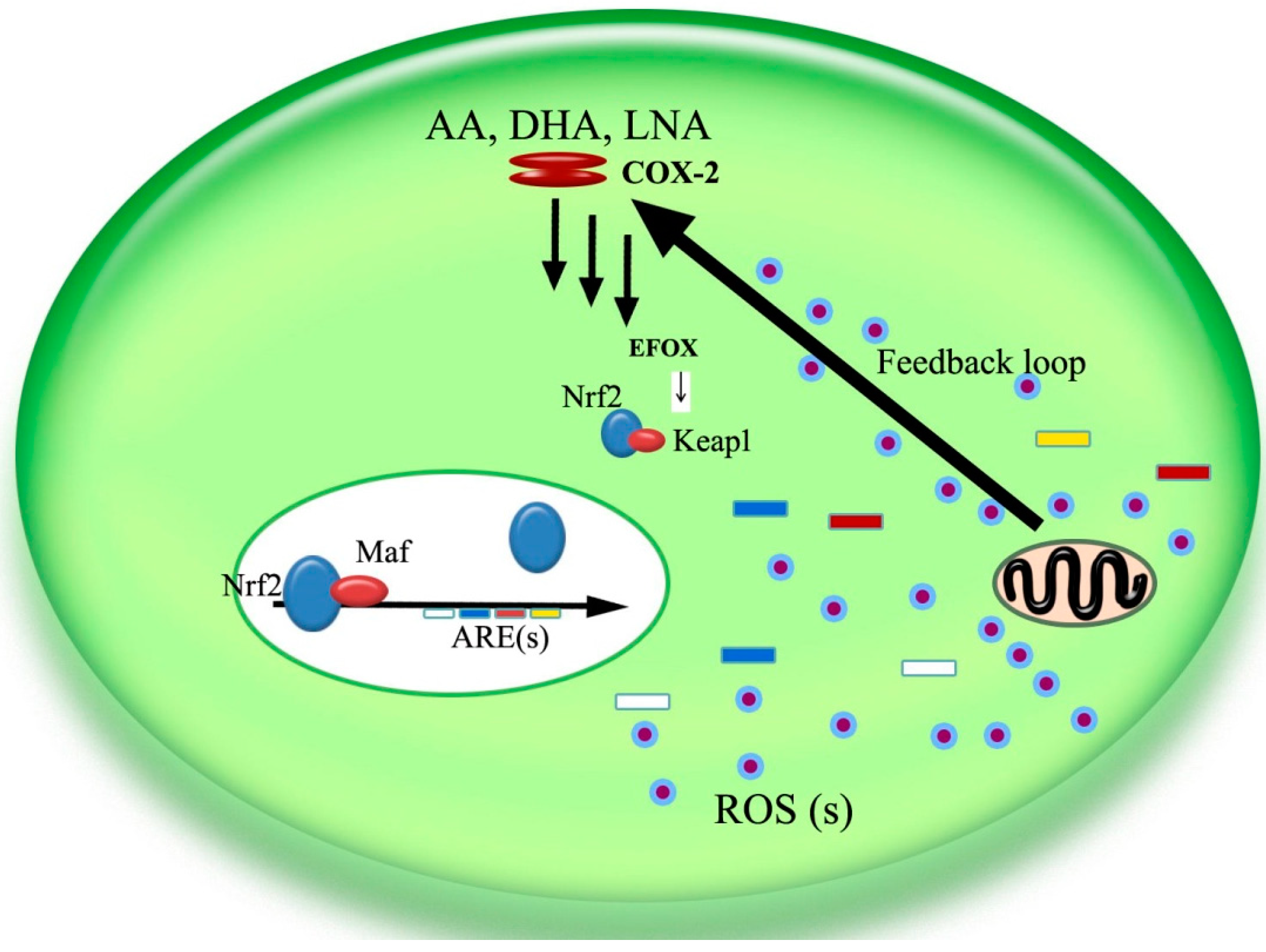
5. The COX-2/Nrf2/ARE Pathway
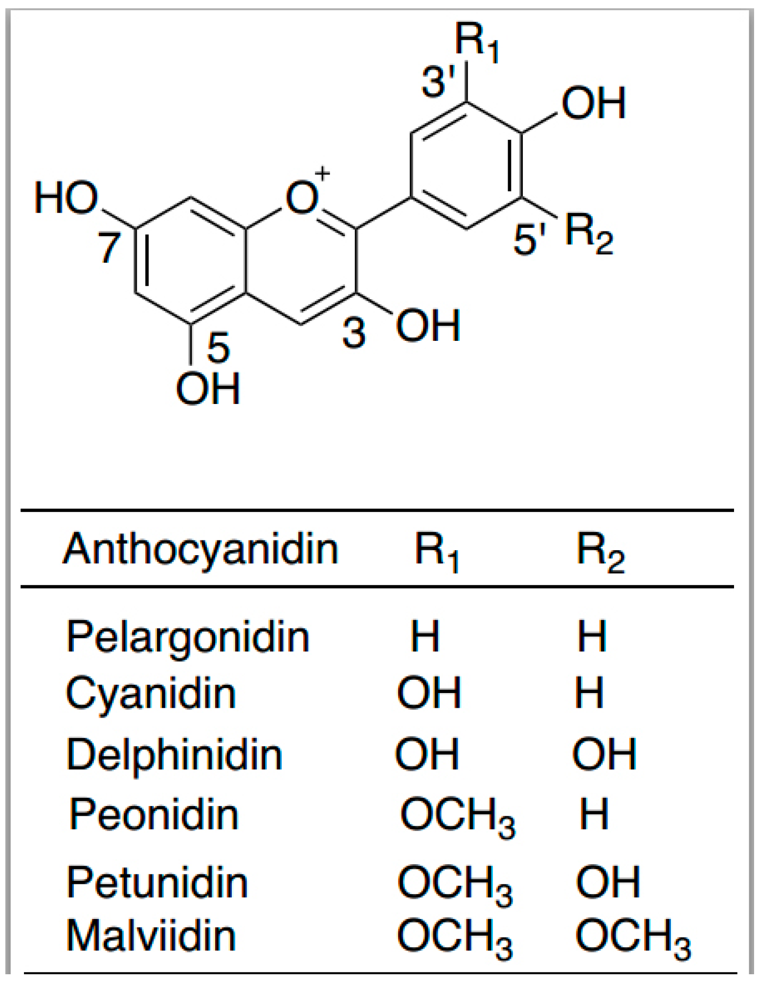
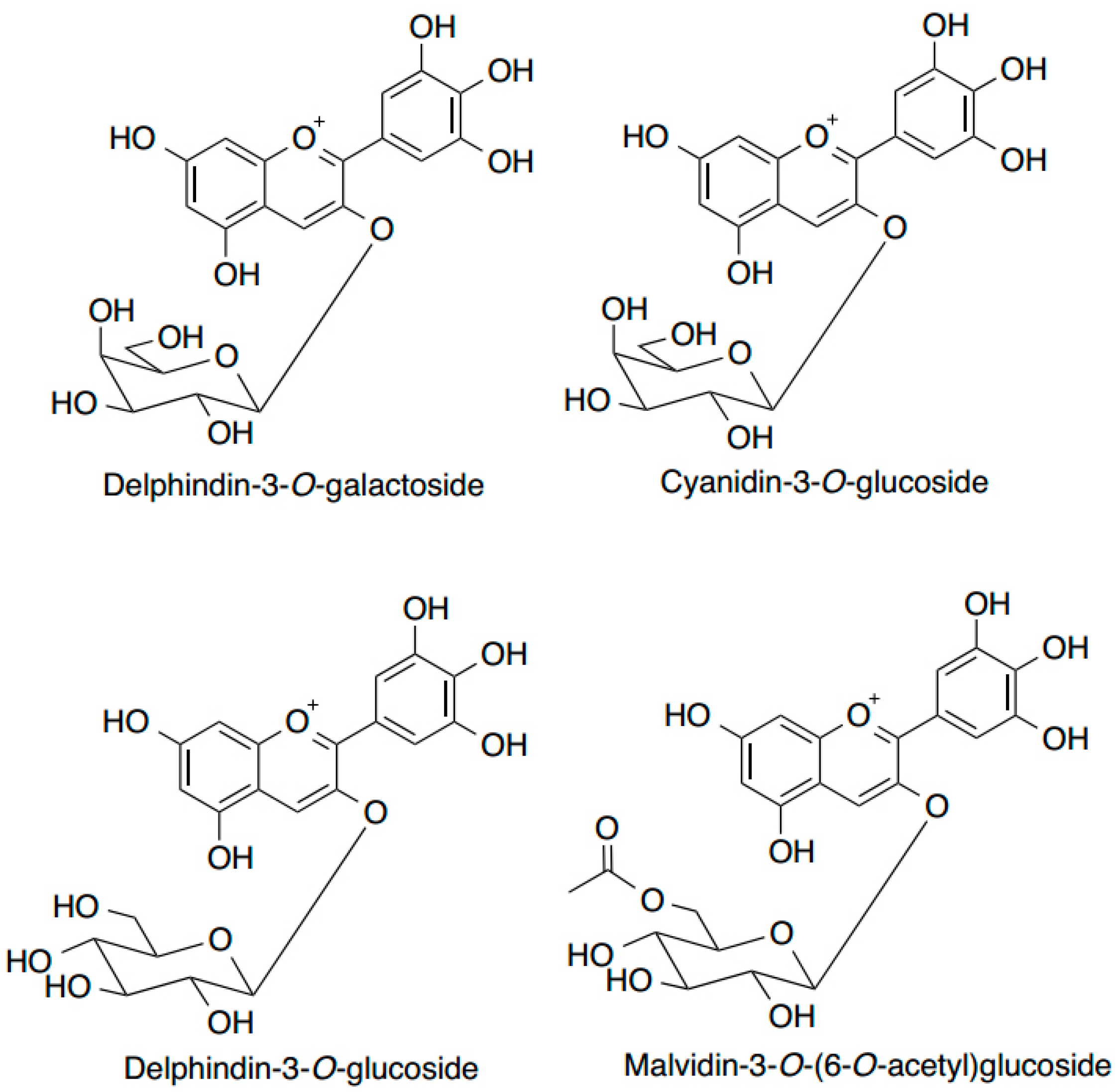
Although the development of tumors and malignancies is complex, carcinogenic events are common. Potentially cancerous cells are produced constantly, but are usually eliminated in a healthy environment. However, carcinogenesis may occur if the body’s internal antioxidant and anti-inflammatory environment changes, or if mechanisms that inhibit abnormal cell proliferation are disrupted. Recent research suggests that antioxidants and anti-inflammatories may actually increase the risk of cancer in an environment where antioxidants involved defense systems are compromised because the internal antioxidative system, the ARE genes may be induced and triggered and eventually increase cell proliferation [61,62,63,64]. However, an antioxidative environment might not curb cancer progression, especially in precancerous cells where COX-2 is overexpressed. A downstream product of COX-2, PGE2, which often causes inflammation, can be modified, and become several short-lived so-called electrophilic oxo-derivative (EFOX) molecules that strongly regulate cell proliferation through the Nrf2/keap1/ARE pathway (Figure 2).
Figure 2. Diagram portraying a potential mechanism of COX-2’s antioxidative effect via EFOX (electrophilic oxo-derivative) molecules, where ROS(s) are hypothesized to trigger the antioxidative and anti-inflammatory effects largely based on the observation that COX-2 expression is increased in aging tissues. Abbreviations: AA, arachidonic acid; DHA, docosahexaenoic acid; EPA, eicosapentaenoic acid; LNA, linolenic acid; ROS, reactive oxygen species [1].
Ijms 15 16226 g002
Figure 2 depicts a potential mechanism of COX-2 antioxidative activity via EFOX molecules, where reactive oxygen species (ROSs) are hypothesized to trigger antioxidative and anti-inflammatory effects. We recently demonstrated that UA, VEPS, APS and their combined application significantly reduced expression of COX-2 and lowered PGE2 concentration in HepG2 cells and inhibiting cellular proliferation. The mechanism for this inhibition may be due to the inhibition of COX-2, which is thought to increase oxidative stress because of the decreased expression of EFOX molecules that mediate gene expression of superoxide dismutase (SOD) and other AREs [65]. This is because the ARE family creates an antioxidative, anti-inflammatory protective environment to increase cell proliferation [66]. Increased malondialdehyde (MDA) concentration, a metabolic product of fatty acids, also indicates an oxidative environment. MDA expression is significantly reduced by UA. In contrast, inhibition of MDA and cell proliferation only occurs at high concentrations of VEPS and APS. Thus, it is possible that inhibition of HepG2 cell proliferation by UA, VEPS, APS and their combined application is attributable to inhibition of COX-2 and the associated decrease in SOD activity, which increases the oxidative environment and induces apoptosis.
These compounds, especially VEPS, may also have cancer-preventive roles in vivo via regulation of the immune system [67,68,69], as VEPS can activate macrophages and lymphocytes without causing severe inflammation or disease. Notably, some correlation between the dietary use of viili and cancer prevention has been suggested [70].

6. Anthocyanins

Anthocyanins are water-soluble pigments derived from 2-phenylbenzopyrylium. They consist of an aglycone (anthocyanidins), sugars and/or acyl groups belonging to the flavonoid group characterized by a C6-C3-C6 skeleton. Major members include pelargonidin, cyanidin, peonidin, delphinidin, petunidin and malvidin (Figure 3), which have diverse patterns of glycosylation and acylation. Owing to the presence of flavylium cations (2-phenylbenzopyrylium), anthocyanins appear as red, blue or violet depending on their concentration, structure and environment [47].
Figure 3. Structural formulas of common anthocyanidins.
Ijms 15 16226 g003
In addition to their widely known use as natural food colorants, anthocyanins are also used as human nutritional supplements, as accumulating evidence has connected the intake of anthocyanin-rich foods with a reduced risk of chronic diseases such as cancer, cardiovascular diseases and Alzheimer’s. As these diseases may result from oxidative stress, it is understandable that the nutraceutical properties of anthocyanins are mainly attributed to their antioxidant activities including free radical scavenging, metal chelating and protein binding [71]. However, the relevance of their antioxidant activity is challenged by in vivo findings that circulating flavonoids have low concentrations (0.1–1 μM), and that antioxidant activity, especially the hydrogen-donating property of phenolic hydroxyl groups, can be impaired by conjugation with other molecules [72,73]. Therefore, more research is needed to understand the health benefits of plant anthocyanins and their potential chemopreventive mechanisms [74,75].
Blueberries have enjoyed worldwide popularity owing to increasing awareness of their health benefits. We place particular emphasis on the highbush blueberry (Vaccinium corymbosum L.) in this review, because it is common to the northern hemisphere and the focus of our research. Blueberries are one of the richest sources of plant anthocyanins among common fruits and vegetables in terms of both variety and content [76,77]. Up to 25 anthocyanins have been identified in wildtype highbush blueberry, and glucosides of delphinidin, cyanidin and malvidin are the predominant fractions among these (Figure 4) [78]. This is in accordance with earlier findings [79,80]. The total anthocyanin content in highbush blueberries of different genotypes ranges from 25–495 mg/100 g fresh fruit [79]. Such high levels are strongly correlated (r = 0.90) with their antioxidant activity, which is three-fold higher than strawberries and raspberries as determined by the oxygen radical absorbing capacity (ORAC) assay [81]. Based on this same method, blueberry anthocyanins were found to contribute to over 50% of the total antioxidant activity, while ascorbate accounted for less than 10% of the total activity [76,82].
Figure 4. Examples of the major anthocyanins present in highbush blueberries (Vaccinium corymbosum L.). All examples have a sugar moiety at position 3.
Ijms 15 16226 g004

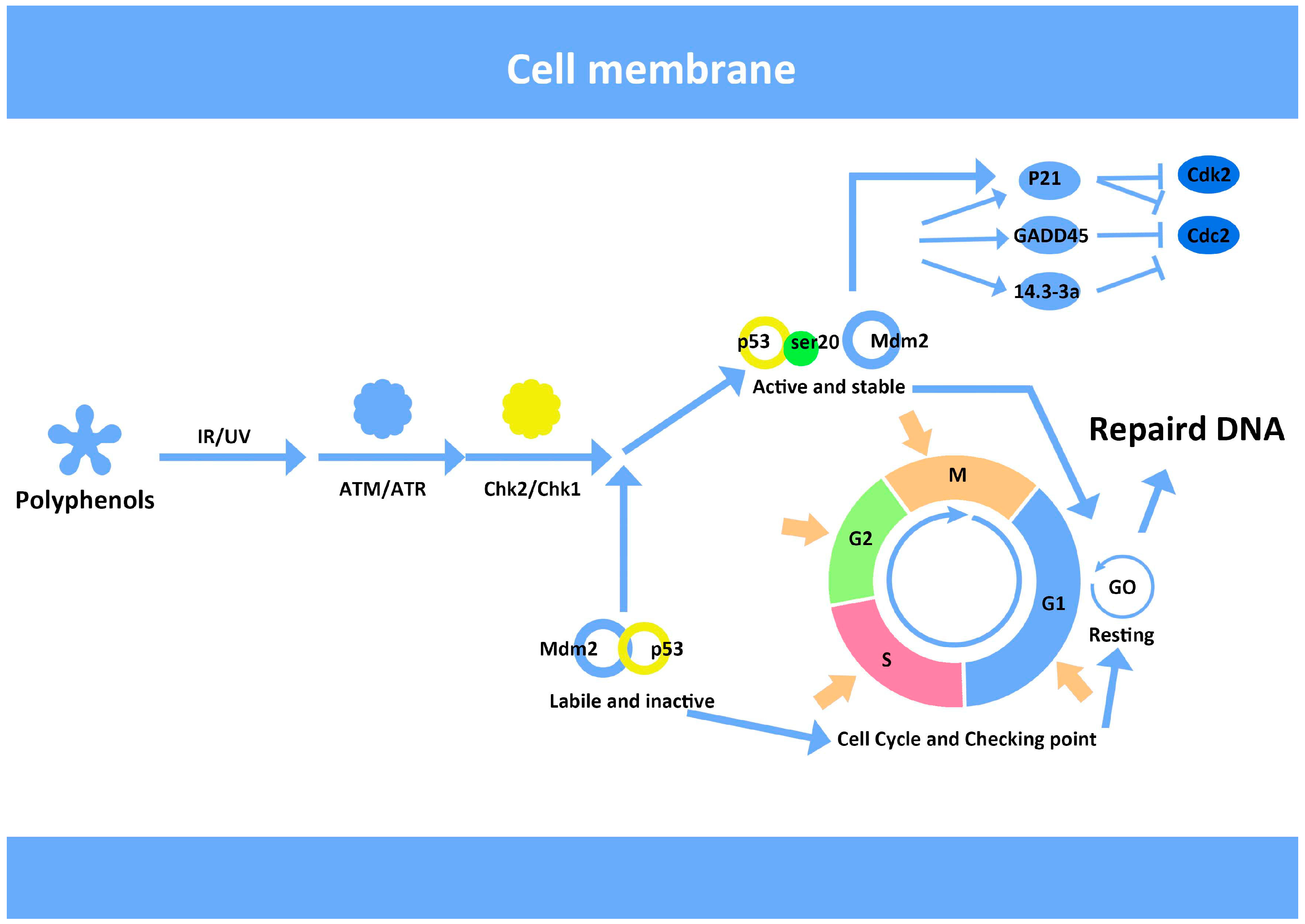
7. Anthocyanins and Modulation of DNA Repair

Oxidative damage to DNA frequently leads to gene mutation and the potential initiation of carcinogenesis. Base repair in DNA damage is a simple process, but the repair of extensive DNA damage requires a complex set of molecular controls in mammalian cells. Cell cycle checkpoints in DNA replication and a complex network of sensor proteins ensure DNA fidelity [83,84]. In response to double-strand DNA breakage, deletion or fragmentation, the proteins ATM and H2AX are triggered and bind to unhelixed DNA between chromatin, where the complex of DNA repair machinery will then bind [85]. Signal transducers and pathways influenced during different parts of the cell cycle determine cell cycle arrest, DNA repair or apoptosis [86,87]. The fate of vertebrate somatic cells is decided in the G1 phase of the cell division cycle. We have reported that blueberry anthocyanins (BA) have a protective effect in UV-irradiated cells, which may be related to their antioxidant activity. Our findings demonstrated that DNA was significantly damaged after UV irradiation, with increased expression of p53 and p21 protein. In contrast, cells pre-treated with blueberry anthocyanin, had decreased p53 and p21 protein expression [88], thus indicating that BA can influence the DNA repair machinery (Figure 5).
Figure 5. Hypothetical mechanism of DNA repair activation facilitated by blueberry anthocyanins (BA). Following DNA damage after ionizing or ultraviolet radiation (UV), the protein kinases ATM/ATR are activated, which modifies checkpoint-signaling pathways in the cell cycle. This is where BA possibly regulates cellular processes. The diagram is adapted from multiple illustrations based on publicly available materials, particularly a netbook [89].
Ijms 15 16226 g005
Apoptosis induced by UV irradiation and its reversal by BA as observed in our study is associated with oxidation and antioxidants [90,91]. The oxidative environment created by the UV dose was not lethal even though typical apoptotic blebs were observed by scanning electron microscopy. Similarly, DNA fragmentation was observed by the comet assay, however the degree of fragmentation was relatively small compared with the whole nuclear genome. The effect of BA on gene and protein expression was also analyzed. Gadd45 and MDM2 were persistently overexpressed beyond 24 h; which made it possible for BA-assisted DNA repair to occur to reverse apoptosis.
It is widely known that p53 is closely associated with DNA damage and repair via the regulation of its downstream genes in reaction to DNA damage [92]. When DNA damage is induced by UV light or ionizing radiation, p53 activates the expression of genes, such as p21 and Gadd45. The expression of Gadd45 allows cells to arrest in the G1/S phase to induce DNA repair and regulates apoptosis signaling pathways and survival [93]. Meanwhile, MDM2 is activated by p53 and then phosphorylated and ubiquitinated by protein kinases to reduce inhibition of p53 by MDM2 [94]. Until the DNA damage is repaired, MDM2 is resynthesized to inhibit and degrade p53 protein and to promote cell cycle to return to the normal condition (Figure 5).
We confirmed that HepG2 cells were arrested in the G1 phase at a dose of 30 mJ/cm2 UV irradiation. Gadd45 and MDM2 proteins were significantly increased 12 h after irradiation and reduced by pretreatment with BA. We speculate that the protective effect of blueberry anthocyanins on DNA damage by UV irradiation is owing to its antioxidant activity, ability to scavenge free radicals and the regulation of relevant DNA repair proteins.

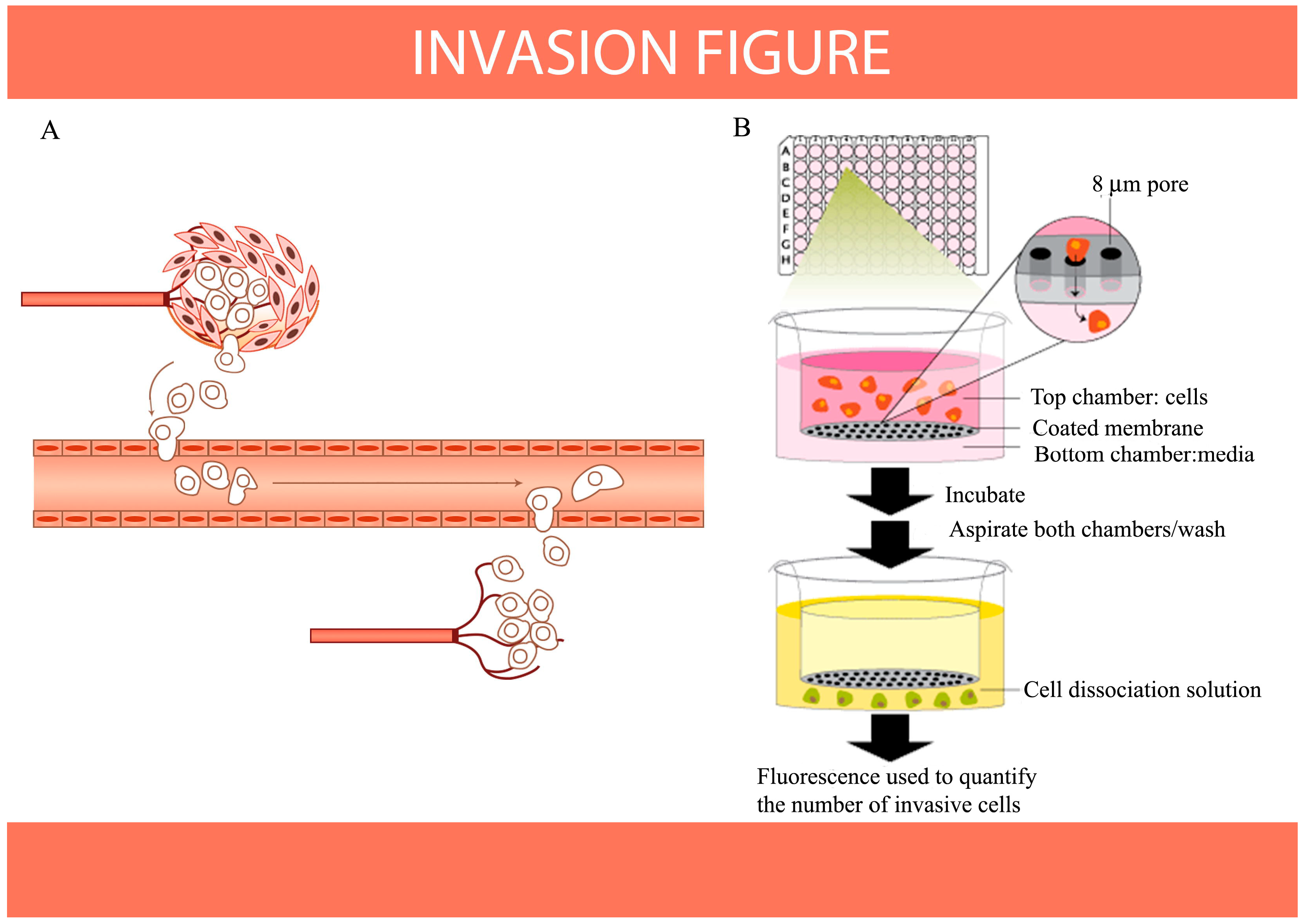
8. Antioxidants Inhibit Migration and Invasion of Cancer Cells as Indicated by HIF-1 and S100A4 Expression in Vitro

It recent years, a greater understanding of the mechanisms regulating cell migration and invasion has been gained. Although in vivo conditions cannot be replicated perfectly in vitro, the molecular mechanisms should be largely similar. We previously used a cell wound scratch assay and Transwell migration assay [95] to observe cell migration and invasion in vitro, investigating HIF-1 and S100A4 activity respectively. HIF-1 is associated with cell migration as hypoxia is a growth-promoting factor that activates fermentation and migration factors. The HIF-1α protein is the primary regulator and its active subunits are strictly regulated by oxygen concentrations. As almost all solid tumors are hypoxic, the inhibition of HIF-1 involves a chemotherapy approach. A staurosporine derivative (UCN-01) was previously found to block HIF-1 in prostate cancer cells by promoter trans-activation, thereby blocking the formation of new blood vessels. Colchicine and vincristine also inhibit HIF-1. Moreover, three types of dietary lignans were shown to reduce the proliferation of human breast cancer T47D cells via the selective inhibition of HIF-1α [96].
According to recent findings, the proteasome pathway also accelerates tumor and HIF-1α protein degradation, followed by the down-regulation of VEGF mRNA transcription and protein secretion. These results thus revealed a new angiogenesis inhibition pathway [97]. Saponins are amphipathic glycosides, which are soap-like foaming agents with both hydrophilic and hydrophobic properties. They are widely distributed in several different types of plants and have many biological activities including anti-tumor, anti-inflammatory, immune regulation, anti-viral and anti-fungal properties. Some saponins, such as ginseng saponins and anax notoginseng saponins, have been shown to regulate HIF-1 expression. For example, ginseng saponins significantly enhanced HIF-1α protein expression in the mouse cerebral cortex under hypoxic conditions [98].
We previously used ginseng saponin and the saponin from the leaf of the herb Gynostemma pentaphyllum to conduct a wound scratch migration assay in HepG2 cells and observed inhibition of HIF-1α under hypoxic conditions (unpublished data). Similarly, the inhibition of invasion by coix polysaccharides was also demonstrated using a Transwell assay in hepatocarcinoma and non-small cell lung cancer cells (NSCLC). The inhibition of migration and invasion of human A549 NSCLC cells was correlated with the in vitro down-regulation of S100A4 [99].
The mechanisms of cancer cell migration and invasion have been well studied. Cancer therapeutics designed to target adhesion receptors or proteases have proven not to be effective in slowing tumor progression in clinical trials. This may be owing to the fact that cancer cells can modify their migration or invasion mechanisms in response to changes in environmental conditions. However, as the processes of cell migration and invasion are integral to embryonic development and the functioning of organisms (Figure 6A), we have continued to investigate the mechanisms of inhibition of migration and invasion in cancer prevention and protection (Figure 6B). Cancer metastasis is coordinated by cell-cell communication and signaling, which may possibly be inhibited by dietary compounds, even though the details of the processes and mechanisms remain largely unknown.
Figure 6. Aggressive malignant cancer cells are able to invade other tissues or organs with microvessels and can also induce angiogenesis. The progression from primary to metastatic tumor is a multigenic, multistep process that involves cell-cell and cell-extracellular matrix adhesion, tissue invasion and/or migration and angiogenesis. (A) Diagram of metastatic cancer invasion (adapted from [100] with permission from Nature Publishing Group, copyright 2005); (B) diagram of the in vitro invasion assay.
Ijms 15 16226 g006

9. Conclusions

A healthy diet is known to be beneficial in helping to prevent disease. Naturally derived polysaccharides from coix and viili, anthocyanins from blueberry, saponins from ginseng and ursolic acid are all similar to other antioxidants in that they are thought to contribute to good health via multiple mechanisms (Figure 7), including the modulation of gene expression. It is scientifically and culturally accepted that a balanced diet is able to lower the risk of disease. However, caution regarding the use of dietary antioxidants in health care and disease prevention remain, although there are clearly proven benefits. However, the multiple functions of dietary antioxidants frequently results in adverse effects, thus caution is needed in their application. For example, ginseng can alter blood glucose concentrations, therefore it is not advised for use in diabetic patients. As most mechanisms of these multiple functions of dietary antioxidants remain unclear, further studies are required.
Figure 7. Commonly recognized functions of dietary antioxidants divided into four main types. Effect 1: Prevention or protection against inflammatory diseases such as urinary tract infections (UTI); Effect 2: Improvement of blood circulation, including cardiovascular diseases (CVD); Effect 3: Prevention of and protection against carcinogenesis; Effect 4: Improvement of the function of the gastric and intestinal systems.
Ijms 15 16226 g007
The antioxidants we tested in vitro have been shown to significantly influence cell growth, DNA repair and mitochondrial membrane-mediated apoptosis. Although the cellular environment in vivo is much more complex, our studies suggest that the signals observed in vitro may provide clues for potential therapies in patients with chronic diseases, and also reveal the challenges facing application of dietary antioxidants.

Acknowledgments

This work was supported by the National Natural Science Foundation of China (No. 81171572), National Major Scientific and Technological Special Project for “Major New Drugs Innovation and Development” (2014ZX09101041), Guangdong Innovative Research Team Program (No. 2009010058) and the China Postdoctoral Science Foundation.

Author Contributions

Likui Wang, Shijuan Gao, Wei Jiang and Maonian Xu collected and analyzed literature and wrote the manuscript. Cheng Luo, Lars Bohlin and Wenlin Huang took responsibility for corrections throughout the review. Markus Rosendahl partly contributed to the production of innovative graphic work.

Conflicts of Interest

The authors declare no conflict of interest.

References

  1. Luo, C.; Urgard, E.; Vooder, T.; Metspalu, A. The role of COX-2 and nrf2/are in anti-inflammation and antioxidative stress: Aging and anti-aging. Med. Hypotheses 2011, 77, 174–178. [Google Scholar]
  2. Jideani, I.A.; Jideani, V.A. Developments on the cereal grains Digitaria exilis (acha) and Digitaria iburua (iburu). J. Food Sci. Technol. 2011, 48, 251–259. [Google Scholar] [CrossRef]
  3. Hsia, S.M.; Chiang, W.; Kuo, Y.H.; Wang, P.S. Downregulation of progesterone biosynthesis in rat granulosa cells by adlay (Coix lachryma-jobi L. var. Ma-yuen Stapf.) bran extracts. Int. J. Impot. Res. 2006, 18, 264–274. [Google Scholar]
  4. Manosroi, J.; Khositsuntiwong, N.; Manosroi, A. Biological activities of fructooligosaccharide (fos)-containing Coix lachryma-jobi Linn. Extract. J. Food Sci. Technol. 2014, 51, 341–346. [Google Scholar] [CrossRef]
  5. Apirattananusorn, S.; Tongta, S.; Cui, S.W.; Wang, Q. Chemical, molecular, and structural characterization of alkali extractable nonstarch polysaccharides from Job’s tears. J. Agric. Food Chem. 2008, 56, 8459–8557. [Google Scholar] [CrossRef]
  6. Akhtar, M.; Cheng, Y.; Magno, R.M.; Ashktorab, H.; Smoot, D.T.; Meltzer, S.J.; Wilson, K.T. Promoter methylation regulates helicobacter pylori-stimulated cyclooxygenase-2 expression in gastric epithelial cells. Cancer Res. 2001, 61, 2399–2403. [Google Scholar]
  7. Hung, W.C.; Chang, H.C. Methanolic extract of adlay seed suppresses COX-2 expression of human lung cancer cells via inhibition of gene transcription. J. Agric. Food Chem. 2003, 51, 7333–7337. [Google Scholar] [CrossRef]
  8. Yu, F.; Gao, J.; Zeng, Y.; Liu, C.X. Inhibition of Coix seed extract on fatty acid synthase, a novel target for anticancer activity. J. Ethnopharmacol. 2008, 119, 252–258. [Google Scholar] [CrossRef]
  9. Shih, C.K.; Chiang, W.; Kuo, M.L. Effects of adlay on azoxymethane-induced colon carcinogenesis in rats. Food Chem. Toxicol. 2004, 42, 1339–1347. [Google Scholar] [CrossRef]
  10. Takahashi, M.; Konno, C.; Hikino, H. Isolation and hypoglycemic activity of coixan-a, coixan-b and coixan-c, glycans of Coix lachryma-jobi var. ma-yuen seeds. Planta Med. 1986, 52, 64–65. [Google Scholar]
  11. Ooi, V.E.C.; Liu, F. Immunomodulation and anti-cancer activity of polysaccharide-protein complexes. Curr. Med. Chem. 2000, 7, 715–729. [Google Scholar] [CrossRef]
  12. Lu, X.Y.; Liu, W.; Wu, J.H.; Li, M.X.; Wang, J.C.; Wu, J.H.; Luo, C. A polysaccharide fraction of adlay seed (Coix lachryma-jobi L.) induces apoptosis in human non-small cell lung cancer a549 cells. Biochem. Biophys. Res. Commun. 2013, 430, 846–851. [Google Scholar]
  13. Singh, N.P.; McCoy, M.T.; Tice, R.R.; Schneider, E.L. A simple technique for quantitation of low levels of DNA damage in individual cells. Exp. Cell Res. 1988, 175, 184–191. [Google Scholar] [CrossRef]
  14. Fairbairn, D.W.; Olive, P.L.; O’Neill, K.L. The comet assay: A comprehensive review. Mutat. Res. 1995, 339, 37–59. [Google Scholar] [CrossRef]
  15. Lowe, S.W.; Lin, A.W. Apoptosis in cancer. Carcinogenesis 2000, 21, 485–495. [Google Scholar] [CrossRef]
  16. Hengartner, M.O. The biochemistry of apoptosis. Nature 2000, 407, 770–776. [Google Scholar] [CrossRef]
  17. McNeish, I.A.; Bell, S.; McKay, T.; Tenev, T.; Marani, M.; Lemoine, N.R. Expression of smac/diablo in ovarian carcinoma cells induces apoptosis via a caspase-9-mediated pathway. Exp. Cell Res. 2003, 286, 186–198. [Google Scholar] [CrossRef]
  18. Yakovlev, A.G.; Knoblach, S.M.; Fan, L.; Fox, G.B.; Goodnight, R.; Faden, A.I. Activation of cpp32-like caspases contributes to neuronal apoptosis and neurological dysfunction after traumatic brain injury. J. Neurosci. 1997, 17, 7415–7424. [Google Scholar]
  19. Wu, J.; Zhou, J.; Lang, Y.; Yao, L.; Xu, H.; Shi, H.; Xu, S. A polysaccharide from Armillaria mellea exhibits strong in vitro anticancer activity via apoptosis-involved mechanisms. Int. J. Biol. Macromol. 2012, 51, 663–667. [Google Scholar] [CrossRef]
  20. Nakajima, H.; Hirota, T.; Toba, T.; Itoh, T.; Adachi, S. Structure of the extracellular polysaccharide from slime-forming Lactococcus lactis subsp. Cremoris SBT 0495. Carbohydr. Res. 1992, 224, 245–253. [Google Scholar]
  21. Higashimura, M.; Mulder-Bosman, B.W.; Reich, R.; Iwasaki, T.; Robijn, G.W. Solution properties of viilian, the exopolysaccharide from Lactococcus lactis subsp. Cremoris SBT 0495. Biopolymers 2000, 54, 143–158. [Google Scholar]
  22. Kitazawa, H.; Yamaguchi, T.; Miura, M.; Saito, T.; Itoh, T. B-Cell mitogen produced by slime-forming, encapsulated Lactococcus lactis ssp. Cremoris isolated from ropy sour milk, viili. J. Dairy Sci. 1993, 76, 1514–1519. [Google Scholar]
  23. Kekkonen, R.A.; Kajasto, E.; Miettinen, M.; Veckman, V.; Korpela, R.; Julkunen, I. Probiotic Leuconostoc mesenteroides ssp. Cremoris and Streptococcus thermophilus induce IL-12 and IFN-γ production. World J. Gastroenterol. 2008, 14, 1192–1203. [Google Scholar]
  24. Lee, M.Y.; Lee, J.A.; Seo, C.S.; Ha, H.; Lee, H.; Son, J.K.; Shin, H.K. Anti-inflammatory activity of Angelica dahurica ethanolic extract on raw 264.7 cells via upregulation of heme oxygenase-1. Food Chem. Toxicol. 2011, 49, 1047–1055. [Google Scholar]
  25. Shin, H.Y.; Shin, C.H.; Shin, T.Y.; Lee, E.J.; Kim, H.M. Effect of Bojungikki-tang on Lipopolysaccharide-Induced Cytokine Production from Peripheral Blood Mononuclear Cells of Chronic Fatigue Syndrome Patients. Immunopharmacol. Immunotoxicol. 2003, 25, 491–501. [Google Scholar] [CrossRef]
  26. Ohshima, H.; Bartsch, H. Chronic infections and inflammatory processes as cancer risk factors: Possible role of nitric oxide in carcinogenesis. Mutat. Res. Fundam. Mol. Mech. Mutagen. 1994, 305, 253–264. [Google Scholar] [CrossRef]
  27. Kroncke, K.D.; Fehsel, K.; Kolb-Bachofen, V. Inducible nitric oxide synthase in human diseases. Clin. Exp. Immunol. 1998, 113, 147–156. [Google Scholar] [CrossRef]
  28. Korhonen, R.; Lahti, A.; Kankaanranta, H.; Moilanen, E. Nitric oxide production and signaling in inflammation. Curr. Drug Targets Inflamm. Allergy 2005, 4, 471–479. [Google Scholar] [CrossRef]
  29. Valko, M.; Leibfritz, D.; Moncol, J.; Cronin, M.T.; Mazur, M.; Telser, J. Free radicals and antioxidants in normal physiological functions and human disease. Int. J. Biochem. Cell Biol. 2007, 39, 44–84. [Google Scholar] [CrossRef]
  30. Wu, J.; Li, M.; Liu, L.; An, Q.; Zhang, J.; Zhang, J.; Li, M.; Duan, W.; Liu, D.; Li, Z.; et al. Nitric oxide and interleukins are involved in cell proliferation of RAW264.7 macrophages activated by viili exopolysaccharides. Inflammation 2013, 36, 954–961. [Google Scholar]
  31. Tohno, M.; Kitazawa, H.; Shimosato, T.; Matsumoto, M.; Katoh, S.; Kawai, Y.; Saito, T. A swine toll-like receptor 2-expressing transfectant as a potential primary screening system for immunobiotic microorganisms. FEMS Immunol. Med. Microbiol. 2005, 44, 283–288. [Google Scholar] [CrossRef]
  32. Tohno, M.; Shimazu, T.; Ueda, W.; Anzawa, D.; Aso, H.; Nishimura, J.; Kawai, Y.; Saito, Y.; Saito, T.; Kitazawa, H. Molecular cloning of porcine rp105/md-1 involved in recognition of extracellular phosphopolysaccharides from Lactococcus lactis ssp. Cremoris. Mol. Immunol. 2007, 44, 2566–2577. [Google Scholar] [CrossRef]
  33. Lyons, C.R.; Orloff, G.J.; Cunningham, J.M. Molecular cloning and functional expression of an inducible nitric oxide synthase from a murine macrophage cell line. J. Boil. Chem. 1992, 267, 6370–6374. [Google Scholar]
  34. Manucha, W.; Oliveros, L.; Carrizo, L.; Seltzer, A.; Valles, P. Losartan modulation on NOS isoforms and COX-2 expression in early renal fibrogenesis in unilateral obstruction. Kidney Int. 2004, 65, 2091–2107. [Google Scholar] [CrossRef]
  35. Jin, M.; Suh, S.-J.; Yang, J.H.; Lu, Y.; Kim, S.J.; Kwon, S.; Jo, T.H.; Kim, J.W.; Park, Y.I.; Ahn, G.W. Anti-inflammatory activity of bark of Dioscorea batatas DECNE through the inhibition of iNOS and COX-2 expressions in RAW264.7 cells via NF-κB and ERK1/2 inactivation. Food Chem. Toxicol. 2010, 48, 3073–3079. [Google Scholar]
  36. Lau, F.C.; Joseph, J.A.; McDonald, J.E.; Kalt, W. Attenuation of iNOS and COX2 by blueberry polyphenols is mediated through the suppression of NF-κB activation. J. Funct. Foods 2009, 1, 274–283. [Google Scholar] [CrossRef]
  37. Luo, C.; Kallajoki, M.; Gross, R.; Mulari, M.; Teros, T.; Ylinen, L.; Makinen, M.; Laine, J.; Simell, O. Cellular distribution and contribution of cyclooxygenase (COX)-2 to diabetogenesis in NOD mouse. Cell Tissue Res. 2002, 310, 169–175. [Google Scholar] [CrossRef]
  38. Mogensen, T.H. Pathogen recognition and inflammatory signaling in innate immune defenses. Clin. Microbiol. Rev. 2009, 22, 240–273. [Google Scholar] [CrossRef]
  39. Schmidt, A.; Caron, E.; Hall, A. Lipopolysaccharide-Induced Activation of β2-Integrin Function in Macrophages Requires Irak Kinase Activity, p38 Mitogen- Activated Protein Kinase, and the Rap1 GTPase. Mol. Cell. Biol. 2001, 21, 438–448. [Google Scholar] [CrossRef]
  40. Begum, R.; Nur-E-Kamal, M.S.A.; Zaman, M.A. The role of Rho GTPases in the regulation of the rearrangement of actin cytoskeleton and cell movement. Exp. Mol. Med. 2004, 36, 358–366. [Google Scholar] [CrossRef]
  41. Lodato, F.; Mazzella, G.; Festi, D.; Azzaroli, F.; Colecchia, A.; Roda, E. Hepatocellular carcinoma prevention: A worldwide emergence between the opulence of developed countries and the economic constraints of developing nations. World J. Gastroenterol. 2006, 12, 7239–7249. [Google Scholar]
  42. El-Serag, H.B. Epidemiology of hepatocellular carcinoma. Clin. Liver Dis. 2001, 5, 87–107. [Google Scholar]
  43. Goodgame, B.; Shaheen, N.J.; Galanko, J.; el-Serag, H.B. The risk of end stage liver disease and hepatocellular carcinoma among persons infected with hepatitis C virus: Publication bias? Am. J. Gastroenterol. 2003, 98, 2535–2542. [Google Scholar]
  44. Simonetti, R.G.; Camma, C.; Fiorello, F.; Politi, F.; D’Amico, G.; Pagliaro, L. Hepatocellular carcinoma. A worldwide problem and the major risk factors. Dig. Dis. Sci. 1991, 36, 962–972. [Google Scholar]
  45. Liu, J. Oleanolic acid and ursolic acid: Research perspectives. J. Ethnopharmacol. 2005, 100, 92–94. [Google Scholar] [CrossRef]
  46. Hsu, Y.L.; Kuo, P.L.; Lin, C.C. Proliferative inhibition, cell-cycle dysregulation, and induction of apoptosis by ursolic acid in human non-small cell lung cancer a549 cells. Life Sci. 2004, 75, 2303–2316. [Google Scholar] [CrossRef]
  47. Aggarwal, B.B.; Shishodia, S. Molecular targets of dietary agents for prevention and therapy of cancer. Biochem. Pharmacol. 2006, 71, 1397–1421. [Google Scholar] [CrossRef]
  48. Choi, Y.H.; Baek, J.H.; Yoo, M.A.; Chung, H.Y.; Kim, N.D.; Kim, K.W. Induction of apoptosis by ursolic acid through activation of caspases and down-regulation of c-IAPs in human prostate epithelial cells. Int. J. Oncol. 2000, 17, 565–571. [Google Scholar]
  49. Huang, M.T.; Ho, C.T.; Wang, Z.Y.; Ferraro, T.; Lou, Y.R.; Stauber, K.; Ma, W.; Georgiadis, C.; Laskin, J.D.; Conney, A.H. Inhibition of skin tumorigenesis by rosemary and its constituents carnosol and ursolic acid. Cancer Res. 1994, 54, 701–708. [Google Scholar]
  50. Nishino, H.; Nishino, A.; Takayasu, J.; Hasegawa, T.; Iwashima, A.; Hirabayashi, K.; Iwata, S.; Shibata, S. Inhibition of the tumor-promoting action of 12-O-tetradecanoylphorbol-13-acetate by some oleanane-type triterpenoid compounds. Cancer Res. 1988, 48, 5210–5215. [Google Scholar]
  51. Cha, H.J.; Bae, S.K.; Lee, H.Y.; Lee, O.H.; Sato, H.; Seiki, M.; Park, B.C.; Kim, K.W. Anti-invasive activity of ursolic acid correlates with the reduced expression of matrix metalloproteinase-9 (MMP-9) in HT1080 human fibrosarcoma cells. Cancer Res. 1996, 56, 2281–2284. [Google Scholar]
  52. Saxelin, M.-L.; Nurmiaho-Lassila, E.-L.; Meriläinen, V.T.; Forsén, R.I. Ultrastructure and Host Specificity of Bacteriophages of Streptococcus cremoris, Streptococcus lactis subsp. diacetylactis, and Leuconostoc cremoris from Finnish Fermented Milk “Viili”. Appl. Environ. Microb. 1986, 52, 771–777. [Google Scholar]
  53. Liu, L.; Wu, J.; Zhang, J.; Li, Z.; Wang, C.; Chen, M.; Wang, Y.; Sun, Y.; Wang, L.; Luo, C. A compatibility assay of ursolic acid and foodborne microbial exopolysaccharides by antioxidant power and anti-proliferative properties in hepatocarcinoma cells. J. Food Agric. Environ. 2012, 10, 111–114. [Google Scholar]
  54. Kitazawa, H.; Yamaguchi, T.; Itoh, T. B-cell mitogenic activity of slime products produced from slime-forming, encapsulated Lactococcus lactis ssp. cremoris. J. Dairy Sci. 1992, 75, 2946–2951. [Google Scholar] [CrossRef]
  55. Shao, B.-M.; Xu, W.; Dai, H.; Tu, P.; Li, Z.; Gao, X.-M. A study on the immune receptors for polysaccharides from the roots of Astragalus membranaceus, a chinese medicinal herb. Biochem. Biophys. Res. Commun. 2004, 320, 1103–1111. [Google Scholar] [CrossRef]
  56. Ross, R. Atherosclerosis—An inflammatory disease. N. Engl. J. Med. 1999, 340, 115–126. [Google Scholar] [CrossRef]
  57. Shariat, S.F.; Kim, J.-H.; Ayala, G.E.; Kho, K.; Wheeler, T.M.; Lerner, S.P. Cyclooxygenase-2 is highly expressed in carcinoma in situ and T1 transitional cell carcinoma of the bladder. J. Urol. 2003, 169, 938–942. [Google Scholar] [CrossRef]
  58. Leng, J.; Han, C.; Demetris, A.J.; Michalopoulos, G.K.; Wu, T. Cyclooxygenase-2 promotes hepatocellular carcinoma cell growth through AKT activation: Evidence for AKT inhibition in celecoxib-induced apoptosis. Hepatology 2003, 38, 756–768. [Google Scholar] [CrossRef]
  59. Baek, J.Y.; Hur, W.; Wang, J.S.; Bae, S.H.; Yoon, S.K. Selective COX-2 inhibitor, NS-398, suppresses cellular proliferation in human hepatocellular carcinoma cell lines via cell cycle arrest. World J. Gastroenterol. 2007, 13, 1175–1181. [Google Scholar] [CrossRef]
  60. Liu, L.; Zhang, J.; Li, M.; Zhang, X.; Zhang, J.; Li, Z.; Wang, L.; Wu, J.; Luo, C. Inhibition of hepg2 cell proliferation by ursolic acid and polysaccharides via the downregulation of cyclooxygenase-2. Mol. Med. Rep. 2014, 9, 2505–2511. [Google Scholar]
  61. Coussens, L.M.; Werb, Z. Inflammation and cancer. Nature 2002, 420, 860–867. [Google Scholar] [CrossRef]
  62. Mantovani, A.; Allavena, P.; Sica, A.; Balkwill, F. Cancer-related inflammation. Nature 2008, 454, 436–444. [Google Scholar] [CrossRef]
  63. Eapen, C.; Madesh, M.; Balasubramanian, K.; Pulimood, A.; Mathan, M.; Ramakrishna, B. Mucosal mitochondrial function and antioxidant defences in patients with gastric carcinoma. Scand. J. Gastroenterol. 1998, 33, 975–981. [Google Scholar] [CrossRef]
  64. Ozturk, H.S.; Karaayvaz, M.; Kacmaz, M.; Kavutcu, M.; Akgul, H.; Durak, I. Activities of the enzymes participating in purine and free-radical metabolism in cancerous human colorectal tissues. Cancer Biochem. Biophys. 1998, 16, 157–168. [Google Scholar]
  65. Chen, C. Cox-2’s new role in inflammation. Nat. Chem. Biol. 2010, 6, 401–402. [Google Scholar] [CrossRef]
  66. Groeger, A.L.; Cipollina, C.; Cole, M.P.; Woodcock, S.R.; Bonacci, G.; Rudolph, T.K.; Rudolph, V.; Freeman, B.A.; Schopfer, F.J. Cyclooxygenase-2 generates anti-inflammatory mediators from omega-3 fatty acids. Nat. Chem. Biol. 2010, 6, 433–441. [Google Scholar] [CrossRef]
  67. Elein, K.; Zucker, R. Comparison of cellular and nuclear flow cytometric techniques for diseriminating apoptotic subpopulation. Exp. Cell Res. 1994, 211, 332–331. [Google Scholar] [CrossRef]
  68. Kerr, J.F.; Winterford, C.M.; Harmon, B.V. Apoptosis. Its significance in cancer and cancer therapy. Cancer 1994, 73, 2013–2026. [Google Scholar]
  69. Lebeer, S.; Claes, I.J.; Verhoeven, T.L.; Vanderleyden, J.; de Keersmaecker, S.C. Exopolysaccharides of Lactobacillus rhamnosus GG form a protective shield against innate immune factors in the intestine. Microb. Biotechnol. 2011, 4, 368–374. [Google Scholar] [CrossRef]
  70. Goodman, M.T.; Wu, A.H.; Tung, K.H.; McDuffie, K.; Kolonel, L.N.; Nomura, A.M.; Terada, K.; Wilkens, L.R.; Murphy, S.; Hankin, J.H. Association of dairy products, lactose, and calcium with the risk of ovarian cancer. Am. J. Epidemiol. 2002, 156, 148–157. [Google Scholar] [CrossRef]
  71. Satué-Gracia, M.T.; Heinonen, M.; Frankel, E.N. Anthocyanins as antioxidants on human low-density lipoprotein and lecithin-liposome systems. J. Agric. Food Chem. 1997, 45, 3362–3367. [Google Scholar]
  72. Dangles, O.; Dufour, C. Flavonoid-protein interactions. In Flavonoids: Chemistry, Biochemistey and Applications; Andersen, Ø.M., Markham, K.R., Eds.; 2006; pp. 443–469. [Google Scholar]
  73. Serafini, M.; Testa, M.F.; Villaño, D.; Pecorari, M.; van Wieren, K.; Azzini, E.; Brambilla, A.; Maiani, G. Antioxidant activity of blueberry fruit is impaired by association with milk. Free Radic. Biol. Med. 2009, 46, 769–774. [Google Scholar] [CrossRef]
  74. Hou, D.X. Potential mechanisms of cancer chemoprevention by anthocyanins. Curr. Mol. Med. 2003, 3, 149–159. [Google Scholar] [CrossRef]
  75. Tomas-Barberan, F.A.; Andres-Lacueva, C. Polyphenols and health: Current state and progress. J. Agric. Food Chem. 2012, 60, 8773–8775. [Google Scholar]
  76. Prior, R.L.; Cao, G.; Martin, A.; Sofic, E.; McEwen, J.; O’Brien, C.; Lischner, N.; Ehlenfeldt, M.; Kalt, W.; Krewer, G. Antioxidant capacity as influenced by total phenolic and anthocyanin content, maturity, and variety of Vaccinium species. J. Agric. Food Chem. 1998, 46, 2686–2693. [Google Scholar] [CrossRef]
  77. Neto, C.C. Cranberry and blueberry: Evidence for protective effects against cancer and vascular diseases. Mol. Nutr. Food Res. 2007, 51, 652–664. [Google Scholar] [CrossRef]
  78. Barnes, J.S.; Nguyen, H.P.; Shen, S.; Schug, K.A. General method for extraction of blueberry anthocyanins and identification using high performance liquid chromatography-electrospray ionization-ion trap-time of flight-mass spectrometry. J. Chromatogr. A 2009, 1216, 4728–4735. [Google Scholar] [CrossRef]
  79. Mazza, G.; Miniati, E. Anthocyanins in Fruits, Vegetables, and Grains; CRC Press: Boca Raton, FL, USA, 1993. [Google Scholar]
  80. Gao, L.; Mazza, G. Quantitation and distribution of simple and acylated anthocyanins and other phenolics in blueberries. J. Food Sci. 1994, 59, 1057–1059. [Google Scholar] [CrossRef]
  81. Kalt, W.; Forney, C.F.; Martin, A.; Prior, R.L. Antioxidant capacity, vitamin C, phenolics, and anthocyanins after fresh storage of small fruits. J. Agric. Food Chem. 1999, 47, 4638–4644. [Google Scholar] [CrossRef]
  82. Zheng, W.; Wang, S.Y. Oxygen radical absorbing capacity of phenolics in blueberries, cranberries, chokeberries, and lingonberries. J. Agric. Food Chem. 2003, 51, 502–509. [Google Scholar] [CrossRef]
  83. Nyberg, K.A.; Michelson, R.J.; Putnam, C.W.; Weinert, T.A. Toward maintaining the genome: DNA damage and replication checkpoints. Annu. Rev. Genet. 2002, 36, 617–656. [Google Scholar] [CrossRef]
  84. Kim, S.H.; Hur, Y.J.; Lee, S.J.; Kim, S.J.; Park, C.G.; Oh, Y.K.; Jung, W.W.; Seo, J.B.; Nam, M.H.; Choi, I.; et al. E6 and E7 fusion immunoglobulin from human papilloma virus 16 induces dendritic cell maturation and antigen specific activation of T helper 1 response. Biotechnol. Lett. 2011, 33, 663–671. [Google Scholar] [CrossRef]
  85. Khosravi, R.; Maya, R.; Gottlieb, T.; Oren, M.; Shiloh, Y.; Shkedy, D. Rapid ATM-dependent phosphorylation of MDM2 precedes p53 accumulation in response to DNA damage. Proc Natl. Acad. Sci. USA 1999, 96, 14973–14977. [Google Scholar] [CrossRef]
  86. Iliakis, G.; Wang, Y.; Guan, J.; Wang, H. DNA damage checkpoint control in cells exposed to ionizing radiation. Oncogene 2003, 22, 5834–5847. [Google Scholar] [CrossRef]
  87. Yang, J.; Yu, Y.; Hamrick, H.E.; Duerksen-Hughes, P.J. ATM, ATR and DNA-PK: Initiators of the cellular genotoxic stress responses. Carcinogenesis 2003, 24, 1571–1580. [Google Scholar] [CrossRef]
  88. Liu, W.; Lu, X.; He, G.; Gao, X.; Li, M.; Wu, J.; Li, Z.; Wu, J.; Wang, J.; Luo, C. Cytosolic protection against ultraviolet induced DNA damage by blueberry anthocyanins and anthocyanidins in hepatocarcinoma HepG2 cells. Biotechnol. Lett. 2013, 35, 491–498. [Google Scholar] [CrossRef]
  89. DNA Repair. Available online: http://www.web-books.com/ MoBio/Free/Ch4Hp53.htm (accessed on 5 September 2014).
  90. Serrano, M.C.; Pagani, R.; Manzano, M.; Comas, J.V.; Portoles, M.T. Mitochondrial membrane potential and reactive oxygen species content of endothelial and smooth muscle cells cultured on poly(epsilon-caprolactone) films. Biomaterials 2006, 27, 4706–4714. [Google Scholar] [CrossRef]
  91. Long, X.; Goldenthal, M.J.; Marin-Garcia, J. Oxidative stress enhances phosphorylation of p53 in neonatal rat cardiomyocytes. Mol. Cell. Biochem. 2007, 303, 167–174. [Google Scholar] [CrossRef]
  92. Gadbois, D.M.; Crissman, H.A.; Nastasi, A.; Habbersett, R.; Wang, S.-K.; Chen, D.; Lehnert, B.E. Alterations in the progression of cells through the cell cycle after exposure to alpha particles or gamma rays. Radiat. Res. 1996, 146, 414–424. [Google Scholar] [CrossRef]
  93. Bae, I.; Fan, S.; Bhatia, K.; Kohn, K.W.; Fornace, A.J.; O’Connor, P.M. Relationships between G1 arrest and stability of the p53 and p21Cip1/Waf1 proteins following γ-irradiation of human lymphoma cells. Cancer Res. 1995, 55, 2387–2393. [Google Scholar]
  94. Chen, C.-Y.; Oliner, J.D.; Zhan, Q.; Fornace, A.J.; Vogelstein, B.; Kastan, M.B. Interactions between p53 and mdm2 in a mammalian cell cycle checkpoint pathway. Proc. Natl. Acad. Sci. USA 1994, 91, 2684–2688. [Google Scholar] [CrossRef]
  95. Cory, G. Scratch-wound assay. Methods Mol. Biol. 2011, 769, 25–30. [Google Scholar]
  96. Dai, J.; Fishback, J.A.; Zhou, Y.-D.; Nagle, D.G. Sodwanone and yardenone triterpenes from a south african species of the marine sponge Axinella inhibit hypoxia-inducible factor-1 (HIF-1) activation in both breast and prostate tumor cells. J. Nat. Prod. 2006, 69, 1715–1720. [Google Scholar] [CrossRef]
  97. Wang, Z.; Wang, N.; Han, S.; Wang, D.; Mo, S.; Yu, L.; Huang, H.; Tsui, K.; Shen, J.; Chen, J. Dietary compound isoliquiritigenin inhibits breast cancer neoangiogenesis via VEGF/VEGFR-2 signaling pathway. PLoS One 2013, 8, e68566. [Google Scholar] [CrossRef] [Green Version]
  98. Kim, H.J.; Kim, P.; Shin, C.Y. A comprehensive review of the therapeutic and pharmacological effects of ginseng and ginsenosides in central nervous system. J. Ginseng Res. 2013, 37, 8–29. [Google Scholar] [CrossRef]
  99. b>Li, M.L.; Zhang, J.K.; Li, Z.J.; Zhang, X.Q.; Luo, C. Influences of S100A4 gene expression, and migration and invasion of A549 Lung Cancer Cells by Coix Polysaccharides in vitro. Priv. Commun. 2014. submitted. [Google Scholar]
  100. Liu, F.-T.; Rabinovich, G.A. Galectins as modulators of tumour progression. Nat. Rev. Cancer 2005, 5, 29–41. [Google Scholar] [CrossRef]

