Article

The Anticoagulant Effect of PGI2S and tPA in Transgenic Umbilical Vein Endothelial Cells Is Linked to Up-Regulation of PKA and PKC

1
Cardiovascular Surgery Department, Second Peopleʼs Hospital of Guangdong Province, 1 Shi-liu Gang Road, Guangzhou 510317, China
2
State Key Laboratory of Oncology in Southern China, Sun Yat-sen University Cancer Center, 651 Dongfeng Road East, Guangzhou 510060, China
3
Department of Gynecology, Sun Yat-sen University Cancer Center, 651 Dongfeng Road East, Guangzhou 510060, China
4
Department of Chest, Sun Yat-sen University Cancer Center, 651 Dongfeng Road East, Guangzhou 510060, China
5
Department of Chest, Second People's Hospital of Guangdong Province, 1 Shi-liu Gang Road, Guangzhou 510317, China
*
Authors to whom correspondence should be addressed.
These authors contributed equally to this work.
Int. J. Mol. Sci. 2014, 15(2), 2826-2839; https://doi.org/10.3390/ijms15022826
Submission received: 23 November 2013 / Revised: 10 February 2014 / Accepted: 12 February 2014 / Published: 19 February 2014

Abstract

:
The selection of vascular grafts for coronary artery bypass surgery is crucial for a positive outcome. This study aimed to establish a novel line of vascular endothelial cells with a potent anticoagulant effect. A lentiviral vector was used to stably transfect human umbilical vein endothelial cells (HUVECs) with PGI2S alone (HUVEC-PGI2S) or both PGI2S and tPA (HUVEC-PGI2S-tPA). Both HUVEC-PGI2S and HUVEC-PGI2S-tPA cells over-expressing PGI2S and tPA were compared to mock-transfected cells. The enzyme-linked immuno sorbent assay (ELISAs) demonstrated that the anticoagulation components, ATIII and PLG, were up-regulated and coagulation factor FVIII was down-regulated in both cell lines. QRT-PCR and western blotting demonstrated the vasodilation and platelet disaggregation proteins PKA, PKC, and PTGIR were up-regulated in both cell lines, but MAPK expression was not altered in either cell line. However, cell viability and colony formation assays and cell cycle analysis demonstrated that both cell lines had a lower rate of cell growth and induced G1 phase arrest. HUVEC-PGI2S and HUVEC-PGI2S-tPA cells have a potent anticoagulant effect and their use in vascular heterografts may decrease the risk of thrombosis.

1. Introduction

Since the first successful coronary artery bypass operation was performed by Goetz et al. in 1960 [1,2], coronary artery bypass surgery, also known as coronary artery bypass graft (CABG) surgery, has become the most common surgical method of treating coronary heart disease [3]. The type of conduit used for the bypass is crucial for a positive outcome, and affects the subsequent quality of life of the patient. Currently, four main classes of coronary artery bypass conduits are used: veins or arteries from autografts, allografts, heterografts, or artificial blood vessels [4,5]. Vascular grafts using autologous blood vessels are not ideal, due to the damage that the operation can cause and the potential for formation of vascular graft lesions. Allograft conduits have limited sources and introduce the problem of rejection by the recipient. Artificial blood vessels, made of endothelial cells adhering to the inner wall of an artificial blood vessel, do not always meet the physiological criteria required for transplantation due to the unsuitability of the materials used. Currently, heterografts are the most favorable option, as they possess physically suitable structures and are readily available. However, unprocessed heterografts can induce a strong rejection reaction in the recipient. Therefore, replacement of the epithelial cells with cells that have lower immunogenicity and stronger anticoagulation effect may enable the development of more suitable heterografts as conduits for CABG.
Vascular endothelial cells play a role in immunological rejection and the coagulation process, both of in which can lead to thrombosis and ultimately even graft failure. Endothelial cell disorders can result in blood clots, as the vascular endothelium forms a barrier between the blood and the vascular wall that isolates antigens from the host immune system, and the coagulation system from the coagulation cascade promoter. Vascular endothelial cells can also generate anticoagulation components [6]. In 1979, Herring et al. [7,8] implanted endothelial cells into artificial blood vessels, and then transferred these vessels into the arterial system of dogs. With the implanted endothelial cells in the blood vessels, the postoperative non-thrombotic area reached 70%, compared to 20% in vessels without implanted cells [7,8]. Other reports have confirmed that seeding artificial blood vessels with endothelial cells can significantly improve the patency rates of grafts, and also reduce platelet aggregation [9,10]. Thrombosis is the most common complication of CABG, and leads to vascular clogging and even graft failure [6,11]. Therefore, the use of artificially-modified epithelial cells with reduced immunogenicity and increased anticoagulant activity to replace the endothelium of a vascular allograft or heterograft could produce a novel type of conduit that does not share the limitations of the options currently available [10,12].
The fibrinolytic system and clotting system operate a system of checks and balances to ensure that thrombosis functions normally. It is possible that artificially increasing the expression of anticoagulants and decreasing the expression of proteins involved in fibrinolysis in endothelial cells may improve their use in artificial vascular grafts. Some reports have shown that when the tissue plasminogen activator (tPA) gene, a promoter of the fibrinolytic system which acts to prevent thrombosis, is transfected into aortic endothelial cells, expression of tPA in the blood remains elevated for several hours, and can effectively prevent formation of a thrombosis in a graft. Dicheck et al. reported that intravascular stents seeded with tPA-transfected vascular endothelial cells could secrete tPA in vitro and prevent coagulation induced by stents [13]. Surface-retained tPA on vascular endothelial cells has also been show to be essential for effective fibrinolysis on vascular endothelial cells [14,15].
Prostacyclin (PGI2) is an important inhibitor of platelet activation, and can inhibit formation of the platelet plug during primary hemostasis, and also acts as a vasodilator to reduce the clotting process. PGI2 is predominantly synthesized in vascular endothelial cells from prostaglandin H2 by PGI2 Synthase (PGI2S; also called prostaglandin-endoperoxide synthase 2), a member of the cytochrome P450 isomerase family [1618]. Some reports have shown that mutations in the PGI2S gene increase the risk of cardiovascular disease [19,20]. Aspirin is often commonly administered after CABG, and is thought to inhibit platelet function by inhibiting prostaglandin-endoperoxide synthase 1 (COX-1), and the resulting inhibition of PGI2 synthesis is also thought to disturb the balance of the coagulation system [3]. Therefore, PGI2S may represent a vital component in the creation of endothelial cells with enhanced anticoagulant activity. It may be possible to produce hybrid endothelial cells with increased anticoagulant activity by transfecting endothelial cells with other potent anticoagulant factors: either PGI2S on its own or in combination with tPA. These hybrid cells may have the potential to provide an ideal source of endothelial cells for vascular grafts.
In this study, we used human umbilical vein endothelial cells (HUVECs), an endothelial cell line with relatively low immunogenicity [21,22]. We established two stable HUVEC cell lines: the first, HUVEC-PGI2S, was transfected solely with the PGI2S gene, and the second, HUVEC-PGI2S-tPA, was transfected with both the PGI2S and tPA genes, to explore any possible synergistic anticoagulant effect. The effects of over-expressing PGI2S and tPA on cell proliferation were examined. Furthermore, the effects of over-expressing PGI2S and tPA on several anticoagulant signaling pathway proteins and the expression levels of anticoagulant and coagulation factors were examined, in order to investigate the potential anticoagulant properties of these cells.

2. Results

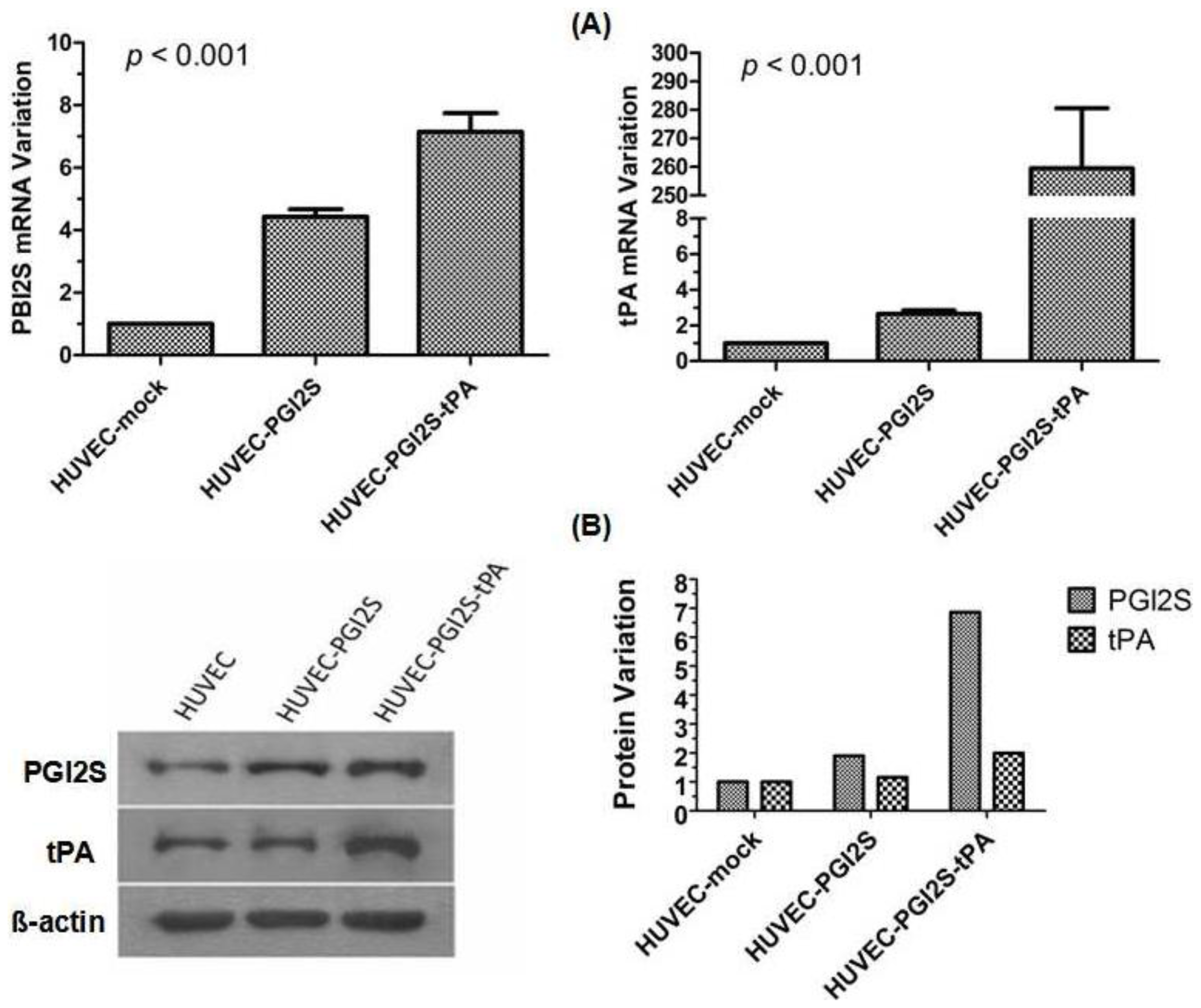
2.1. PGI2S and tPA Are Over-Expressed in HUVEC-PGI2S and HUVEC-PGI2S-tPA Cell Lines

After cloning the PGI2S and tPA cDNAs from the human liver cell line L-02, the lentiviral backbone vector, pSin-EF2-puro-oligo was utilized to construct HUVEC cell lines stably over-expressing PGI2S (HUVEC-PGI2S), or PGI2S and tPA (HUVEC-PGI2S-tPA). A negative control cell line, HUVEC-mock, was created using the empty lentivirus backbone vector.
The mRNA and protein expression of PGI2S and tPA were analyzed by qRT-PCR and Western blotting. Compared to HUVEC-mock cells, PGI2S mRNA expression was approximately 4.4-fold higher in HUVEC-PGI2S cells, and approximately 7.1-fold higher when simultaneously over-expressed with tPA in HUVEC-PGI2S-tPA cells (p < 0.001); tPA mRNA expression was approximately 2.6-fold and 259.5-fold higher in HUVEC-PGI2S and HUVEC-PGI2S-tPA cells, respectively, compared to HUVEC-mock cells (p < 0.001; Figure 1A). These increases in mRNA expression were matched by similar increases in PGI2S and tPA protein expression. Densitometry revealed that the protein expression levels of PGI2S were 1.9-fold higher in HUVEC-PGI2S cells and 6.9-fold higher in HUVEC-PGI2S-tPA cells compared to HUVEC-mock cells. The protein expression levels of tPA were not increased in HUVEC-PGI2S cells but were 2.0-fold higher in HUVEC-PGI2S-tPA cells compared to HUVEC-mock cells (Figure 1B).

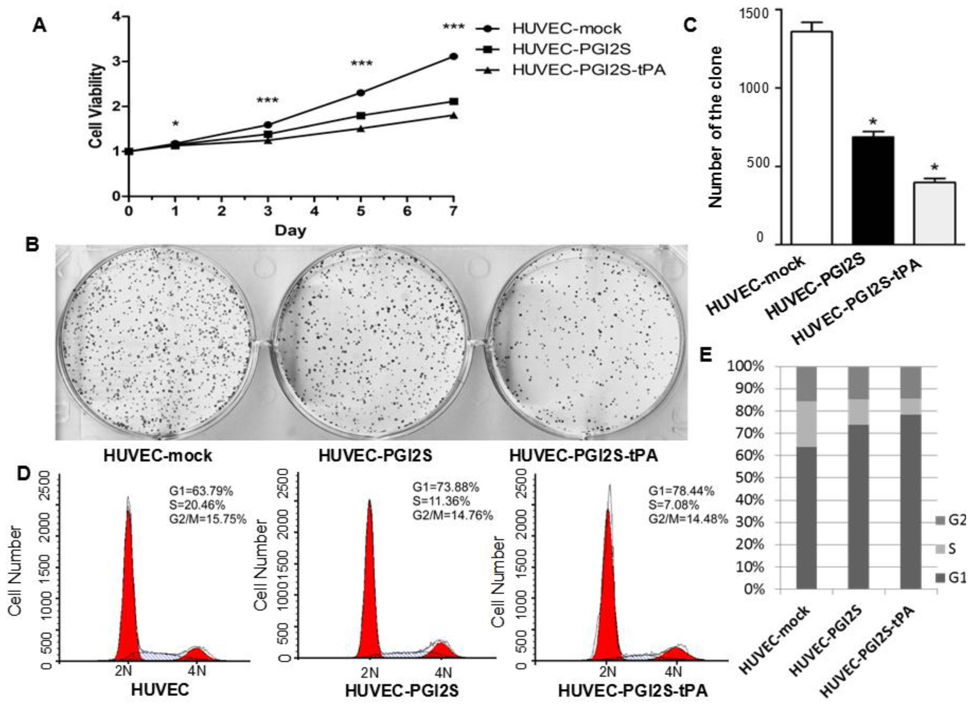
2.2. Cell Growth Is Restricted in HUVEC-PGI2S and HUVEC-PGI2S-tPA Cell Lines

The result from the MTT assay showed that the proliferation of HUVEC-PGI2S cells was significantly reduced compared to the negative control HUVEC-mock cells, and the proliferation of HUVEC-PGI2S-tPA cells was even slower than that of HUVEC-PGI2S cells (Figure 2A). Figure 2B,C shows that the colony formation ability of both HUVEC-PGI2S and HUVEC-PGI2S-tPA cells was also reduced compared to HUVEC-mock cells. Flow cytometric examination of the cell cycle indicated G1 phase arrest occurred in both HUVEC-PGI2S and HUVEC-PGI2S-tPA cells; the extent of G1 phase arrest was more significant in HUVEC-PGI2S-tPA cells (Figure 2D,E). These results indicate that both the proliferation and growth ability of HUVEC-PGI2S and HUVEC-PGI2S-tPA cells were reduced in comparison to HUVEC-mock cells.

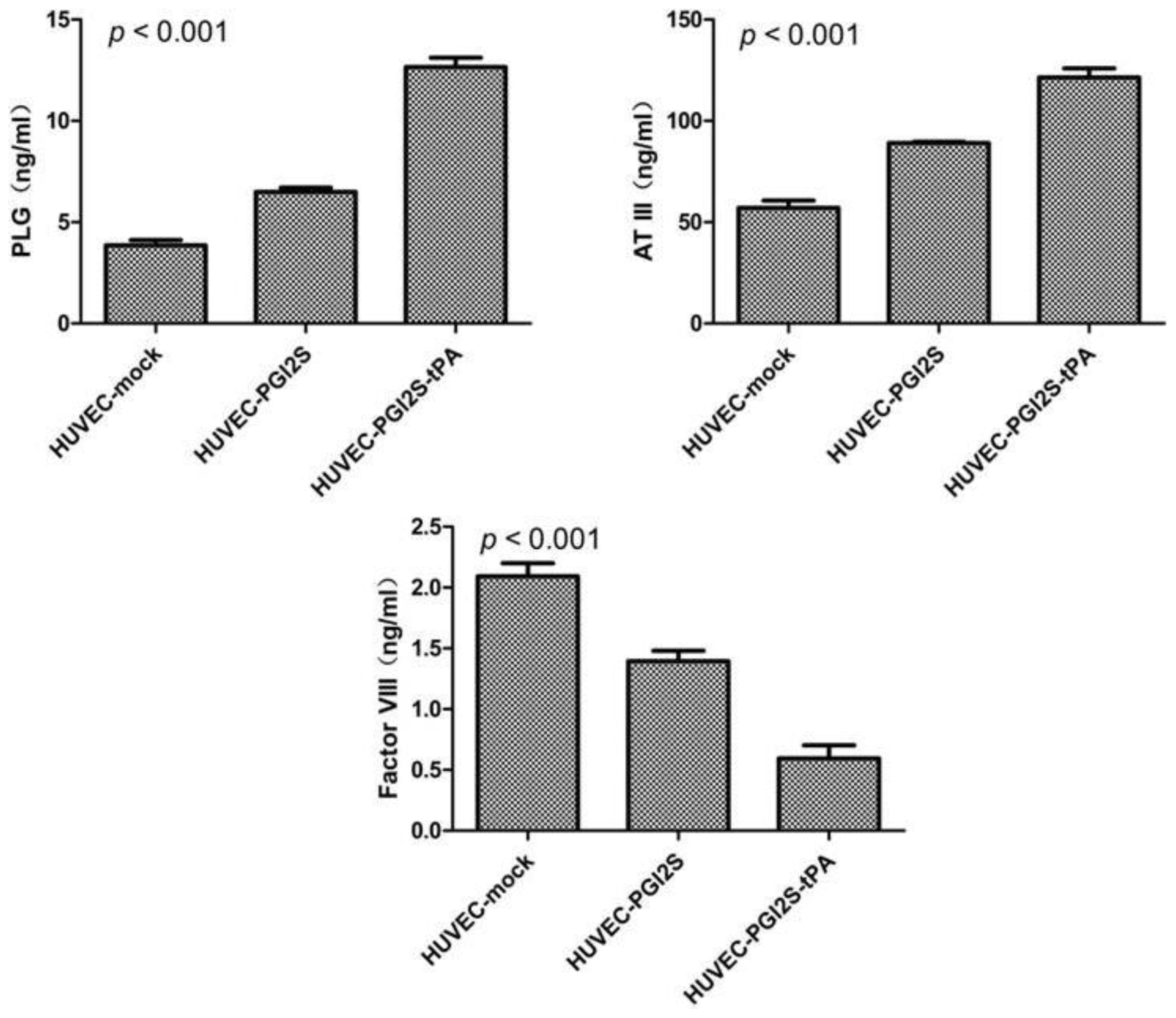
2.3. HUVEC-PGI2S and HUVEC-PGI2S-tPA Cell Lines Expressed Higher Levels of Anticoagulation Factors

The protein expression levels of ATIII, PLG and FVIII were examined using ELISA assays (Figure 3). ATIII acts as an inhibitor of several coagulation factors, and its expression was up-regulated from 57 ng/mL in HUVEC-mock cells to 89 ng/mL in HUVEC-PGI2S cells and 121 ng/mL in HUVEC-PGI2S-tPA cells (p < 0.001). PLG plays a vital role in fibrinolysis, and, in vivo, is activated by tPA in plasma. The expression of PLG was elevated by 2-fold and 4-fold in HUVEC-PGI2S and HUVEC-PGI2S-tPA cells, respectively, compared to the negative control cells (p < 0.001). On the other hand, FVIII, a participant in both the intrinsic and extrinsic coagulation pathways, was down-regulated by 45% and 70% in HUVEC-PGI2S and HUVEC-PGI2S-tPA cells compared to the negative control cells, respectively (p < 0.001).

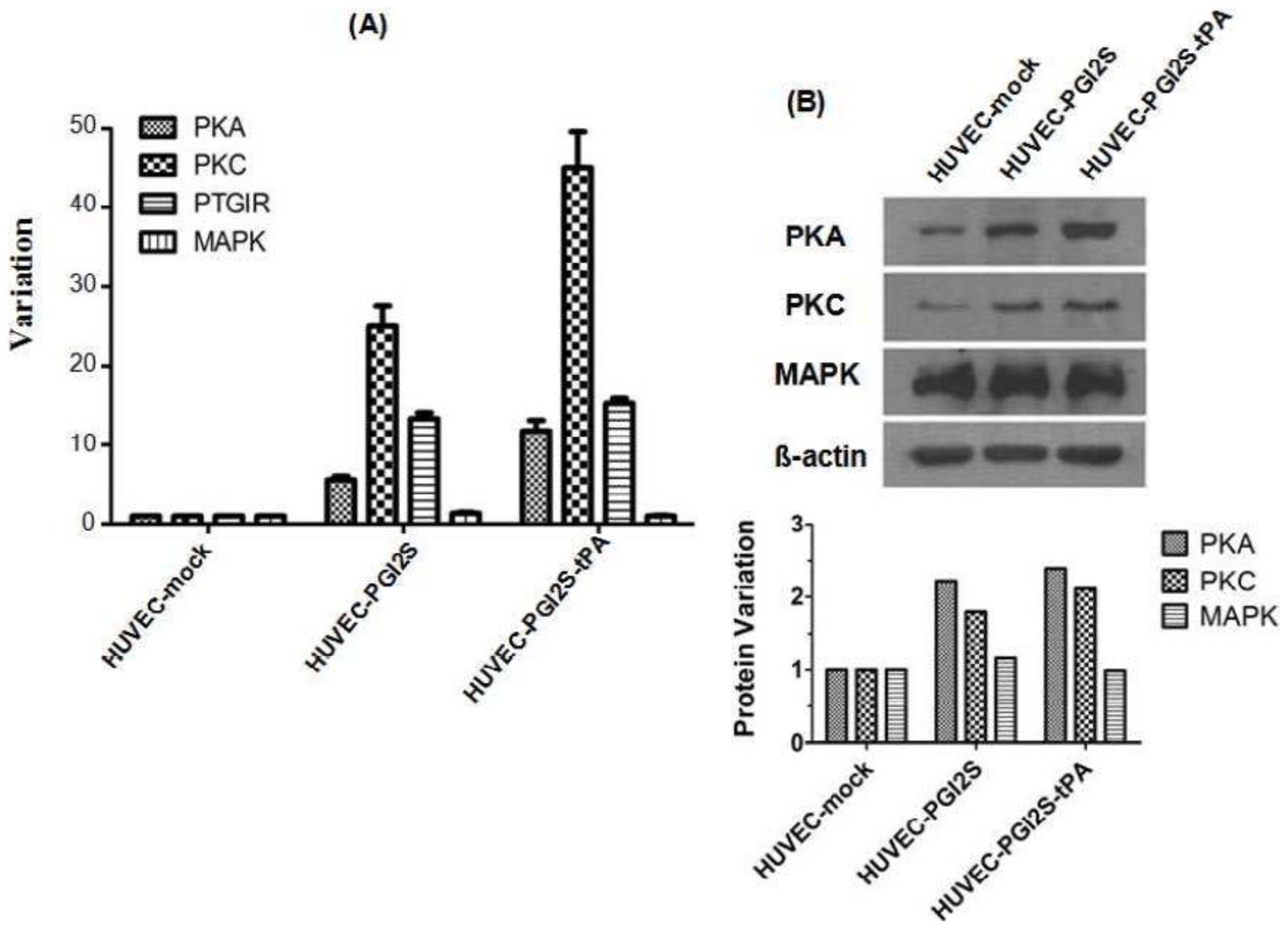
2.4. Signaling Pathways Involved in Vasodilation and Platelet Disaggregation Are Activated in HUVEC-PGI2S and HUVEC-PGI2S-tPA Cell Lines

PGI2, whose synthesis is catalyzed by PGI2S, combines with the PTGIR to activate PKA, resulting in vasodilation and platelet disaggregation. The PKC pathway acts upstream of PGI2S, and PKC can also activate platelets via other signaling proteins, including MAPK. PKA, PKC, PTGIR and MAPK mRNA expression were analyzed by qRT-PCR. The expression levels of all of these signaling molecules, except for MAPK, were up-regulated in HUVEC-PGI2S cells compared to HUVEC-mock cells, and even higher in HUVEC-PGI2S-tPA cells (Figure 4A). Up-regulation of PKA, PKC except for MAPK protein expression was confirmed by Western blotting (Figure 4B). These results indicate that over-expression of PGI2S improves the anticoagulant effect of HUVEC cells, and that these effects were more potent when PGI2S was over-expressed in combination with tPA.

3. Discussion

CABG is an effective therapy for coronary artery disease. However, the conduits used for the bypass play an important role in the success of CABG. The use of heterograft vessels constructed from endothelial cells with improved anticoagulant ability represents a promising area of research in CABG. HUVEC cells are reported to have a relatively low immunogenicity [21,22], suggesting that transgenic HUVEC cells may be the ideal epithelial cells for developing heterograft vessels. The data presented in this study indicates that our novel transgenic HUVEC cell lines, HUVEC-PGI2S and HUVEC-PGI2S-tPA cells, may have a more potent anticoagulant effect, which may enable their future development as cells for bypass conduits.
To explore the anticoagulant effect of HUVEC-PGI2S and HUVEC-PGI2S-tPA cells, we examined their expression of vascular endothelial cell anticoagulant and coagulation factors using ELISAs. ATIII and PLG are the key components of the anticoagulation and antifibrinolytic systems. ATIII, a serine protease inhibitor belonging to the humoral anticoagulant system, plays an integral role in the coagulation system by inhibiting thrombin and other clotting factors [2325]. PLG dissolves fibrin in blood clots and is an important protease involved in a variety of other cellular processes [26,27]. Another key protein, Factor VIII (FVIII), is involved in both the intrinsic and extrinsic coagulation pathways [28,29]. Our results showed that the coagulation factor inhibitors ATIII and PLG were up-regulated, and FVIII, the main coagulation pathway promoter, was down-regulated in both HUVEC-PGI2S and HUVEC-PGI2S-tPA cells. Therefore, HUVEC-PGI2S and HUVEC-PGI2S-tPA cells have a more potent anticoagulant effect than HUVEC cells.
Additionally, the expression of proteins involved in the signaling pathways which regulate vasodilation and platelet disaggregation were investigated. Epithelial growth factor (EGF), vascular endothelial growth factor (VEGF), Bradykinin (BK), epinephrine and angiotensin II all activate the PKC-MAPK signaling pathway, via phosphorylation of MAPK, which results in increased formation of PGI2 [30,31]. Thus, up-regulation and activation of PKC are key upstream events required for synthesis of PGI2. Our qRT-PCR and Western blotting results demonstrated that expression of PKC increased in both HUVEC-PGI2S and HUVEC-PGI2S-tPA cells, PKC has been reported to be widely expressed in various types of tissues and plays a role in cardiovascular disease. The expression of MAPK remained unchanged in both HUVEC-PGI2S and HUVEC-PGI2S-tPA cells compared to HUVEC-mock cells, perhaps reflecting the fact that activation of MAPK may only require it to be phosphorylated rather than over-expressed.
Generation of cAMP and activation of PKA also form part of another important signaling pathway which regulates the biological functions of PTGIR activation, which in the presence of PGI2, results in vasodilation and inhibition of platelet aggregation [32]. In this study, PKA was observed to be over-expressed in both HUVEC-PGI2S and HUVEC-PGI2S-tPA cells, implying activation of this signaling pathway. Furthermore, PTGIR was also found to be over-expressed in both cell lines, indicating an increased sensitivity to PGI2. A recent study indicated that activation of PTGIR could protect the vascular epithelium from arteriosclerosis obliterans, the condition in which vascular grafts develop atherosclerosis a few years after CABG [33]. We hypothesize that grafts constructed from HUVEC-PGI2S and HUVEC-PGI2S-tPA cells may have a lower risk of developing atherosclerosis.
However, the results of the cell viability assay and colony formation assay showed that HUVEC-PGI2S and HUVEC-PGI2S-tPA cells are not currently fit for clinical use. Over-expression of PGI2S and tPA in HUVEC cells reduced their proliferation and growth ability. Cell cycle assays indicated that the limited cell growth and proliferation in HUVEC-PGI2S and HUVEC-PGI2S-tPA cells were due to increased G1 phase arrest. The mechanisms involved in this process are not clear, and further study is needed. Additionally, increased cell proliferation ability would be required in order for these transgenic endothelial cells to be used in clinical applications.

4. Experimental Section

4.1. Cell Culture

All HUVEC cells were cultured in RPMI-1640 medium (Life Technologies, Carlsbad, CA, USA) supplemented with 10% fetal bovine serum (FBS; Thermo Fisher Scientific, Waltham, MA, USA). HUVEC cells were stably transfected with PGI2S, or PGI2S and tPA, to establish the HUVEC-PGI2S and HUVEC-PGI2S-tPA cell lines, respectively. HUVEC cells were also stably transfected with lentivirus packed with pSin-EF2-puro-oligo to generate the HUVEC-mock negative control cell line. After infection, HUVEC-mock HUVEC-PGI2S, and HUVEC-PGI2S-tPA cells were treated with puromycin for four weeks to select for stably-expressing cells. Expression of PGI2S and tPA was confirmed after the four-week selection process before the other experiments were performed.

4.2. Plasmid Construction

Primers specific for PGI2S cDNA incorporating BamHI and EcoRI restriction sites (forward: 5′-CGGGATCCATGCTCGCCCGCGCCCTGCT-3′ and reverse: 5′-CGGAATTCCTACAGTTCAGT CGAACGTTCTT-3′) and, primers specific for tPA cDNA incorporating with BamHI and SpeI restriction sites (forward: 5′-CGGGATCCATGGATGCAATGAAGAGAGGGCT-3′ and reverse: 5′-GGACTAGTTCACGGTCGCATGTTGTCAC-3′) were designed and constructed. PCR amplifications of the PGI2S and tPA cDNAs were performed with the specific primers using PrimeSTAR HS (TAKARA, Otsu, Japan) using human liver cell line L-02 (Chinese Academy of Sciences, Shanghai, China) cDNA as the template. The purified PCR products and pSin-EF2-puro-oligo plasmid (James Thomson Lab, Madison, WI, USA) were digested with the corresponding enzymes, and then ligated.

4.3. RNA Preparation and Quantitative Real-Time PCR (qRT-PCR)

Total RNA was extracted from HUVEC, HUVEC-PGI2S and HUVEC-PGI2S-tPA cells using TRIzol reagent (Life Technologies, Carlsbad, CA, USA) and reverse transcription was performed using the SuperScript First-Strand Synthesis System for RT-PCR (Life Technologies, Carlsbad, CA, USA), according to the manufacturer’s instruction. After reverse transcription, real-time-PCR was performed with Platinum SYBRGreen PCR SuperMix-UDG (Life Technologies, Carlsbad, CA, USA) using the CFX96™ Real-Time PCR Detection System and Bio-Rad CFX Manager (Bio-Rad, Hercules, CA, USA) following the manufacturer’s protocol. Briefly, 2 μg of total RNA was amplified in 20 μL reactions, containing 0.5 μL M-MLV reverse transcriptase, 0.5 μL RNasin inhibitor, 0.5 μg random hexamers, 4 μL of 5× RT buffer, and RNase free water. qRT-PCR was performed in total reaction volumes of 20 μL, containing 1 μL cDNA sample, 10 μL of 2× Platinum SYBRGreen PCR SuperMix-UDG, primers, and nuclease-free water. The three-step reaction protocol was: 95 °C for 2 min, followed by 40 cycles at 95 °C for 30 s, 60 °C for 35 s and 72 °C for 30 s. β-actin mRNA was amplified as an internal control. The primers used were as follows: β-actin, forward 5′-TGGCAC CCAGCACAATGAA-3′ and reverse 5′-CTAAGTCATAGTCCGCCTAGAAGCA-3′; PGI2S, forward 5′-TTGCTGGCAGGGTTGCTGGTGGTA-3′ and reverse 5′-AAGATGGCATCTGGCCGAGGCTT3′; tPA, forward 5′-AAGCATGAGGCCTTGTCTCCTT-3′ and reverse 5′-TGCGGCCATCGTTCAGACA C-3′; protein kinase A (PKA), 5′-CGGAAGGTTCAGTGAGCCCCATGC-3′ and reverse 5′-GCAGATT CTCCGGCTTCAGGTC-3′; protein kinase C-alpha (PKC), forward 5′-TTATGCTGTCATGTCCCG GAACAG-3′ and reverse 5′-AGGAGGCGATGATGATGAGGAC-3′; PGI2 Receptor (PTGIR), forward 5′-CAGAGCTTGAGTCGCTGGAA-3′ and reverse 5′-CCTCTCACGATCCGCTGCTT-3′; mitogen-activated protein kinase (MAPK), forward 5′-GGCATGGTGTGCTCTGCTTA-3′ and reverse 5′-CGATGGTTGGTGCTCGAATA-3′.

4.4. Western Blotting

Protein samples were extracted from HUVEC, HUVEC-PGI2S and HUVEC-PGI2S-tPA cells using ProteoJET Mammalian Cell Lysis Reagent (Thermo Fisher Scientific, Waltham, MA, USA) according to the manufacturer’s instruction, mixed with DualColor Protein Loading Buffer (Thermo Fisher Scientific, Waltham, MA, USA) and boiled for 5 min. Sodium dodecyl sulfate-polyacrylamide gel electrophoresis (SDS-PAGE) was performed on 4% spacer and 10% separation gels; 30 μg protein was loaded into each well. Afterwards, the proteins were transferred to polyvinylidene fluoride (PVDF) membranes (EMD Millipore, Billerica, MA, USA), blocked with 5% non-fat dry milk for 1 h at room temperature, then incubated with primary antibodies specific for PGI2S (52 kDa; 1:200; Santa Cruz Biotechnology, Santa Cruz, CA, USA), tPA (65 kDa; 1:500; Bioworld, St. Louis Park, CA, USA), PKA (43 kDa; 1:500; Bioworld), PKC (80 kDa; 1:2000; Epitomics, Burlingame, CA, USA) and MAPK (44 kDa; 1:1000; Cell Signaling Technology, Danvers, MA, USA) or β-actin (45 kDa; 1:1000; Cell Signaling Technology) for 12 h at 4 °C. Membranes were incubated with secondary antibodies labeled with horseradish peroxidase for 1 h, imaged using an ECL system (Pierce, Madison, WI, USA) and densitometric analyses was performed by normalization against the internal loading control β-actin using Image J software (NIH, Bethesda, MD, USA).

4.5. Cell Proliferation Assays

Growing cells were seeded into 96-well plates (5 × 10³ cells in 200 μL medium per well), cultured for 0, 1, 3, 5 or 7 days, and then 20 μL of sterile 5 mg/mL 5-dimethylthiazol-2-yl-2,5-diphenyltetrazolium bromide (MTT; Sigma-Aldrich, St. Louis, MO, USA) was added per well, and the cells were incubated for 4 h at 37 °C. After removing the medium, 150 μL dimethyl sulfoxide (DMSO, Sigma-Aldrich, St. Louis, MO, USA) was added to each well, mixed thoroughly for 15 min, and the optical density (OD) values were read at 490 nm using a Bio-Rad 3500 Microplate Reader (Bio-Rad, Hercules, CA, USA). The relative numbers of cells were expressed as percentages of the readings taken on day 0. All experiments were performed in triplicate.

4.6. Plate Colony Formation Assays

Cells were seeded into six-well plates (1 × 10³ per well) and cultured for two weeks. The colonies were fixed in methanol for 10 min and stained with 1% crystal violet for 1 min. The plate colony formation assay was performed in triplicate for each group of cells.

4.7. Cell Cycle Analysis

Cell cycle analysis was performed using propidium iodide (PI, Sigma-Aldrich, St. Louis, MO, USA) staining. The cultured HUVEC, HUVEC-PGI2S and HUVEC-PGI2S-tPA cells were digested with 0.25% trypsin (Life Technologies, Carlsbad, CA, USA), washed in phosphate-saline buffer (PBS), fixed in 1 mL cold 70% ethanol for 1 h, washed and centrifuged. The cell pellet was resuspended in 500 μL PI solution (50 μg/mL PI, 0.1 mg/mL RNase A, 0.05% Triton X-100 in PBS) and incubated for 40 min at 37 °C, then the cells were centrifuged and resuspended in 500 μL PBS for flow cytometric analysis using the Cytomics™ FC500 Flow Cytometry (Beckman Coulter, Brea, CA, USA).

4.8. Enzyme-Linked Immunosorbent Assays

The cultured cells were digested, resuspended in PBS and stored overnight at −20 °C. After two freeze-thaw cycles, the cells were centrifuged, and the resulting supernatants were used as the cell homogenates. Enzyme-linked immunosorbent assays (ELISAs) were performed on the cell homogenates using the Human Coagulation Factor VIII (FVIII) ELISA kit (Cusabio, Wuhan, China), Human Antithrombin III (ATIII) ELISA kit (Cusabio) and ELISA kit for plasminogen (USCN, Wuhan, China) following the manufacturer’s protocol. Standard curves were constructed and used to determine the expression level of each molecule.

5. Conclusions

The development of heterograft conduits is one of the most promising new sources of CABG grafts. Although our transgenic HUVEC cells still have a number of issues preventing their use in clinical applications, this study provides strong evidence to suggest that transgenic HUVEC-PGI2S and HUVEC-PGI2S-tPA vascular endothelial cells have the potential to be developed as ideal cells for the creation of heterograft vascular conduits.

Acknowledgments

This work was supported by grants from the Guangdong Provincial Medical Research Foundation (No. A2009143), the National Natural Science Foundation of China (No. 81171948), the Key Program of Natural Science Foundation of Guangdong Province, China (No. S2012020011060), and the Project of State Key Laboratory of Oncology in South China (No. 030041060004, to M. Zheng).

Conflicts of Interest

The authors declare no conflict of interest.
  • Author ContributionsJian-Hua Wang contributed to the study design, data collection and analysis, manuscript writing and revisions to the manuscript. Lin-Jing Yuan and Zhi-Min Zhong contributed to the study design, experimentation and manuscript writing. Zhe-Sheng Wen contributed to the study design, data collection and analysis. Jian-Ming Deng and Rong-Xin Liang contributed to the experimentation. Min Zheng had the idea of the study, was responsible for its design and coordination, contributed to analysis and interpretation of the data, as well as manuscript writing and revisions to the manuscript. All authors read and approved the final manuscript.

References

  1. Mehta, N.J.; Khan, I.A. Cardiology’s 10 greatest discoveries of the 20th century. Tex. Heart Inst. J 2002, 29, 164–171. [Google Scholar]
  2. Goetz, R.H.; Rohman, M.; Haller, J.D.; Dee, R.; Rosenak, S.S. Internal mammary-coronary artery anastomosis: A nonsuture method employing tantalum rings. J. Thorac. Cardiovasc. Surg 1961, 41, 378–386. [Google Scholar]
  3. Jeremy, J.Y.; Zacharowski, K.; Shukla, N.; Wan, S. Pharmacological strategies aimed at reducing complications associated with coronary artery bypass graft surgery. Curr. Opin. Pharmacol 2012, 12, 111–113. [Google Scholar]
  4. Englberger, L.; Noti, J.; Immer, F.F.; Stalder, M.; Eckstein, F.S.; Carrel, T.P. The shelhigh no-react bovine internal mammary artery: A questionable alternative conduit in coronary bypass surgery? Eur. J. Cardiothorac. Surg 2008, 33, 222–224. [Google Scholar]
  5. Cleary, M.A.; Geiger, E.; Grady, C.; Best, C.; Naito, Y.; Breuer, C. Vascular tissue engineering: The next generation. Trends Mol. Med 2012, 18, 394–404. [Google Scholar]
  6. Kim, F.Y.; Marhefka, G.; Ruggiero, N.J.; Adams, S.; Whellan, D.J. Saphenous vein graft disease: Review of pathophysiology, prevention, and treatment. Cardiol. Rev 2013, 21, 101–109. [Google Scholar]
  7. Herring, M.; Gardner, A.; Glover, J. Seeding endothelium onto canine arterial prostheses: The effects of graft design. Arch. Surg 1979, 114, 679–682. [Google Scholar]
  8. Herring, M.B.; Dilley, R.; Jersild, R.A., Jr.; Boxer, L.; Gardner, A.; Glover, J. Seeding arterial prostheses with vascular endothelium: The nature of the lining. Ann. Surg 1979, 190, 84–90. [Google Scholar]
  9. LaFayette, N.G.; Skrzypchak, A.M.; Merz, S.; Bartlett, R.H.; Annich, G.M. An in vitro method for assessing biomaterial-associated platelet activation. ASAIO J 2007, 53, 159–162. [Google Scholar]
  10. McGuigan, A.P.; Sefton, M.V. The influence of biomaterials on endothelial cell thrombogenicity. Biomaterials 2007, 28, 2547–2571. [Google Scholar]
  11. Bednar, F.; Tencer, T.; Plasil, P.; Paluch, Z.; Sadilkova, L.; Prucha, M.; Kopa, M. Evaluation of aspirin’s effect on platelet function early after coronary artery bypass grafting. J. Cardiothorac. Vasc. Anesth 2012, 26, 575–580. [Google Scholar]
  12. Balyasnikova, I.V.; Danilov, S.M.; Muzykantov, V.R.; Fisher, A.B. Modulation of angiotensin-converting enzyme in cultured human vascular endothelial cells. In Vitro Cell. Dev. Biol 1998, 34, 545–554. [Google Scholar]
  13. Dichek, D.A.; Neville, R.F.; Zwiebel, J.A.; Freeman, S.M.; Leon, M.B.; Anderson, W.F. Seeding of intravascular stents with genetically engineered endothelial cells. Circulation 1989, 80, 1347–1353. [Google Scholar]
  14. Degen, S.J.; Rajput, B.; Reich, E. The human tissue plasminogen activator gene. J. Biol. Chem 1986, 261, 6972–6985. [Google Scholar]
  15. MacDonald, M.E.; van Zonneveld, A.J.; Pannekoek, H. Functional analysis of the human tissue-type plasminogen activator protein: The light chain. Gene 1986, 42, 59–67. [Google Scholar]
  16. Wu, K.K.; Liou, J.Y. Cellular and molecular biology of prostacyclin synthase. Biochem. Biophys. Res. Commun 2005, 338, 45–52. [Google Scholar]
  17. Nakayama, T. Prostacyclin analogues: Prevention of cardiovascular diseases. Cardiovasc. Hematol. Agents Med. Chem 2006, 4, 351–359. [Google Scholar]
  18. Brash, A.R. Mechanistic aspects of CYP74 allene oxide synthases and related cytochrome P450 enzymes. Phytochemistry 2009, 70, 1522–1531. [Google Scholar]
  19. Nakayama, T.; Soma, M.; Watanabe, Y.; Hasimu, B.; Sato, M.; Aoi, N.; Kosuge, K.; Kansmatsuse, K.; Kokubun, S.; Marrow, J.D.; et al. Splicing mutation of the prostacyclin synthase gene in a family associated with hypertension. Biochem. Biophys. Res. Commun 2002, 297, 1135–1139. [Google Scholar]
  20. Suhara, H.; Sawa, Y.; Fukushima, N.; Kagisaki, K.; Yokoyama, C.; Tanabe, T.; Ohtake, S.; Matsuda, H. Gene transfer of human prostacyclin synthase into the liver is effective for the treatment of pulmonary hypertension in rats. J. Thorac. Cardiovasc. Surg 2002, 123, 855–861. [Google Scholar]
  21. Dengler, T.J.; Johnson, D.R.; Pober, J.S. Human vascular endothelial cells stimulate a lower frequency of alloreactive CD8+ pre-CTL and induce less clonal expansion than matching B lymphoblastoid cells: Development of a novel limiting dilution analysis method based on CFSE labeling of lymphocytes. J. Immunol 2001, 166, 3846–3854. [Google Scholar]
  22. Waldman, W.J.; Knight, D.A.; Adams, P.W.; Orosz, C.G.; Sedmak, D.D. In vitro induction of endothelial HLA class II antigen expression by cytomegalovirus-activated CD4+ T cells. Transplantation 1993, 56, 1504–1512. [Google Scholar]
  23. Grundy, C.B.; Thomas, F.; Millar, D.S.; Krawczak, M.; Melissari, E.; Lindo, V.; Moffat, E.; Kakkar, V.V.; Cooper, D.N. Recurrent deletion in the human antithrombin III gene. Blood 1991, 78, 1027–1032. [Google Scholar]
  24. Blajchman, M.A.; Austin, R.C.; Fernandez-Rachubinski, F.; Sheffield, W.P. Molecular basis of inherited human antithrombin deficiency. Blood 1992, 80, 2159–2171. [Google Scholar]
  25. Labarrere, C.A.; Pitts, D.; Halbrook, H.; Faulk, W.P. Natural anticoagulant pathways in normal and transplanted human hearts. J. Heart Lung Transplant 1992, 11, 342–347. [Google Scholar]
  26. Aoki, N.; Moroi, M.; Sakata, Y.; Yoshida, N.; Matsuda, M. Abnormal plasminogen: A hereditary molecular abnormality found in a patient with recurrent thrombosis. J. Clin. Invest 1978, 61, 1186–1195. [Google Scholar]
  27. Petersen, T.E.; Martzen, M.R.; Ichinose, A.; Davie, E.W. Characterization of the gene for human plasminogen, a key proenzyme in the fibrinolytic system. J. Biol. Chem 1990, 265, 6104–6111. [Google Scholar]
  28. Della-Morte, D.; Beecham, A.; Dong, C.; Wang, L.; McClendon, M.S.; Gardener, H.; Blanton, S.H.; Sacco, R.L.; Rundek, T. Association between variations in coagulation system genes and carotid plaque. J. Neurol. Sci 2012, 323, 93–98. [Google Scholar]
  29. Starke, R.D.; Paschalaki, K.E.; Dyer, C.E.; Harrison-Lavoie, K.J.; Cutler, J.A.; McKinnon, T.A.J.; Millar, C.M.; Cutler, D.F.; Laffan, M.A.; Randi, A.M. Cellular and molecular basis of von Willebrand disease: Studies on blood outgrowth endothelial cells. Blood 2013, 121, 2773–2784. [Google Scholar]
  30. Gliki, G.; Abu-Ghazaleh, R.; Jezequel, S.; Wheeler-Jones, C.; Zachary, I. Vascular endothelial growth factor-induced prostacyclin production is mediated by a protein kinase C (PKC)-dependent activation of extracellular signal-regulated protein kinases 1 and 2 involving PKC-δ and by mobilization of intracellular Ca2+. Biochem. J 2001, 353, 503–512. [Google Scholar]
  31. Mustonen, P.; van Willigen, G.; Lassila, R. Epinephrine—Via activation of p38-MAPK—Abolishes the effect of aspirin on platelet deposition to collagen. Thromb. Res 2001, 104, 439–449. [Google Scholar]
  32. Webb, J.G.; Tan, Y.; Jaffa, M.A.; Jaffa, A.A. Evidence for prostacyclin and cAMP up-regulation by bradykinin and insulin-like growth factor 1 in vascular smooth muscle cells. J. Recept. Signal Transduct. Res 2010, 30, 61–71. [Google Scholar]
  33. Muto, A.; Nishibe, T.; Miyauchi, Y.; Kondo, Y.; Yamamoto, Y.; Dardik, A.; Shigematsu, H. Prostaglandin receptors EP2 and IP are detectable in atherosclerotic arteries and plaques. Int. Angiol 2010, 29, 43–48. [Google Scholar]
Figure 1. Expression of PGI2S and tPA in HUVEC-mock, HUVEC-PGI2S, and HUVEC-PGI2S-tPA cell lines. (A) PGI2S and tPA mRNA expression were analyzed by quantitative real-time PCR and expressed relative to the HUVEC-mock cell line. Values are means ± S.D. of triplicates. p values were calculated using one-way ANOVA; (B) Analysis of PGI2S and tPA protein expression by Western blotting in HUVEC-mock, HUVEC-PGI2S, and HUVEC-PGI2S-tPA cell lines. The Western blotting analyses were performed in triplicate.
Ijms 15 02826f1
Figure 2. Cell growth and colony formation ability of HUVEC-mock, HUVEC-PGI2S and HUVEC-PGI2S-tPA cell lines. (A) Cell viability of HUVEC-PGI2S and HUVEC-PGI2S-tPA cells determined by the MTT assay. Growth curves are shown; data is expressed relative to cell viability on day 0. p values were calculated using independent samples t-test, * p < 0.05, and *** p < 0.001; (B) and (C) The plate colony formation assay was performed over 14 days, initially 1000 cells were seeded per well. The numbers of colonies formed were shown; three independent experiments were performed and P values were calculated using the independent samples t-test; (D) and (E) Cell cycle analysis of HUVEC-mock, HUVEC-PGI2S and HUVEC-PGI2S-tPA cell lines, and the percentage of cells at different phases of the cell cycle.
Ijms 15 02826f2
Figure 3. Expression of anticoagulation factors by HUVEC-mock, HUVEC-PGI2S, and HUVEC-PGI2S-tPA cells, ELISA were performed to measure the levels of ATIII, PLG and Factor VIII in cell homogenates, p values were calculated using one-way ANOVA.
Ijms 15 02826f3
Figure 4. Vasodilation and platelet disaggregation signaling pathway factors in HUVEC-mock, HUVEC-PGI2S and HUVEC-PGI2S-tPA cell lines. (A) Analysis of PKA, PKC, PTGIR and MAPK mRNA expression by qRT-PCR; (B) Analysis of PKA, PKC and MAPK protein expression by Western blotting. All analysis were performed triplicated.
Ijms 15 02826f4

