Review

Bacterial Cellular Engineering by Genome Editing and Gene Silencing

1
Bioproduction Research Institute, National Institute of Advanced Industrial Sciences and Technology (AIST), 2-17-2-1 Tsukisamu-Higashi, Toyohira-ku, Sapporo 062-8517, Japan
2
Biomass Refinery Research Center, National Institute of Advanced Industrial Sciences and Technology (AIST), 3-11-32, Kagamiyama, Higashi-Hiroshima, Hiroshima 739-0046, Japan
3
Department of Medical Genome Sciences, Graduate School of Frontier Sciences, the University of Tokyo, 2-17-2-1 Tsukisamu-higashi, Toyohira-ku, Sapporo, Hokkaido 062-8517, Japan
*
Author to whom correspondence should be addressed.
Int. J. Mol. Sci. 2014, 15(2), 2773-2793; https://doi.org/10.3390/ijms15022773
Submission received: 23 December 2013 / Revised: 27 January 2014 / Accepted: 28 January 2014 / Published: 18 February 2014
(This article belongs to the Special Issue Molecular Cut and Paste)

Abstract

:
Genome editing is an important technology for bacterial cellular engineering, which is commonly conducted by homologous recombination-based procedures, including gene knockout (disruption), knock-in (insertion), and allelic exchange. In addition, some new recombination-independent approaches have emerged that utilize catalytic RNAs, artificial nucleases, nucleic acid analogs, and peptide nucleic acids. Apart from these methods, which directly modify the genomic structure, an alternative approach is to conditionally modify the gene expression profile at the posttranscriptional level without altering the genomes. This is performed by expressing antisense RNAs to knock down (silence) target mRNAs in vivo. This review describes the features and recent advances on methods used in genomic engineering and silencing technologies that are advantageously used for bacterial cellular engineering.

1. Introduction

Microorganisms have been used since ancient times for the production of fermented food products, such as cheese, sourdough, beer, wine, and vinegar, and humans have enjoyed the benefits of this biotechnology in terms of transforming raw materials to value-added products with superior flavor, texture, and longevity. Microorganisms can be found in almost every natural environment on our planet [1,2]. The traditional method for obtaining microorganisms that are useful for human life is to search for such microorganisms in the natural environment. Once the strain has been identified, it is cultured in the laboratory, and the original strain is often bred for higher performance through a randomized process. Subculturing has been employed to select for better performing progeny strains arising during cultivation. This “forward” genetics approach has been widely used in biotechnological processes for the production of, e.g., enzymes, foods, amino acids, and fine chemicals.
In recent years, an opposite “reverse” genetics approach has emerged. In this approach, genetic perturbations (mutations) are introduced into a particular gene of interest, and its impact is investigated through functional analysis [3]. Concomitant with the rapid accumulation of available genetic information, this reverse genetics approach is increasingly used for strain improvement in this synthetic biology age [4]. Specific genes residing in bacterial genomes (or chromosomal DNA) are targeted by various mutations, including knockout (disruption), knock-in (insertion), and allelic exchange [5]. All of these genomic modifications can be carried out following a similar experimental technique based on homologous recombination.
In addition to methods that directly (or permanently) modify genomic sequences, a novel approach has emerged that does not alter the genomic sequences, but rather alters the gene expression profile through conditionally repressing expression of mRNA at the translation level. This gene silencing or gene knockdown is carried out by antisense RNAs (asRNAs). The advantage of this silencing technology is its wide applicability, especially for genes essential to growth and thus not ready for disruption. In this review, we describe the principles, procedures, and important points for the above-mentioned methods from the perspective of bacterial cellular engineering.

2. Conventional Genome Editing
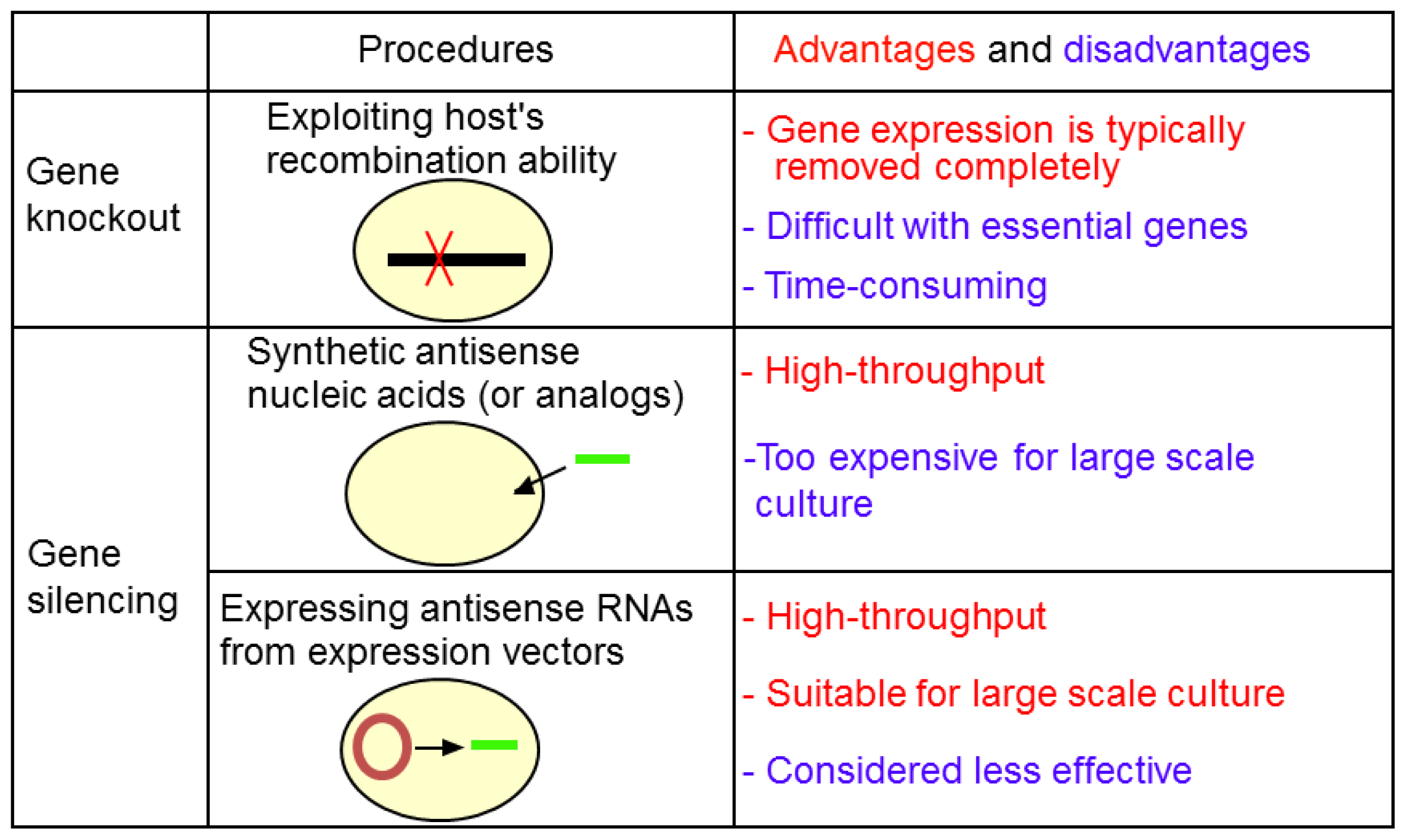
2.1. Gene Knockout

In 1989, a plasmid-based gene knockout method was developed in Escherichia coli that is achieved through two recombination events (i.e., integration and resolution) [6]. A plasmid carrying a replacing gene fragment containing homologous ends is integrated into a target gene locus through homologous recombination. To this end, the replacing gene fragment is cloned into a plasmid containing a temperature-sensitive replication origin, pSC101ts, which replicates at low temperatures (e.g., 30 °C), but not high temperatures (e.g., 43 °C ; Step (a) in Figure 1). Using the plasmid, E. coli is transformed and colonies are selected at a permissive, low temperature in the presence of the appropriate antibiotic. Transformants are then transferred to fresh medium and grown at a high temperature to prohibit self-replication of the plasmid. By selecting transformants in the presence of the antibiotic, the modified genomic fragment harbored on the plasmid is forced to integrate into the targeted genome locus through homologous recombination (Step (b) in Figure 1). After integration, the second recombination event takes place; resolved (disintegrated) progeny is selected at low, permissive temperatures at which the plasmid is able to replicate. During the resolving step, the specific region between the homologous sequences is deleted from the genome (Step (c) in Figure 1). This approach has been successfully applied to replace the bolA gene in the E. coli genome. However, the frequency to obtain the desired clone (“hit-rate”) was rather low and thus labor-intensive, especially at the resolution step [6]. It was hence improved by adding a counterselective marker gene onto the vector. The marker was a sucrose-sensitive suicide sacB gene of Bacillus subtilis; recombinants carrying the gene cannot grow on sucrose-containing plates, which are effective for the enrichment of clones lacking the gene during resolution. This improved method has been applied to various bacteria, including E. coli [69], Myxococcus xanthus [10], Corynebacterium glutamicum [11], Rhodococcus spp. [12], and Pseudomonas putida [13].
In 2000, Datsenko and colleagues developed a recombination-dependent, but slightly different, method for deletion of a genomic segment from E. coli using linear DNAs and λ-red recombinase [14]. Since then, this method has been applied to various bacteria including Salmonella spp. [15], Mycobacterium tuberculosis [16], Streptomyces spp. [17], and B. subtilis [18]. In brief, cells in which Gam, Bet, and Exo proteins of λ phage are expressed are transformed with linear DNAs containing homologous DNA sequences, two FRT sequences and a selection marker [14]. Then, the selection marker is removed by expressing the FLP recombinase that causes recombination between two FRT sequences [14].
Features of two representative gene knockout methods above are summarized in Table 1. The key feature of both methods is that no selection marker is left on the genomes, allowing multiple rounds of knockout. Note, however, that in the case of the original λ-red recombinase method by Datsenko and colleagues, a “scar” sequence of 81–85 base pairs (bp) in length is retained in the genome. When multiple rounds of disruption are performed, a risk exists of recombination between the scar sequences [19]. Furthermore, the scar sequence limits the precision of possible genomic modifications, which is easy with the pSC101ts-sacB method. The λ-red recombinase method has been widespread, but the pSC101ts-sacB method is still preferred for its several advantages, including the sequence-specificity and easiness for allelic exchange and knock-in, as described in Sections 2.2 and 2.3. A combination of these two methods has also been reported [20], and other variations are also known [7,2123].

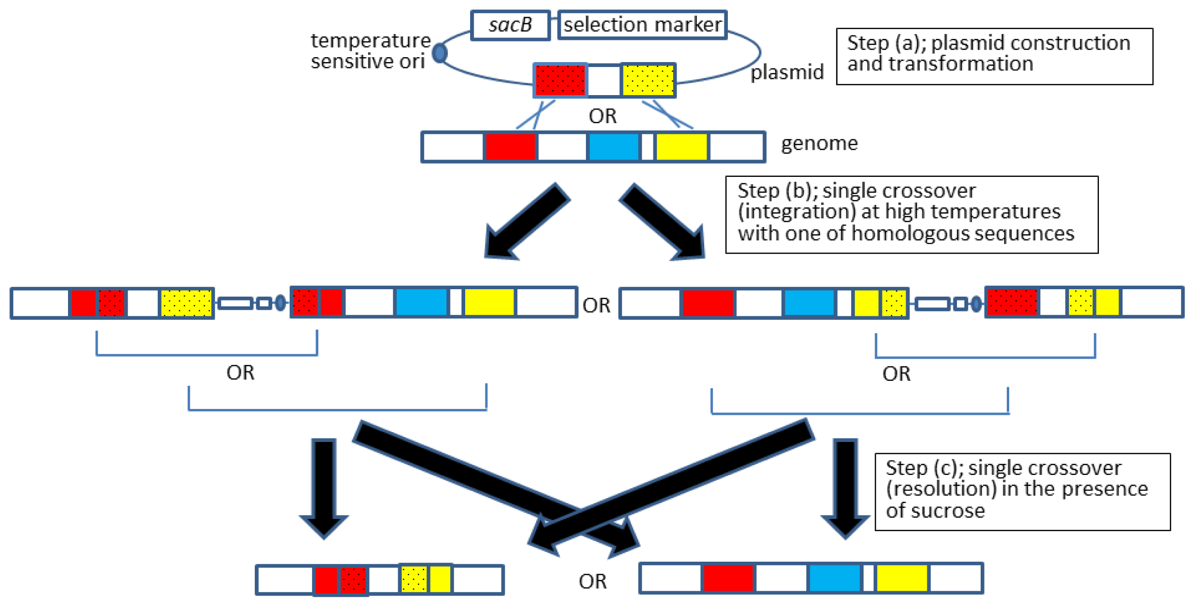
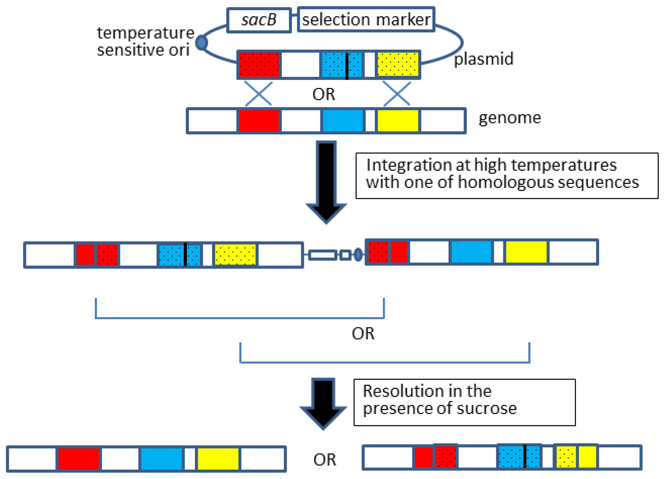
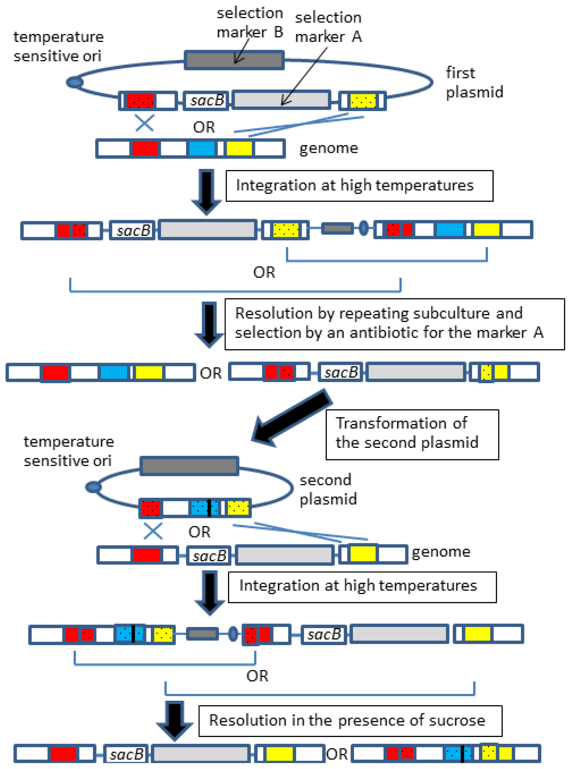
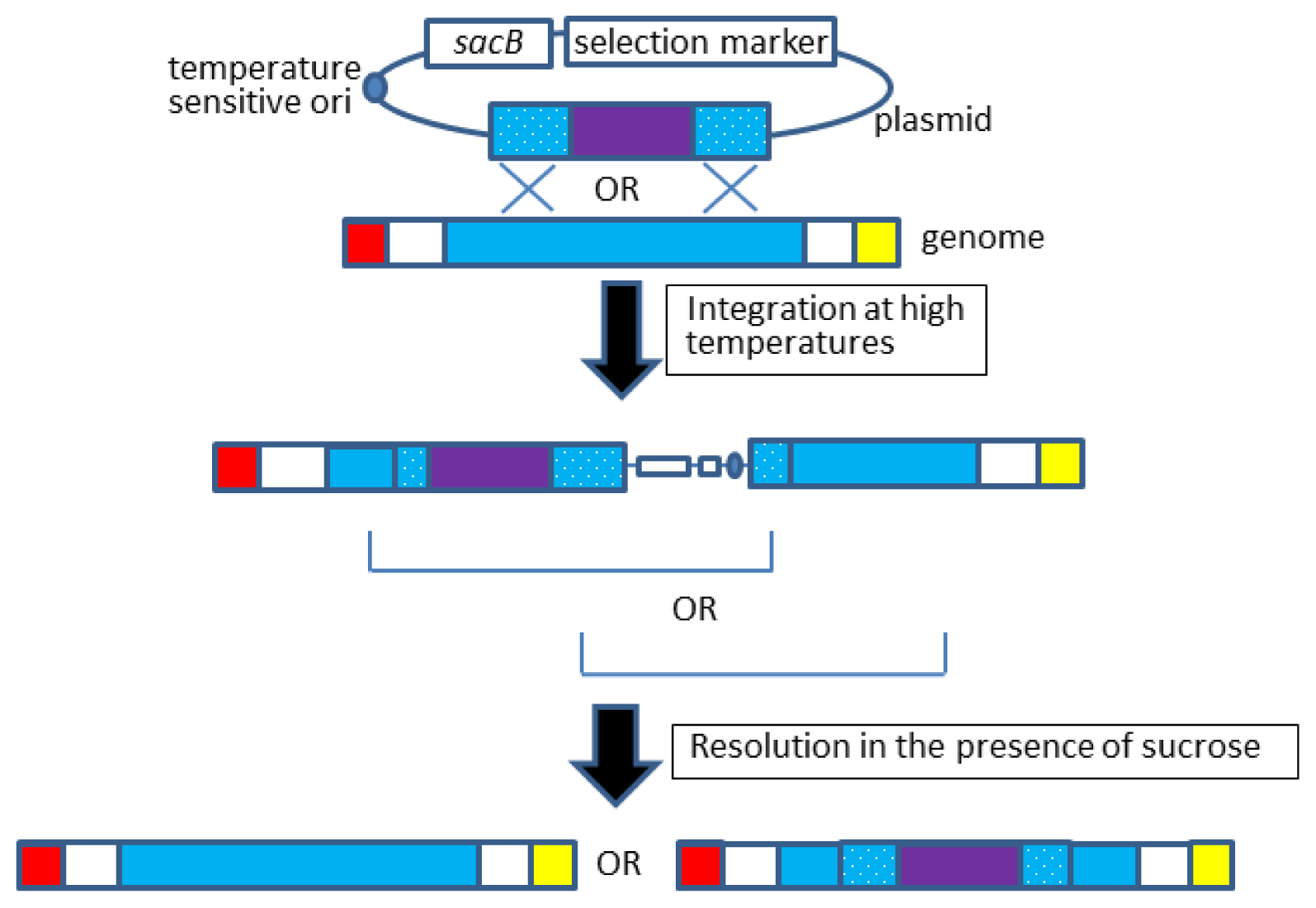
2.2. Allelic Exchange

Allelic exchange is a modified version of gene knockout that can be carried out by following the same procedure for gene knockout (Section 2.1), which is illustrated in Figure 2 [8,9].
Overall, the gene knockout and allelic exchange procedures are quite similar; however, the latter requires an observable phenotypic change because confirmation of fragment exchange is not readily achievable by simple methods, such as colony polymerase chain reaction (PCR), but instead requires DNA sequencing. Furthermore, by this method, we failed to introduce an mlc* allele, which contained a point mutation in the mlc gene of E. coli [24]. This was probably due to the low rate of proper resolution to obtain desired recombinants, which was a common problem in the gene knockout experiments as described above. To overcome these problems, Emmerson and colleagues applied two mechanisms for the resolution step: antibiotic selection and sucrose-based counterselection (Figure 3) [7], which was originally conducted by single sucrose-counterselection. Although Emmerson’s method requires an additional plasmid and, thus, is rather labor-intensive, this modification is effective for recovering the desired allelic exchange of various genes (e.g., mlc [24], crp [24], and lee4 [9]).
Another allelic exchange method is getting popular, which is mediated with single-stranded oligonucleotides and the λ-red recombinase [25]. For this method, only a Beta protein of three λ-red proteins is required, and the Beta protein binds single-stranded DNAs and promotes annealing to the homologous DNAs [25]. The important feature is high efficiency, allowing a recombination without any selection. In one report, 25% of the E. coli cell population was successfully recombined in the absence of antibiotic marker selection and any selection pressure [25]. Furthermore, because no plasmid constructions are required, multiple-rounds of allelic exchange are easily achieved. However, there is a serious limitation for usage in E. coli; the mismatch repair system of host cells should be removed for high efficiency, because mismatched nucleotides always occur at the initial step of recombination. Therefore, occurrence rate of undesirable mutations is increased [25]. To circumvent this disadvantage, it is reported that usage of modified (unnatural) bases at the mismatched sites are effective [26], and other improvements for usability are also reported [27,28].
In bacteria other than E. coli, this method has been used in M. tuberculosis [29], Pseudomonas syringae [30], Lactobacillus reuteri [31], and Lactococcus lactis [31].

2.3. Gene Knock-in

The gene knock-in procedure is very similar to that of gene knockout and allelic exchange [3234], as outlined in Figure 4. For example, our group knocked-in a “doxycycline inducible promoter–T7 RNA polymerase gene” cassette into the lacZ locus of E. coli [32]; the lacZ locus was chosen because it has no or little effect on cell growth. When the resulting strain was transformed with plasmids having a “T7 promoter-gene of interest” cassette, it was successful in expressing the gene of interest in a doxycycline-dependent manner [32].
Gene knock-in is most advantageously employed when the use of plasmid vectors is inadequate or when the copy number of the gene of interest should be kept low. Note also that knocked-in genes are more stably inherited to progenies compared to genes on plasmids [35].

3. New Technologies for Genome Editing

3.1. Gene Knockout with Mobile Group II Introns

In 2001, a novel method to knockout bacterial genes was reported, which uses mobile group II introns [36]. Group II introns are naturally occurring genetic elements found in eubacteria, mitochondria, and plastids [3739]. L. lactis Ll.LtrB is the most studied group II intron, which is a ribonucleoprotein (RNP) consisting of an intron RNA and an LtrA protein [40,41]. The intron RNA excises itself from the RNA transcript through a lariat structure and also encodes LtrA protein, a reverse transcriptase. Ll.LtrB selects a DNA site to integrate into the intron RNA through both protein–DNA interactions and RNA–DNA base pairings [4244]. Redesigning the intron RNA in Ll.LtrB allows any gene to be targeted [43]. When host cells are transformed with a vector expressing a redesigned intron RNA and intact LtrA, the redesigned Ll.LtrB recognizes the target DNA sequence and integrates itself (both intron RNA and LtrA protein participate in target-site recognition). Then, LtrA reverse transcribes the integrated sequence, and the DNA repair system of host cells repairs the target site, completing gene knockout.
Computer-aided design is carried out for Ll.LtrB intron RNAs [43,45] via the Sigma-Aldrich TargeTron Design Site for purchasers of the TargeTron Gene Knockout System ( http://www.sigmaaldrich.com/life-science/functional-genomics-and-rnai/targetron.html) [43] and the Targetronics web site ( http://www.targetrons.com/). Additionally, it is available to the research community free of charge at http://clostron.com [46].
Advantages of this method compared to conventional methods are its high efficiency, high specificity (low ectopic integration rate), and applicability to a broad range of bacteria. It has been applied to knockout genes in E. coli [36,47], Shigella flexneri [36], Salmonella enterica [36], L. lactis [48], Clostridium spp. [46,49,50], Staphylococcus spp. [5153], Pseudomonas spp. [47,54], and Agrobacterium tumefaciens [47,54], Azospirillum brasiliense [55], Francisella tularensis [56], Listeria monocytogenes [57], Paenibacillus alvei [58], Pasteurella multocida [59], Ralstonia eutropha [60], Yersinia pseudotuberculosis [61,62], Sodalis glossinidius [63], and Bacillus anthracis [64]. This method is most extensively applied for the modification of Clostridium spp. genomes as artificial homologous recombination events are difficult to cause in this genus [46]. In addition, a thermophilic targetron system has been reported in Clostridium thermocellum [49]. In E. coli, a library of randomized target site recognition sequences has been constructed and used to introduce insertion mutation throughout the genome [65].
In 2013, an improved method was reported in which mobile group II introns and Cre/lox recombination system was combined (GETR, Genome Editing via Targetrons and Recombinases) [66]. The method was developed for applicability in broad bacterial host range and usage in gene knockout, knock-in, and other large-scale genome modifications. At least, the genomes of E. coli, S. aureus, B. subtilis, and Shewanella oneidensis are amenable to this method [66].

3.2. RNA Guided-, Artificial Endonuclease Mediated-, and Peptide Nucleic Acid Stimulated-Recombination

Very recently, a novel mechanism was discovered in Streptococcus spp.; two small RNAs interacted with a Cas9 endonuclease and guided the enzyme to a specific DNA sequence through DNA–RNA hybridization [67,68]. Next, the targeted DNA underwent blunt-ended and double-stranded breakage by Cas9. This mechanism is called clustered regularly interspaced short palindromic repeats (CRISPR)/CRISPR-associated (Cas) systems. Bacteria possess this mechanism to protect themselves from potentially toxic intruders, such as viruses and plasmids. The small RNAs can be redesigned to guide Cas9 to virtually any DNA sequence, and this finding have paved a possibility of a new method for genome editing in both eukaryotes and prokaryotes [67,6973]. In 2013, the CRISPR-Cas system has been applied for allelic exchange in Streptococcus pneumoniae and E. coli with high efficiency [74]; 100% and 65% of resulting colonies had expected mutations in S. pneumoniae and E. coli, respectively. This high efficiency is due to cytotoxic nature of the CRISPR-Cas gene cassette that has targeted to genomic loci, and cells having the mutated target loci can escape from the cytotoxicity [75]. However, we would like to emphasize that construction and design of complex plasmids are necessary as a disadvantage of the method.
In eukaryotes, genome editing using zinc finger nucleases (ZFNs) or transcription activator-like effector nucleases (TALENs) is becoming increasingly popular [7577]. TALENs are artificial endonucleases created by fusing DNA-binding domain of transcription activator-like effector protein (secreted by Xanthomonas spp.) and the DNA cleavage domain of the FokI restriction enzyme [78]. Importantly, these nucleases can easily be engineered to cleave any DNA sequence [79,80]. Cleaved DNAs are repaired by nonhomologous end-joining, which is stimulated by double-stranded breaks [81], causing base replacement or deletion. This method can be combined with homologous recombination to cause gene knock-in by introducing exogenous DNA fragments in parallel [80,82].
Peptide nucleic acids (PNAs) are synthetic nucleotides. Oligomeric PNAs can hybridize to DNAs or RNAs as natural oligonucleotides [8385]. Unlike natural nucleic acid oligomers that are connected by phosphodiester bonds, PNA oligomers are connected by peptide bonds and thus resistant to both nucleases and proteases. Furthermore, PNAs form more stable PNA–DNA and PNA–RNA duplexes than DNA–DNA and DNA–RNA duplexes because PNAs do not have a negative charge on their backbone, and electrostatic repulsion between complementary strands is absent. PNAs can also form PNA–DNA–PNA triplexes [86]. Studies have shown that PNA–DNA–PNA triplexes at specific genomic sites can stimulate the DNA repair machinery and homologous recombination in mammalian cells [87]. Indeed, successful knockout has been reported at the CCR5 gene locus [88].

3.3. Possibility of the Brand-New Methods for the Future

Several new methods for genome editing are described in the above two sections, but these methods have not been applied to bacteria widely. Efficiency of genome editing in bacteria is summarized and listed in Table 2. In the case where the artificial nuclease method was applied to knock-out genes in murine embryonic stem cells, 8% of cells had the disrupted gene in maximum [89]. This high efficiency allowed identifying disrupted cells without using any selection marker. All the conventional methods in bacteria require selection markers, as occurrence frequency of correct homologous recombination is usually low (Table 2). Therefore, the new methods are expected to become popular if a method without selection markers would be established. Some bacteria have too low homologous recombination frequencies to apply conventional methods [90], and genomes of such bacteria may be manipulated only by the new methods. The other important point is reducing off-target knockouts that are confirmed for artificial nucleases [91]. In some bacteria, expression vectors and transformation procedures have not been established yet and plasmid-less methods should be developed. We further would like to point out that nuclease-based methods involve double stranded DNA brakes that are difficult to be repaired in bacteria and may cause problem of cytotoxicity [74].

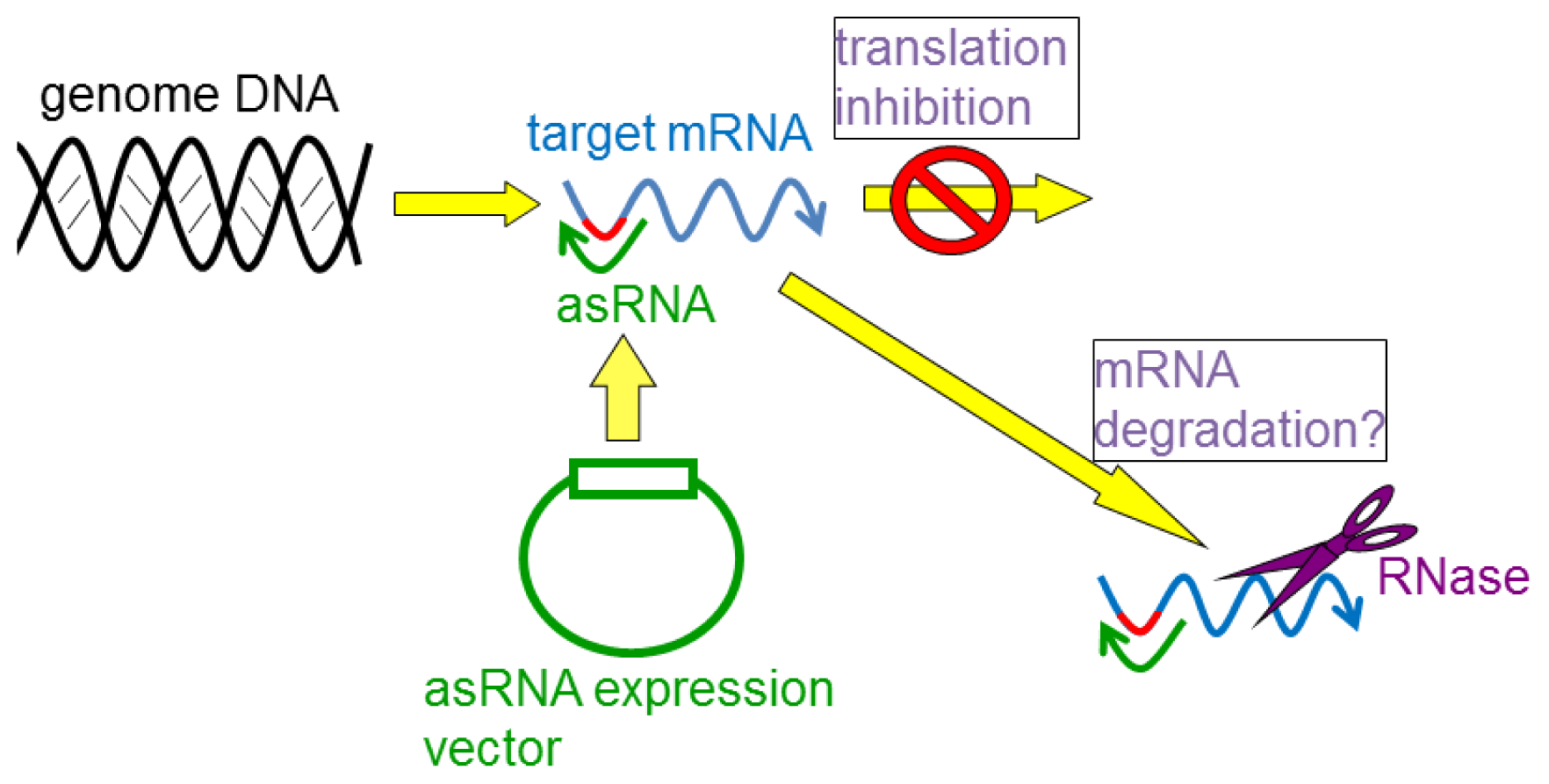
4. Gene Silencing Using asRNAs

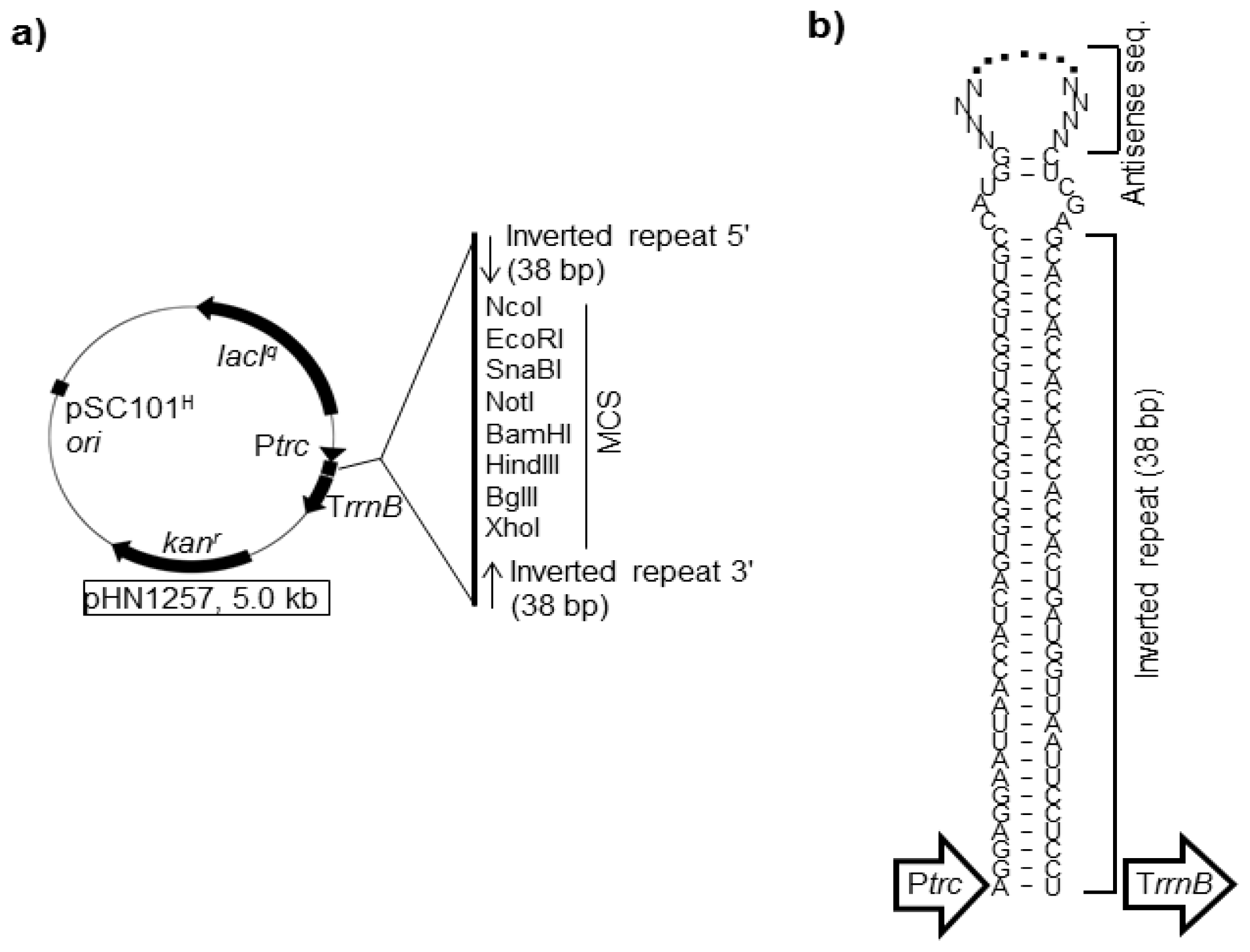
4.1. asRNAs Expressed from Expression Vectors

One can change the nature of bacteria without editing the genome. One method is to silencing target mRNAs by expressing asRNAs [9399]. This method was first reported in 1984 in E. coli [100]. The largest advantage of using asRNAs is the conditionality of the silencing effect, making it possible to apply to genes essential for growth. Creating expression vectors for asRNAs is less laborious than gene knockout methods.
In many cases, asRNAs are designed to hybridize to the ribosome-binding site (RBS) and the start codon region of the target mRNAs (Figure 5) [98]. This is because translation initiation is the limiting step in the translational process; thus, preventing the ribosome from binding to the RBS site of target mRNAs is most critical for its efficacy. However, the factors that are most affected by knocking down the targeted mRNAs is still unclear, although some mechanisms have been proposed. In E. coli, the level of target mRNAs decrease following silencing as well as protein level [9395,101,102]. It is, thus, likely that target mRNAs that are masked with asRNAs tend to be rapidly degraded in the cell. Probably, asRNA-targeted mRNAs are free from ribosomes (so-called naked mRNAs) and easily accessible to the nucleases compared to mRNAs in the polysome states [103].
Until recently, the asRNA-mediated gene silencing method was disadvantageous, especially in E. coli; the silencing efficacy varied greatly depending on the targeted gene and was generally low [104]. We and other groups attempted to increase the efficacy by redesigning the expression system of asRNAs. In 2006, we found that asRNAs combined by a hairpin structure (hairpin asRNAs; HPasRNAs), had much higher silencing efficacies than those lacking the hairpin structure in E. coli (Figure 6) [95]. The hairpin structure improves stability of the asRNAs and extending the lifetime in cells. Indeed, for several genes (e.g., fabI and ackA) [94], expected phenotypes did not appear upon expression of asRNAs lacking the hairpin structure, but clearly appeared upon expression of HPasRNAs. The HPasRNA expression plasmid (pHN1257) contained the trc promoter (Ptrc) and the lactose repressor gene (lacIq), which drive conditional expression of HPasRNAs with IPTG. When DNA fragments containing the reverse complements of RBS and start codon sequences of target genes are cloned under the control of the Ptrc in the multiple cloning site (MCS) of pHN1257, HPasRNAs containing antisense sequences at the loop region are expressed. Antisense sequences of 80–150 nucleotides in length are sufficient for specific hybridization of mRNAs-HPasRNAs [95]. Furthermore, an additional three plasmids harboring different selection markers and replication origins were constructed [94]. These four plasmids, including pHN1257, were co-transformable (compatible) in any combination and were used to silencing up to four genes simultaneously [94]. We confirmed that four genes were silenced simultaneously, and the silencing efficacy of each gene was comparable to those of their respective single silencing [94].
To observe a distinct phenotype by asRNA-mediated gene silencing, the key parameter is the amount of the cellular asRNA level over target mRNAs [95,103]. In bacteria, transcription and translation take place simultaneously in the same location [103]. For successful gene silencing, the expressed asRNA must bind the target mRNA before the ribosome. As the ribosome is the most abundant molecule in cells, one must maximize the expression of asRNAs by using strong transcriptional promoters. In our studies, a strong Ptrc was used, which showed improved results compared to weaker promoters, such as arabinose-inducible and tetracycline-inducible promoters [32,95]. The vector copy number also affects the efficiency, and using a higher copy number plasmid gave better results [94]. Detailed procedures for constructing fine silencing plasmids have been described earlier [93]. Expression vectors for asRNAs developed by us have been distributed to many researchers. Table 3 summarizes the selected applications using the vectors.
The asRNA-mediated gene silencing approach has been proven to be effective not only in E. coli but also in various other bacteria, including S. aureus [108], Clostridium spp. [109], Bacillus megaterium [110], Streptomyces spp. [111], Lactobacillus rhamnosus [112], and Mycobacterium spp. [113,114]. In one outstanding report in 2006, a genome-wide shotgun library of asRNAs was constructed by cloning genomic fragments of S. aureus downstream of the tetracycline-inducible promoter [108]. This systematic approach led to the identification of growth essential genes, which can be a promising target for new antibiotics. Presently, all bacteria are thought to have naturally occurring small RNAs that act as asRNAs [115], and therefore, this method should be applicable to other bacteria.
Recently, two studies have been published where the asRNA-mediated gene silencing has been applied for rationally designing metabolic pathway of E. coli [116,117]. In both reports, over 70 genes were silenced to screen gene targets that increase productivity of valuable compounds. Such large scale screens are feasible with this method but not with gene knock out, because any E. coli strain is used and multiple silencing is easy.

4.2. Antisnese Oligonucleotides Synthesized in Vitro

Antisense oligonucleotides (both DNAs and RNAs) that are synthesized in vitro are convenient to silence target RNAs, because they can be added directly to bacterial cultures whenever desired, without constructing plasmids [118]. Therefore, synthetic asRNAs are suitable for bacteria for which expression vectors have not been developed. Stability of antisense oligonucleotides in bacterial cells (in other words, tolerance to nuclease) can be improved by incorporating unnatural modified nucleotides, such as Locked Nucleic Acids [119] or phosphoroathioate oligonucleotides [120]. As PNAs (see Section 3.2) have high stability in cells and high target site specificity than natural oligonucleotides, they also work as effective antisense silencers [83,84].
However, synthetic asRNAs are costly and hardly fits to large-scale cultures compared to expressed asRNAs. Permeability of synthetic asRNAs across bacterial cell membranes should be taken into account. When PNAs are applied to E. coli, permeability of PNAs should be increased by attaching “cell penetrating peptides” to PNAs [83,84]. Figure 7 summarizes features of expressed and synthetic antisense methods as well as gene knockout method.

5. Conclusions

Here, we described the methods for genome editing and gene silencing, including conventional and new ones. The research trend of this area is correspondence to high-throughput and large-scale analyses. To this end, high recombination efficiency and selection-free approaches (e.g., not using antibiotic markers) are required for genome editing. Gene silencing with expressed asRNAs well suites to high-throughput analyses and indeed come into usage [116,117]. Once the asRNA expression libraries that cover whole bacterial genome are established, then everyone can use the libraries as valuable research resources almost permanently. In addition to the above points, the methods that can be used in many bacteria universally are required for the future.
We believe that the further development of genome editing and gene silencing methods are necessary for understanding cellular functions as a system and for altering metabolic functions as desired.

Acknowledgments

We thank the members of our research group for their assistance. This work was partly supported by the Japan Society for the Promotion of Science (JSPS) Grant-in-Aid for Young Scientist 23780096 (to Nobutaka Nakashima), Grant-in-Aid for Scientific Research (B) 23380197 (to Kentaro Miyazaki), Grant-in-Aid for Scientific Research on Innovative Areas 24119515 (to Kentaro Miyazaki), and Grant-in-Aid for Challenging Exploratory Research 24651231 (to Kentaro Miyazaki).

Conflicts of Interest

The authors declare no conflict of interest.

References

  1. Curtis, T.P.; Sloan, W.T.; Scannell, J.W. Estimating prokaryotic diversity and its limits. Proc. Natl. Acad. Sci. USA 2002, 99, 10494–10499. [Google Scholar]
  2. Ward, B.B. How many species of prokaryotes are there? Proc. Natl. Acad. Sci. USA 2002, 99, 10234–10236. [Google Scholar]
  3. Hardy, S.; Legagneux, V.; Audic, Y.; Paillard, L. Reverse genetics in eukaryotes. Biol. Cell 2010, 102, 561–580. [Google Scholar] [Green Version]
  4. Collins, J.J.; Endy, D.; Hutchison, C.A.; Roberts, R.J. Editorial-synthetic biology. Nucleic Acids Res 2010, 38, 2513. [Google Scholar]
  5. Reyrat, J.M.; Pelicic, V.; Gicquel, B.; Rappuoli, R. Counterselectable markers: Untapped tools for bacterial genetics and pathogenesis. Infect. Immun 1998, 66, 4011–4017. [Google Scholar]
  6. Hamilton, C.M.; Aldea, M.; Washburn, B.K.; Babitzke, P.; Kushner, S.R. New method for generating deletions and gene replacements in Escherichia coli. J. Bacteriol 1989, 171, 4617–4622. [Google Scholar]
  7. Emmerson, J.R.; Gally, D.L.; Roe, A.J. Generation of gene deletions and gene replacements in Escherichia coli O157: H7 using a temperature sensitive allelic exchange system. Biol. Proced. Online 2006, 8, 153–162. [Google Scholar]
  8. Blomfield, I.C.; Vaughn, V.; Rest, R.F.; Eisenstein, B.I. Allelic exchange in Escherichia coli using the Bacillus subtilis sacB gene and a temperature-sensitive pSC101 replicon. Mol. Microbiol 1991, 5, 1447–1457. [Google Scholar]
  9. Link, A.J.; Phillips, D.; Church, G.M. Methods for generating precise deletions and insertions in the genome of wild-type Escherichia coli: Application to open reading frame characterization. J. Bacteriol 1997, 179, 6228–6237. [Google Scholar]
  10. Wu, S.S.; Kaiser, D. Markerless deletions of pil genes in Myxococcus xanthus generated by counterselection with the Bacillus subtilis sacB gene. J. Bacteriol 1996, 178, 5817–5821. [Google Scholar]
  11. Okibe, N.; Suzuki, N.; Inui, M.; Yukawa, H. Efficient markerless gene replacement in Corynebacterium glutamicum using a new temperature-sensitive plasmid. J. Microbiol. Methods 2011, 85, 155–163. [Google Scholar]
  12. Van der Geize, R.; Hessels, G.I.; van Gerwen, R.; Vrijbloed, J.W.; van der Meijden, P.; Dijkhuizen, L. Targeted disruption of the kstD gene encoding a 3-ketosteroid Δ1-dehydrogenase isoenzyme of Rhodococcus erythropolis strain SQ1. Appl. Environ. Microbiol 2000, 66, 2029–2036. [Google Scholar]
  13. Graf, N.; Altenbuchner, J. Development of a method for markerless gene deletion in Pseudomonas putida. Appl. Environ. Microbiol 2011, 77, 5549–5552. [Google Scholar]
  14. Datsenko, K.A.; Wanner, B.L. One-step inactivation of chromosomal genes in Escherichia coli K-12 using PCR products. Proc. Natl. Acad. Sci. USA 2000, 97, 6640–6645. [Google Scholar]
  15. Solano, C.; Garcia, B.; Valle, J.; Berasain, C.; Ghigo, J.M.; Gamazo, C.; Lasa, I. Genetic analysis of Salmonella enteritidis biofilm formation: Critical role of cellulose. Mol. Microbiol 2002, 43, 793–808. [Google Scholar]
  16. Van Kessel, J.C.; Hatfull, G.F. Recombineering in Mycobacterium tuberculosis. Nat. Methods 2007, 4, 147–152. [Google Scholar]
  17. Gust, B.; Chandra, G.; Jakimowicz, D.; Tian, Y.Q.; Bruton, C.J.; Chater, K.F. λ red-mediated genetic manipulation of antibiotic-producing Streptomyces. Adv. Appl. Microbiol 2004, 54, 107–128. [Google Scholar]
  18. Wang, Y.; Weng, J.; Waseem, R.; Yin, X.H.; Zhang, R.F.; Shen, Q.R. Bacillus subtilis genome editing using ssDNA with short homology regions. Nucleic Acids Res 2012, 40, e91. [Google Scholar]
  19. Blank, K.; Hensel, M.; Gerlach, R.G. Rapid and highly efficient method for scarless mutagenesis within the Salmonella enterica chromosome. PLoS One 2011, 6, e15763. [Google Scholar]
  20. Geng, S.Z.; Jiao, X.A.; Pan, Z.M.; Chen, X.J.; Zhang, X.M.; Chen, X. An improved method to knock out the asd gene of Salmonella enterica serovar Pullorum. J. Biomed. Biotechnol 2009, 2009, 646380. [Google Scholar]
  21. Murphy, K.C.; Campellone, K.G.; Poteete, A.R. PCR-mediated gene replacement in Escherichia coli. Gene 2000, 246, 321–330. [Google Scholar]
  22. Sun, W.; Wang, S.; Curtiss, R., III. Highly efficient method for introducing successive multiple scarless gene deletions and markerless gene insertions into the Yersinia pestis chromosome. Appl. Environ. Microbiol 2008, 74, 4241–4245. [Google Scholar]
  23. Ried, J.L.; Collmer, A. An nptI-sacB-sacR cartridge for constructing directed, unmarked mutations in gram-negative bacteria by marker exchange-eviction mutagenesis. Gene 1987, 57, 239–246. [Google Scholar]
  24. Nakashima, N.; Tamura, T. A new carbon catabolite repression mutation of Escherichia coli, mlc*, and its use for producing isobutanol. J. Biosci. Bioeng 2012, 114, 38–44. [Google Scholar]
  25. Costantino, N.; Court, D.L. Enhanced levels of λ Red mediated recombinants in mismatch repair mutants. Proc. Natl. Acad. Sci. USA 2003, 100, 15748–15753. [Google Scholar]
  26. Wang, H.H.; Xu, G.; Vonner, A.J.; Church, G. Modified bases enable high-efficiency oligonucleotide-mediated allelic replacement via mismatch repair evasion. Nucleic Acids Res 2011, 39, 7336–7347. [Google Scholar]
  27. Wang, H.H.; Isaacs, F.J.; Carr, P.A.; Sun, Z.Z.; Xu, G.; Forest, C.R.; Church, G.M. Programming cells by multiplex genome engineering and accelerated evolution. Nature 2009, 460, 894–898. [Google Scholar]
  28. Isaacs, F.J.; Carr, P.A.; Wang, H.H.; Lajoie, M.J.; Sterling, B.; Kraal, L.; Tolonen, A.C.; Gianoulis, T.A.; Goodman, D.B.; Reppas, N.B.; et al. Precise manipulation of chromosomes in vivo enables genome-wide codon replacement. Science 2011, 333, 348–353. [Google Scholar]
  29. Van Kessel, J.C.; Hatfull, G.F. Efficient point mutagenesis in mycobacteria using single-stranded DNA recombineering: Characterization of antimycobacterial drug targets. Mol. Microbiol 2008, 67, 1094–1107. [Google Scholar]
  30. Swingle, B.; Bao, Z.; Markel, E.; Chambers, A.; Cartinhour, S. Recombineering using RecTE from Pseudomonas syringae. Appl. Environ. Microbiol 2010, 76, 4960–4968. [Google Scholar]
  31. Van Pijkeren, J.P.; Britton, R.A. High efficiency recombineering in lactic acid bacteria. Nucleic Acids Res 2012, 40, e76. [Google Scholar]
  32. Nakashima, N.; Tamura, T. Gene silencing in Escherichia coli using antisense RNAs expressed from doxycycline-inducible vectors. Lett. Appl. Microbiol 2013, 56, 436–442. [Google Scholar]
  33. Stolworthy, T.S.; Krabbenhoft, E.; Black, M.E. A novel Escherichia coli strain allows functional analysis of guanylate kinase drug resistance and sensitivity. Anal. Biochem 2003, 322, 40–47. [Google Scholar]
  34. Yamada, J.; Yamasaki, S.; Hirakawa, H.; Hayashi-Nishino, M.; Yamaguchi, A.; Nishino, K. Impact of the RNA chaperone Hfq on multidrug resistance in Escherichia coli. J. Antimicrob. Chemother 2010, 65, 853–858. [Google Scholar]
  35. Tyo, K.E.; Ajikumar, P.K.; Stephanopoulos, G. Stabilized gene duplication enables long-term selection-free heterologous pathway expression. Nat. Biotechnol 2009, 27, 760–765. [Google Scholar]
  36. Karberg, M.; Guo, H.T.; Zhong, J.; Coon, R.; Perutka, J.; Lambowitz, A.M. Group II introns as controllable gene targeting vectors for genetic manipulation of bacteria. Nat. Biotechnol 2001, 19, 1162–1167. [Google Scholar]
  37. Simon, D.M.; Clarke, N.A.C.; McNeil, B.A.; Johnson, I.; Pantuso, D.; Dai, L.X.; Chai, D.G.; Zimmerly, S. Group II introns in eubacteria and archaea: ORF-less introns and new varieties. RNA 2008, 14, 1704–1713. [Google Scholar]
  38. Martínez-Abarca, F.; Toro, N. Group II introns in the bacterial world. Mol. Microbiol 2000, 38, 917–926. [Google Scholar]
  39. Michel, F.; Ferat, J.L. Structure and activities of group-II introns. Annu. Rev. Biochem 1995, 64, 435–461. [Google Scholar]
  40. Quiroga, C.; Kronstad, L.; Ritlop, C.; Filion, A.; Cousineau, B. Contribution of base-pairing interactions between group II intron fragments during trans-splicing in vivo. RNA 2011, 17, 2212–2221. [Google Scholar]
  41. Plante, I.; Cousineau, B. Restriction for gene insertion within the Lactococcus lactis Ll.LtrB group II intron. RNA 2006, 12, 1980–1992. [Google Scholar]
  42. Guo, H.T.; Zimmerly, S.; Perlman, P.S.; Lambowitz, A.M. Group II intron endonucleases use both RNA and protein subunits for recognition of specific sequences in double-stranded DNA. EMBO J 1997, 16, 6835–6848. [Google Scholar]
  43. Perutka, J.; Wang, W.J.; Goerlitz, D.; Lambowitz, A.M. Use of computer-designed group II introns to disrupt Escherichia coli DExH/D-box protein and DNA helicase genes. J. Mol. Biol 2004, 336, 421–439. [Google Scholar]
  44. Belhocine, K.; Mak, A.B.; Cousineau, B. Trans-splicing of the Ll.LtrB group II intron in Lactococcus lactis. Nucleic Acids Res 2007, 35, 2257–2268. [Google Scholar]
  45. Jones, J.P., III; Kierlin, M.N.; Coon, R.G.; Perutka, J.; Lambowitz, A.M.; Sullenger, B.A. Retargeting mobile group II introns to repair mutant genes. Mol. Ther 2005, 11, 687–694. [Google Scholar]
  46. Heap, J.T.; Kuehne, S.A.; Ehsaan, M.; Cartman, S.T.; Cooksley, C.M.; Scott, J.C.; Minton, N.P. The ClosTron: Mutagenesis in Clostridium refined and streamlined. J. Microbiol. Methods 2010, 80, 49–55. [Google Scholar]
  47. Yao, J.; Lambowitz, A.A. Gene targeting in gram-negative bacteria by use of a mobile group II intron (“targetron”) expressed from a broad-host-range vector. Appl. Environ. Microbiol 2007, 73, 2735–2743. [Google Scholar]
  48. Frazier, C.L.; San Filippo, J.; Lambowitz, A.M.; Mills, D.A. Genetic manipulation of Lactococcus lactis by using targeted group II introns: Generation of stable insertions without selection. Appl. Environ. Microbiol 2003, 69, 1121–1128. [Google Scholar]
  49. Mohr, G.; Hong, W.; Zhang, J.; Cui, G.Z.; Yang, Y.; Cui, Q.; Liu, Y.J.; Lambowitz, A.M. A targetron system for gene targeting in thermophiles and its application in Clostridium thermocellum. PLoS One 2013, 8, e69032. [Google Scholar]
  50. Chen, Y.; McClane, B.A.; Fisher, D.J.; Rood, J.I.; Gupta, P. Construction of an alpha toxin gene knockout mutant of Clostridium perfringens type A by use of a mobile group II intron. Appl. Environ. Microbiol 2005, 71, 7542–7547. [Google Scholar]
  51. Yao, J.; Zhong, J.; Fang, Y.; Geisinger, E.; Novick, R.P.; Lambowitz, A.M. Use of targetrons to disrupt essential and nonessential genes in Staphylococcus aureus reveals temperature sensitivity of Ll.LtrB group II intron splicing. RNA 2006, 12, 1271–1281. [Google Scholar]
  52. Corvaglia, A.R.; François, P.; Hernandez, D.; Perron, K.; Linder, P.; Schrenzel, J. A type III-like restriction endonuclease functions as a major barrier to horizontal gene transfer in clinical Staphylococcus aureus strains. Proc. Natl. Acad. Sci. USA 2010, 107, 11954–11958. [Google Scholar]
  53. King, N.P.; Beatson, S.A.; Totsika, M.; Ulett, G.C.; Alm, R.A.; Manning, P.A.; Schembri, M.A. UafB is a serine-rich repeat adhesin of Staphylococcus saprophyticus that mediates binding to fibronectin, fibrinogen and human uroepithelial cells. Microbiology 2011, 157, 1161–1175. [Google Scholar]
  54. Upadhyay, A.; Srivastava, S. Phenazine-1-carboxylic acid is a more important contributor to biocontrol Fusarium oxysporum than pyrrolnitrin in Pseudomonas fluorescens strain Psd. Microbiol. Res 2011, 166, 323–335. [Google Scholar]
  55. Malhotra, M.; Srivastava, S. An ipdC gene knock-out of Azospirillum brasilense strain SM and its implications on indole-3-acetic acid biosynthesis and plant growth promotion. Antonie Van Leeuwen 2008, 93, 425–433. [Google Scholar]
  56. Rodriguez, S.A.; Yu, J.J.; Davis, G.; Arulanandam, B.P.; Klose, K.E. Targeted inactivation of Francisella tularensis genes by group II introns. Appl. Environ. Microbiol 2008, 74, 2619–2626. [Google Scholar]
  57. Alonzo, F.; Port, G.C.; Cao, M.; Freitag, N.E. The posttranslocation chaperone PrsA2 contributes to multiple facets of Listeria monocytogenes pathogenesis. Infect. Immun 2009, 77, 2612–2623. [Google Scholar]
  58. Zarschler, K.; Janesch, B.; Zayni, S.; Schäffer, C.; Messner, P. Construction of a gene knockout system for application in Paenibacillus alvei CCM 2051T, exemplified by the S-layer glycan biosynthesis initiation enzyme WsfP. Appl. Environ. Microbiol 2009, 75, 3077–3085. [Google Scholar]
  59. Steen, J.A.; Steen, J.A.; Harrison, P.; Seemann, T.; Wilkie, I.; Harper, M.; Adler, B.; Boyce, J.D. Fis is essential for capsule production in Pasteurella multocida and regulates expression of other important virulence factors. PLoS Pathog 2010, 6, e1000750. [Google Scholar]
  60. Park, J.M.; Jang, Y.S.; Kim, T.Y.; Lee, S.Y. Development of a gene knockout system for Ralstonia eutropha H16 based on the broad-host-range vector expressing a mobile group II intron. FEMS Microbiol. Rev 2010, 309, 193–200. [Google Scholar]
  61. Palonen, E.; Lindström, M.; Karttunen, R.; Somervuo, P.; Korkeala, H. Expression of signal transduction system encoding genes of Yersinia pseudotuberculosis IP32953 at 28 and 3 °C. PLoS One 2011, 6, e25063. [Google Scholar]
  62. Palonen, E.; Lindström, M.; Somervuo, P.; Johansson, P.; Björkroth, J.; Korkeala, H. Requirement for RNA helicase CsdA for growth of Yersinia pseudotuberculosis IP32953 at low temperatures. Appl. Environ. Microbiol 2012, 78, 1298–1301. [Google Scholar]
  63. Maltz, M.A.; Weiss, B.L.; O’Neill, M.; Wu, Y.; Aksoy, S. OmpA-mediated biofilm formation is essential for the commensal bacterium Sodalis glossinidius to colonize the tsetse fly gut. Appl. Environ. Microbiol 2012, 78, 7760–7768. [Google Scholar]
  64. Akhtar, P.; Khan, S.A. Two independent replicons can support replication of the anthrax toxin-encoding plasmid pXO1 of Bacillus anthracis. Plasmid 2012, 67, 111–117. [Google Scholar]
  65. Zhong, J.; Karberg, M.; Lambowitz, A.M. Targeted and random bacterial gene disruption using a group II intron (targetron) vector containing a retrotransposition-activated selectable marker. Nucleic Acids Res 2003, 31, 1656–1664. [Google Scholar]
  66. Enyeart, P.J.; Chirieleison, S.M.; Dao, M.N.; Perutka, J.; Quandt, E.M.; Yao, J.; Whitt, J.T.; Keatinge-Clay, A.T.; Lambowitz, A.M.; Ellington, A.D. Generalized bacterial genome editing using mobile group II introns and Cre-lox. Mol. Syst. Biol. 2013, 9, 685. [Google Scholar]
  67. Jinek, M.; Chylinski, K.; Fonfara, I.; Hauer, M.; Doudna, J.A.; Charpentier, E. A Programmable dual-RNA-guided DNA endonuclease in adaptive bacterial immunity. Science 2012, 337, 816–821. [Google Scholar]
  68. Gasiunas, G.; Barrangou, R.; Horvath, P.; Siksnys, V. Cas9-crRNA ribonucleoprotein complex mediates specific DNA cleavage for adaptive immunity in bacteria. Proc. Natl. Acad. Sci. USA 2012, 109, E2579–E2586. [Google Scholar]
  69. Hruscha, A.; Krawitz, P.; Rechenberg, A.; Heinrich, V.; Hecht, J.; Haass, C.; Schmid, B. Efficient CRISPR/Cas9 genome editing with low off-target effects in zebrafish. Development 2013, 140, 4982–4987. [Google Scholar]
  70. Chen, K.; Gao, C. Targeted genome modification technologies and their applications in crop improvements. Plant Cell Rep 2013. [Google Scholar] [CrossRef]
  71. Malina, A.; Mills, J.R.; Cencic, R.; Yan, Y.; Fraser, J.; Schippers, L.M.; Paquet, M.; Dostie, J.; Pelletier, J. Repurposing CRISPR/Cas9 for in situ functional assays. Genes Dev 2013, 27, 2602–2614. [Google Scholar]
  72. Beumer, K.J.; Carroll, D. Targeted genome engineering techniques in Drosophila. Methods 2014. [Google Scholar] [CrossRef]
  73. Wang, T.; Wei, J.J.; Sabatini, D.M.; Lander, E.S. Genetic screens in human cells using the CRISPR-Cas9 system. Science 2014, 343, 80–84. [Google Scholar]
  74. Jiang, W.; Bikard, D.; Cox, D.; Zhang, F.; Marraffini, L.A. RNA-guided editing of bacterial genomes using CRISPR-Cas systems. Nat. Biotechnol 2013, 31, 233–239. [Google Scholar]
  75. Wood, A.J.; Lo, T.W.; Zeitler, B.; Pickle, C.S.; Ralston, E.J.; Lee, A.H.; Amora, R.; Miller, J.C.; Leung, E.; Meng, X.D.; et al. Targeted genome editing across species using ZFNs and TALENs. Science 2011, 333, 307–307. [Google Scholar]
  76. Urnov, F.D.; Rebar, E.J.; Holmes, M.C.; Zhang, H.S.; Gregory, P.D. Genome editing with engineered zinc finger nucleases. Nat. Rev. Genet 2010, 11, 636–646. [Google Scholar]
  77. Gupta, A.; Christensen, R.G.; Rayla, A.L.; Lakshmanan, A.; Stormo, G.D.; Wolfe, S.A. An optimized two-finger archive for ZFN-mediated gene targeting. Nat. Methods 2012, 9, 588–590. [Google Scholar]
  78. Cermak, T.; Doyle, E.L.; Christian, M.; Wang, L.; Zhang, Y.; Schmidt, C.; Baller, J.A.; Somia, N.V.; Bogdanove, A.J.; Voytas, D.F. Efficient design and assembly of custom TALEN and other TAL effector-based constructs for DNA targeting. Nucleic Acids Res 2011, 39, e82. [Google Scholar]
  79. Curtin, S.J.; Voytas, D.F.; Stupar, R.M. Genome engineering of crops with designer nucleases. Plant Genome 2012, 5, 42–50. [Google Scholar]
  80. Mussolino, C.; Cathomen, T. TALE nucleases: Tailored genome engineering made easy. Curr. Opin. Biotechnol 2012, 23, 644–650. [Google Scholar]
  81. Christian, M.L.; Demorest, Z.L.; Starker, C.G.; Osborn, M.J.; Nyquist, M.D.; Zhang, Y.; Carlson, D.F.; Bradley, P.; Bogdanove, A.J.; Voytas, D.F. Targeting G with TAL effectors: A comparison of activities of TALENs constructed with NN and NK repeat variable di-Residues. PLoS One 2012, 7, e45383. [Google Scholar]
  82. Li, T.; Huang, S.; Zhao, X.F.; Wright, D.A.; Carpenter, S.; Spalding, M.H.; Weeks, D.P.; Yang, B. Modularly assembled designer TAL effector nucleases for targeted gene knockout and gene replacement in eukaryotes. Nucleic Acids Res 2011, 39, 6315–6325. [Google Scholar]
  83. Good, L.; Nielsen, P.E. Antisense inhibition of gene expression in bacteria by PNA targeted to mRNA. Nat. Biotechnol 1998, 16, 355–358. [Google Scholar]
  84. Good, L.; Awasthi, S.K.; Dryselius, R.; Larsson, O.; Nielsen, P.E. Bactericidal antisense effects of peptide-PNA conjugates. Nat. Biotechnol 2001, 19, 360–364. [Google Scholar]
  85. Lundin, K.E.; Good, L.; Strömberg, R.; Gräslund, A.; Smith, C.I.E. Biological activity and biotechnological aspects of peptide nucleic acid. Adv. Genet 2006, 56, 1–51. [Google Scholar]
  86. Hansen, M.E.; Bentin, T.; Nielsen, P.E. High-affinity triplex targeting of double stranded DNA using chemically modified peptide nucleic acid oligomers. Nucleic Acids Res 2009, 37, 4498–4507. [Google Scholar]
  87. McNeer, N.A.; Chin, J.Y.; Schleifman, E.B.; Fields, R.J.; Glazer, P.M.; Saltzman, W.M. Nanoparticles deliver triplex-forming PNAs for site-specific genomic recombination in CD34+ human hematopoietic progenitors. Mol. Ther 2011, 19, 172–180. [Google Scholar]
  88. Schleifman, E.B.; Bindra, R.; Leif, J.; del Campo, J.; Rogers, F.A.; Uchil, P.; Kutsch, O.; Shultz, L.D.; Kumar, P.; Greiner, D.L.; et al. Targeted disruption of the CCR5 gene inhuman hematopoietic stem cells stimulated by peptide nucleic acids. Chem. Biol 2011, 18, 1189–1198. [Google Scholar]
  89. Osiak, A.; Radecke, F.; Guhl, E.; Radecke, S.; Dannemann, N.; Lütge, F.; Glage, S.; Rudolph, C.; Cantz, T.; Schwarz, K.; et al. Selection-independent generation of gene knockout mouse embryonic stem cells using zinc-finger nucleases. PLoS One 2011, 6, e28911. [Google Scholar]
  90. Vos, M.; Didelot, X. A comparison of homologous recombination rates in bacteria and archaea. ISME J 2009, 3, 199–208. [Google Scholar]
  91. Cradick, T.J.; Ambrosini, G.; Iseli, C.; Bucher, P.; McCaffrey, A.P. ZFN-site searches genomes for zinc finger nuclease target sites and off-target sites. BMC Bioinf 2011, 12, 152. [Google Scholar]
  92. Sawitzke, J.A.; Thomason, L.C.; Costantino, N.; Bubunenko, M.; Datta, S.; Court, D.L. Recombineering: In vivo genetic engineering in E. coli, S. enterica, and beyond. Methods Enzymol 2007, 421, 171–199. [Google Scholar]
  93. Nakashima, N.; Goh, S.; Good, L.; Tamura, T. Multiple-gene silencing using antisense RNAs in Escherichia coli. Methods Mol. Biol 2012, 815, 307–319. [Google Scholar]
  94. Nakashima, N.; Tamura, T. Conditional gene silencing of multiple genes with antisense RNAs and generation of a mutator strain of Escherichia coli. Nucleic Acids Res 2009, 37, e103. [Google Scholar]
  95. Nakashima, N.; Tamura, T.; Good, L. Paired termini stabilize antisense RNAs and enhance conditional gene silencing in Escherichia coli. Nucleic Acids Res 2006, 34, e138. [Google Scholar]
  96. Brantl, S. Antisense-RNA regulation and RNA interference. Biochim. Biophys. Acta 2002, 1575, 15–25. [Google Scholar]
  97. Sharma, V.; Yamamura, A.; Yokobayashi, Y. Engineering artificial small RNAs for conditional gene silencing in Escherichia coli. ACS Synth. Biol 2012, 1, 6–13. [Google Scholar]
  98. Stefan, A.; Schwarz, F.; Bressanin, D.; Hochkoeppler, A. Shine-Dalgarno sequence enhances the efficiency of lacZ repression by artificial anti-lac antisense RNAs in Escherichia coli. J. Biosci. Bioeng 2010, 110, 523–528. [Google Scholar]
  99. Goh, S.; Boberek, J.M.; Nakashima, N.; Stach, J.; Good, L. Concurrent growth rate and transcript analyses reveal essential gene stringency in Escherichia coli. PLoS One 2009, 4, e6061. [Google Scholar]
  100. Pestka, S.; Daugherty, B.L.; Jung, V.; Hotta, K.; Pestka, R.K. Anti-mRNA; specific inhibition of translation of single mRNA molecules. Proc. Natl. Acad. Sci. USA 1984, 81, 7525–7528. [Google Scholar]
  101. Srivastava, R.; Cha, H.J.; Peterson, M.S.; Bentley, W.E. Antisense downregulation of σ32 as a transient metabolic controller in Escherichia coli: Effects on yield of active organophosphorus hydrolase. Appl. Environ. Microbiol 2000, 66, 4366–4371. [Google Scholar]
  102. Krylov, A.A.; Airich, L.G.; Kiseleva, E.M.; Minaeva, N.I.; Biryukova, I.V.; Mashko, S.V. Conditional silencing of the Escherichia coli pykF gene results from artificial convergent transcription protected from rho-dependent termination. J. Mol. Microbiol. Biotechnol 2010, 18, 1–13. [Google Scholar]
  103. Chen, H.; Ferbeyre, G.; Cedergren, R. Efficient hammerhead ribozyme and antisense RNA targeting in a slow ribosome Escherichia coli mutant. Nat. Biotechnol 1997, 15, 432–435. [Google Scholar]
  104. Kim, J.Y.H.; Cha, H.J. Down-regulation of acetate pathway through antisense strategy in Escherichia coli: Improved foreign protein production. Biotechnol. Bioeng 2003, 83, 841–853. [Google Scholar]
  105. Boberek, J.M.; Stach, J.; Good, L. Genetic evidence for inhibition of bacterial division protein FtsZ by berberine. PLoS One 2010, 5, e13745. [Google Scholar]
  106. Meng, J.; Kanzaki, G.; Meas, D.; Lam, C.K.; Crummer, H.; Tain, J.; Xu, H.H. A genome-wide inducible phenotypic screen identifies antisense RNA constructs silencing Escherichia coli essential genes. FEMS Microbiol. Lett 2012, 329, 45–53. [Google Scholar]
  107. Setoyama, D.; Ito, R.; Takagi, Y.; Sekiguchi, M. Molecular actions of Escherichia coli MutT for control of spontaneous mutagenesis. Mutat. Res 2011, 707, 9–14. [Google Scholar]
  108. Ji, Y.D.; Zhang, B.; van Horn, S.F.; Warren, P.; Woodnutt, G.; Burnham, M.K.R.; Rosenberg, M. Identification of critical staphylococcal genes using conditional phenotypes generated by antisense RNA. Science 2001, 293, 2266–2269. [Google Scholar]
  109. Desai, R.P.; Papoutsakis, E.T. Antisense RNA strategies for metabolic engineering of Clostridium acetobutylicum. Appl. Environ. Microbiol 1999, 65, 936–945. [Google Scholar]
  110. Biedendieck, R.; Malten, M.; Barg, H.; Bunk, B.; Martens, J.H.; Deery, E.; Leech, H.; Warren, M.J.; Jahn, D. Metabolic engineering of cobalamin (vitamin B12) production in Bacillus megaterium. Microb. Biotechnol 2010, 3, 24–37. [Google Scholar]
  111. Uguru, G.C.; Mondhe, M.; Goh, S.; Hesketh, A.; Bibb, M.J. Synthetic RNA silencing of actinorhodin biosynthesis in Streptomyces coelicolor A3(2). PLoS One 2013, 8, e67509. [Google Scholar]
  112. Bouazzaoui, K.; LaPointe, G. Use of antisense RNA to modulate glycosyltransferase gene expression and exopolysaccharide molecular mass in Lactobacillus rhamnosus. J. Microbiol. Methods 2006, 65, 216–225. [Google Scholar]
  113. Kaur, P.; Agarwal, S.; Datta, S. Delineating bacteriostatic and bactericidal targets in Mycobacteria using IPTG inducible antisense expression. PLoS One 2009, 4, e5923. [Google Scholar]
  114. Parish, T.; Stoker, N.G. Development and use of a conditional antisense mutagenesis system in Mycobacteria. FEMS Microbiol. Lett 1997, 154, 151–157. [Google Scholar]
  115. Thomason, M.K.; Storz, G. Bacterial antisense RNAs: How many are there, and what are they doing? Annu. Rev. Genet 2010, 44, 167–188. [Google Scholar]
  116. Nakashima, N.; Ohno, S.; Yoshikawa, K.; Shimizu, H.; Tamura, T. A vector library for silencing central carbon metabolism genes with antisense RNAs in Escherichia coli. Appl. Environ. Microbiol 2014, 80, 564–573. [Google Scholar]
  117. Na, D.; Yoo, S.M.; Chung, H.; Park, H.; Park, J.H.; Lee, S.Y. Metabolic engineering of Escherichia coli using synthetic small regulatory RNAs. Nat. Biotechnol 2013, 31, 170–174. [Google Scholar]
  118. Stach, J.E.M.; Good, L. Synthetic RNA silencing in bacteria-antimicrobial discovery and resistance breaking. Front. Microbiol 2011, 2, 185. [Google Scholar]
  119. Bistué, A.; Martín, F.A.; Vozza, N.; Ha, H.; Joaquín, J.C.; Zorreguieta, A.; Tolmasky, M.E. Inhibition of aac(6′)-Ib-mediated amikacin resistance by nuclease-resistant external guide sequences in bacteria. Proc. Natl. Acad. Sci. USA 2009, 106, 13230–13235. [Google Scholar]
  120. Rasmussen, L.C.V.; Sperling-Petersen, H.U.; Mortensen, K.K. Hitting bacteria at the heart of the central dogma: Sequence-specific inhibition. Microb. Cell Fact 2007, 6, 24. [Google Scholar]
Figure 1. Schematic drawing of a gene knockout using the pSC101ts-sacB method [9]. Colored boxes denote open reading frames and blue boxes indicate the gene to be knocked out. The open reading frames that are derived from a plasmid are dotted. As homologous sequences on plasmids, fragments >500 base pairs are typically used. In this drawing, the first recombination (integration) occurs within the red (middle left) or yellow boxes (middle right). The second recombination (resolution) occurs using homologous regions within the genome, causing deletion (bottom left) or reconstruction to original organization (bottom right).
Ijms 15 02773f1
Figure 2. Allelic exchange. Colored boxes denote open reading frames and a black line in blue boxes indicates a point mutation to be introduced. The open reading frames that are derived from a plasmid are dotted. The integration step should occur via one of the two homologous regions, but only one of the two integration patterns is shown here for simplicity.
Ijms 15 02773f2
Figure 3. Improved method for allelic exchange. Light and dark gray boxes indicate different selection marker genes. Other boxes and lines are the same as in Figure 2. The open reading frames that are derived from a plasmid are dotted. The integration step should occur via one of the two homologous regions, but only one of the two integration patterns is shown here for simplicity.
Ijms 15 02773f3
Figure 4. Gene knock-in. Blue box indicates a gene locus to which a target gene is integrated. Red and yellow boxes are neighboring genes, and purple boxes indicate the heterologous gene to be knocked-in. The open reading frames that are derived from a plasmid are dotted. The integration step should occur via one of the two homologous regions, but only one of the two integration patterns is shown here for simplicity.
Ijms 15 02773f4
Figure 5. Mechanism of asRNAs. Red lines indicate a ribosome-binding site (RBS) on the target mRNA.
Ijms 15 02773f5
Figure 6. Schematic map of the HPasRNA expression vector, pHN1270. (a) Arrows indicate open reading frames (lacIq and kanamycin-resistance gene, kanr), the trc promoter (Ptrc), or rrnB terminator (TrrnB), and a box indicates the pSC101H replication origin (high copy version of pSC101). Restriction enzyme sites in the multiple cloning site (MCS) are unique; (b) The structure of HPasRNAs is shown. “NNNN….NNNN” indicates an antisense sequence or a MCS control sequence in the case of an empty plasmid.
Ijms 15 02773f6
Figure 7. Summary of gene knockout and gene silencing.
Ijms 15 02773f7
Table 1. Comparison of two gene knockout methods a.
Compared pointspSC101ts-sacB method by Blomfield et al. [8]λ-Red recombinase method by Datsenko et al. [14]
Recombination viaTwo times of single crossoverOne time of double crossover and FLP (flippase)–FRT recombination
Enzymes for recombinationendogenous enzymesλ Gam, Bet, Exo, and flippase
ReliabilityLow due to resolution of original gene organizationsHigh
Host requirementsOnly recombination-proficient hostsAny
Plasmid constructionNecessaryunnecessary
Transformation efficiency requiredLowHigh
Transformation procedures requiredOnceTwice
Marker gene used for integrationNot retainedNot retained
Unnecessary genome arrangementNoYes, leaving an 81–85-bp “scar” sequence [19]
Bacteria proven to be applicableE. coli [69], M. xanthus [10], C. glutamicum [11], Rhodococcus spp. [12], and P. putida [13]E. coli [14], Salmonella spp. [15], M. tuberculosis [16], Streptomyces spp. [17], and B. subtilis [18]
aAdvantageous features are shown in bold.
Table 2. Comparison of typical efficiency of genome editing.
Host bacteriaMethod usedEfficiencyReference
E. coliλ-red recombinase method, double stranded DNA103 to 104 recombinants per 108 viable cells a[92]
E. coliλ-red recombinase method, single stranded DNA~107 recombinants per 108 viable cells[92]
E. coliλ-red recombinase method, single stranded DNA25% b[25]
L. reuteriλ-red recombinase method, single stranded DNA0.4%–19% b[31]
E. colimobile group II introns1%–80% b[43]
C. thermocellummobile group II introns67%–100% b[49]
S. aureusmobile group II introns37%–100% b[51]
S. pneumoniaeCRISPR-Cas9 system100% b[74]
E. coliCRISPR-Cas9 system65% b[74]
aReplacing the galK gene with a drug cassette;
bEfficiency is calculated as percentage of successful recombination per appeared colonies without any selection pressure.
Table 3. Genes silenced with HPasRNAs in E. coli.
Gene nameGene productSilencing efficacy aObserved phenotypes upon expression of HPasRNAs
lacZβ-galactosidase88% [93]
ackAAcetate kinase78% [94]Reduced acetate production, no growth on minimal acetate media [95]
aceEPyruvate dehydrogenase componentAcetate auxotroph, accumulation of pyruvate [93]
ftsZTubulin-like proteinSevere growth (essential gene), elongated cell [105]
fusAElongation factor GSevere growth (essential gene), sensitization 12-fold to fusidic acid [106]
(Many growth essential genes)Construction of a shotgun genomic library expressing HPasRNAs, identification of growth essential genes [106]
mutTProtein for maintaining DNA replication fidelity>90% [106]Protein level control in a stepwise fashion by changing concentration of expression inducer (IPTG) [107]
mutS, mutD, ndk (triple silencing)Proteins for maintaining DNA replication fidelityIncreased mutation rate by 2000-fold over wild-type cells [94]
aEvaluated as reduced protein activity upon HPasRNA expression.

