Article

[FeFe]-Hydrogenase Abundance and Diversity along a Vertical Redox Gradient in Great Salt Lake, USA

1
Department of Microbiology and Immunology, Montana State University, Bozeman, MT 59717, USA
2
Department of Chemistry and Biochemistry, Montana State University, Bozeman, MT 59717, USA
3
Department of Biology and the Great Salt Lake Institute, Westminster College, Salt Lake City, UT 84105, USA
4
Department of Chemistry and Geochemistry, Colorado School of Mines, Golden, CO 80401, USA
*
Author to whom correspondence should be addressed.
Int. J. Mol. Sci. 2014, 15(12), 21947-21966; https://doi.org/10.3390/ijms151221947
Submission received: 21 October 2014 / Revised: 11 November 2014 / Accepted: 13 November 2014 / Published: 28 November 2014
(This article belongs to the Special Issue Photosynthesis and Biological Hydrogen Production)

Abstract

:
The use of [FeFe]-hydrogenase enzymes for the biotechnological production of H2 or other reduced products has been limited by their sensitivity to oxygen (O2). Here, we apply a PCR-directed approach to determine the distribution, abundance, and diversity of hydA gene fragments along co-varying salinity and O2 gradients in a vertical water column of Great Salt Lake (GSL), UT. The distribution of hydA was constrained to water column transects that had high salt and relatively low O2 concentrations. Recovered HydA deduced amino acid sequences were enriched in hydrophilic amino acids relative to HydA from less saline environments. In addition, they harbored interesting variations in the amino acid environment of the complex H-cluster metalloenzyme active site and putative gas transfer channels that may be important for both H2 transfer and O2 susceptibility. A phylogenetic framework was created to infer the accessory cluster composition and quaternary structure of recovered HydA protein sequences based on phylogenetic relationships and the gene contexts of known complete HydA sequences. Numerous recovered HydA are predicted to harbor multiple N- and C-terminal accessory iron-sulfur cluster binding domains and are likely to exist as multisubunit complexes. This study indicates an important role for [FeFe]-hydrogenases in the functioning of the GSL ecosystem and provides new target genes and variants for use in identifying O2 tolerant enzymes for biotechnological applications.

1. Introduction

Hydrogen (H2) is a diffusible electron carrier with the highest energy content per unit mass of all naturally occurring fuels, and forms the basis of numerous interspecies interactions in natural microbial communities [1,2]. [NiFe]- and [FeFe]-hydrogenase, which differ in the metal composition of their respective active-site clusters, are principally responsible for H2 cycling in natural environments. In general, [FeFe]-hydrogenases are found in anaerobic bacteria and are especially prevalent among fermentative organisms (e.g., firmicutes) where they typically function in the regeneration of reduced electron carriers (NAD(P)H, ferredoxin (Fd)) coupled to the reduction of protons. They are also found in a number of lower eukaryotes including algae and protists, but surprisingly they have yet to be found in cyanobacteria or in archaea [3]. In contrast, [NiFe]-hydrogenases, which are primarily associated with H2 oxidation in energy yielding processes, are frequently found in archaea and bacteria [4,5].
The majority of H2 in natural environments is produced through fermentative processes [6] or through biological N2 fixation [7]. While both [NiFe]- and [FeFe]-hydrogenases can function in the generation of H2 during fermentation [3,4,5], [FeFe]-hydrogenases are generally more efficient H2-producing catalysts [8] and have been the subject of extensive biochemical and structural characterization in an effort to optimize these enzymes for efficient bio-H2 production [9,10,11,12,13]. The best characterized [FeFe]-hydrogenases are monomeric, Fd-dependent enzymes [3,5]. Examples of these enzymes include those from eukaryotic algae such as Chlamydomonas reinhardtii [14,15,16] and clostridial species such as Clostridium pasteurianum [10,17,18]. [FeFe]-hydrogenase diversity is manifested mainly in the number and type of accessory cluster domains that complement the active site H-cluster domain, the latter of which is universally conserved in currently sequenced homologs [3,19]. [FeFe]-hydrogenases from C. reinhardtii are the simplest and exist without additional accessory clusters. In contrast, [FeFe]-hydrogenase from C. pasteurianum have three additional iron-sulfur (Fe-S) cluster binding domains in the N-terminus which presumably function to allow coupling with external electron donors and/or acceptors. Characterization of [FeFe]-hydrogenase homologs in available genome sequences [3,19] noted substantial variation in N- and C-terminal Fe-S cluster and accessory cofactor binding motifs, suggesting potential interactions with a variety of other redox partners. In addition, such analyses performed in conjunction with an evaluation of gene context and biochemical characterization revealed differences in [FeFe]-hydrogenase quaternary structure, including the identification of multimeric homologs that likely form dimers, trimers, or tetramers [3,19].
Biochemical characterization of multimeric [FeFe]-hydrogenase enzymes from Desulfovibrio fructosovorans [20], Thermoanaerobacter tengcongensis [21], and Thermotoga maritima [22] indicate that they are pyridine nucleotide (NAD(P)H- or NADH-) linked H2 producing enzymes. The trimeric hydrogenase from T. maritima was shown to require both reduced Fd and NADH for H2 production [23]. Conversely, the [FeFe]-hydrogenase from Acetobacterium woodii simultaneously reduces NAD+ and Fd during H2 oxidation. The key to the reversible oxidation of H2 and simultaneous NAD+ and Fd reduction is the coupling of their energetics; the exergonic reduction of NAD+ by electrons derived from H2 allows the endergonic reduction of Fd in a process now termed electron bifurcation [24,25]. Bifurcating [FeFe]-hydrogenanse enzymes function to create electrons with a very low redox potential by simultaneously creating an electron with a high redox potential [23,25,26,27]. While the aforementioned examples indicate a directionality in the bifurcating process, [FeFe]-hydrogenases that bifurcate can in principle function in vivo in both the production and oxidation of H2, as was recently shown for a bifurcating hydrogenase in Moorella thermoacetica [27]. During growth on glucose, M. thermoacetica can intermittently produce H2 from Fd and NADH via the bifurcating hydrogenase. Likewise, this same hydrogenase can utilize produced H2 and CO2 to preform acetogenesis. Although M. thermoacetica does not grow well autotrophically on H2 and CO2, it uses the bifurcating hydrogenase to produce reduced Fd to drive acetogenesis. The reversible production of such low potential metabolic electrons through the process of H2 dependent electron bifurcation is of significant biotechnological interest since they could be modulated in vivo and directed toward the controlled production of highly reduced biofuel products.
Multimeric bifurcating [FeFe]-hydrogenase complexes now appear to be commonplace among anaerobic bacteria [3,19]. However, like the monomeric forms of [FeFe]-hydrogenase [28,29,30], multimeric enzymes are O2 sensitive which presents challenges in their application in large-scale production efforts [31]. Numerous biochemical and structural studies have been conducted to shed light on the basis of the O2 sensitivity of [FeFe]-hydrogenase [28,29,32,33,34,35,36,37]. One approach to address this problem is gene shuffling or randomized mutation, which generates a diverse recombinant hydrogenase library to screen for enhanced O2 tolerance and/or stability [12,13,38,39]. Another method is to examine the natural diversity of these enzymes across geochemical gradients (e.g., O2) through the use of PCR or metagenomic-directed approaches [40,41,42,43,44]. A previous application of a PCR-directed approach targeting a fragment of the gene encoding the large subunit (hydA) of [FeFe]-hydrogenase in a phototrophic mat community identified significant variation in the amino acid environment of the active site H-cluster [41] and previously implicated gas channels [39,45,46,47,48]. Substitutions in the amino acids lining these gas channels have been previously shown to support increased O2 tolerance [39,48,49]. This may suggest that natural sequence variations that would adjust the size of this gas channel may provide a biomarker for O2 tolerant [FeFe]-hydrogenase. While the functional implications of these substitutions/insertions have yet to be fully characterized biochemically, such findings provide impetus to continue searching natural systems for potential variants capable of tolerating higher O2 concentrations.
Here, we apply a PCR-directed approach to determine the distribution, abundance, and diversity of hydA gene fragments in a vertical water column environment of Great Salt Lake (GSL), UT that exhibits strong and co-varying gradients in salinity, O2, photosynthetically active radiation (PAR), pH, and temperature [50]. A phylogenetic framework was created for use in predicting the accessory cluster composition and quaternary structure of inferred HydA protein sequences obtained along the vertical gradient. The results indicate an abundance of multimeric [FeFe]-hydrogenase that are putatively involved in electron bifurcating processes. Recovered HydA protein fragments revealed evidence of adaptation to elevated salt concentrations and harbored variations in regions of the protein that form gas channels leading to the O2-labile FeS cubane of the active site. Similarly, novel substitutions were identified in residues that coordinate the O2-labile active site. This study demonstrates that enzyme variants with desired properties can potentially be recovered by examining protein diversity in microbial assemblages that have evolved in the presence of a particular environmental stress.

2. Results

2.1. Water Column Chemistry

Strong and often co-varying physical and chemical gradients were observed in the Utah Division of Wildlife Resources sample site number 3 (DWR3) vertical water column (Table 1). Salinity did not vary substantially over the interval from the surface to a depth of 6.0 meters but increased from 126 parts per thousand (ppt) at 6.0 meters to 203 and 247 ppt at depths of 6.5 and 8.0 meters, respectively. Likewise, water column temperature was nearly constant (23–24 °C) over the 0.0 to 6.0 meters vertical transect, but decreased abruptly at a depth of 6.5 and 8.0 meters (20 and 17 °C, respectively), indicating the presence of an inverted thermocline between depth intervals of 6.0 and 8.0 meters. pH varied from 5.95 to 8.05 over the eight meter depth, with the highest pH observed at a depth of 6.0 meters and the lowest pH observed at 8.0 meters. PAR decreased systematically from 1800 µmol·photons·m−2·s–1 just below the surface to 175 µmol·photons·m−2·s–1 at a depth of 4.0 meters and was below detection at a depth of 8.0 meters. Dissolved O2 varied little over the 0.0 to 4.0 meter vertical transect, but decreased substantially from 5.1 to 2.3 mg·L–1 over the 4.0 to 6.0 meters depth transect.
Table 1. Geographical, physical, and chemical data for the water column depths sampled a.
Depth (m)LatitudeLongitudeSalinity (ppt)DissolvedPAR (µmol·photons·m–2·s−1)Temperature (°C)pH
O2 (mg/L)
0.041.1674600−112.66961171495.46207924.177.21
1.041.1674600−112.66961171515.38121924.167.46
4.041.1674717−112.66961171435.1317523.777.83
6.041.1674717−112.66964171512.327523.378.07
6.541.1674833−112.66962002031.684420.20 b7.10 b
8.041.1674817−112.66960832470.98016.595.95
a: Previously reported in Meuser et al. [50]; b: Data points were not measured, but were interpolated based on observed trends.

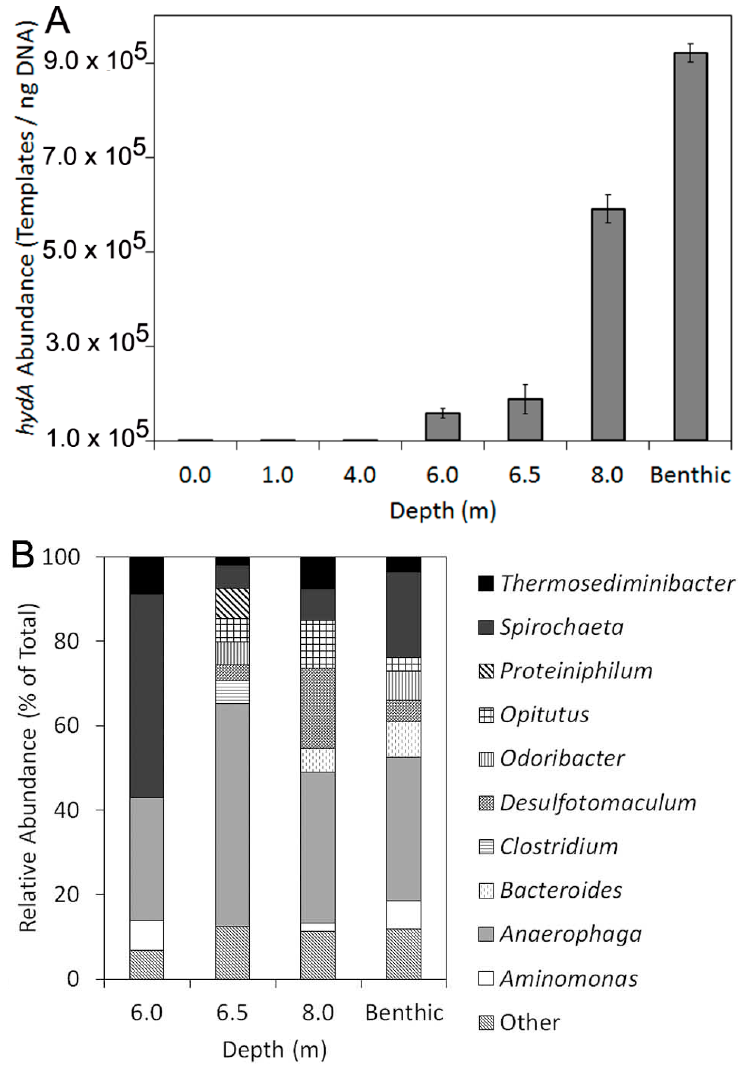
2.2. Abundance, Composition, and Diversity of hydA in GSL Water Column

hydA amplicons were not detected in surface samples (0.0 meters) or from samples collected from depths of 1.0 or 4.0 meters (Figure 2A). In contrast, hydA genes were detected in samples collected from depth intervals of 6.0, 6.5 and 8.0 meters, as well as from benthic sediment samples collected at 8.5 meters. The relative abundance of hydA templates, when normalized to the total quantity of extractable DNA, increased with increasing depth with the highest concentration of hydA genes (9.2 ± 1.9 × 10⁵ templates/ng DNA) detected in association with benthic sediment biomass (Table 2). The distribution of hydA along the DWR3 water column is coincident with the position of the thermocline and with decreasing levels of PAR and dissolved O2 and increasing salinity. The abundance of hydA templates was significantly and inversely correlated with temperature (Pearson R = −0.96, p < 0.01) and dissolved O2 (Pearson R = −0.85, p = 0.03) and was positively correlated with salinity (Pearson R = 0.93, p < 0.01).
A total of 211 hydA sequences (47 to 58 clones per sample) that, when translated, harbored signature motifs characteristic of HydA [3] were obtained in the current study (Table 2). The average hydropathy of amino acids (GRAVY index) comprising the inferred protein sequences was calculated and compared to sequences obtained with the same primer sets from other environmental systems, including microbial mat communities inhabiting salterns at Guerrero Negro (GN) [41] and hot springs in Yellowstone National Park (YNP), USA [40] (Figure 1). The GRAVY index for HydA sequences obtained from the GSL DWR3 water column spanned values ranging from 0.04 (indicative of average hydrophobic amino acid content) to −0.56 (indicative of very low hydrophobic amino acid content). There was no discernible pattern in the average GRAVY index for HydA sequences obtained from the GSL with sampling depth (data not shown). However, the average GRAVY index for sequences obtained from the GSL water column (14.9% to 24.7% salinity) was −0.30 whereas average GRAVY indices obtained from GN (8% salt) and YNP (0.5% to 3.1% salinity) were −0.26 and −0.15, respectively. This indicates that HydA from GSL, on average, comprise a greater number of hydrophilic amino acids than those from GN and YNP.
Table 2. Distribution, abundance, and phylogenetic diversity of hydA as a function of depth at the Great Salt Lake (GSL), Utah Division of Wildlife Resources sample site number 3 (DWR3) vertical transect.
Depth (m)Templates/ng DNASDnaPD b
0.0BD
1.0BD
4.0BD
6.01.6 × 1051.0 × 1045812.5
6.51.9 × 1053.0 × 1045512.5
8.05.9 × 1053.1 × 1045313.9
Benthic c9.2 × 1051.9 × 1045916.0
a: The total number of hydA sequences obtained from a given depth; b: Faith’s index of HydA phylogenetic diversity; c: Benthic sediment was obtained from a depth of 8.5 meters. Abbreviations: BD, below detection; SD, standard deviation.
Faith’s index of phylogenetic diversity (PD), a diversity metric that quantifies the proportion of total branch length in the phylogeny associated with sequences obtained from a given environment when compared to all environments when considered together, increased systematically for HydA assemblages with increasing depth (Table 2). HydA deduced amino acid sequences recovered from the four GSL environments were distantly related to HydA from cultivated organisms, with the average, minimum, and maximum observed sequence identities of 64%, 52%, and 76% (Supplementary Table S1). The majority (99.6%) of the sequences obtained from the 4 GSL environments exhibited affiliation with bacteria, although a single sequence affiliated with a eukaryotic diatom Thalassiosira pseudonana (62% sequence identity) was recovered from the benthic sediments. Despite significant variation in the taxonomic affiliation of HydA assemblages sampled at depth, binning the sequences at the class level did not reveal patterns in the taxonomic composition of assemblages as a function of depth (Figure 2B), consistent with previous studies indicating the tendency for hydA to be horizontally transferred which would likely obscure such patterns [42]. At the order level, HydA sequences affiliated with the Clostridiales were most abundant across all of the sample transects and at the genus level, HydA sequences most closely related to Anaerophaga were the most abundant regardless of sample depth.
Figure 1. Hydropathy values associated with HydA from Great Salt Lake (GSL), Guerrerro Negro (GN), and Yellowstone National Park (YNP), as assessed using the GRAVY index (grand average hydropathy of amino acids). GRAVY indices for sequences from each environment were binned at 0.04 increments (rounded up).
Ijms 15 21947 g001

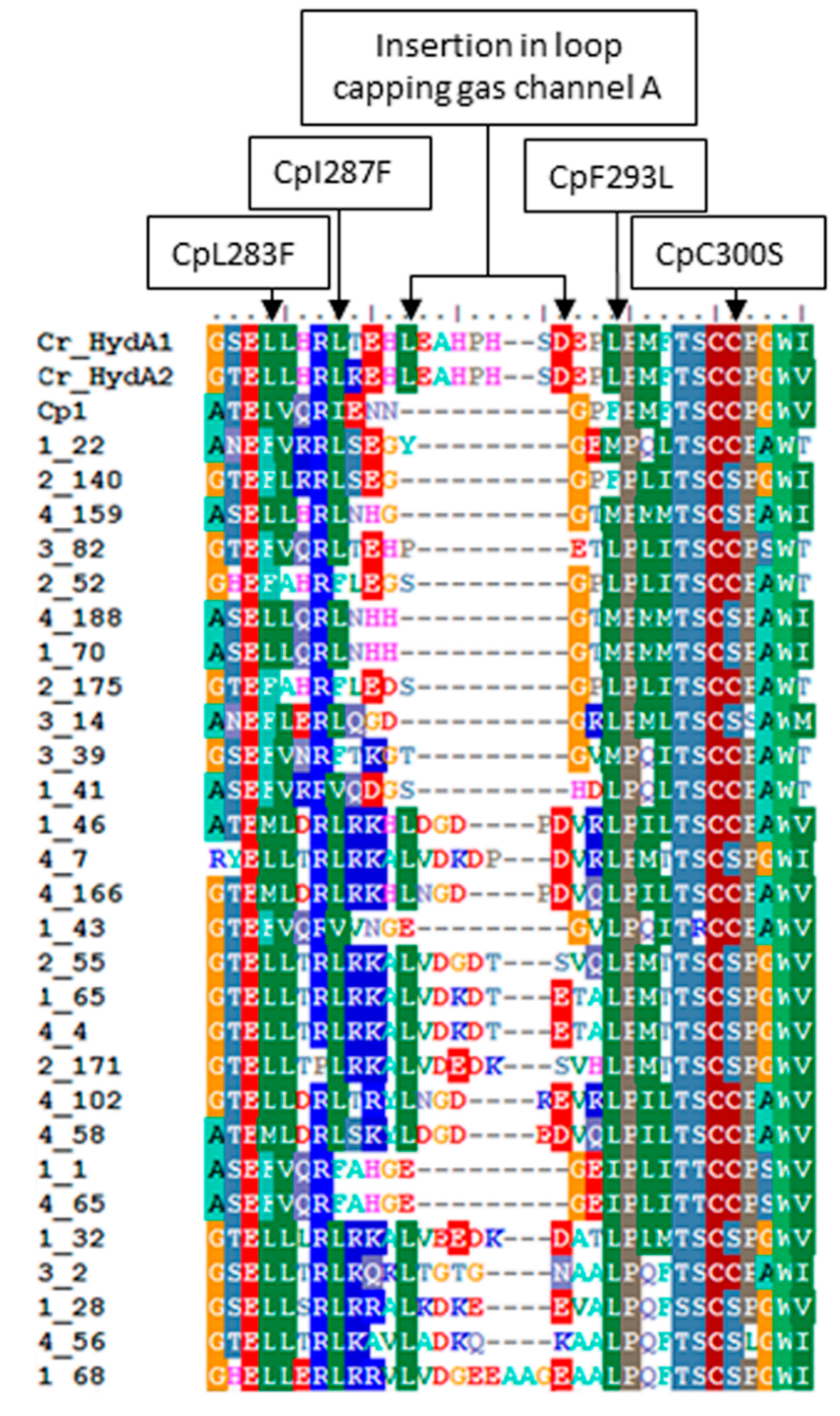
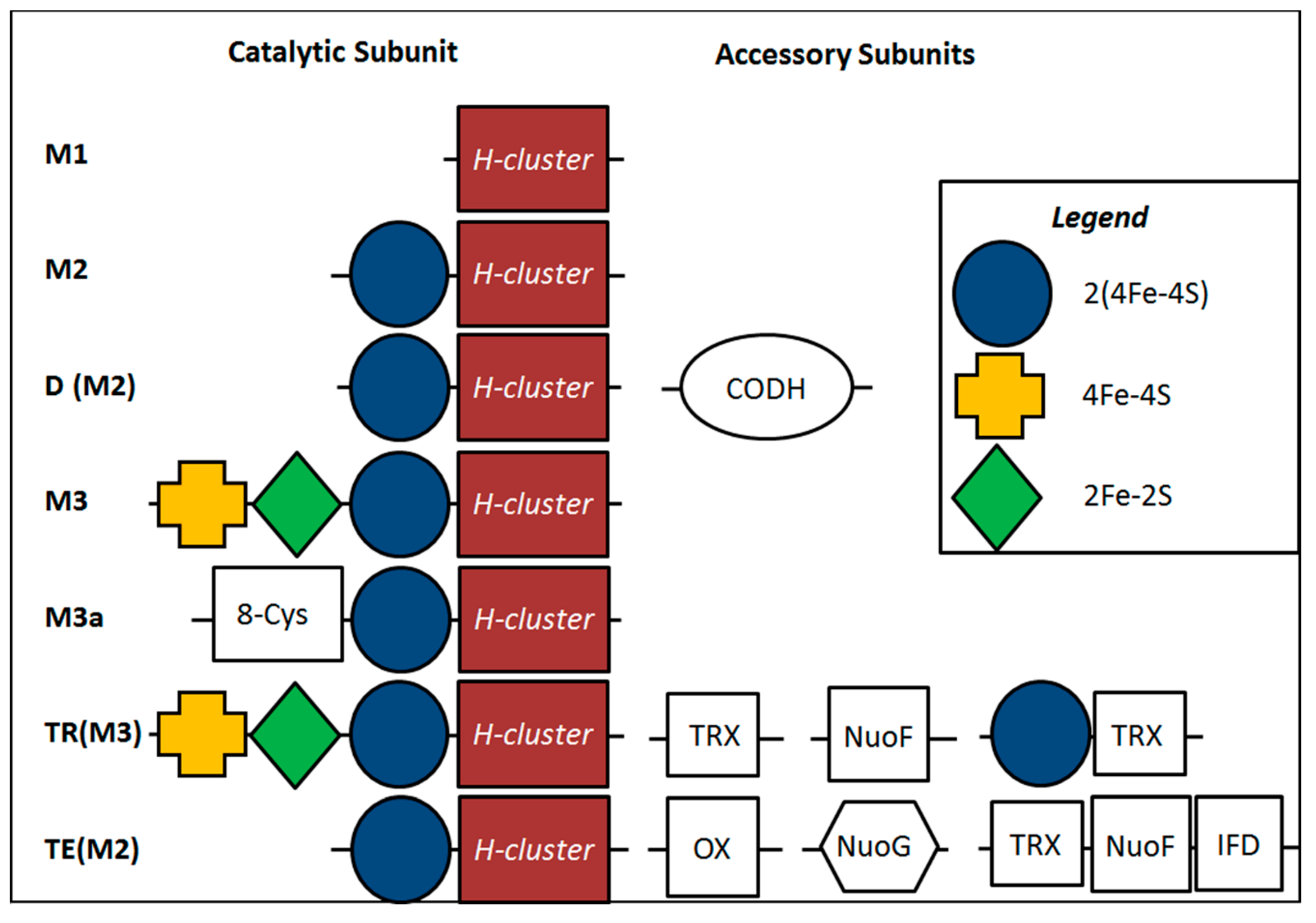
2.3. Variation in H-Cluster Binding Motifs and Putative Gas Channels

Amino acid sequence alignments of the DWR3 HydA sequences with sequences of well-characterized HydA revealed a variety of substitutions in the L1 motif, a series of highly-conserved H-cluster binding residues. The observed substitutions included a Cys to Ser substitution relative to position 300 of the C. pasteurianum (Cp1) HydA sequence (AAA23248). This residue is involved in coordinating the active site H-cluster [10]. Interestingly, a number of substitutions were also observed in residues that line a putative gas channel that leads to the O2 sensitive active site H-cluster (Figure 3) [45]. In particular, substitution of the much larger Phe for Lys and Ile (positions 283 and 287 of Cp1, respectively) were noted in several GSL sequences. Conversely, other proteins exhibited substitution of Lys for Phe (position 293 of Cp1). Intriguingly, a large insertion just upstream from the L1 motif was observed in a number of sequences recovered from GSL [45]. Similar insertions are common in phototrophic algae, although it is unclear what functional significance (if any) that they may have [51].
Figure 2. Abundance of hydA templates as a function of depth at DWR3 (A); Taxonomic composition of HydA protein sequences as determined by BLASTp analysis (B). Further details of the taxonomic composition of HydA are located in Supplementary Table S1.
Ijms 15 21947 g002

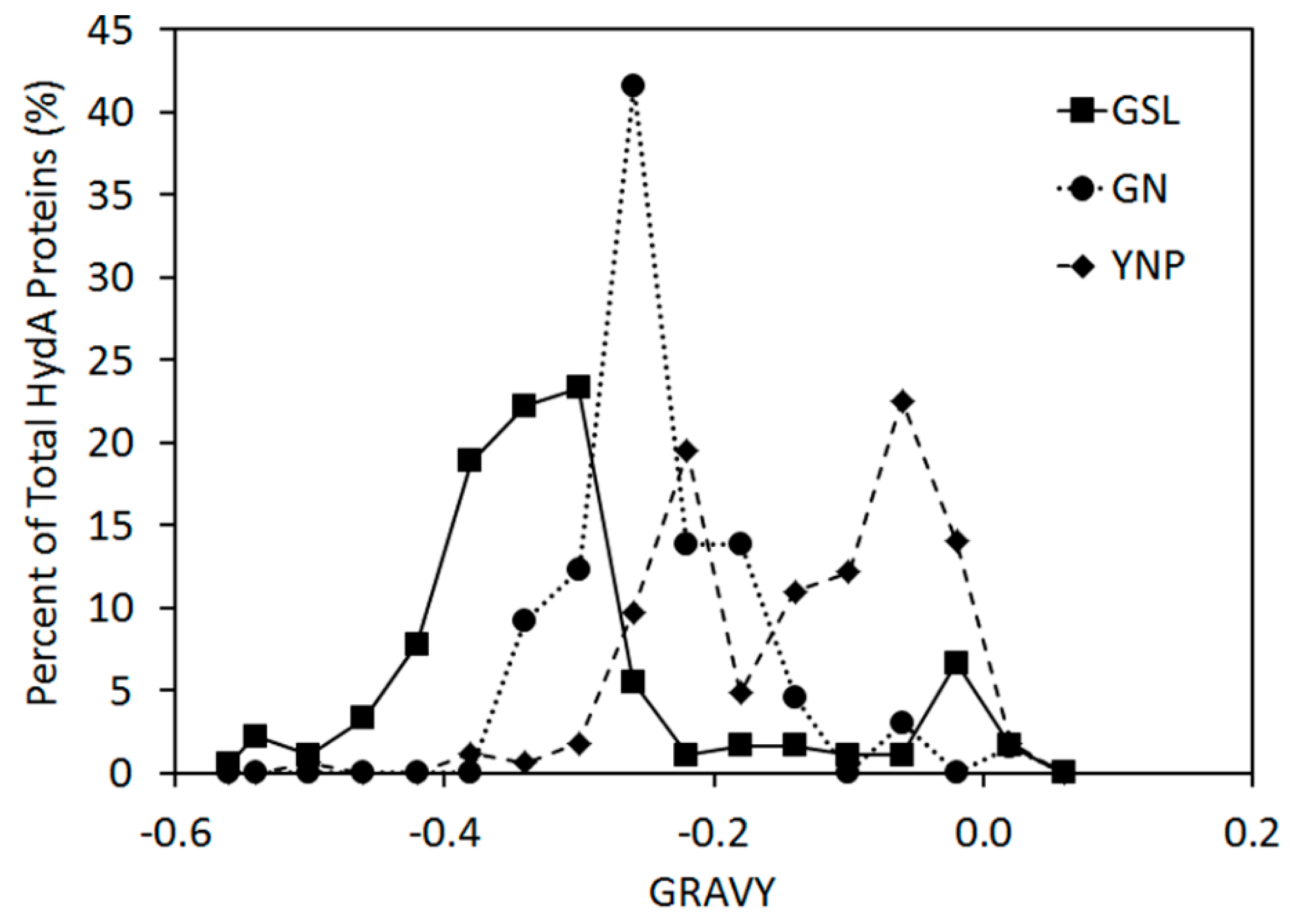
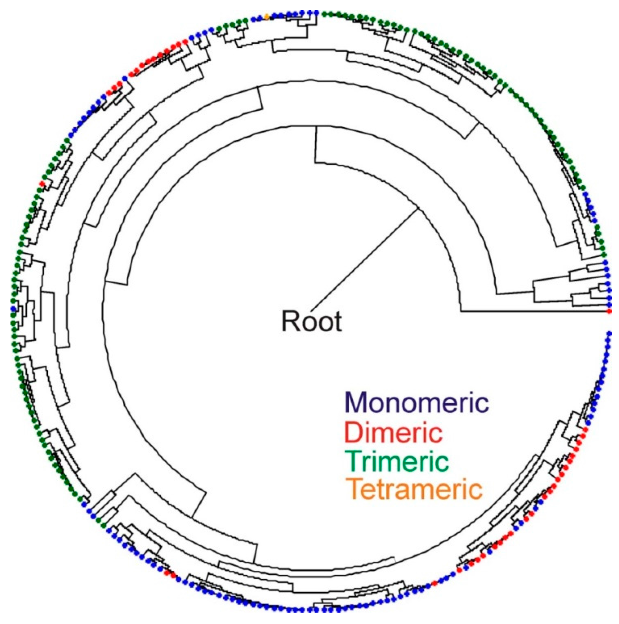
2.4. Inferred Structural Variation of GSL HydA

A database of HydA sequences (Supplementary Table S3) was compiled from complete genome sequences present in GenBank based on previous characterizations of their N- and C-terminal cluster composition (Figure 4) and quaternary structure [3,19]. Additional sequences were obtained based on BLASTp searches and the primary sequence and quaternary structural characteristics were determined as described previously [19]. These sequences and the modular composition of the N- and C-terminal domains (i.e., F- and C-clusters) and their quaternary structure was used to develop a Bayesian phylogenetic framework for predicting these features based on phylogenetic clustering of the HydA fragment amplified with the primers used herein. Similar to previous phylogenetic analyses of the H-cluster domain [3,19], phylogenetic reconstruction of just the H-cluster fragment (positions 280 to 419 of HydA1 from Cp1) in sequences recovered from GenBank revealed clustering that corresponded to primary and quaternary structural characteristics (Figure 5). This indicates that the phylogenetic signal of the amplified fragment is sufficient for predicting the characteristics of HydA from GSL at these levels of consideration. The majority (93.8% of total sequences) of HydA homologs recovered from GSL were nested within or formed clusters with reference sequences that have the M3 architecture and that are likely to form trimers (TR(M3)) (Supplementary Table S1). Only a small fraction of sequences were inferred to have a thermotogae monomeric configuration (5.3% of total sequences). No discernible pattern was observed in the distribution of these sequences as a function of depth in the DWR3 water column (data not shown). A small number of HydA sequences (0.88% of the total) did not cluster or nest phylogenetically with sequences comprising our database preventing prediction of their F- and C-cluster composition or quaternary structure. These sequences generally had low sequence identities to known HydA in genomic databases.
Figure 3. Unique substitutions in the L1 motif and insertions upstream from the L1 motif observed in recovered GSL HydA protein fragments. HydA1 and HydA2 from C. reinhardtii (Cr_HydA1 and Cr_HydA2, respectively) and HydA1 from C. pasteurianum (Cp1) are indicated. Substitutions and insertions are outlined above in reference to the Cp1 sequence.
Ijms 15 21947 g003
Figure 4. Accessary cluster domains associated with diverse HydA. The composition of N- and C-terminal modules (F- and C-clusters, respectively) as identified in Meyer et al., 2007 [3] and as modified by Calusinska et al., 2010 [19] are presented. Abbreviations: M, monomeric; D, dimeric; TR, trimeric; TE, tetrameric; CODH, carbon monoxide dehydrogenase module; TRX, thioredoxin domain; OX, oxidoreductase domain; NuoF, NuoF of complex I domain; NuoG, NuoG of complex I domain; and IFD, indolepyruvate: Fd domain.
Ijms 15 21947 g004

3. Discussion

A diversity of [FeFe]-hydrogenase were identified along the DWR3 vertical gradient in the GSL. The phylogenetic framework developed here indicates that the majority of HydA sequences from GSL form trimers and thus are possibly involved in H2-based electron bifurcation; however, it is unclear if they are involved in the production or consumption of H2. Examples of bifurcating [FeFe]-hydrogenase involved in oxidation and production of H2 have been described. For example, an enzyme that functions to bifurcate electrons derived from the oxidation of H2 coupled to the reduction of Fd and NAD+ has been identified in A. woodii [52] while other enzymes that function to couple the oxidation of Fd and NADH to the formation of H2 have been identified in T. maritima, T. saccharolyticum, and T. tengcongensis [21,23,53]. Moreover, two recent studies reveal the presence of reversible bifurcating complexes. In the case of the acetogen M. thermoacetica, a reversible Fd- and NAD-dependent trimeric [FeFe]-hydrogenase was identified [27].
Most organisms that harbor a bifurcating [FeFe]-hydrogenase harbor additional non-bifurcating enzymes [3,19]. It is therefore a surprise that the majority of sequences recovered in the present examination of HydA diversity in GSL are predicted to be of the bifurcating type. It is possible that the primers used in the present study are biased toward homologs that form trimers and that are thus potentially involved in bifurcation (e.g., TR(M3)) [3,19]. Indeed, a prior comparison of the primers used in this study with other primer designs found that each is biased toward a different spectrum of hydA diversity [42]. Application of untargeted shotgun metagenomic sequencing approaches, which are currently ongoing, will provide a mechanism to: (1) evaluate the potential bias associated with the recovery of predominantly bifurcating enzymes in the present study; and (2) evaluate the predictive capacity of the phylogenetic approaches used here to assign secondary and quaternary structure to the recovered protein sequences. Regardless, the recovery of a number of diverse HydA sequences from the GSL water column, including many exhibiting key amino acid substitutions, indicates a role for the putative bifurcating enzymes in situ.
Figure 5. Bayesian phylogenetic reconstruction of 263 HydA protein sequences compiled from Calusinska et al., 2010 [19] and from GenBank (Supplementary Table S3). The phylogeny was generated using only the region of HydA corresponding to the fragment obtained from the GSL water column and sediments (positions 280 to 419 of HydA1 from C. pasteurianum (AAA23248)). The primary quaternary structure of the homologs (monomeric, dimeric, trimeric, tetrameric) as outlined in Meyer et al., 2007 [3] and more recently in Calusinska et al., 2010 [19] are overlain on the phylogeny. These primary quaternary structural architectures correspond with those presented in Figure 4.
Ijms 15 21947 g005
The distribution of genes encoding putative bifurcating [FeFe]-hydrogenase was strongly influenced by gradients of both O2 and salinity. High salinity, such as that observed along the DWR3 vertical gradient, is generally unfavorable to proteins due to lower water activity, increases in the strength of hydrophobic interactions during protein folding, and a reduction in electrostatic interactions in proteins residing in the cytoplasm [54,55]. HydA recovered from the hypersaline GSL (15%–24% salt) water column and sediments were enriched in hydrophilic amino acids as indicated by significantly lower GRAVY indices when compared to HydA sequences obtained from lower salinity mats inhabiting GN salterns (8.0% salt) and YNP hot springs (0.5%–3.1% salt). This finding is consistent with the results of previous investigations of the proteomes of a variety of halophilic and non-halophilic organisms which indicate lower hydrophobicity indices than for non-halophilic counterparts. Lower hydrophobicity is a feature that is hypothesized to reduce the effects of salt-driven non-specific inter- or intra-molecular interactions in hydrophobic side-chains, which may lead to protein mis-folding and/or aggregation [54,56,57,58,59,60]. Similar results—lower hydrophobicity in proteins from halophilic organisms—were obtained from a comparative genomic and proteomic analysis of 6 halophilic and 24 non-halophilic isolates, spanning both the bacterial and archaeal domains [61]. Thus, the lower hydrophobicity observed in GSL HydA sequences suggests that these proteins are uniquely adapted to the high salt conditions present in their natural environment.
HydA was identified in transects of the DWR3 water column where salt levels were high (15%–24%) but where dissolved O2 was depleted relative to surface waters (dissolved O2 range 0.98–2.32 mg/L). The hypoxic nature of the DWR3 transects where hydA was detected implies either anaerobic microniches that allow anaerobes to exist in otherwise sub-oxic conditions, a lower effective exposure of the organism or its O2-sensitive enzymes to O2, or enhanced tolerance to O2. The solubility and bioavailability of O2 is lower in hypersaline solutions [62], which may allow anaerobic organisms to inhabit otherwise hypoxic conditions. Intriguingly, many of the HydA sequences recovered in this study exhibit novel substitutions in the L1 sequence motifs which are involved in the coordination of the O2-labile [4Fe-4S] subcluster of the H-cluster of HydA [10,63,64]. Substitutions in this region may have implications for the redox properties of this [4Fe-4S] cluster and thus may be important in conferring stability in the presence of O2 [11].
In particular, substitution of Ser for Cys in the L1 motif was commonly observed both in the GSL HydA fragments as well as HydA fragments obtained from GN [41]. Ser coordination of Fe-S clusters is uncommon in nature. The only well-characterized enzyme displaying such coordination is the P-cluster of nitrogenase where the Ser is thought to stabilize the cluster in its oxidized state [65,66]. It is possible that the L1 motif Cys-to-Ser variant observed in a number of the GSL and GN HydA sequences (corresponding to position 300 in the [FeFe]-hydrogenase from C. pasteurianum) may also have a stabilizing effect on the H-cluster, functioning to stabilize a more oxidized state of the active site in the presence of O2.
The variations in the residues forming the gas channel in the GSL HydA sequences may also play a role in conferring O2 tolerance. The most striking variations, CpL283F and CpI287F (positions in reference to CpHydA; Figure 3) may help to act as a molecular sieve [45,67]. Here, the larger sized side chain of Phe is expected to effectively decrease the rate of O2 diffusion but have little effect on rates of H2 diffusion due in principle to the smaller size of H2 relative to O2. Lastly the large insertion observed in numerous HydA homologs identified in GSL near the opening of gas channel A may also function as a molecular sieve or could even act as cap, both of which may slow diffusion of O2 into the enzyme.
The detection of abundant putative trimeric bifurcating [FeFe]-hydrogenase along the GSL DWR3 vertical gradient coupled with the numerous studies that continue to expand the functional capabilities of these enzymes [23,26,27,52] may have a significant application in biofuel production. One limitation for the production of highly reduced bioproducts is that a large proportion of the metabolic electron flow is directed towards reduction of pyridine nucleotides (i.e., to NAD(P)H). Thermodynamically the reduced pyridine nucleotides are not sufficient in energy for the production of a number of desirable, highly reduced biofuel products. [FeFe]-hydrogenase catalyzed electron bifurcation potentially overcomes this thermodynamic challenge by combining electrons from the reduced pyridine nucleotides with other lower potential electrons from reduced Fd [23,25,26,27]. This increases the reducing potential of electrons from reduced pyridine nucleotides such that a larger flux is directed toward the production of desired biofuels (i.e., reduced products). That other electron donors (e.g., CO, formate) [26] can potentially feed into these complexes expands the potential use of these enzymes in directing the flux of metabolic electrons toward desired biofuels. Efforts aimed at isolating representatives of the dominant, yet uncharacterized organisms harboring the unique [FeFe]-hydrogenase enzymes identified herein should continue to expand our understanding of the diversity of biochemical reactions catalyzed by this unique class of enzyme, which may prove useful in overcoming key bioengineering barriers.

4. Experimental Section

4.1. Site Description and Sample Collection

Samples were collected from the Division of Wildlife Resources site 3 (DWR3) (Latitude 41.16746, Longitude−112.6696117) in June 2007 (surface, 1.0, 4.0, 6.0, 6.5 and 8.0 meters depths and benthic sediments), by lowering a 2 L Kemmerer collection bottle (Wildlife Supply, Yulee, FL, USA) to each depth sampled, with deionized water rinses between each sampling, as described previously [50]. Respective samples were transferred to sterile bottles leaving a minimum of gaseous headspace and then immersed in ice until further workup in the laboratory. Cells were then collected by centrifugation (4000× g for 15 min at 3 °C) and the supernatant removed. Cell pellets were resuspended in 1 mL of supernatant and the resuspension was frozen at −80 °C until used for DNA extraction.

4.2. Physical and Chemical Analysis

Chemical and physical measurements were compiled as a part of a previous study conducted on the DWR3 water column [50]. Briefly, photosynthetic active radiation (PAR, 400–700 µmol·photons·m−2·s−1) was determined using a Li-Cor LI-193 underwater spherical quantum sensor (Li-Cor Biosciences, Lincoln, NE, USA) and Li-Cor LI-250A light meter (Li-Cor Biosciences). A Troll 9500 multi-sensor (In-Situ Inc., Fort Collins, CO, USA) was used to determine pH, temperature, salinity, and dissolved O2.

4.3. DNA Extraction, Amplification, Cloning, and Sequencing of hydA

Total DNA was extracted from 200 mg aliquots of biomass using the PowerSoil DNA Isolation Kit (MoBio Inc., Carlsbad, CA, USA) following the manufacturer’s instructions. DNA was quantified fluorometrically as previously described [67]. All DNA extracts were screened for the presence of 16S rRNA genes to ensure the presence of PCR-amplifiable DNA using 10 ng of DNA and primers bacterial 1070F (5'-ATGGCTGTCGTCAGCT-3') and universal 1492R (5'-GGTTACCTTGTTACGACTT-3') [68]. Approximately 500 bp fragments of hydA were PCR-amplified in triplicate from 10 ng of environmental genomic DNA as template with primer pair FeFe-272F and FeFe-427R using previously established reagent concentrations and reaction conditions [41]. Equal volumes of each replicate amplification were pooled and purified using the Wizard PCR Preps DNA purification system (Promega, Madison, WI, USA), quantified using the Low DNA Mass Ladder (Invitrogen, Carlsbad, CA, USA), cloned using the pGem-T Easy Vector System (Promega), and sequenced using the M13F-M13R primer pair as previously described [69]. A total of 215 hydA gene sequences were sequenced in the present study and representatives of each phylotype (Supplementary Table S1) have been deposited in the GenBank, DDBJ, and EMBL databases under the accession numbers HM636647–HM636798.

4.4. Primary Sequence Analysis

MEGA (version. 4.0.1) [69] was used to translate and align hydA sequences specifying the Gonnet 250 protein weight matrix with a pairwise alignment gap opening penalty of 13 and gap extension penalty of 0.05. The aligned HydA sequences were screened for the presence of the L1 ((FLI)TSC(C/S)P(GAS)W(VIQH)) and L2 ((IVLF)MPCx(ASRD)K(KQ)xE) (conserved residues are in bold and underlined, and bracketed positions indicate “semiconserved” residues at that position) as outlined previously [3,5]. ClustalX [70] was also used to create a pairwise sequence identity matrix which was subsequently imported into DOTUR [71] to identify and group operational taxonomic units (OTUs) and to perform rarefaction analyses at a sequence identity threshold of 0.01. Indices describing the hydrophobicity of inferred protein sequences (Aliphatic Index (AI) and Grand Average of Hydropathicity (GRAVY)) were computed using the ProtParam tool available on the ExPASy proteomics server (http://au.expasy.org/tools/protparam.html) [72]. The AI is the relative volume occupied by aliphatic amino acid side chains (alanine, valine, isoleucine, and leucine) in a given protein or protein fragment and is calculated according to the formula:
Aliphatic index = X(Ala) + a × X(Val) + b × (X(Ile) + X(Leu))
where X(Ala), X(Val), X(Ile), and X(Leu) are mole percent (100 × mole fraction) of alanine, valine, isoleucine, and leucine and a and b are the relative volume coefficients for valine (a = 2.9) and Leu/Ile (b = 3.9) relative to alanine [73], with higher indices indicative of a higher content of aliphatic side chains in the protein. The GRAVY index is the sum of the hydropathy values for each amino acid in a sequence divided by the number of residues [73], with positive GRAVY indices indicative of hydrophobicity and negative GRAVY indices indicative of hydrophilicity. For comparison, aliphatic and GRAVY indices were computed for putative HydA previously recovered from microbial mats inhabiting salterns from GN (GenBank: FJ623894–FJ623958) [41] and from geothermal springs in YNP (GenBank: GU362773–GU362867) [40] (Supplementary Table S2). Intervals of 0.04 GRAVY index units were empirically selected for use in binning GRAVY indices for sequences recovered from the three geochemically-distinct environments (GSL, YNP, GN). Indices were rounded downward to the nearest 0.04 unit interval prior to binning.

4.5. Prediction of Environmental HydA Accessary Cluster Composition

The sequences used to develop the F- and C-cluster classification and tertiary structure scheme developed by Meyer et al., 2007 [3] and further augmented by Calusinska et al., 2010 [19] were compiled and used to create a database for use in predicting the accessory cluster composition and tertiary structure of putative HydA fragments recovered from the GSL water column (Supplementary Table S3). Additional sequences representing the closest BLASTp hits for each OTU were also compiled and characterized for F- and C-cluster composition and tertiary structure using the criteria of Calusinska et al., 2010 [19] and the Conserved Domain Database as implemented with BLASTp [74]. The classification of these proteins, along with those reported by Calusinska et al., 2010 [19], are reported in Supplementary Table S3 and a schematic illustrating their F- and C-cluster structural variation is presented in Figure 3.
HydA protein sequences from GSL and from our database were aligned, trimmed to the length of the amplified protein fragment (positions 272 to 427 in HydA from Clostridium pasteurianum), and subjected to Bayesian phylogenetic reconstruction as described previously [40]. The C- and F-cluster composition and tertiary structure of environmental HydA was then predicted by phylogenetic clustering with HydA comprising our database. Sequences that formed lineages without a representative from our database were classified as “unknown” with respect to F- and C-cluster composition and tertiary structure. Alignments of HydA fragments of reference sequences used to construct the predictive framework can be obtained by email correspondence with the authors.

4.6. hydA Quantitative PCR (qPCR)

qPCR was used to estimate the number of hydA templates in GSL DNA extracts according to previously described methods [75]. Standard curves were generated from plasmid DNA containing hydA amplified from the GSL sample sites. Three plasmid clones were used in generating a standard curve that relates template copy number to threshold qPCR amplification signal. The threshold amplification signal as a function of copy number varied by less than 1 cycle for each of the three plasmid clones; thus, the standard curves generated using each plasmid clone were averaged for use in calculating the average template abundances and standard deviation in template abundances from replicate qPCRs. A standard curve was generated over 6 orders of magnitude ranging from 91 to 1.1 × 10⁷ copies of template per assay (R2 = 0.995).
qPCR assays were performed in a Rotor-Gene 300 quantitative real-time PCR machine (Qiagen, Valencia, CA, USA) in 0.5 mL optically clear PCR tubes (Qiagen) using a SsoFast EvaGreen Supermix qPCR Kit (Bio-Rad Laboratories, Hercules, CA, USA). Assay reactions were amended to a final concentration of 0.4 mg·mL−1 molecular-grade bovine serum albumin (Roche, Indianapolis, IN, USA). qPCR cycling conditions were as follows: Initial denaturation (95 °C for 10 min) followed by 40 cycles of denaturation (95 °C for 10 s), annealing (56.5 °C for 15 s), and extension (72 °C for 20 s). Specificity of the qPCR assays was verified by melt curve analysis. The reported template abundances are the average and standard deviation of qPCR assays performed in triplicate.

4.7. HydA Phylogenetic Diversity

The phylogenetic position of hydA homologs was assessed using PhyML-aBayes [76] with the nuclear prelamin A recognition factor-like protein-encoding gene (XM_414836), a distantly related homolog of hydA [3], from Gallus gallus serving as the outgroup. The General Time Reversible (GTR) substitution model with gamma-shaped rate variation with a proportion of invariable sites was specified in the calculation, as recommended by Modeltest (version 3) [77]. The consensus phylogram was rate-smoothed using the multidimensional version of Rambaut’s parameterization as implemented in PAUP (version 4.0) [78]. Rate-smoothing was performed according to the parameters identified using Modeltest (version 3). This included the identification of the substitution model, the gamma distribution of rate variation across sites, the proportion of invariant sites, nucleobase frequencies, and the rate matrix for each phylogram. Phylocom (version 4.0.1) [79] was used to calculate Faith’s index of phylogenetic diversity (PD) using the rate smoothed Bayesian chronogram. PD is the proportion of total branch length in the phylogeny associated with the taxa in a given sample or assemblage. A higher PD index for an assemblage is indicative of higher phylogenetic diversity (phylogenetic richness) relative to the total sequence pool.

Supplementary Materials

Supplementary tables can be found at https://www.mdpi.com/1422-0067/15/12/21947/s1.

Acknowledgments

This work was supported by the United States Air Force Office of Scientific Research grants FA9550-11-1-0218 (John W. Peters), FA9550-11-1-0211 (Matthew C. Posewitz), and FA9550-14-1-0147 (Eric S. Boyd, John W. Peters, Matthew C. Posewitz).

Author Contributions

Eric S. Boyd and Trinity L. Hamilton generated the sequence data from the Great Salt Lake; Eric S. Boyd, Kevin D. Swanson and Alta E. Howells analyzed the data; Jonathan E. Meuser and Matthew C. Posewitz collected water samples from GSL; and all authors contributed to the writing of this manuscript.

Conflicts of Interest

The authors declare no conflict of interest.

References

  1. McInerney, M.J.; Sieber, J.R.; Gunsalus, R.P. Syntrophy in anaerobic global carbon cycles. Curr. Opin. Biotechnol. 2009, 20, 623–632. [Google Scholar]
  2. Schink, B. Energetics of syntrophic cooperation in methanogenic degradation. Microbiol. Mol. Biol. Rev. 1997, 61, 262–280. [Google Scholar]
  3. Meyer, J. [FeFe] hydrogenases and their evolution: A genomic perspective. Cell Mol. Life Sci. 2007, 64, 1063–1084. [Google Scholar]
  4. Vignais, P.M.; Billoud, B. Occurrence, classification, and biological function of hydrogenases: An overview. Chem. Rev. 2007, 107, 4206–4272. [Google Scholar]
  5. Vignais, P.M.; Billoud, B.; Meyer, J. Classification and phylogeny of hydrogenases. FEMS Microbiol. Rev. 2001, 25, 455–501. [Google Scholar]
  6. Conrad, R. Soil microorganisms as controllers of atmospheric trace gases (H2, CO, CH4, OCS, N2O, and NO). Microbiol. Rev. 1996, 60, 609–640. [Google Scholar]
  7. Robson, R.L.; Postgate, J.R. Oxygen and hydrogen in biological nitrogen fixation. Annu. Rev. Microbiol. 1980, 34, 183–207. [Google Scholar]
  8. Cammack, R. Bioinorganic chemistry: Hydrogenase sophistication. Nature 1999, 397, 214–215. [Google Scholar]
  9. Mulder, D.W.; Boyd, E.S.; Sarma, R.; Lange, R.K.; Endrizzi, J.A.; Broderick, J.B.; Peters, J. W. Stepwise [FeFe]-hydrogenase H-cluster assembly revealed in the structure of HydAΔEFG. Nature 2010, 465, 248–251. [Google Scholar]
  10. Peters, J.W.; Lanzilotta, W.N.; Lemon, B.J.; Seefeldt, L.C. X-ray crystal structure of the Fe-only hydrogenase (CpI) from Clostridium pasteurianum to 1.8 angstrom resolution. Science 1998, 282, 1853–1858. [Google Scholar]
  11. Posewitz, M.C.; Mulder, D.W.; Peters, J.W. New frontiers in hydrogenase structure and biosynthesis. Curr. Chem. Biol. 2008, 2, 178–199. [Google Scholar]
  12. Stapleton, J.A.; Swartz, J.R. A cell-free microtiter plate screen for improved [FeFe] hydrogenases. PLoS One 2010, 5, e10554. [Google Scholar]
  13. Stapleton, J.A.; Swartz, J.R. Development of an in vitro compartmentalization screen for high-throughput directed evolution of [FeFe] hydrogenases. PLoS One 2010, 5, e15275. [Google Scholar] [PubMed]
  14. Happe, T.; Mosler, B.; Naber, J.D. Induction, localization and metal content of hydrogenase in the green alga Chlamydomonas reinhardtii. Eur. J. Biochem. 1994, 222, 769–774. [Google Scholar]
  15. Ghirardi, M.L.; Posewitz, M.C.; Maness, P.C.; Dubini, A.; Yu, J.; Seibert, M. Hydrogenases and hydrogen photoproduction in oxygenic photosynthetic organisms. Annu. Rev. Plant Biol. 2007, 58, 71–91. [Google Scholar]
  16. Forestier, M.; King, P.; Zhang, L.; Posewitz, M.; Schwarzer, S.; Happe, T.; Ghirardi, M.L.; Seibert, M. Expression of two [Fe]-hydrogenases in Chlamydomonas reinhardtii under anaerobic conditions. Eur. J. Biochem. 2003, 270, 2750–2758. [Google Scholar]
  17. Adams, M.W.W. The structure and mechanism of iron-hydrogenases. Biochim. Biophys. Acta 1990, 1020, 115–145. [Google Scholar]
  18. Chen, J.S.; Mortenson, L.E. Purification and properties of hydrogenase from Clostridium pasteurianum W5. Biochim. Biophys. Acta 1974, 371, 283–298. [Google Scholar]
  19. Calusinska, M.; Happe, T.; Joris, B.; Wilmotte, A. The surprising diversity of clostridial hydrogenases: A comparative genomic perspective. Microbiology 2010, 156, 1575–1588. [Google Scholar]
  20. Malki, S.; Saimmaime, I.; de Luca, G.; Rousset, M.; Dermoun, Z.; Belaich, J.P. Characterization of an operon encoding an NADP-reducing hydrogenase in Desulfovibrio fructosovorans. J. Bacteriol. 1995, 177, 2628–2636. [Google Scholar]
  21. Soboh, B.; Linder, D.; Hedderich, R. A multisubunit membrane-bound [NiFe] hydrogenaseand an NADH-dependent Fe-only hydrogenase in the fermenting bacterium Thermoanaerobacter tengcongensis. Microbiology 2004, 150, 2451–2463. [Google Scholar]
  22. Verhagen, M.F.; Adams, M.W. Fe-only hydrogenase from Thermotoga maritima. Methods Enzymol. 2001, 331, 216–226. [Google Scholar]
  23. Schut, G.J.; Adams, M.W. The iron-hydrogenase of Thermotoga maritima utilizes ferredoxin and NADH synergistically: A new perspective on anaerobic hydrogen production. J. Bacteriol. 2009, 191, 4451–4457. [Google Scholar]
  24. Herrmann, G.; Jayamani, E.; Mai, G.; Buckel, W. Energy conservation via electron-transferring flavoprotein in anaerobic bacteria. J. Bacteriol. 2008, 190, 784–791. [Google Scholar]
  25. Buckel, W.; Thauer, R.K. Energy conservation via electron bifurcating ferredoxin reduction and proton/Na+ translocating ferredoxin oxidation. Biochim. Biophys. Acta 2013, 1827, 94–113. [Google Scholar]
  26. Wang, S.; Huang, H.; Kahnt, J.; Mueller, A.P.; Kopke, M.; Thauer, R.K. NADP-specific electron-bifurcating [FeFe]-hydrogenase in a functional complex with formate dehydrogenase in Clostridium autoethanogenum grown on CO. J. Bacteriol. 2013, 195, 4373–4386. [Google Scholar]
  27. Wang, S.; Huang, H.; Kahnt, J.; Thauer, R.K. A reversible electron-bifurcating ferredoxin- and NAD-dependent [FeFe]-hydrogenase (HydABC) in Moorella thermoacetica. J Bacteriol. 2013, 195, 1267–1275. [Google Scholar]
  28. Stripp, S.T.; Goldet, G.; Brandmayr, C.; Sanganas, O.; Vincent, K.A.; Haumann, M.; Armstrong, F.A.; Happe, T. How oxygen attacks [FeFe] hydrogenases from photosynthetic organisms. Proc. Natl. Acad. Sci. USA 2009, 106, 17331–17336. [Google Scholar]
  29. Lambertz, C.; Leidel, N.; Havelius, K.G.V.; Noth, J.; Chernev, P.; Winkler, M.; Happe, T.; Haumann, M. O2-reactions at the six-iron activesite (H-cluster) in [FeFe]-hydrogenase. J. Biol. Chem. 2011, 286, 40614–40623. [Google Scholar]
  30. Stripp, S.; Sanganas, O.; Happe, T.; Haumann, M. The structure of the active site H-cluster of [FeFe] hydrogenase from the green alga Chlamydomonas reinhardtii studied by X-ray absorption spectroscopy. Biochemistry 2009, 48, 5042–5049. [Google Scholar]
  31. Juszczak, A.; Aono, S.; Adams, M.W. The extremely thermophilic eubacterium, Thermotoga maritima, contains a novel iron-hydrogenase whose cellular activity is dependent upon tungsten. J. Biol. Chem. 1991, 266, 13834–13841. [Google Scholar]
  32. Baffert, C.; Demuez, M.; Cournac, L.; Burlat, B.; Guigliarelli, B.; Bertrand, P.; Girbal, L.; Léger, C. Hydrogen-activating enzymes: Activity does not correlate with oxygen sensitivity. Angew. Chem. (Int. Ed. Engl.) 2008, 47, 2052–2054. [Google Scholar]
  33. Bruska, M.K.; Stiebritz, M.T.; Reiher, M. Regioselectivity of H-cluster oxidation. J. Am. Chem. Soc. 2011, 133, 20588–20603. [Google Scholar]
  34. Stiebritz, M.T.; Reiher, M. Hydrogenases and oxygen. Chem. Sci. 2012, 3, 1739–1751. [Google Scholar]
  35. Kubas, A.; de Sancho, D.; Best, R.B.; Blumberger, J. Aerobic damage to [FeFe]-hydrogenases: Activation barriers for the chemical attachment of O2. Angew. Chem. (Int. Ed. Engl.) 2014, 126, 4165–4168. [Google Scholar]
  36. Fourmond, V.; Greco, C.; Sybirna, K.; Baffert, C.; Wang, P.H.; Ezanno, P.; Montefiori, M.; Bruschi, M.; Meynial-Salles, I.; Soucaille, P.; et al. The oxidative inactivation of FeFe hydrogenase reveals the flexibility of the H-cluster. Nat. Chem. 2014, 6, 336–342. [Google Scholar]
  37. Liebgott, P.P.; Leroux, F.; Burlat, B.; Dementin, S.; Baffert, C.; Lautier, T.; Fourmond, V.; Ceccaldi, P.; Cavazza, C.; Meynial-Salles, I.; et al. Relating diffusion along the substrate tunnel and oxygen sensitivity in hydrogenase. Nat. Chem. Biol. 2010, 6, 63–70. [Google Scholar]
  38. Nagy, L.; Meuser, J.; Plummer, S.; Seibert, M.; Ghirardi, M.; King, P.; Ahmann, D.; Posewitz, M. Application of gene-shuffling for the rapid generation of novel [FeFe]-hydrogenase libraries. Biotechnol. Lett. 2007, 29, 421–430. [Google Scholar]
  39. Bingham, A.S.; Smith, P.R.; Swartz, J.R. Evolution of an [FeFe] hydrogenase with decreased oxygen sensitivity. Int. J. Hydrogen. Energy 2012, 37, 2965–2976. [Google Scholar]
  40. Boyd, E.S.; Hamilton, T.L.; Spear, J.R.; Lavin, M.; Peters, J.W. [FeFe]-hydrogenase in Yellowstone National Park: Evidence for dispersal limitation and phylogenetic niche conservatism. ISME J. 2010, 4, 1485–1495. [Google Scholar]
  41. Boyd, E.S.; Spear, J.R.; Peters, J.W. [FeFe]-hydrogenase genetic diversity provides insight into molecular adaptation in a saline microbial mat community. Appl. Environ. Microbiol. 2009, 75, 4620–4623. [Google Scholar]
  42. Schmidt, O.; Drake, H.L.; Horn, M.A. Hitherto unknown [FeFe]-hydrogenase gene diversity in anaerobes and anoxic enrichments from a moderately acidic fen. Appl. Environ. Microbiol. 2010, 76, 2027–2031. [Google Scholar]
  43. Chivian, D.; Brodie, E.L.; Alm, E.J.; Culley, D.E.; Dehal, P.S.; de Santis, T.Z.; Gihring, T.M.; Lapidus, A.; Lin, L.H.; Lowry, S.R.; et al. Environmental genomics reveals a single-species ecosystem deep within Earth. Science 2008, 322, 275–278. [Google Scholar]
  44. Warnecke, F.; Luginbuhl, P.; Ivanova, N.; Ghassemian, M.; Richardson, T.H.; Stege, J.T.; Cayouette, M.; McHardy, A.C.; Djordjevic, G.; Aboushadi, N.; et al. Metagenomic and functional analysis of hindgut microbiota of a wood-feeding higher termite. Nature 2007, 450, 560–565. [Google Scholar]
  45. Cohen, J.; Kim, K.; King, P.; Seibert, M.; Schulten, K. Finding gas diffusion pathways in proteins: Application to O2 and H2 transport in CpI [FeFe]-hydrogenase and the role of packing defects. Structure 2005, 13, 1321–1329. [Google Scholar]
  46. Goldet, G.; Brandmayr, C.; Stripp, S.T.; Happe, T.; Cavazza, C.; Fontecilla-Camps, J.C.; Armstrong, F.A.J. Electrochemical kinetic investigations of the reactions of [FeFe]-hydrogenases with carbon monoxide and oxygen: Comparing the importance of gas tunnels and active-site electronic/redox effects. J. Am. Chem. Soc. 2009, 131, 14979–14989. [Google Scholar]
  47. King, P.W.; Svedruzic, D.; Cohen, J.; Schulten, K.; Seibert, M.; Ghirardi, M.L. Structural and functional investigations of biological catalysts for optimization of solar-driven, H2 production systems. In Proceedings of the SPIE Optics and Photonics, San Diego, CA, USA, 13–17 August 2006; p. 9.
  48. Lautier, T.; Ezanno, P.; Baffert, C.; Fourmond, V.; Cournac, L.; Fontecilla-Camps, J.C.; Soucaille, P.; Bertrand, P.; Meynial-Salles, I.; Leger, C. The quest for a functional substrate access tunnel in FeFe hydrogenase. Faraday Discuss. 2011, 148, 385–407. [Google Scholar]
  49. Ghirardi, M.L.; Cohen, J.; King, P.; Schulten, K.; Kim, K.; Seibert, M. [FeFe]-hydrogenases and photobiological hydrogen production. In Proceedings of the SPIE Optics and Photonics, San Diego, CA, USA, 13–17 August 2006; p. 6.
  50. Meuser, J.E.; Baxter, B.K.; Spear, J.R.; Peters, J.W.; Posewitz, M.C.; Boyd, E.S. Contrasting patterns of community assembly in the stratified water column of Great Salt Lake, Utah. Microb. Ecol. 2013, 66, 268–280. [Google Scholar]
  51. Beer, L.L.; Boyd, E.S.; Peters, J.W.; Posewitz, M.C. Engineering algae for biohydrogen and biofuel production. Curr. Opin. Biotechnol. 2009, 20, 1–8. [Google Scholar]
  52. Schuchmann, K.; Müller, V. A bacterial electron-bifurcating hydrogenase. J. Biol. Chem. 2012, 287, 31165–31171. [Google Scholar]
  53. Shaw, A.J.; Hogsett, D.A.; Lynd, L.R. Identification of the [FeFe]-hydrogenase responsible for hydrogen generation in Thermoanaerobacterium saccharolyticum and demonstration of increased ethanol yield via hydrogenase knockout. J. Bacteriol. 2009, 191, 6457–6464. [Google Scholar]
  54. Lanyi, J.K. Salt-dependent properties of proteins from extremely halophilic bacteria. Bacteriol. Rev. 1974, 38, 272–290. [Google Scholar]
  55. Dennis, P.P.; Shimmin, L.C. Evolutionary divergence and salinity-mediated selection in halophilic archaea. Microbiol. Mol. Biol. Rev. 1997, 61, 90–104. [Google Scholar]
  56. Reistad, R. On the composition and nature of the bulk protein of the extremely halophilic bacteria. Arch. Mikrobiol. 1970, 71, 353–360. [Google Scholar]
  57. Spahr, P.F. Amino acid composition of ribosomes from Escherichia coli. J. Mol. Biol. 1962, 4, 395–406. [Google Scholar]
  58. Visentin, L.P.; Chow, C.; Matheson, A.T.; Yaguchi, M.; Rollin, F. Halobacterium cutirubrum ribosomes: Properties of the ribosomal proteins and ribonucleic acid. Biochem. J. 1972, 130, 103–110. [Google Scholar]
  59. Hutcheon, G.W.; Vasisht, N.; Bolhuis, A. Characterization of a highly stable alpha-amylase from the halophilic archaeon Haloarcula hispanica. Extremophiles 2005, 9, 487–495. [Google Scholar]
  60. Joo, W.A.; Kim, C.W. Proteomics of halophilic archaea. J. Chromatogr. B Anal. Technol. Biomed. Life Sci. 2005, 815, 237–250. [Google Scholar]
  61. Paul, S.; Bag, S.; Das, S.; Harvill, E.; Dutta, C. Molecular signature of hypersaline adaptation: Insights from genome and proteome composition of halophilic prokaryotes. Genome Biol. 2008, 9, R70. [Google Scholar]
  62. Lange, R.; Staaland, H.; Mostad, A. The effect of salinity and temperature on solubility of oxygen and respiratory rate in oxygen-dependent marine invertebrates. J. Exp. Mar. Biol. Ecol. 1972, 9, 217–229. [Google Scholar]
  63. Nicolet, Y.; Lemon, B.J.; Fontecilla-Camps, J.C.; Peters, J.W. A novel FeS cluster in Fe-only hydrogenases. Trends Biochem. Sci. 2000, 25, 138–143. [Google Scholar]
  64. Nicolet, Y.; Piras, C.; Legrand, P.; Hatchikian, C.E.; Fontecilla-Camps, J.C. Desulfovibrio desulfuricans iron hydrogenase: The structure shows unusual coordination to an active site Fe binuclear center. Struct. Fold. Des. 1999, 7, 13–23. [Google Scholar]
  65. Rees, D.C. Great Metalloclusters in Enzymology. Annu. Rev. Biochem. 2002, 71, 221–246. [Google Scholar]
  66. Meyer, J. Iron-sulfur protein folds, iron-sulfur chemistry, and evolution. J. Biol. Inorg. Chem. 2008, 13, 157–170. [Google Scholar]
  67. Boyd, E.S.; Jackson, R.A.; Encarnacion, G.; Zahn, J.A.; Beard, T.; Leavitt, W.D.; Pi, Y.; Zhang, C.L.; Pearson, A.; Geesey, G.G. Isolation, characterization, and ecology of sulfur-respiring Crenarchaea inhabiting acid-sulfate-chloride geothermal springs in Yellowstone National Park. Appl. Environ. Microbiol. 2007, 73, 6669–6677. [Google Scholar]
  68. Boyd, E.S.; Leavitt, W.D.; Geesey, G.G. CO2 uptake and fixation by a thermoacidophilic microbial community attached to precipitated sulfur in a geothermal spring. Appl. Environ. Microbiol. 2009, 75, 4289–4296. [Google Scholar]
  69. Tamura, K.; Dudley, J.; Nei, M.; Kumar, S. MEGA4: Molecular evolutionary genetics analysis (MEGA) software version 4.0. Mol. Biol. Evol. 2007, 24, 1596–1599. [Google Scholar]
  70. Larkin, M.A.; Blackshields, G.; Brown, N.P.; Chenna, R.; McGettigan, P.A.; McWilliam, H.; Valentin, F.; Wallace, I.M.; Wilm, A.; Lopez, R.; et al. Clustal W and Clustal X version 2.0. Bioinformatics 2007, 23, 2947–2948. [Google Scholar]
  71. Schloss, P.D.; Handelsman, J. Introducing DOTUR, a computer program for defining operational taxonomic units and estimating species richness. Appl. Environ. Microbiol. 2005, 71, 1501–1506. [Google Scholar]
  72. Gasteiger, E.; Hoogland, C.; Gattiker, A.; Duvaud, S.; Wilkins, M.R.; Appel, R.D.; Bairoch, A. Protein identification and analysis tools on the ExPASy server. In The Proteomics Protocols Handbook; Walker, J.M., Ed.; Humana Press: New York, NY, USA, 2005; pp. 571–607. [Google Scholar]
  73. Kyte, J.; Doolittle, R.F. A simple method for displaying the hydropathic character of a protein. J. Mol. Biol. 1982, 157, 105–132. [Google Scholar]
  74. Marchler-Bauer, A.; Anderson, J.B.; Chitsaz, F.; Derbyshire, M.K.; de Weese-Scott, C.; Fong, J.H.; Geer, L.Y.; Geer, R.C.; Gonzales, N.R.; Gwadz, M.; et al. CDD: Specific functional annotation with the Conserved Domain Database. Nucleic Acids Res. 2009, 37, D205–D210. [Google Scholar]
  75. Boyd, E.S.; Lange, R.K.; Mitchell, A.C.; Havig, J.R.; Lafrenière, M.J.; Hamilton, T.L.; Shock, E.L.; Peters, J.W.; Skidmore, M. Diversity, abundance, and potential activity of nitrifying and nitrate-reducing microbial assemblages in a subglacial ecosystem. Appl. Environ. Microbiol. 2011, 77, 4778–4787. [Google Scholar]
  76. Anisimova, M.; Gil, M.; Dufayard, J.F.; Dessimoz, C.; Gascuel, O. Survey of branch support methods demonstrates accuracy, power, and robustness of fast likelihood-based approximation schemes. Syst. Biol. 2011, 60, 685–699. [Google Scholar]
  77. Posada, D. ModelTest Server: A web-based tool for the statistical selection of models of nucleotide substitution online. Nucleic Acids Res. 2006, 34, W700–W703. [Google Scholar]
  78. Swofford, D.L. PAUP *: Kembel, S.W. Phylocom: Software for the Analysis of Phylogenetic Community Structure and Character Evolution. Phylogenetic Analysis Using Parsimony (* and Other Methods). version 4, Sinauer Associate: Sunderland, MA, USA, 2001. [Google Scholar]
  79. Webb, C.O.; Ackerly, D.D.; Kembel, S.W. Phylocom: Software for the analysis of phylogenetic community structure and character evolution. Bioinformatics 2008, 24, 2098–2100. [Google Scholar]

