<?xml version="1.0" encoding="UTF-8"?>
<!DOCTYPE article PUBLIC "-//NLM//DTD Journal Publishing DTD v2.3 20070202//EN" "journalpublishing.dtd">
<article xmlns:mml="http://www.w3.org/1998/Math/MathML" xmlns:xlink="http://www.w3.org/1999/xlink" xml:lang="en" article-type="research-article">
<front>
<journal-meta>
<journal-id journal-id-type="publisher-id">ijms</journal-id>
<journal-title>International Journal of Molecular Sciences</journal-title>
<abbrev-journal-title>Int. J. Mol. Sci.</abbrev-journal-title>
<issn pub-type="epub">1422-0067</issn>
<publisher>
<publisher-name>Molecular Diversity Preservation International (MDPI)</publisher-name></publisher></journal-meta>
<article-meta>
<article-id pub-id-type="doi">10.3390/ijms14023050</article-id>
<article-id pub-id-type="publisher-id">ijms-14-03050</article-id>
<article-categories>
<subj-group>
<subject>Article</subject></subj-group></article-categories>
<title-group>
<article-title>Sodium Dodecyl Sulfate (SDS)-Loaded Nanoporous Polymer as Anti-Biofilm Surface Coating Material</article-title></title-group>
<contrib-group>
<contrib contrib-type="author">
<name><surname>Li</surname><given-names>Li</given-names></name><xref ref-type="aff" rid="af1-ijms-14-03050">1</xref></contrib>
<contrib contrib-type="author">
<name><surname>Molin</surname><given-names>Soeren</given-names></name><xref ref-type="aff" rid="af2-ijms-14-03050">2</xref></contrib>
<contrib contrib-type="author">
<name><surname>Yang</surname><given-names>Liang</given-names></name><xref ref-type="aff" rid="af2-ijms-14-03050">2</xref><xref ref-type="aff" rid="af3-ijms-14-03050">3</xref><xref ref-type="aff" rid="af4-ijms-14-03050">4</xref><xref ref-type="corresp" rid="c1-ijms-14-03050">*</xref></contrib>
<contrib contrib-type="author">
<name><surname>Ndoni</surname><given-names>Sokol</given-names></name><xref ref-type="aff" rid="af5-ijms-14-03050">5</xref><xref ref-type="corresp" rid="c1-ijms-14-03050">*</xref></contrib></contrib-group>
<aff id="af1-ijms-14-03050">
<label>1</label>Danish Polymer Centre, Department of Chemical and Biochemical Engineering, Technical University of Denmark, Kgs. Lyngby DK-2800, Denmark; E-Mail: <email>li.li@nanotech.dtu.dk</email></aff>
<aff id="af2-ijms-14-03050">
<label>2</label>Department of Systems Biology, Technical University of Denmark, Kgs. Lyngby DK-2800, Denmark; E-Mail: <email>sm@bio.dtu.dk</email></aff>
<aff id="af3-ijms-14-03050">
<label>3</label>Singapore Centre on Environmental Life Sciences Engineering (SCELSE), Nanyang Technological University, Singapore 637551, Singapore</aff>
<aff id="af4-ijms-14-03050">
<label>4</label>School of Biological Sciences, Nanyang Technological University, Singapore 639798, Singapore</aff>
<aff id="af5-ijms-14-03050">
<label>5</label>Department of Micro and Nanotechnology, Technical University of Denmark, Kgs. Lyngby DK-2800, Denmark</aff>
<author-notes>
<corresp id="c1-ijms-14-03050">
<label>*</label>Authors to whom correspondence should be addressed; E-Mails: <email>yangliang@ntu.edu.sg</email> (L.Y.); <email>sond@nanotech.dtu.dk</email> (S.N.); Tel.: +65-6592-3085 (L.Y.); Fax: +65-6515-6751 (L.Y.).</corresp></author-notes>
<pub-date pub-type="collection">
<year>2013</year></pub-date>
<pub-date pub-type="epub">
<day>01</day>
<month>02</month>
<year>2013</year></pub-date>
<volume>14</volume>
<issue>2</issue>
<fpage>3050</fpage>
<lpage>3064</lpage>
<history>
<date date-type="received">
<day>18</day>
<month>01</month>
<year>2013</year></date>
<date date-type="rev-recd">
<day>25</day>
<month>01</month>
<year>2013</year></date>
<date date-type="accepted">
<day>29</day>
<month>01</month>
<year>2013</year></date></history>
<permissions>
<copyright-statement>© 2013 by the authors; licensee Molecular Diversity Preservation International, Basel, Switzerland.</copyright-statement>
<copyright-year>2013</copyright-year>
<license license-type="open-access" xlink:href="http://creativecommons.org/licenses/by/3.0">
<p>This article is an open-access article distributed under the terms and conditions of the Creative Commons Attribution license (http://creativecommons.org/licenses/by/3.0/).</p></license></permissions>
<abstract>
<p>Biofilms cause extensive damage to industrial settings. Thus, it is important to improve the existing techniques and develop new strategies to prevent bacterial biofilm formation. In the present study, we have prepared nanoporous polymer films from a self-assembled 1,2-polybutadiene-<italic>b</italic>-polydimethylsiloxane (1,2-PB-<italic>b</italic>-PDMS) block copolymer via chemical cross-linking of the 1,2-PB block followed by quantitative removal of the PDMS block. Sodium dodecyl sulfate (SDS) was loaded into the nanoporous 1,2-PB from aqueous solution. The SDS-loaded nanoporous polymer films were shown to block bacterial attachment in short-term (3 h) and significantly reduce biofilm formation in long-term (1 week) by gram-negative bacterium <italic>Escherichia coli</italic>. Tuning the thickness or surface morphology of the nanoporous polymer films allowed to extent the anti-biofilm capability.</p></abstract>
<kwd-group>
<kwd>nanoporous polymer</kwd>
<kwd>SDS</kwd>
<kwd>biofilm</kwd>
<kwd><italic>Escherichia coli</italic></kwd></kwd-group></article-meta></front>
<body>
<sec sec-type="intro">
<title>1. Introduction</title>
<p>Most pathogens in the natural environment reside in surface-attached communities known as biofilms [<xref ref-type="bibr" rid="b1-ijms-14-03050">1</xref>]. Biofilms can protect pathogens from disinfection and allow microorganisms injured by environmental stress and disinfectants to recover and grow [<xref ref-type="bibr" rid="b2-ijms-14-03050">2</xref>]. Once biofilms are formed, it is almost impossible to eradicate them. Biofilms cause extensive damage to industrial settings and place a huge burden on our society [<xref ref-type="bibr" rid="b3-ijms-14-03050">3</xref>].</p>
<p>Microbial attachment to a surface is a universal phenomenon in nature and is essential for biofilm formation. In the past years, a series of coating methods have been developed to reduce microbial attachment [<xref ref-type="bibr" rid="b4-ijms-14-03050">4</xref>–<xref ref-type="bibr" rid="b6-ijms-14-03050">6</xref>]. For example, poly (ethylene oxide) brushes [<xref ref-type="bibr" rid="b7-ijms-14-03050">7</xref>], quaternary ammonium salt (QAS) moieties [<xref ref-type="bibr" rid="b8-ijms-14-03050">8</xref>] and hydrophilic polyurethanes [<xref ref-type="bibr" rid="b9-ijms-14-03050">9</xref>] are reported to reduce microbial attachment as passive coatings. However, the effectiveness of passive coatings for reducing microbial attachment varies greatly since their physicochemical properties can be masked by an adsorbed conditioning film.</p>
<p>Recently, coatings that actively release antimicrobial agents are widely recognized as alternative approach to reduce microbial attachment [<xref ref-type="bibr" rid="b10-ijms-14-03050">10</xref>]. Various active coating strategies have been developed with different complexity and these active coatings can release fluxes of antimicrobial agents for long periods (weeks to months) [<xref ref-type="bibr" rid="b11-ijms-14-03050">11</xref>–<xref ref-type="bibr" rid="b13-ijms-14-03050">13</xref>]. However, certain limitations exist in these active coatings due to their chemical and physical properties. For example, some of these active coatings have low delivery efficiency since a large part of the drug is not able to diffuse through the pores of the polymer [<xref ref-type="bibr" rid="b11-ijms-14-03050">11</xref>]. Also, the preparation of many of these active coatings is quite specific and is only suitable for a certain class of antimicrobial agents.</p>
<p>The aim of this work is to develop a universal and simple anti-biofilm coating strategy, providing a potentially better alternative for the existing surface coating systems. The principle is to use nanoporous polymers that are derived from self-organized block copolymers, which can be repeatedly loaded with the desired anti-biofilm agents, as surface coating materials. Block copolymers, due to the chemical incompatibility of the covalently bonded blocks, can self-assemble into well-defined structures, e.g., spheres, cylinders, lamellae, <italic>etc.</italic>, with controllable sizes in the range of 5–100 nm. The nanoporous matrices can be created by totally or partially removing one of the blocks with different methods, as reviewed by Hillmyer [<xref ref-type="bibr" rid="b14-ijms-14-03050">14</xref>]. With some attractive features, e.g., large interior surface area, tunable pore size, narrow pore size distribution, and adjustable chemical and mechanical properties, this kind of nanoporous polymers is being extensively explored for a wide range of applications, for instance, templates for nanostructured hybrid materials [<xref ref-type="bibr" rid="b15-ijms-14-03050">15</xref>], substrates for catalysis [<xref ref-type="bibr" rid="b16-ijms-14-03050">16</xref>], filtration membranes for sustainable water treatment [<xref ref-type="bibr" rid="b17-ijms-14-03050">17</xref>], and use in medical diagnostics [<xref ref-type="bibr" rid="b18-ijms-14-03050">18</xref>].</p>
<p>In the present work, we explored the potential as anti-biofilm materials of nanoporous polymers templated from block copolymers and loaded with a common anti-biofilm detergent, sodium dodecyl sulfate (SDS). Nanoporous films of different thickness and surface morphology were first loaded with SDS from aqueous solutions. We examined then how efficiently the SDS release from the different nanoporous films in fact prevents the formation of <italic>Escherichia coli</italic> and <italic>Staphylococcus epidermidis</italic> biofilms. SDS as an anti-biofilm agent is directly relevant for industrial applications. Of course, for medical applications other agents should be used instead of SDS and in that context the present serves as a model system.</p></sec>
<sec sec-type="results|discussion">
<title>2. Results and Discussion</title>
<sec>
<title>
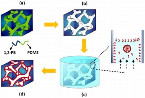
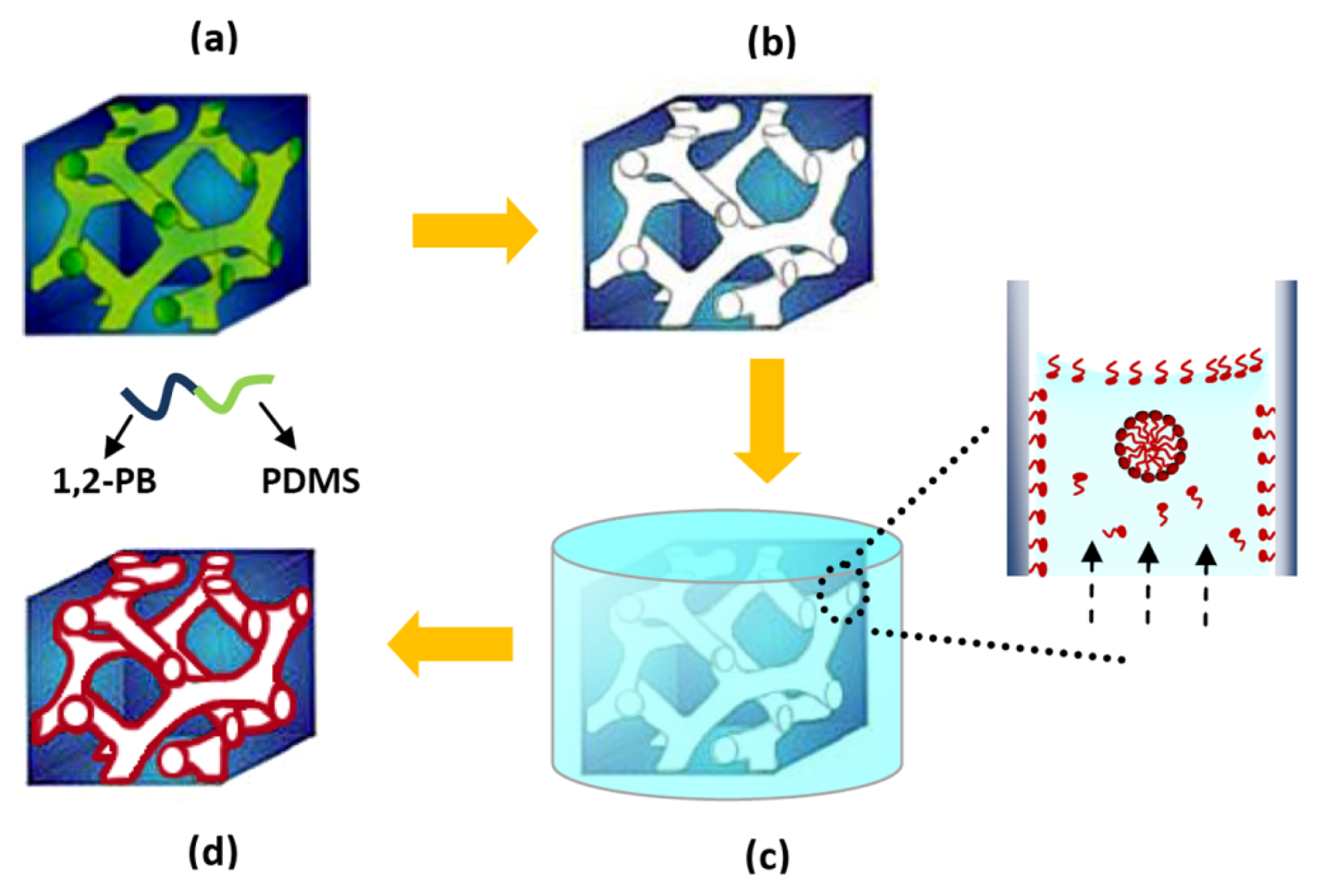
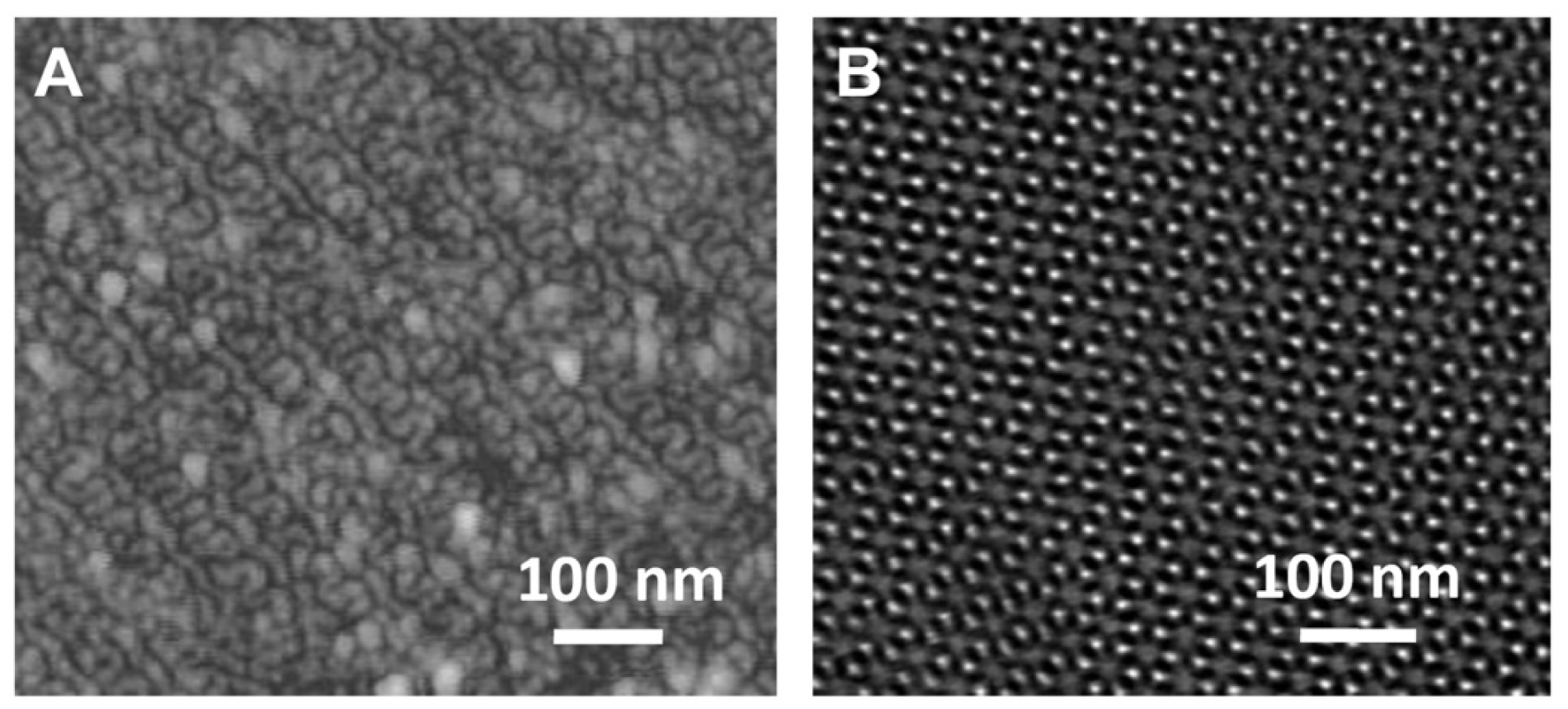
2.1. Preparation of Nanoporous Polymer</title>
<p>As depicted in <xref ref-type="fig" rid="f1-ijms-14-03050">Figure 1</xref>, a gyroid nanoporous matrix was prepared from self-assembled 1,2-polybutadiene-<italic>b</italic>-polydimethylsiloxane (1,2-PB-<italic>b</italic>-PDMS) via quantitative and selective removal of the PDMS block (<xref ref-type="fig" rid="f1-ijms-14-03050">Figure 1a,b</xref>). A gyroid nanostructure was designed from the polymer synthesis stage [<xref ref-type="bibr" rid="b19-ijms-14-03050">19</xref>] in order to ensure isotropic percolation with no need for structure alignment. The composition of the nanoporous matrix is essentially hydrocarbonic and is therefore hydrophobic, showing a static contact angle of 119° with water [<xref ref-type="bibr" rid="b20-ijms-14-03050">20</xref>]. Therefore water cannot spontaneously wet and fill the nanoporous volume, while methanol can [<xref ref-type="bibr" rid="b19-ijms-14-03050">19</xref>,<xref ref-type="bibr" rid="b20-ijms-14-03050">20</xref>]. For this reason SDS loading was realized by first conditioning the nanoporous 1,2-PB film in methanol before dipping it into SDS aqueous solution (<xref ref-type="fig" rid="f1-ijms-14-03050">Figure 1c</xref>). We have recently reported a detailed study on the load-release of SDS into-from nanoporous cross-linked 1,2-PB [<xref ref-type="bibr" rid="b21-ijms-14-03050">21</xref>]. At the loading conditions applied in the present study SDS is adsorbed onto the hydrophobic pore walls creating a dense monolayer as depicted in the inset of <xref ref-type="fig" rid="f1-ijms-14-03050">Figure 1c</xref>. At equilibrium more than 99% of the SDS inside the pores is in the adsorbed state, the rest being either free molecules or organized in micelles [<xref ref-type="bibr" rid="b21-ijms-14-03050">21</xref>]. After SDS loading the nanoporous film (<xref ref-type="fig" rid="f1-ijms-14-03050">Figure 1d</xref>) was then challenged in the culture medium with the biofilm model organism <italic>E. coli</italic> to evaluate its efficiency on preventing bacterial attachment and biofilm formation. As already mentioned virtually all SDS inside the nanopores is adsorbed on the pore walls; its release in the presence of water is slow and happens by multiple release-adsorption steps [<xref ref-type="bibr" rid="b21-ijms-14-03050">21</xref>]. More information on the SDS release kinetics will be given in the Experimental Section.</p>
<p>Typical AFM and TEM images of nanoporous 1,2-PB films are presented in <xref ref-type="fig" rid="f2-ijms-14-03050">Figure 2</xref>, showing a gyroid nanoporosity in the matrix. Two projections of gyroid morphology, the so-called knitting view [<xref ref-type="bibr" rid="b22-ijms-14-03050">22</xref>] (<xref ref-type="fig" rid="f2-ijms-14-03050">Figure 2A</xref>) and wagon-wheel view [<xref ref-type="bibr" rid="b22-ijms-14-03050">22</xref>] (<xref ref-type="fig" rid="f2-ijms-14-03050">Figure 2B</xref>) were observed. Both images show regular patterns with uniform pore size of ~10 nm. The films used in the present work were identical in the bulk morphology, porosity, pore size and size distribution.</p></sec>
<sec>
<title>
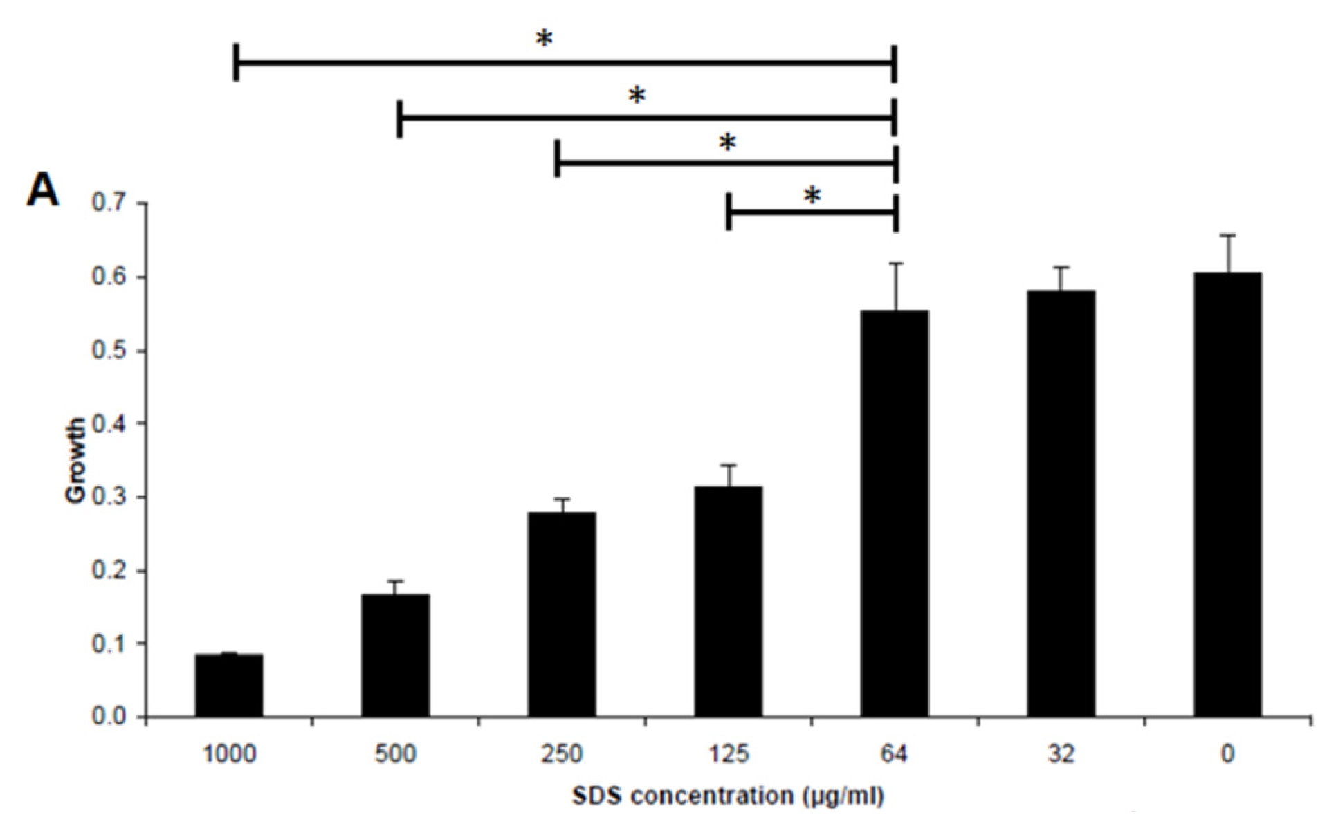
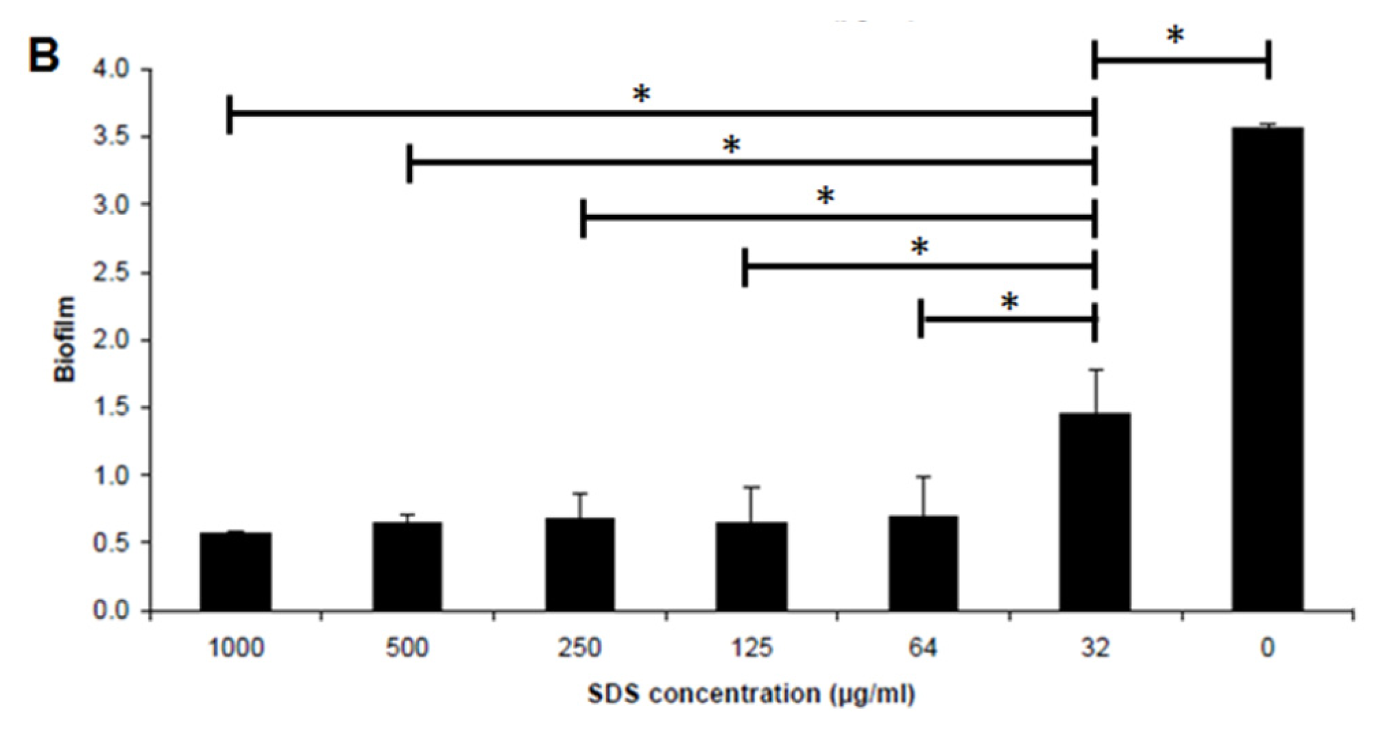
2.2. Effect of SDS on Inhibiting Biofilm Formation by <italic>E. coli</italic> Sar18</title>
<p>SDS is an anionic surfactant used in many cleaning and hygiene products and is shown by several studies to inhibit bacterial biofilm formation and disperse mature biofilms [<xref ref-type="bibr" rid="b23-ijms-14-03050">23</xref>–<xref ref-type="bibr" rid="b25-ijms-14-03050">25</xref>]. We firstly grew the biofilm model organism <italic>E. coli</italic> Sar18 in microtitre tray in medium supplemented with different concentrations of SDS. In our experimental conditions the minimum inhibitory concentration (MIC) and minimum biofilm inhibitory concentration (MBIC) of SDS to <italic>E. coli</italic> Sar18 were 1000 μg/mL and 64 μg/mL, respectively (<xref ref-type="fig" rid="f3-ijms-14-03050">Figure 3</xref>). SDS at subinhibitory concentration (32 μg/mL) could significantly inhibit biofilm formation by <italic>E. coli</italic> Sar18 (<xref ref-type="fig" rid="f3-ijms-14-03050">Figure 3B</xref>).</p></sec>
<sec>
<title>
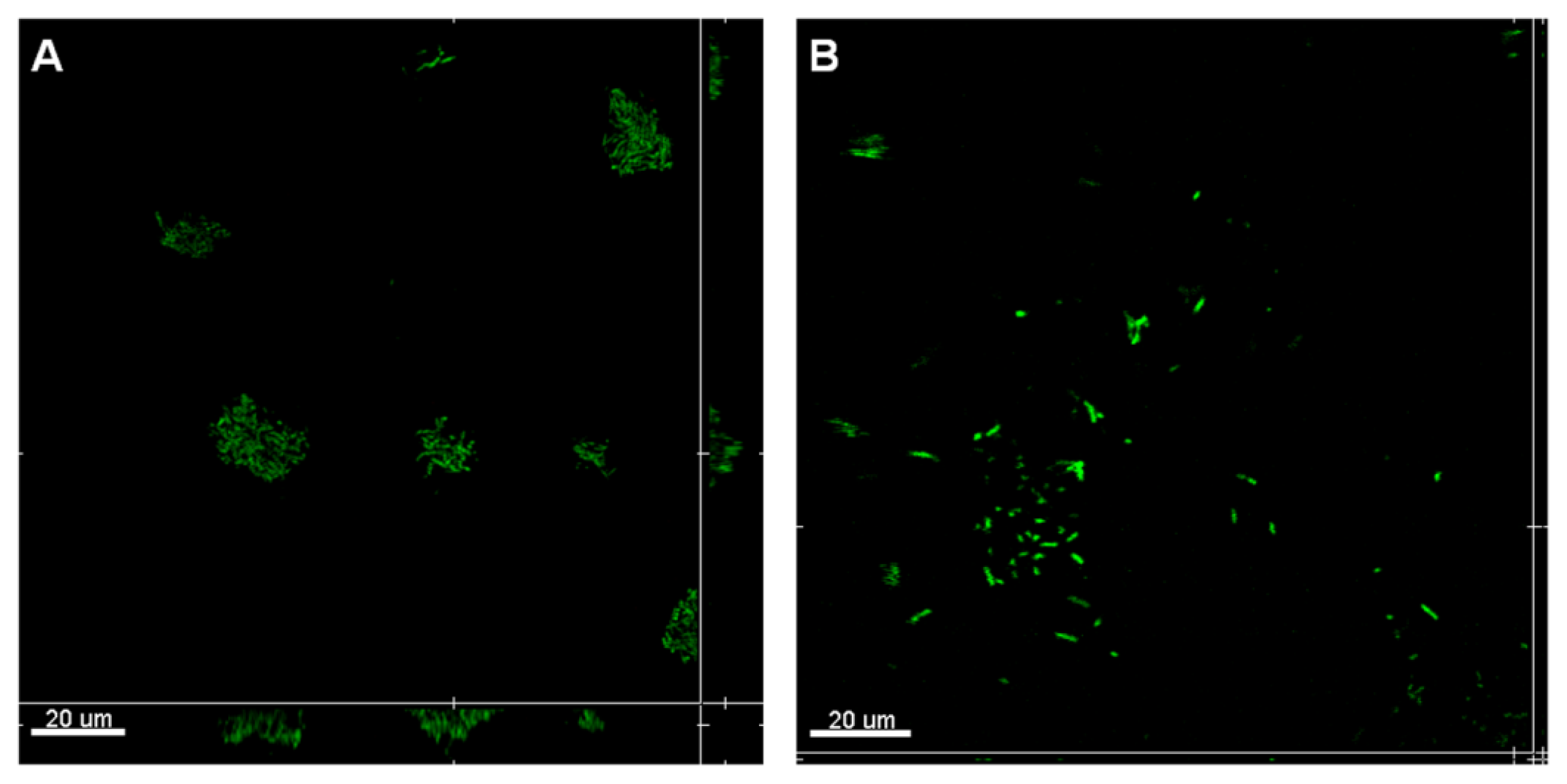
2.3. Attachment Inhibition by SDS-Loaded Nanoporous Film</title>
<p>Shortly after 3 h, <italic>E. coli</italic> Sar18 was able to attach to the surface of control sample T<sub>1</sub> and start to form microcolony structures (<xref ref-type="fig" rid="f4-ijms-14-03050">Figure 4A</xref>). The microcolony formation is often the essential step for biofilm maturation, which can further lead to formation of thick and resistant biofilms [<xref ref-type="bibr" rid="b26-ijms-14-03050">26</xref>,<xref ref-type="bibr" rid="b27-ijms-14-03050">27</xref>]. In contrast, the SDS-loaded T<sub>1</sub> sample could significantly inhibit the formation of microcolony structures by <italic>E. coli</italic> Sar18 (<xref ref-type="fig" rid="f4-ijms-14-03050">Figure 4B</xref>), reducing biofilm development and maturation.</p>
<p>The results of the attachment assay (<xref ref-type="fig" rid="f4-ijms-14-03050">Figure 4</xref>) clearly showed the capabilities of SDS detergent in inhibiting the growth of <italic>E. coli</italic> Sar18 cells, preventing the formation of microcolony structures as well as detaching the cells from the surface. SDS can disrupt bacterial cell-to-cell communications through pili and nanotubes, which are required for aggregation and biofilm formation [<xref ref-type="bibr" rid="b28-ijms-14-03050">28</xref>–<xref ref-type="bibr" rid="b30-ijms-14-03050">30</xref>]. As a detergent, SDS might also extract or induce denaturation of bacterial surface proteins required for attachment and biofilm formation. SDS is widely used in industrial cleaners and household detergents and thus SDS-loaded nanoporous films might be a potential anti-fouling coating material.</p></sec>
<sec>
<title>
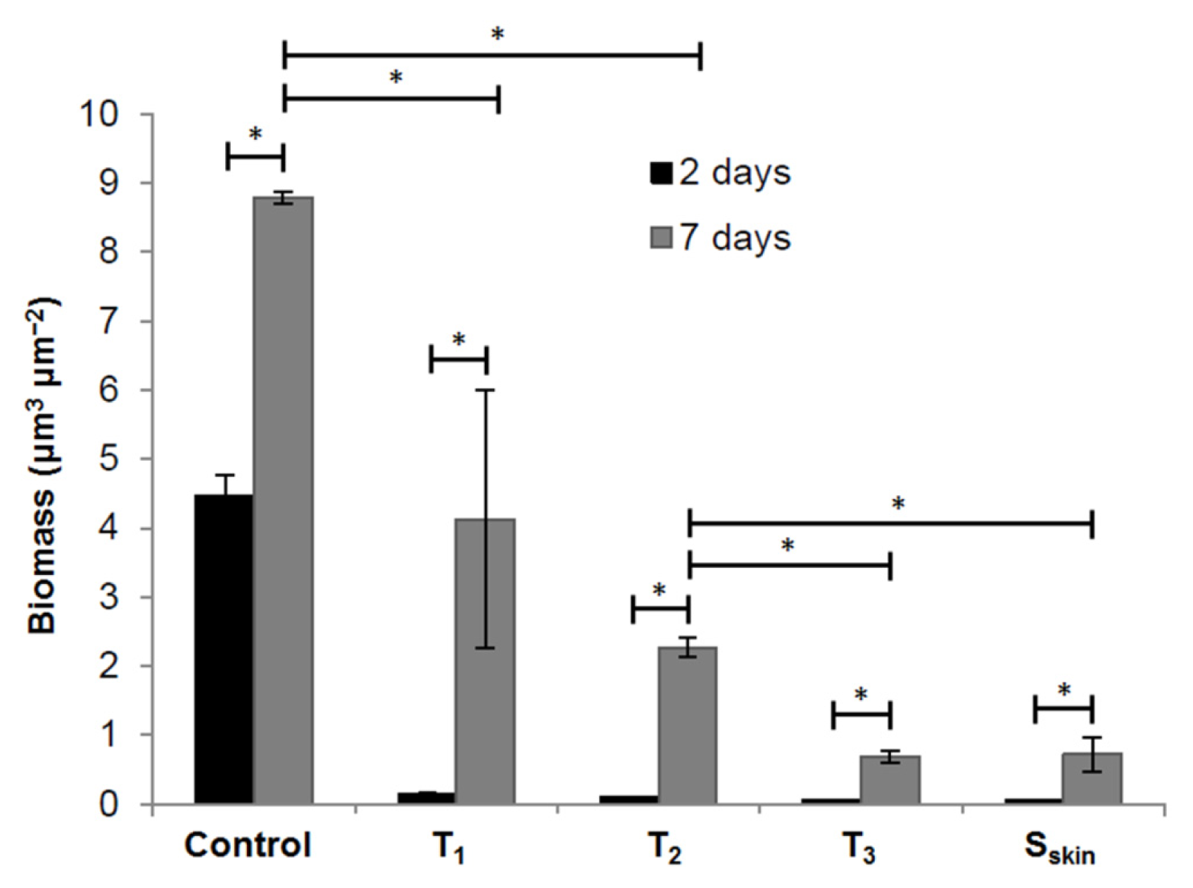
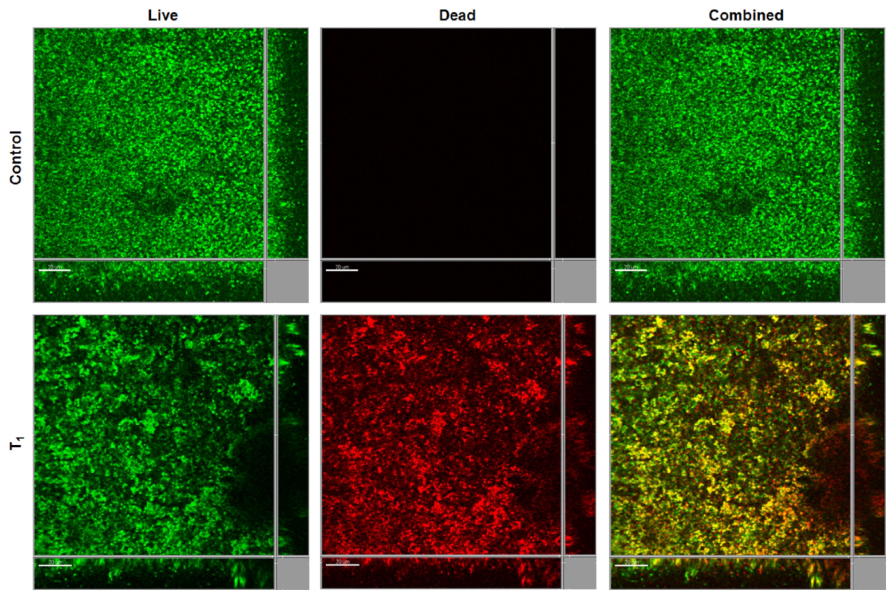
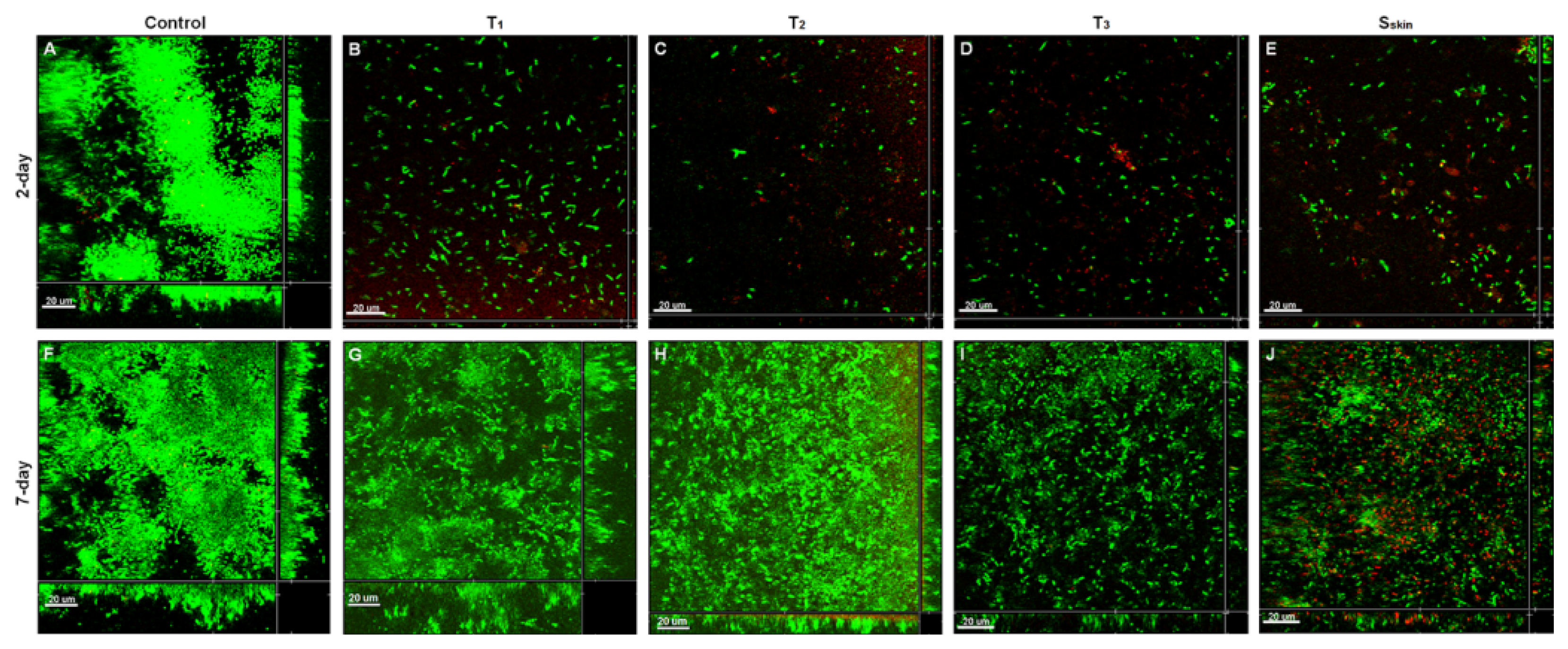
2.4. Biofilm Inhibition by SDS-Loaded Nanoporous Film</title>
<p>To evaluate the impact of release of SDS-loaded nanoporous film on biofilm formation<italic>,</italic> we tested SDS-loaded nanoporous films of different thickness (T<sub>1</sub> 0.5 mm, T<sub>2</sub> 1.0 mm and T<sub>3</sub> 1.5 mm). Besides, two samples with thickness of 1.0 mm (referred to as S<sub>skin</sub>) were glued in the opposite way relative to the other samples, <italic>i.e.</italic>, with the skin-layer surface on the upper side, thus exposing only the dense surface to the culture medium.</p>
<p>After 2-day cultivation, the control sample showed large continuous <italic>E. coli</italic> Sar18 biofilms with dense ball-shaped microcolonies on the surface (<xref ref-type="fig" rid="f5-ijms-14-03050">Figure 5A</xref>). However, only thin layers of <italic>E. coli</italic> Sar18 cells were formed on the surface of the SDS-loaded samples for all the different thickness (T<sub>1</sub>, T<sub>2</sub> and T<sub>3</sub>) and the skin surface (S<sub>skin</sub>) as shown in <xref ref-type="fig" rid="f5-ijms-14-03050">Figure 5B–E</xref>. A large portion of these cells were dead, as demonstrated by the red color. These results indicate that all of the tested SDS-loaded nanoporous films were able to release SDS and create surface concentrations high enough to prevent the biofilm formation on the surface after a contact time of 2 days.</p>
<p>After 7-day release, all the SDS-loaded samples T<sub>1</sub>, T<sub>2</sub>, T<sub>3</sub> and S<sub>skin</sub> showed cell attachment and biofilm formation on their surfaces (<xref ref-type="fig" rid="f5-ijms-14-03050">Figure 5G–J</xref>). However, compared to the control sample without the incorporation of SDS (<xref ref-type="fig" rid="f5-ijms-14-03050">Figure 5F</xref>), the SDS-loaded samples were able to reduce the biofilm formation (<xref ref-type="fig" rid="f5-ijms-14-03050">Figures 5G–J</xref> and <xref ref-type="fig" rid="f6-ijms-14-03050">6</xref>). <italic>E. coli</italic> Sar18 formed least biofilm on the samples T<sub>3</sub> and S<sub>skin</sub> (<xref ref-type="fig" rid="f5-ijms-14-03050">Figures 5I–J</xref> and <xref ref-type="fig" rid="f6-ijms-14-03050">6</xref>). Particularly, a certain amount of dead cells were visible in biofilms formed on the sample S<sub>skin</sub> (<xref ref-type="fig" rid="f5-ijms-14-03050">Figure 5J</xref>).</p>
<p>The nanoporous matrix plays a positive role in both providing large storage space for SDS molecules due to the large interior surface area and regulating the transport at the molecular level thus offering opportunity of sustainable or even controllable release. The gyroid nanoporosity of the films has the advantage of isotropic percolation which ensures high delivery efficiency. This is definitely supported by the results from the 2-day incubation (<xref ref-type="fig" rid="f5-ijms-14-03050">Figure 5</xref>), which reveal the efficiency of the SDS-releasing nanoporous 1,2-PB films against bacterial attachment and biofilm formation by <italic>E. coli</italic>. Remarkable reduction in bacterial attachment and no visible biofilm formation were found compared with the control sample. It therefore can be expected that the surface concentration of SDS could retain a value within 2 days, which is at least comparable to the MBIC value of 64 μg/mL. This can actually be confirmed by the estimation of the initial SDS surface concentration and the SDS-released concentration after 48 h, as described in “Materials and Methods” section. The estimated value of <italic>C</italic><sub>os</sub> (<italic>t</italic> = 0) is almost identical to the MIC value, and 15 times higher than MBIC. The size of a bacterium is usually of the order of few micrometers and at such near-surface zone the concentration can be very close to <italic>C</italic><italic><sub>os</sub></italic>. Therefore, the expectation from the above estimation is that the starting SDS concentration is sufficient to kill bacteria in the near-surface zone and to totally inhibit biofilm formation. The estimated average SDS concentration after 48 h is close to the MBIC value, and it’s therefore consistent with the absence of biofilm formation in the 48 h experiment.</p>
<p>Unfortunately, the present SDS-releasing nanoporous systems showed weaker ability against the biofilm formation in 7-day anti-biofilm assay (50% reduction, T<sub>1</sub>) compared to 2- day anti-biofilm assay (95% reduction, T<sub>1</sub>, <xref ref-type="fig" rid="f6-ijms-14-03050">Figure 6</xref>). This can be ascribed to low release rate of SDS after 72 h, as already reported in [<xref ref-type="bibr" rid="b21-ijms-14-03050">21</xref>]. However, increasing the film thickness or slowing down the release rate by exposing the skin layer side to the solution did reduce long-term biofilm formation (74% reduction for T<sub>2</sub>, 92% reduction for T<sub>3</sub> and S<sub>skin</sub>, <xref ref-type="fig" rid="f6-ijms-14-03050">Figure 6</xref>). The improvement provided by the S<sub>skin</sub> samples is particularly interesting. In a comparative study of glucose permeability through 1,2-PB nanoporous membranes [<xref ref-type="bibr" rid="b31-ijms-14-03050">31</xref>], we have observed that the presence of skin layer diminishes the effective diffusion coefficient by a factor of 5.7 compared to the porous surface. This effect is correlated to decreased surface porosity due to the compact nature of the skin layer, irregularly interrupted by porous defects [<xref ref-type="bibr" rid="b31-ijms-14-03050">31</xref>].</p>
<p>To evaluate the biofilm inhibitory effect of the SDS-loaded nanoporous films on Gram-positive bacteria, we tested the biofilm formation of <italic>Staphylococcus epidermidis</italic> RP62A [<xref ref-type="bibr" rid="b32-ijms-14-03050">32</xref>] on the control and SDS-loaded nanoporous T<sub>1</sub> films. <italic>S. epidermidis</italic> RP62A formed thick biofilms on both control and SDS-loaded nanoporous T<sub>1</sub> films after 1 day cultivation (<xref ref-type="fig" rid="f7-ijms-14-03050">Figure 7</xref>). However, SDS-loaded nanoporous T<sub>1</sub> film killed most of the attached <italic>S. epidermidis</italic> RP62A cells (<xref ref-type="fig" rid="f7-ijms-14-03050">Figure 7</xref>). This result indicates that SDS is not a good agent for controlling biofilms formed by <italic>S. epidermidis</italic>. Further test is required to identify agents suitable for preparing anti-<italic>S. epidermidis</italic> nanoporous films.</p></sec></sec>
<sec>
<title>3. Experimental Section</title>
<sec>
<title>3.1. Preparation of Nanoporous Film</title>
<p>The 1,2-polybutadiene-<italic>b</italic>-polydimethylsiloxane (1,2-PB-<italic>b</italic>-PDMS) diblock copolymer was synthesized by living anionic polymerization as previously reported [<xref ref-type="bibr" rid="b19-ijms-14-03050">19</xref>]. The general procedure to prepare a nanoporous 1,2-PB matrix was as follows. <italic>A tetrahydrofuran</italic> or oxacyclopentane (THF, Sigma-Aldrich, Copenhagen, Denmark) solution of 1,2-PB-<italic>b</italic>-PDMS and cross-linker (dicumyl peroxide, DCP, Sigma-Aldrich) was solvent casted onto a flat bottom glass Petri-dish. The molar amount of DCP was 1% relative to that of the double bonds in 1,2-PB. After complete evaporation of THF, the cast copolymer film was cross-linked at 140 °C for 2 h under nitrogen atmosphere. The cross-linked copolymer film was subsequently immersed in a tetra-n-butylammonium fluoride solution (TBAF, Sigma-Aldrich) in THF at room temperature for 36 h to selectively and quantitatively remove the PDMS block. The molar amount of TBAF was twice that of Si–O bonds in PDMS. The etched film was rinsed in a mixture of THF and methanol and finally dried under nitrogen flow at room temperature.</p>
<p>As reported previously [<xref ref-type="bibr" rid="b31-ijms-14-03050">31</xref>], the outer surface of the resultant nanoporous film showed two different features. A 30 nm dense skin layer was observed on the free outer surface that during cross-linking was in contact with the nitrogen atmosphere; while the surface in contact with the glass petri-dish was nanoporous with a porosity of approximately 40%. In the subsequent biofilm formation assay experiments the tested samples had the porous surface side exposed to the culture medium for releasing SDS, except for one set of samples referred to as S<sub>skin</sub> that had the surface with the skin layer in contact with the culture medium. In this study, we prepared nanoporous 1,2-PB films with three different thicknesses: 0.5 ± 0.1 mm, 1.0 ± 0.1 mm, and 1.5 ± 0.1 mm. They are denoted in the manuscript as T<sub>1</sub>, T<sub>2</sub> and T<sub>3</sub> respectively.</p></sec>
<sec>
<title>3.2. Characterization of Nanoporous Films</title>
<p>The morphology of nanoporous membranes was checked by Atomic Force Microscopy (AFM) and Transmission Electron Microscopy (TEM). AFM was conducted at ambient air using NanoMan AFM in tapping mode, with NANOSENSORS™ SSS-NCH AFM probe. TEM was performed on a FEI TECNAI T20 at an acceleration voltage of 200 kV.</p>
<p>The fracture cross-section of a nanoporous film was trimmed and microtomed to a flat surface for AFM measurement. Ninety nm slices were sectioned from the film and deposited onto a holey carbon copper grid for TEM measurements. The microtoming process was completed on a Leica ultramicrotome with a cryo 35_diamond knife (DIATOME) at room temperature.</p></sec>
<sec>
<title>
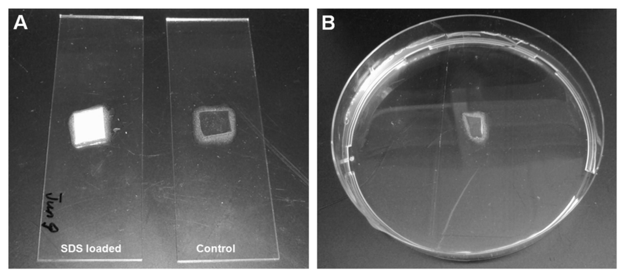
3.3. Loading of Nanoporous Films with SDS</title>
<p>A systematic study of SDS infiltration from aqueous solutions with SDS concentration between 0.5 and 50 mM into the nanoporous 1,2-PB matrix has been recently reported [<xref ref-type="bibr" rid="b21-ijms-14-03050">21</xref>]. It has been shown that the SDS adsorption onto the inner surface of nanoporous 1,2-PB film saturates at concentrations above 6.8 mM. In the present work we used a 10 mM SDS aqueous solution to ensure adsorption saturation. The nanoporous 1,2-PB film was pre-wet with methanol for 10 minutes and then kept under shaking for 15 h in 15 mL of SDS aqueous solution. The SDS-loaded samples were gently wiped with a tissue and dried under nitrogen flow without further treatment.</p></sec>
<sec>
<title>3.4. Minimum Biofilm Inhibitory Concentration (MBIC) Assay</title>
<p>The microtitre tray biofilm formation assay was performed as previously described [<xref ref-type="bibr" rid="b33-ijms-14-03050">33</xref>] to measure the minimum biofilm inhibitory concentration of SDS to <italic>E. coli</italic>. Overnight cultures were grown in AB minimal medium [<xref ref-type="bibr" rid="b33-ijms-14-03050">33</xref>] supplemented with 5 g/L glucose and diluted to optical density OD 600 nm = 0.001 with fresh medium. SDS was added to culture medium at different concentrations from 0 to 1000 μg/mL. The diluted cultures were transferred to the wells of microtitre trays (150 μL per well) and incubated for 24 h at 37 °C. The growth of bacterium was recorded by measuring absorbance at OD 600 nm for minimum inhibitory concentration (MIC) determination. The medium was then removed from the wells and these were washed twice with 0.9% NaCl, stained with 0.1% crystal violet and again washed twice with 0.9% NaCl; the crystal violet-stained biofilms were then resuspended in 96% ethanol, and biofilm cell-associated dye was measured as absorbance at OD 600 nm by the use of a Wallac microplate reader. Six replicates were used for each SDS concentration.</p></sec>
<sec>
<title>3.5. Attachment Assay</title>
<p>Nanoporous 1,2-PB films T<sub>1</sub> (0.5 ± 0.1 mm) with the incorporated SDS were tested in the attachment assay, while T<sub>1</sub> samples without SDS were used as control samples. As shown in <xref ref-type="fig" rid="f8-ijms-14-03050">Figure 8A</xref>, the samples were attached onto the surface of glass slides with glue (Epoxy Universal 335, high strength 2-component expoxy adhesive) surrounding the side surfaces. The glue was immediately cured at 65 °C for 2.5 h to allow a complete curing process. As a result, the tested samples were fixed on the glass slides and only the upper surface was exposed for effective release. In this experiment, the upper surface was the porous surface. Prior to be ready for the attachment assay, all the samples were sterilized under UV exposure for 30 min. The biofilm model organism gram-negative <italic>Escherichia coli</italic> Sar18 [<xref ref-type="bibr" rid="b34-ijms-14-03050">34</xref>] was used in this study. The samples were submerged into bacterial cultures in Petri dishes as illustrated in <xref ref-type="fig" rid="f8-ijms-14-03050">Figure 8B</xref>. <italic>E. coli</italic> was cultivated in 25 mL AB minimal medium supplemented with 5 g/L glucose at 37 °C for 3 h. After that the slides were taken out from Petri dishes and washed with fresh AB minimal medium in order to remove floating bacterial cells. Three 1,2-PB films were used for each analysis and two confocal images were taken at different positions of each 1,2-PB film.</p></sec>
<sec>
<title>3.6. Biofilm Formation Assay</title>
<p>In the biofilm formation assay we selected different thicknesses of the SDS-loaded nanoporous films for the 2-day and 7-day tests, T<sub>1</sub> (0.5 ± 0.1 mm), T<sub>2</sub> (1.0 ± 0.1 mm) and T<sub>3</sub> (1.5 ± 0.1 mm). The T<sub>2</sub> and T<sub>3</sub> films have a higher capacity for SDS than T<sub>1</sub> enabling longer SDS release periods (data not shown). Film pieces not containing SDS were taken as control samples. All the samples exposed the porous surface to the culture medium for effective release. For comparison we tested two samples S<sub>skin</sub> (1.0 ± 0.1 mm thick) with the skin-layer side exposed to the <italic>E. coli</italic> culture. For the 2-day assay, nanoporous 1,2-PB films T<sub>1</sub>, T<sub>2</sub> and T<sub>3</sub> loaded with SDS as well as a control T<sub>1</sub> without SDS were glued on the surface of glass slides and submerged into bacterial cultures in Petri dishes as described above. <italic>E. coli</italic> was cultivated in AB minimal medium supplemented with 5 g/L glucose at 37 °C for 2 days. After that the slides were taken out from petri dishes and washed with fresh AB minimal medium in order to remove floating bacterial cells. Three 1,2-PB films were used for each analysis and two confocal images were taken at different positions of each 1,2-PB film. For the 7-day test, we glued the SDS-loaded samples T<sub>1</sub>, T<sub>2</sub>, T<sub>3</sub> and S<sub>skin</sub> on glass as above and left them in fresh AB minimal medium for 5 days (SDS-containing medium was replaced by fresh medium every 2 days) and then submerged into bacterial cultures in Petri dishes for additional 2 days. After that the slides were taken out from the Petri dishes and again washed by fresh AB minimal medium in order to get rid of floating bacterial cells. Three 1,2-PB films were used for each analysis and two confocal images were taken at different positions of each 1,2-PB film.</p>
<p>For comparison, biofilm formation by Gram-positive bacterium <italic>Staphylococcus epidermidis</italic> RP62A on the control and SDS-loaded nanoporous T<sub>1</sub> films was also investigated as described above.</p></sec>
<sec>
<title>3.7. CLSM Observation</title>
<p>The LIVE/DEAD<sup>®</sup> Bacterial Viability Kit was used to stain live (appears green under fluorescence microscopy) and dead (appears red under fluorescence microscopy) bacterial cells on the surface of all of tested the nanoporous 1,2-PB films. The nanoporous 1,2-PB films were observed under a Carl Zeiss LSM510 META Confocal Laser scanning Microscope (CLSM, company, Jena, Germany) for biofilms. Images were obtained using a 63×/1.4 objective. Simulated three-dimensional images and sections were generated using the IMARIS software package (Bitplane AG: Zurich, Switzerland, 2010).</p></sec>
<sec sec-type="methods">
<title>3.8. COMSTAT Analysis</title>
<p>CLSM images were analyzed by use of the computer program COMSTAT for calculating the biomass of biofilms [<xref ref-type="bibr" rid="b35-ijms-14-03050">35</xref>]. A fixed threshold value and connected volume filtration were used for all image stacks. Six images were used for analysis of each sample.</p></sec>
<sec>
<title>3.9. Estimation of the SDS Concentration Near the Surface</title>
<p>We estimated the SDS concentration at the outer surface of the nanoporous samples from data reported in a recent publication [<xref ref-type="bibr" rid="b21-ijms-14-03050">21</xref>] on the kinetics and equilibrium of SDS load—release in samples similar to the T<sub>1</sub> samples of this study. Then we compared the estimated values with the minimum inhibitoryconcentration, MIC and with the minimum biofilm inhibitory concentration, MBIC. The equilibrium amount of SDS adsorbed into nanoporous 1,2-PB films immersed in excess SDS aqueous solutions was 23% of the 1,2-PB matrix mass, independent of SDS concentration in the concentration range of 6–50 mM. The adsorbed amount corresponds to a monolayer of SDS onto the pore walls. At the beginning of a release experiment, the SDS concentration at the very outer surface <italic>C</italic><sub>os</sub> (<italic>t</italic> = 0) of a nanoporous film fully loaded with SDS can be estimated from the concentration of SDS in the porous volume at the immediate vicinity of the outer surface <italic>C</italic><sub>is</sub> (<italic>t</italic> = 0), reduced by a factor equal to the surface porosity α.</p>
<disp-formula id="FD1">
<label>(1)</label>
<mml:math id="mm1" display="block">
<mml:semantics id="sm1">
<mml:mrow>
<mml:msub>
<mml:mrow>
<mml:mi>C</mml:mi></mml:mrow>
<mml:mrow>
<mml:mi>o</mml:mi>
<mml:mi>s</mml:mi></mml:mrow></mml:msub>
<mml:mo stretchy="false">(</mml:mo>
<mml:mi>t</mml:mi>
<mml:mo>=</mml:mo>
<mml:mn>0</mml:mn>
<mml:mo stretchy="false">)</mml:mo>
<mml:mo>=</mml:mo>
<mml:mi mathvariant="normal">α</mml:mi>
<mml:mo>·</mml:mo>
<mml:msub>
<mml:mrow>
<mml:mi>C</mml:mi></mml:mrow>
<mml:mrow>
<mml:mi>i</mml:mi>
<mml:mi>s</mml:mi></mml:mrow></mml:msub>
<mml:mo stretchy="false">(</mml:mo>
<mml:mi>t</mml:mi>
<mml:mo>=</mml:mo>
<mml:mn>0</mml:mn>
<mml:mo stretchy="false">)</mml:mo>
<mml:mo>≈</mml:mo>
<mml:mi mathvariant="normal">α</mml:mi>
<mml:mo>·</mml:mo>
<mml:mi>C</mml:mi>
<mml:mi>M</mml:mi>
<mml:mi>C</mml:mi>
<mml:mo>=</mml:mo>
<mml:mn>0.4</mml:mn>
<mml:mo>·</mml:mo>
<mml:mn>8.1</mml:mn>
<mml:mi> </mml:mi>
<mml:mtext>mM</mml:mtext>
<mml:mo>=</mml:mo>
<mml:mn>3.24</mml:mn>
<mml:mi> </mml:mi>
<mml:mtext>mM</mml:mtext>
<mml:mo>=</mml:mo>
<mml:mn>934</mml:mn>
<mml:mi> </mml:mi>
<mml:mi mathvariant="normal">μ</mml:mi>
<mml:mtext>g</mml:mtext>
<mml:mo>/</mml:mo>
<mml:msup>
<mml:mrow>
<mml:mrow>
<mml:mtext>cm</mml:mtext></mml:mrow></mml:mrow>
<mml:mn>3</mml:mn></mml:msup></mml:mrow></mml:semantics></mml:math></disp-formula>
<p>The approximation is justified by the already mentioned concentration independence of equilibrium adsorption in a wide range of concentrations, with the lowest value being just below the critical micelle concentration (CMC) of SDS in water (CMC = 8.1 mM). The surface porosity α for the porous outer surface was 0.4 [<xref ref-type="bibr" rid="b21-ijms-14-03050">21</xref>]. We used the molar mass of SDS, <italic>M</italic><italic><sub>SDS</sub></italic> = 288.4 g/mol in order to convert the molar concentration into a mass concentration at the end of the above expression. In the time interval 0 ≤ <italic>t</italic> ≤ 72 h (= 259,200 s) the experimental data on the kinetics of SDS release in excess of distilled water could be accurately described by a power low expression:</p>
<disp-formula id="FD2">
<label>(2)</label>
<mml:math id="mm2" display="block">
<mml:semantics id="sm2">
<mml:mrow>
<mml:mi>M</mml:mi>
<mml:mo stretchy="false">(</mml:mo>
<mml:mi>t</mml:mi>
<mml:mo stretchy="false">)</mml:mo>
<mml:mo>=</mml:mo>
<mml:msub>
<mml:mrow>
<mml:mi>M</mml:mi></mml:mrow>
<mml:mn>0</mml:mn></mml:msub>
<mml:mo stretchy="false">[</mml:mo>
<mml:mn>1</mml:mn>
<mml:mo>-</mml:mo>
<mml:mn>0.0114</mml:mn>
<mml:mo>·</mml:mo>
<mml:mi>t</mml:mi>
<mml:msup>
<mml:mrow>
<mml:mrow>
<mml:mo stretchy="false">(</mml:mo>
<mml:mi>s</mml:mi>
<mml:mo stretchy="false">)</mml:mo></mml:mrow></mml:mrow>
<mml:mrow>
<mml:mn>1</mml:mn>
<mml:mo>/</mml:mo>
<mml:mn>3</mml:mn></mml:mrow></mml:msup>
<mml:mo stretchy="false">]</mml:mo></mml:mrow></mml:semantics></mml:math></disp-formula>
<p>where <italic>M</italic> (<italic>t</italic>)/<italic>M</italic><sub>0</sub> is the mass fraction of SDS remaining inside the nanopores at release time <italic>t</italic>. About 70% of the SDS adsorbed could be released after 72 h [<xref ref-type="bibr" rid="b21-ijms-14-03050">21</xref>]. The initial mass of adsorbed SDS was 23% of the 1,2-PB matrix, which in the present case was <italic>m</italic><sub>PB</sub> = 20 mg, therefore <italic>M</italic><sub>0</sub> = 4.6 mg.</p>
<p>The presented SDS release data were obtained under shaking and in the presence of large excess of water. The release experiments of the present study were done in Petri dishes containing 50 mL aqueous solution with stirring. We illustrate the use of the above relation by considering the 48 h release experiment. At the end of the experiment a total of 2.9 mg of SDS was released in the surrounding solution and the corresponding average concentration was 58 μg/mL. The estimated values of initial <italic>C</italic><sub>s</sub> and the SDS concentration at different times of release will be compared with the experimental values of MIC and MBIC in the ‘Discussion’ section. The consumption of released SDS by the <italic>E. coli</italic> bacteria was not taken into account in the course of the presented estimates.</p></sec></sec>
<sec sec-type="conclusions">
<title>4. Conclusions</title>
<p>In the present work, we explored the potential as anti-biofilm materials of nanoporous polymers templated from block copolymers and loaded with a common anti-biofilm detergent, sodium dodecyl sulfate (SDS). The presented results are promising and constitute a basis for the development of a simple and generic strategy for the fabrication of anti-biofilm coatings. Supplementary research efforts are necessary in order to further improve the release profile from the nanoporous polymers, thus making our system competitive in practical applications. Guided by the promising results from the skin layer surface we can try to further reduce the pore size of the nanoporous polymer, or to better control the morphology of the skin layer and its defects, thus enabling to decrease the release rate.</p></sec></body>
<back>
<ack>
<title>Acknowledgments</title>
<p>This work was supported by a grant from the Danish Research Council for Independent Research (09-073917) to L.Y. and a Ph.D. studentship funded by the Radiometer Medical ApS to L.L.</p></ack>
<fn-group><fn id="fn1-ijms-14-03050">
<p><bold>Conflict of Interest</bold></p>
<p>The authors declare no conflict of interest.</p></fn></fn-group>
<ref-list>
<title>References</title>
<ref id="b1-ijms-14-03050"><label>1</label><citation citation-type="journal"><person-group person-group-type="author"><name><surname>Costerton</surname><given-names>J.W.</given-names></name><name><surname>Lewandowski</surname><given-names>Z.</given-names></name><name><surname>Caldwell</surname><given-names>D.E.</given-names></name><name><surname>Korber</surname><given-names>D.R.</given-names></name><name><surname>Lappin-Scott</surname><given-names>H.M.</given-names></name></person-group><article-title>Microbial biofilms</article-title><source>Annu. Rev. Microbiol</source><year>1995</year><volume>49</volume><fpage>711</fpage><lpage>745</lpage><pub-id pub-id-type="doi">10.1146/annurev.mi.49.100195.003431</pub-id><pub-id pub-id-type="pmid">8561477</pub-id></citation></ref>
<ref id="b2-ijms-14-03050"><label>2</label><citation citation-type="journal"><person-group person-group-type="author"><name><surname>Yang</surname><given-names>L.</given-names></name><name><surname>Liu</surname><given-names>Y.</given-names></name><name><surname>Wu</surname><given-names>H.</given-names></name><name><surname>Song</surname><given-names>Z.</given-names></name><name><surname>Hoiby</surname><given-names>N.</given-names></name><name><surname>Molin</surname><given-names>S.</given-names></name><name><surname>Givskov</surname><given-names>M.</given-names></name></person-group><article-title>Combating biofilms</article-title><source>FEMS Immunol. Med. Microbiol</source><year>2012</year><volume>65</volume><fpage>146</fpage><lpage>157</lpage><pub-id pub-id-type="doi">10.1111/j.1574-695X.2011.00858.x</pub-id><pub-id pub-id-type="pmid">22066868</pub-id></citation></ref>
<ref id="b3-ijms-14-03050"><label>3</label><citation citation-type="journal"><person-group person-group-type="author"><name><surname>Yang</surname><given-names>L.</given-names></name><name><surname>Liu</surname><given-names>Y.</given-names></name><name><surname>Wu</surname><given-names>H.</given-names></name><name><surname>Hoiby</surname><given-names>N.</given-names></name><name><surname>Molin</surname><given-names>S.</given-names></name><name><surname>Song</surname><given-names>Z.J.</given-names></name></person-group><article-title>Current understanding of multi-species biofilms</article-title><source>Int. J. Oral Sci</source><year>2011</year><volume>3</volume><fpage>74</fpage><lpage>81</lpage><pub-id pub-id-type="doi">10.4248/IJOS11027</pub-id><pub-id pub-id-type="pmid">21485311</pub-id></citation></ref>
<ref id="b4-ijms-14-03050"><label>4</label><citation citation-type="journal"><person-group person-group-type="author"><name><surname>De Prijck</surname><given-names>K.</given-names></name><name><surname>Nelis</surname><given-names>H.</given-names></name><name><surname>Coenye</surname><given-names>T.</given-names></name></person-group><article-title>Efficacy of silver-releasing rubber for the prevention of <italic>Pseudomonas aeruginosa</italic> biofilm formation in water</article-title><source>Biofouling</source><year>2007</year><volume>23</volume><fpage>405</fpage><lpage>411</lpage><pub-id pub-id-type="doi">10.1080/08927010701647861</pub-id><pub-id pub-id-type="pmid">17934912</pub-id></citation></ref>
<ref id="b5-ijms-14-03050"><label>5</label><citation citation-type="journal"><person-group person-group-type="author"><name><surname>Olsen</surname><given-names>S.M.</given-names></name><name><surname>Pedersen</surname><given-names>L.T.</given-names></name><name><surname>Laursen</surname><given-names>M.H.</given-names></name><name><surname>Kiil</surname><given-names>S.</given-names></name><name><surname>Dam-Johansen</surname><given-names>K.</given-names></name></person-group><article-title>Enzyme-based antifouling coatings: A review</article-title><source>Biofouling</source><year>2007</year><volume>23</volume><fpage>369</fpage><lpage>383</lpage><pub-id pub-id-type="doi">10.1080/08927010701566384</pub-id><pub-id pub-id-type="pmid">17852071</pub-id></citation></ref>
<ref id="b6-ijms-14-03050"><label>6</label><citation citation-type="journal"><person-group person-group-type="author"><name><surname>Almaguer-Flores</surname><given-names>A.</given-names></name><name><surname>Ximenez-Fyvie</surname><given-names>L.A.</given-names></name><name><surname>Rodil</surname><given-names>S.E.</given-names></name></person-group><article-title>Oral bacterial adhesion on amorphous carbon and titanium films: Effect of surface roughness and culture media</article-title><source>J. Biomed. Mater. Res. B Appl. Biomater</source><year>2010</year><volume>92</volume><fpage>196</fpage><lpage>204</lpage><pub-id pub-id-type="pmid">19810113</pub-id></citation></ref>
<ref id="b7-ijms-14-03050"><label>7</label><citation citation-type="journal"><person-group person-group-type="author"><name><surname>Kaper</surname><given-names>H.J.</given-names></name><name><surname>Busscher</surname><given-names>H.J.</given-names></name><name><surname>Norde</surname><given-names>W.</given-names></name></person-group><article-title>Characterization of poly(ethylene oxide) brushes on glass surfaces and adhesion of <italic>Staphylococcus epidermidis</italic></article-title><source>J. Biomater. Sci. Polym. Ed</source><year>2003</year><volume>14</volume><fpage>313</fpage><lpage>324</lpage><pub-id pub-id-type="doi">10.1163/156856203321478847</pub-id><pub-id pub-id-type="pmid">12747672</pub-id></citation></ref>
<ref id="b8-ijms-14-03050"><label>8</label><citation citation-type="journal"><person-group person-group-type="author"><name><surname>Majumdar</surname><given-names>P.</given-names></name><name><surname>Lee</surname><given-names>E.</given-names></name><name><surname>Patel</surname><given-names>N.</given-names></name><name><surname>Ward</surname><given-names>K.</given-names></name><name><surname>Stafslien</surname><given-names>S.J.</given-names></name><name><surname>Daniels</surname><given-names>J.</given-names></name><name><surname>Chisholm</surname><given-names>B.J.</given-names></name><name><surname>Boudjouk</surname><given-names>P.</given-names></name><name><surname>Callow</surname><given-names>M.E.</given-names></name><name><surname>Callow</surname><given-names>J.A.</given-names></name><etal/></person-group><article-title>Combinatorial materials research applied to the development of new surface coatings IX: An investigation of novel antifouling/fouling-release coatings containing quaternary ammonium salt groups</article-title><source>Biofouling</source><year>2008</year><volume>24</volume><fpage>185</fpage><lpage>200</lpage><pub-id pub-id-type="doi">10.1080/08927010801894660</pub-id><pub-id pub-id-type="pmid">18368587</pub-id></citation></ref>
<ref id="b9-ijms-14-03050"><label>9</label><citation citation-type="journal"><person-group person-group-type="author"><name><surname>Nagel</surname><given-names>J.A.</given-names></name><name><surname>Dickinson</surname><given-names>R.B.</given-names></name><name><surname>Cooper</surname><given-names>S.L.</given-names></name></person-group><article-title>Bacterial adhesion to polyurethane surfaces in the presence of pre-adsorbed high molecular weight kininogen</article-title><source>J. Biomater. Sci. Polym. Ed</source><year>1996</year><volume>7</volume><fpage>769</fpage><lpage>780</lpage><pub-id pub-id-type="doi">10.1163/156856296X00110</pub-id><pub-id pub-id-type="pmid">8773881</pub-id></citation></ref>
<ref id="b10-ijms-14-03050"><label>10</label><citation citation-type="journal"><person-group person-group-type="author"><name><surname>Hetrick</surname><given-names>E.M.</given-names></name><name><surname>Schoenfisch</surname><given-names>M.H.</given-names></name></person-group><article-title>Reducing implant-related infections: Active release strategies</article-title><source>Chem. Soc. Rev</source><year>2006</year><volume>35</volume><fpage>780</fpage><lpage>789</lpage><pub-id pub-id-type="doi">10.1039/b515219b</pub-id><pub-id pub-id-type="pmid">16936926</pub-id></citation></ref>
<ref id="b11-ijms-14-03050"><label>11</label><citation citation-type="journal"><person-group person-group-type="author"><name><surname>Price</surname><given-names>J.S.</given-names></name><name><surname>Tencer</surname><given-names>A.F.</given-names></name><name><surname>Arm</surname><given-names>D.M.</given-names></name><name><surname>Bohach</surname><given-names>G.A.</given-names></name></person-group><article-title>Controlled release of antibiotics from coated orthopedic implants</article-title><source>J. Biomed. Mater. Res</source><year>1996</year><volume>30</volume><fpage>281</fpage><lpage>286</lpage><pub-id pub-id-type="doi">10.1002/(SICI)1097-4636(199603)30:3&lt;281::AID-JBM2&gt;3.0.CO;2-M</pub-id><pub-id pub-id-type="pmid">8698690</pub-id></citation></ref>
<ref id="b12-ijms-14-03050"><label>12</label><citation citation-type="journal"><person-group person-group-type="author"><name><surname>Schierholz</surname><given-names>J.M.</given-names></name><name><surname>Steinhauser</surname><given-names>H.</given-names></name><name><surname>Rump</surname><given-names>A.F.</given-names></name><name><surname>Berkels</surname><given-names>R.</given-names></name><name><surname>Pulverer</surname><given-names>G.</given-names></name></person-group><article-title>Controlled release of antibiotics from biomedical polyurethanes: Morphological and structural features</article-title><source>Biomaterials</source><year>1997</year><volume>18</volume><fpage>839</fpage><lpage>844</lpage><pub-id pub-id-type="doi">10.1016/S0142-9612(96)00199-8</pub-id><pub-id pub-id-type="pmid">9184747</pub-id></citation></ref>
<ref id="b13-ijms-14-03050"><label>13</label><citation citation-type="journal"><person-group person-group-type="author"><name><surname>Lee</surname><given-names>D.</given-names></name><name><surname>Cohen</surname><given-names>R.E.</given-names></name><name><surname>Rubner</surname><given-names>M.F.</given-names></name></person-group><article-title>Antibacterial properties of Ag nanoparticle loaded multilayers and formation of magnetically directed antibacterial microparticles</article-title><source>Langmuir</source><year>2005</year><volume>21</volume><fpage>9651</fpage><lpage>9659</lpage><pub-id pub-id-type="doi">10.1021/la0513306</pub-id><pub-id pub-id-type="pmid">16207049</pub-id></citation></ref>
<ref id="b14-ijms-14-03050"><label>14</label><citation citation-type="journal"><person-group person-group-type="author"><name><surname>Hillmyer</surname><given-names>M.A.</given-names></name></person-group><article-title>Nanoporous materials from block copolymer precursors</article-title><source>Adv. Polym. Sci.</source><year>2005</year><fpage>137</fpage><lpage>181</lpage></citation></ref>
<ref id="b15-ijms-14-03050"><label>15</label><citation citation-type="journal"><person-group person-group-type="author"><name><surname>Orilall</surname><given-names>M.C.</given-names></name><name><surname>Wiesner</surname><given-names>U.</given-names></name></person-group><article-title>Block copolymer based composition and morphology control in nanostructured hybrid materials for energy conversion and storage: Solar cells, batteries, and fuel cells</article-title><source>Chem. Soc. Rev</source><year>2011</year><volume>40</volume><fpage>520</fpage><lpage>535</lpage><pub-id pub-id-type="doi">10.1039/c0cs00034e</pub-id><pub-id pub-id-type="pmid">21152638</pub-id></citation></ref>
<ref id="b16-ijms-14-03050"><label>16</label><citation citation-type="journal"><person-group person-group-type="author"><name><surname>Logar</surname><given-names>N.Z.</given-names></name><name><surname>Kaucic</surname><given-names>V.</given-names></name></person-group><article-title>Nanoporous materials: From catalysis and hydrogen storage to wastewater treatment</article-title><source>Acta Chim. Slov</source><year>2006</year><volume>53</volume><fpage>117</fpage><lpage>135</lpage></citation></ref>
<ref id="b17-ijms-14-03050"><label>17</label><citation citation-type="journal"><person-group person-group-type="author"><name><surname>Shannon</surname><given-names>M.A.</given-names></name><name><surname>Bohn</surname><given-names>P.W.</given-names></name><name><surname>Elimelech</surname><given-names>M.</given-names></name><name><surname>Georgiadis</surname><given-names>J.G.</given-names></name><name><surname>Marinas</surname><given-names>B.J.</given-names></name><name><surname>Mayes</surname><given-names>A.M.</given-names></name></person-group><article-title>Science and technology for water purification in the coming decades</article-title><source>Nature</source><year>2008</year><volume>452</volume><fpage>301</fpage><lpage>310</lpage><pub-id pub-id-type="doi">10.1038/nature06599</pub-id><pub-id pub-id-type="pmid">18354474</pub-id></citation></ref>
<ref id="b18-ijms-14-03050"><label>18</label><citation citation-type="journal"><person-group person-group-type="author"><name><surname>Jackson</surname><given-names>E.A.</given-names></name><name><surname>Hillmyer</surname><given-names>M.A.</given-names></name></person-group><article-title>Nanoporous membranes derived from block copolymers: From drug delivery to water filtration</article-title><source>ACS Nano</source><year>2010</year><volume>4</volume><fpage>3548</fpage><lpage>3553</lpage><pub-id pub-id-type="doi">10.1021/nn1014006</pub-id><pub-id pub-id-type="pmid">20695511</pub-id></citation></ref>
<ref id="b19-ijms-14-03050"><label>19</label><citation citation-type="journal"><person-group person-group-type="author"><name><surname>Schulte</surname><given-names>L.</given-names></name><name><surname>Grydgaard</surname><given-names>A.</given-names></name><name><surname>Jakobsen</surname><given-names>M.R.</given-names></name><name><surname>Szewczykowski</surname><given-names>P.P.</given-names></name><name><surname>Guo</surname><given-names>F.X.</given-names></name><name><surname>Vigild</surname><given-names>M.E.</given-names></name><name><surname>Berg</surname><given-names>R.H.</given-names></name><name><surname>Ndoni</surname><given-names>S.</given-names></name></person-group><article-title>Nanoporous materials from stable and metastable structures of 1,2-PB-b-PDMS block copolymers</article-title><source>Polymer</source><year>2011</year><volume>52</volume><fpage>422</fpage><lpage>429</lpage><pub-id pub-id-type="doi">10.1016/j.polymer.2010.11.038</pub-id></citation></ref>
<ref id="b20-ijms-14-03050"><label>20</label><citation citation-type="journal"><person-group person-group-type="author"><name><surname>Guo</surname><given-names>F.</given-names></name><name><surname>Jankova</surname><given-names>K.</given-names></name><name><surname>Schulte</surname><given-names>L.</given-names></name><name><surname>Vigild</surname><given-names>M.E.</given-names></name><name><surname>Ndoni</surname><given-names>S.</given-names></name></person-group><article-title>Surface modification of nanoporous 1,2-polybutadiene by atom transfer radical polymerization or click chemistry</article-title><source>Langmuir</source><year>2010</year><volume>26</volume><fpage>2008</fpage><lpage>2013</lpage><pub-id pub-id-type="doi">10.1021/la9025443</pub-id><pub-id pub-id-type="pmid">20099923</pub-id></citation></ref>
<ref id="b21-ijms-14-03050"><label>21</label><citation citation-type="journal"><person-group person-group-type="author"><name><surname>Li</surname><given-names>L.</given-names></name><name><surname>Wang</surname><given-names>Y.</given-names></name><name><surname>Vigild</surname><given-names>M.E.</given-names></name><name><surname>Ndoni</surname><given-names>S.</given-names></name></person-group><article-title>Physisorption of SDS in a hydrocarbon nanoporous polymer</article-title><source>Langmuir</source><year>2010</year><volume>26</volume><fpage>13457</fpage><lpage>13465</lpage><pub-id pub-id-type="doi">10.1021/la1003403</pub-id><pub-id pub-id-type="pmid">20617826</pub-id></citation></ref>
<ref id="b22-ijms-14-03050"><label>22</label><citation citation-type="journal"><person-group person-group-type="author"><name><surname>Hamersky</surname><given-names>M.W.</given-names></name><name><surname>Hillmyer</surname><given-names>M.A.</given-names></name><name><surname>Tirrell</surname><given-names>M.</given-names></name><name><surname>Bates</surname><given-names>F.S.</given-names></name><name><surname>Lodge</surname><given-names>T.P.</given-names></name><name><surname>von Meerwall</surname><given-names>E.D.</given-names></name></person-group><article-title>Block copolymer self-diffusion in the gyroid and cylinder morphologies</article-title><source>Macromolecules</source><year>1998</year><volume>31</volume><fpage>5363</fpage><lpage>5370</lpage><pub-id pub-id-type="doi">10.1021/ma971476e</pub-id></citation></ref>
<ref id="b23-ijms-14-03050"><label>23</label><citation citation-type="journal"><person-group person-group-type="author"><name><surname>Gawande</surname><given-names>P.V.</given-names></name><name><surname>LoVetri</surname><given-names>K.</given-names></name><name><surname>Yakandawala</surname><given-names>N.</given-names></name><name><surname>Romeo</surname><given-names>T.</given-names></name><name><surname>Zhanel</surname><given-names>G.G.</given-names></name><name><surname>Cvitkovitch</surname><given-names>D.G.</given-names></name><name><surname>Madhyastha</surname><given-names>S.</given-names></name></person-group><article-title>Antibiofilm activity of sodium bicarbonate, sodium metaperiodate and SDS combination against dental unit waterline-associated bacteria and yeast</article-title><source>J. Appl. Microbiol</source><year>2008</year><volume>105</volume><fpage>986</fpage><lpage>992</lpage><pub-id pub-id-type="doi">10.1111/j.1365-2672.2008.03823.x</pub-id><pub-id pub-id-type="pmid">18422552</pub-id></citation></ref>
<ref id="b24-ijms-14-03050"><label>24</label><citation citation-type="journal"><person-group person-group-type="author"><name><surname>Allesen-Holm</surname><given-names>M.</given-names></name><name><surname>Barken</surname><given-names>K.B.</given-names></name><name><surname>Yang</surname><given-names>L.</given-names></name><name><surname>Klausen</surname><given-names>M.</given-names></name><name><surname>Webb</surname><given-names>J.S.</given-names></name><name><surname>Kjelleberg</surname><given-names>S.</given-names></name><name><surname>Molin</surname><given-names>S.</given-names></name><name><surname>Givskov</surname><given-names>M.</given-names></name><name><surname>Tolker-Nielsen</surname><given-names>T.</given-names></name></person-group><article-title>A characterization of DNA release in <italic>Pseudomonas aeruginosa</italic> cultures and biofilms</article-title><source>Mol. Microbiol</source><year>2006</year><volume>59</volume><fpage>1114</fpage><lpage>1128</lpage><pub-id pub-id-type="doi">10.1111/j.1365-2958.2005.05008.x</pub-id><pub-id pub-id-type="pmid">16430688</pub-id></citation></ref>
<ref id="b25-ijms-14-03050"><label>25</label><citation citation-type="journal"><person-group person-group-type="author"><name><surname>Furukawa</surname><given-names>S.</given-names></name><name><surname>Akiyoshi</surname><given-names>Y.</given-names></name><name><surname>Komoriya</surname><given-names>M.</given-names></name><name><surname>Ogihara</surname><given-names>H.</given-names></name><name><surname>Morinaga</surname><given-names>Y.</given-names></name></person-group><article-title>Removing <italic>Staphylococcus aureus</italic> and <italic>Escherichia coli</italic> biofilms on stainless steel by cleaning-in-place (CIP) cleaning agents</article-title><source>Food Control</source><year>2010</year><volume>21</volume><fpage>669</fpage><lpage>672</lpage><pub-id pub-id-type="doi">10.1016/j.foodcont.2009.10.005</pub-id></citation></ref>
<ref id="b26-ijms-14-03050"><label>26</label><citation citation-type="journal"><person-group person-group-type="author"><name><surname>Yang</surname><given-names>L.</given-names></name><name><surname>Hu</surname><given-names>Y.</given-names></name><name><surname>Liu</surname><given-names>Y.</given-names></name><name><surname>Zhang</surname><given-names>J.</given-names></name><name><surname>Ulstrup</surname><given-names>J.</given-names></name><name><surname>Molin</surname><given-names>S.</given-names></name></person-group><article-title>Distinct roles of extracellular polymeric substances in <italic>Pseudomonas aeruginosa</italic> biofilm development</article-title><source>Environ. Microbiol</source><year>2011</year><volume>13</volume><fpage>1705</fpage><lpage>1717</lpage><pub-id pub-id-type="doi">10.1111/j.1462-2920.2011.02503.x</pub-id><pub-id pub-id-type="pmid">21605307</pub-id></citation></ref>
<ref id="b27-ijms-14-03050"><label>27</label><citation citation-type="journal"><person-group person-group-type="author"><name><surname>Yang</surname><given-names>L.</given-names></name><name><surname>Nilsson</surname><given-names>M.</given-names></name><name><surname>Gjermansen</surname><given-names>M.</given-names></name><name><surname>Givskov</surname><given-names>M.</given-names></name><name><surname>Tolker-Nielsen</surname><given-names>T.</given-names></name></person-group><article-title>Pyoverdine and PQS mediated subpopulation interactions involved in <italic>Pseudomonas aeruginosa</italic> biofilm formation</article-title><source>Mol. Microbiol</source><year>2009</year><volume>74</volume><fpage>1380</fpage><lpage>1392</lpage><pub-id pub-id-type="doi">10.1111/j.1365-2958.2009.06934.x</pub-id><pub-id pub-id-type="pmid">19889094</pub-id></citation></ref>
<ref id="b28-ijms-14-03050"><label>28</label><citation citation-type="journal"><person-group person-group-type="author"><name><surname>Dubey</surname><given-names>G.P.</given-names></name><name><surname>Ben-Yehuda</surname><given-names>S.</given-names></name></person-group><article-title>Intercellular nanotubes mediate bacterial communication</article-title><source>Cell</source><year>2011</year><volume>144</volume><fpage>590</fpage><lpage>600</lpage><pub-id pub-id-type="doi">10.1016/j.cell.2011.01.015</pub-id><pub-id pub-id-type="pmid">21335240</pub-id></citation></ref>
<ref id="b29-ijms-14-03050"><label>29</label><citation citation-type="journal"><person-group person-group-type="author"><name><surname>Achtman</surname><given-names>M.</given-names></name><name><surname>Morelli</surname><given-names>G.</given-names></name><name><surname>Schwuchow</surname><given-names>S.</given-names></name></person-group><article-title>Cell-cell interactions in conjugating <italic>Escherichia coli</italic>: Role of F pili and fate of mating aggregates</article-title><source>J. Bacteriol</source><year>1978</year><volume>135</volume><fpage>1053</fpage><lpage>1061</lpage><pub-id pub-id-type="pmid">357413</pub-id></citation></ref>
<ref id="b30-ijms-14-03050"><label>30</label><citation citation-type="journal"><person-group person-group-type="author"><name><surname>Ghigo</surname><given-names>J.M.</given-names></name></person-group><article-title>Natural conjugative plasmids induce bacterial biofilm development</article-title><source>Nature</source><year>2001</year><volume>412</volume><fpage>442</fpage><lpage>445</lpage><pub-id pub-id-type="doi">10.1038/35086581</pub-id><pub-id pub-id-type="pmid">11473319</pub-id></citation></ref>
<ref id="b31-ijms-14-03050"><label>31</label><citation citation-type="journal"><person-group person-group-type="author"><name><surname>Li</surname><given-names>L.</given-names></name><name><surname>Schulte</surname><given-names>L.</given-names></name><name><surname>Clausen</surname><given-names>L.D.</given-names></name><name><surname>Hansen</surname><given-names>K.M.</given-names></name><name><surname>Jonsson</surname><given-names>G.E.</given-names></name><name><surname>Ndoni</surname><given-names>S.</given-names></name></person-group><article-title>Gyroid nanoporous membranes with tunable permeability</article-title><source>ACS Nano</source><year>2011</year><volume>5</volume><fpage>7754</fpage><lpage>7766</lpage><pub-id pub-id-type="doi">10.1021/nn200610r</pub-id><pub-id pub-id-type="pmid">21866958</pub-id></citation></ref>
<ref id="b32-ijms-14-03050"><label>32</label><citation citation-type="journal"><person-group person-group-type="author"><name><surname>Gill</surname><given-names>S.R.</given-names></name><name><surname>Fouts</surname><given-names>D.E.</given-names></name><name><surname>Archer</surname><given-names>G.L.</given-names></name><name><surname>Mongodin</surname><given-names>E.F.</given-names></name><name><surname>Deboy</surname><given-names>R.T.</given-names></name><name><surname>Ravel</surname><given-names>J.</given-names></name><name><surname>Paulsen</surname><given-names>I.T.</given-names></name><name><surname>Kolonay</surname><given-names>J.F.</given-names></name><name><surname>Brinkac</surname><given-names>L.</given-names></name><name><surname>Beanan</surname><given-names>M.</given-names></name><etal/></person-group><article-title>Insights on evolution of virulence and resistance from the complete genome analysis of an early methicillin-resistant <italic>Staphylococcus aureus</italic> strain and a biofilm-producing methicillin-resistant <italic>Staphylococcus epidermidis</italic> strain</article-title><source>J. Bacteriol</source><year>2005</year><volume>187</volume><fpage>2426</fpage><lpage>2438</lpage><pub-id pub-id-type="doi">10.1128/JB.187.7.2426-2438.2005</pub-id><pub-id pub-id-type="pmid">15774886</pub-id></citation></ref>
<ref id="b33-ijms-14-03050"><label>33</label><citation citation-type="journal"><person-group person-group-type="author"><name><surname>Yang</surname><given-names>L.</given-names></name><name><surname>Barken</surname><given-names>K.B.</given-names></name><name><surname>Skindersoe</surname><given-names>M.E.</given-names></name><name><surname>Christensen</surname><given-names>A.B.</given-names></name><name><surname>Givskov</surname><given-names>M.</given-names></name><name><surname>Tolker-Nielsen</surname><given-names>T.</given-names></name></person-group><article-title>Effects of iron on DNA release and biofilm development by <italic>Pseudomonas aeruginosa</italic></article-title><source>Microbiology</source><year>2007</year><volume>153</volume><fpage>1318</fpage><lpage>1328</lpage><pub-id pub-id-type="doi">10.1099/mic.0.2006/004911-0</pub-id><pub-id pub-id-type="pmid">17464046</pub-id></citation></ref>
<ref id="b34-ijms-14-03050"><label>34</label><citation citation-type="journal"><person-group person-group-type="author"><name><surname>Reisner</surname><given-names>A.</given-names></name><name><surname>Haagensen</surname><given-names>J.A.</given-names></name><name><surname>Schembri</surname><given-names>M.A.</given-names></name><name><surname>Zechner</surname><given-names>E.L.</given-names></name><name><surname>Molin</surname><given-names>S.</given-names></name></person-group><article-title>Development and maturation of <italic>Escherichia coli</italic> K-12 biofilms</article-title><source>Mol. Microbiol</source><year>2003</year><volume>48</volume><fpage>933</fpage><lpage>946</lpage><pub-id pub-id-type="doi">10.1046/j.1365-2958.2003.03490.x</pub-id><pub-id pub-id-type="pmid">12753187</pub-id></citation></ref>
<ref id="b35-ijms-14-03050"><label>35</label><citation citation-type="journal"><person-group person-group-type="author"><name><surname>Heydorn</surname><given-names>A.</given-names></name><name><surname>Nielsen</surname><given-names>A.T.</given-names></name><name><surname>Hentzer</surname><given-names>M.</given-names></name><name><surname>Sternberg</surname><given-names>C.</given-names></name><name><surname>Givskov</surname><given-names>M.</given-names></name><name><surname>Ersboll</surname><given-names>B.K.</given-names></name><name><surname>Molin</surname><given-names>S.</given-names></name></person-group><article-title>Quantification of biofilm structures by the novel computer program COMSTAT</article-title><source>Microbiology</source><year>2000</year><volume>146</volume><fpage>2395</fpage><lpage>2407</lpage><pub-id pub-id-type="pmid">11021916</pub-id></citation></ref></ref-list>
<sec sec-type="display-objects">
<title>Figures</title>
<fig id="f1-ijms-14-03050" position="float">
<label>Figure 1</label>
<caption>
<p>Schematic illustration of the fabrication of nanoporous 1,2-PB polymer and Sodium dodecyl sulfate (SDS) loading process by diffusion-controlled mechanism: (<bold>a</bold>) the precursor 1,2-polybutadiene-<italic>b</italic>-polydimethylsiloxane (1,2-PB-<italic>b</italic>-PDMS) copolymer self-assembles into gyroid morphology at the cross-linking temperature; (<bold>b</bold>) a nanoporous matrix template from the cross-linked copolymer by selectively and quantitatively removal of PDMS; (<bold>c</bold>) nanoporous 1,2-PB matrix in contact with SDS aqueous solution and the loading process shown in the enlarged window; (<bold>d</bold>) SDS-loaded nanoporous 1,2-PB matrix; the red color indicates a SDS adsorption layer.</p></caption>
<graphic xlink:href="ijms-14-03050f1.gif"/></fig>
<fig id="f2-ijms-14-03050" position="float">
<label>Figure 2</label>
<caption>
<p>(<bold>A</bold>) Atomic Force Microscopy (AFM) image of film’s cross-section showing the “knitting” projection of gyroid morphology; (<bold>B</bold>) Transmission Electron Microscopy (TEM) micrograph of an ultrathin section of a nanoprous film showing the “wagon-wheel” projection of gyroid morphology.</p></caption>
<graphic xlink:href="ijms-14-03050f2.gif"/></fig>
<fig id="f3-ijms-14-03050" position="float">
<label>Figure 3</label>
<caption>
<p>Minimum inhibitoryconcentration (MIC) (<bold>A</bold>) and minimum biofilm inhibitory concentration (MBIC) (<bold>B</bold>) of SDS to <italic>E. coli</italic> Sar18. Bacterium was cultivated in 96 well microtitre tray in AB minimal medium supplemented with 5 g/L glucose at 37 °C for 24 h before measurement. Data are the means and SDs from six replicate wells. * <italic>p</italic> &lt; 0.01.</p></caption>
<graphic xlink:href="ijms-14-03050f3a.gif"/>
<graphic xlink:href="ijms-14-03050f3b.gif"/></fig>
<fig id="f4-ijms-14-03050" position="float">
<label>Figure 4</label>
<caption>
<p>Three-hour attachment assay by <italic>E. coli</italic> Sar18 on T<sub>1</sub> nanoporous films without (<bold>A</bold>) and with (<bold>B</bold>) loaded SDS. The central pictures show horizontal optical sections, and the flanking pictures show vertical optical sections. Bars, 20 μm.</p></caption>
<graphic xlink:href="ijms-14-03050f4.gif"/></fig>
<fig id="f5-ijms-14-03050" position="float">
<label>Figure 5</label>
<caption>
<p>Two-day (<bold>A</bold>–<bold>E</bold>) and 7-day (<bold>F</bold>–<bold>J</bold>) biofilm formation by <italic>E. coli</italic> Sar18 on nanoporous films with (<bold>B</bold>–<bold>E</bold>, <bold>G</bold>–<bold>J</bold>) and without (<bold>A</bold>, <bold>F</bold>) loaded SDS. Green and red cells indicate live and dead cells respectively. The central pictures in each frame show horizontal optical sections, and the flanking pictures show vertical optical sections. Bars, 20 μm. <bold>A</bold>–<bold>E</bold> are freshly prepared control T<sub>1</sub> nanoporous film, SDS-loaded T<sub>1</sub> nanoporous film, SDS-loaded T<sub>2</sub> nanoporous film, SDS-loaded T<sub>3</sub> nanoporous film, and SDS-loaded S<sub>skin</sub> nanoporous film, respectively. <bold>F</bold>–<bold>J</bold> are 7-day samples, control T<sub>1</sub> nanoporous film, SDS-loaded T1 nanoporous film, SDS-loaded T<sub>2</sub> nanoporous film, SDS-loaded T<sub>3</sub> nanoporous film, and SDS-loaded S<sub>skin</sub> nanoporous film, respectively.</p></caption>
<graphic xlink:href="ijms-14-03050f5.gif"/></fig>
<fig id="f6-ijms-14-03050" position="float">
<label>Figure 6</label>
<caption>
<p>Quantification of biofilms by COMSTAT. The results are means of datasets obtained from analysis of six Confocal Laser scanning Microscope (CLSM) images acquired at random positions in each of the biofilms. Data are the means and SDs from six CLSM images. * <italic>p</italic> &lt; 0.01.</p></caption>
<graphic xlink:href="ijms-14-03050f6.gif"/></fig>
<fig id="f7-ijms-14-03050" position="float">
<label>Figure 7</label>
<caption>
<p>One-day biofilm formation by <italic>S. epidermidis</italic> RP62A on control and SDS-loaded nanoporous T<sub>1</sub> films. Green and red cells indicate live and dead cells respectively. The central pictures in each frame show horizontal optical sections, and the flanking pictures show vertical optical sections. Bars, 20 μm.</p></caption>
<graphic xlink:href="ijms-14-03050f7.gif"/></fig>
<fig id="f8-ijms-14-03050" position="float">
<label>Figure 8</label>
<caption>
<p>Nanoporous 1,2-PB films with (left) and without (right) the incorporation of SDS glued on the surface of glass slides (<bold>A</bold>) and submerged into bacterial culture in a petri dish (<bold>B</bold>).</p></caption>
<graphic xlink:href="ijms-14-03050f8.gif"/></fig></sec></back></article>
