Article

Molecular Cloning and Characterization of a P-Glycoprotein from the Diamondback Moth, Plutella xylostella (Lepidoptera: Plutellidae)

1
Department of Plant Protection, Institute of Vegetables and Flowers, Chinese Academy of Agricultural Sciences, Beijing 100081, China
2
Department of Entomology, University of Kentucky, Lexington, KY 40546-0091, USA
*
Authors to whom correspondence should be addressed.
Int. J. Mol. Sci. 2013, 14(11), 22891-22905; https://doi.org/10.3390/ijms141122891
Submission received: 9 September 2013 / Revised: 1 October 2013 / Accepted: 18 October 2013 / Published: 20 November 2013
(This article belongs to the Section Biochemistry)

Abstract

:
Macrocyclic lactones such as abamectin and ivermectin constitute an important class of broad-spectrum insecticides. Widespread resistance to synthetic insecticides, including abamectin and ivermectin, poses a serious threat to the management of diamondback moth, Plutella xylostella (L.) (Lepidoptera: Plutellidae), a major pest of cruciferous plants worldwide. P-glycoprotein (Pgp), a member of the ABC transporter superfamily, plays a crucial role in the removal of amphiphilic xenobiotics, suggesting a mechanism for drug resistance in target organisms. In this study, PxPgp1, a putative Pgp gene from P. xylostella, was cloned and characterized. The open reading frame (ORF) of PxPgp1 consists of 3774 nucleotides, which encodes a 1257-amino acid peptide. The deduced PxPgp1 protein possesses structural characteristics of a typical Pgp, and clusters within the insect ABCB1. PxPgp1 was expressed throughout all developmental stages, and showed the highest expression level in adult males. PxPgp1 was highly expressed in midgut, malpighian tubules and testes. Elevated expression of PxPgp1 was observed in P. xylostella strains after they were exposed to the abamectin treatment. In addition, the constitutive expressions of PxPgp1 were significantly higher in laboratory-selected and field-collected resistant strains in comparison to their susceptible counterpart.

1. Introduction

The diamondback moth, Plutella xylostella (L.) (Lepidoptera: Plutellidae), is one of the major lepidopteran pests of cruciferous vegetables worldwide. A recent study estimated that with the intensification of agriculture, the annual crop losses and management costs caused by P. xylostella have increased from US$1 billion to US$4–5 billion over the past two decades [13]. Apart from its agricultural and economic importance, P. xylostella is known for its ability to develop resistance to almost all classes of insecticides, including macrocyclic lactones (MLs), potent nematicidal and insecticidal compounds derived from Streptomyces spp. Avermectins, an important subgroup of the MLs, consist of abamectin for pest control and ivermectin for parasite control. In the early 1990s, a 195-fold abamectin resistance was documented in P. xylostella field populations from Malaysia [4]. Recently, a field-derived population collected in 2007 from Yunnan Province, China, exhibited about 5000-fold resistance to abamectin and a laboratory-selected strain developed 23,670-fold abamectin resistance [5]. Abamectin resistance in laboratory-selected strains of P. xylostella was reported to be incompletely recessive, autosomally inherited and possibly controlled by multiple genes [6].
Although mechanisms of P. xylostella resistance to abamectin have not yet been fully elucidated, a number of hypotheses have been proposed. Major mechanisms include metabolic resistance, which involves the phase I, and II detoxification enzymes, including cytochrome P450 monooxygenases (P450s) and glutathione S-transferases (GSTs) [79]; reduced cuticle penetration of insecticides [10]; and target-site insensitivity, such as conformational changes in ligand-gated chloride channels [1116]. Recent studies have focused on ATP-binding cassette (ABC) transporters acting in a phase III detoxification process. This can actively export conjugated toxins out of the cell and can contribute to xenobiotic resistance in insects [1719]. Recently, 53 ABC transporters have been uncovered in the midgut of Bt resistant P. xylostella larvae [20], in which ABCC2, a member of ABC transporter, has been implicated in Bt resistance [21].
ABC transporters constitute a large protein superfamily and exist in all organisms from prokaryotes to eukaryotes [2224]. Based on their sequence similarity, these proteins have been divided into eight subfamilies, designated A to H [19,23,25]. ABC transporters can not only translocate a wide variety of substrates but also take on other roles, including cell signaling and ribosome assembly and translation [24,26]. Moreover, the subfamilies B, C, and G contain pumps that are capable of mediating drug transport [19,23,2628]. Considerable evidence has shown that the absorption, distribution and elimination of MLs in hosts and parasites are under the control of multidrug resistance (MDR) transporters, a group of ABC transporters that includes P-glycoproteins (Pgps) [29].
P-glycoprotein (Pgp), encoded by MDR1 or ABCB1, was first discovered in the ovary cells of colchicine-resistant Chinese hamster in 1976 [30]. It plays a crucial role in protecting tissues from toxic xenobiotics and endogenous metabolites, and also affects the uptake and distribution of many important drugs [31,32]. Broad substrate specificity is a hallmark of Pgp, and naturally occurring abamectin and ivermectin are suitable substrates for Pgp [3336]. Pgp has been implicated in avermectin resistance in a number of parasites and insects [3740]. Moreover, overexpression of Pgp has been documented in the resistant strains of several insect species, including the spider mite, Tetranychus urticae, the salmon louse, Lepeophtheirus salmonis, the tobacco budworm, Heliothis virescens, and the cotton bollworm, Helicoverpa armigera, which is consistent with the drug-resistant nematodes [28,3739,4145]. Most recently, Luo (2013) [46] suggested that elevated expression of Pgp plays a crucial role in abamectin resistance in Drosophila. To study the function of Pgp in P. xylostella, we cloned a cDNA encoding a Pgp in P. xylostella, quantified the mRNA expression profiles in different tissues and developmental stages and investigated the transcriptional response of PxPgp1 after exposure to abamectin.

2. Results and Discussion

2.1. Molecular Cloning of PxPgp1

The full-length cDNA sequence of a Pgp gene from P. xylostella was obtained and named PxPgp1. It has a 3774 bp open reading frame (ORF), a 133 bp 5′-untranslated region (UTR) containing a TATA box, and a 258 bp 3′-UTR containing a 32 bp poly-A tail. A classic polyadenylation signal, AATAAA [47], is located 18 bp upstream of the poly-A tail. The PxPgp1 cDNA encodes a 1257-amino acid peptide with a molecular weight of 137.775 kDa and an isoelectric point of 5.71. The deduced protein has two distinct sections, which mirror each other and is comprised of a transmembrane domain (TMD) containing multiple transmembrane regions and a nucleotide-binding domain (NBD). The TMDs and NBDs were arranged in the N- to C-terminus order of TMD-NBD-TMD-NBD, which is the classical domain architecture of a full transporter. By coupling and hydrolyzing ATP, NBDs provide energy and work together with TMDs to remove excessive substrates. Generally, two molecules of ATP are consumed during a single transportation cycle [48,49].
Based on a preferred model from TMpred, N-terminus inside, the primary structure of PxPgp1 contains 12 transmembrane helices. However, it did not have a signal peptide at the N-terminal (Figure 1). The secondary structure has two sections, each one includes an ABC signature motif, a Walker A motif, a Walker B motif, a D-loop, a Q-loop and an H-loop (Figure 1) [50]. Although the tertiary structure of PxPgp1 could not be fully resolved due to a lack of an optimal peptide template (Figure S2), the majority of the transmembrane regions and the second NBD were simulated based on the crystal structure of a multidrug transporter P-glycoprotein, 3g5u, from Caenorhabditis elegans [33,51]. The two unmatched structural features, the sixth transmembrane domain (TM6) and the first NBD (NBD1) might be unique to P. xylostella (Figure S2). Sheps et al. [27] suggested that full-transporters generally evolved from half-transporters, whereas half-transporters evolved from duplicated genes; the ABCB subfamily contains both full- and half-transporters. It was also hypothesized that duplications of ABCB genes gave rise to the Pgp genes [27]. Although the full-length sequences of PxPgp1 in the resistant and susceptible strains differed at some nucleotide sites, no consistent differences were identified [27].

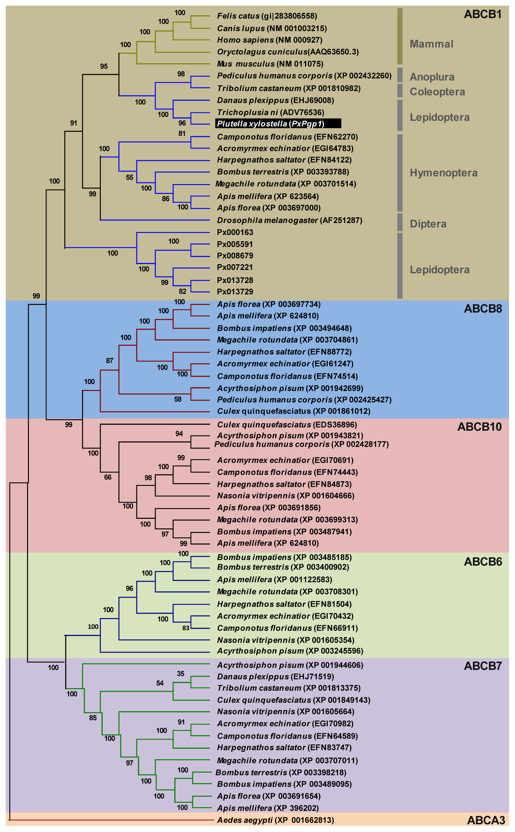
2.2. Phylogenetic Relationship of PxPgp1 with Other Insect Pgps

Phylogenetic analysis clustered PxPgp1 with ABCB1 genes from P. xylostella genome, other insect species and mammal Pgps from the ABCB subfamily, within the ABCB1 subgroup (Figure 2). Among them, PxPgp1 had the highest sequence similarity with two other lepidopteran species, the cabbage looper, T. ni (74%) and the monarch butterfly, D. plexippus (71%), which are substantially higher than its similarity with Pgps from other insect orders, including 10 hymenopteran insects (44%–48%), the human body louse, Pediculus humanus corporis (49%) and the red flour beetle, T. castaneum (51%).

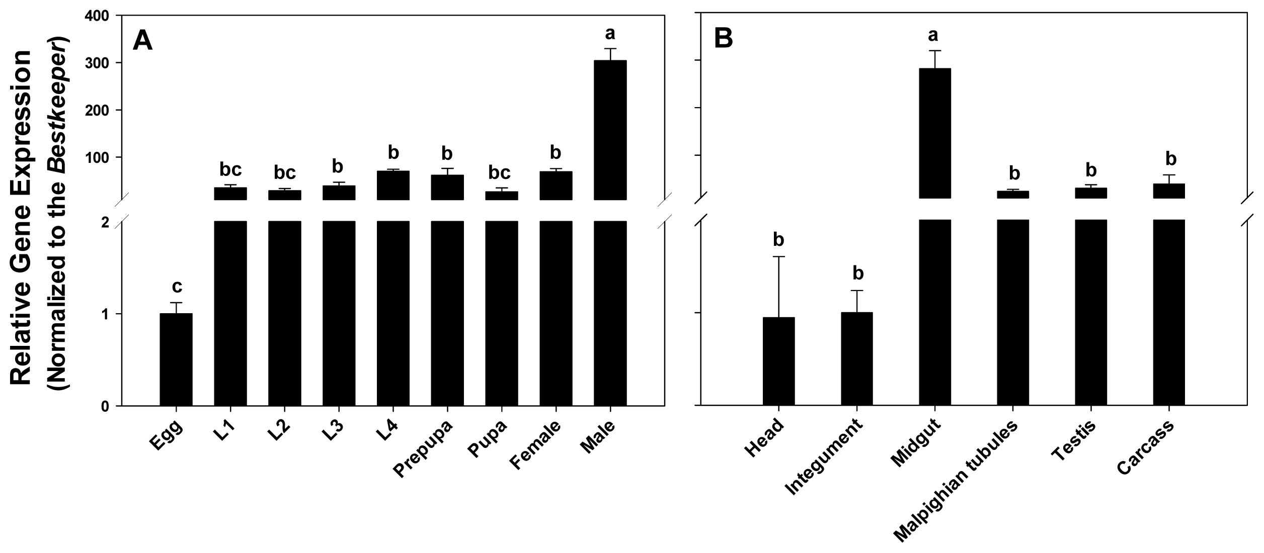
2.3. Expression Profiling of PxPgp1 in Different Developmental Stages and Tissues

PxPgp1 was constantly expressed during the entire life cycle of P. xylostella and the expression level increased continually during the larval stage, which was consistent with the expression profile in Heliothis virescens [42]. Similar to the expression pattern of an orthologue in the salmon louse L. salmonis [41], PxPgp1 in adult males were significantly higher than that in other developmental stages (ANOVA and Tukey’s, p < 0.05), which were 7.82, 4.33, 4.94 and 4.35 times higher than that in the third-instar larvae, fourth-instar larvae, prepupae and adult females, respectively (Figure 3A).
The relative expression level of PxPgp1 were highest in the midgut (p < 0.05), which was about 11.83-, 9.37-, and 7.24-fold higher than that in the malpighian tubules, testes and carcass, respectively (Figure 3B). PxPgp1 expressions in the head and integument were equally low. This is consistent with the other lepidopteran, the cabbage looper, T. ni, in which Pgp was most abundant in the midgut and followed by the malpighian tubules [54]. Pgp was also highly expressed in the malpighian tubules of Manduca sexta larvae [55]. Two Pgp genes in D. melanogaster, mdr49 and mdr65, were located in the brain and gut tissues, respectively [56]. Spatial distribution of these multidrug resistant proteins limits xenobiotic absorption and decreases the penetration of xenobiotics from the systemic circulation, thus protecting vital structures such as the brain or testes against toxins [57,58].

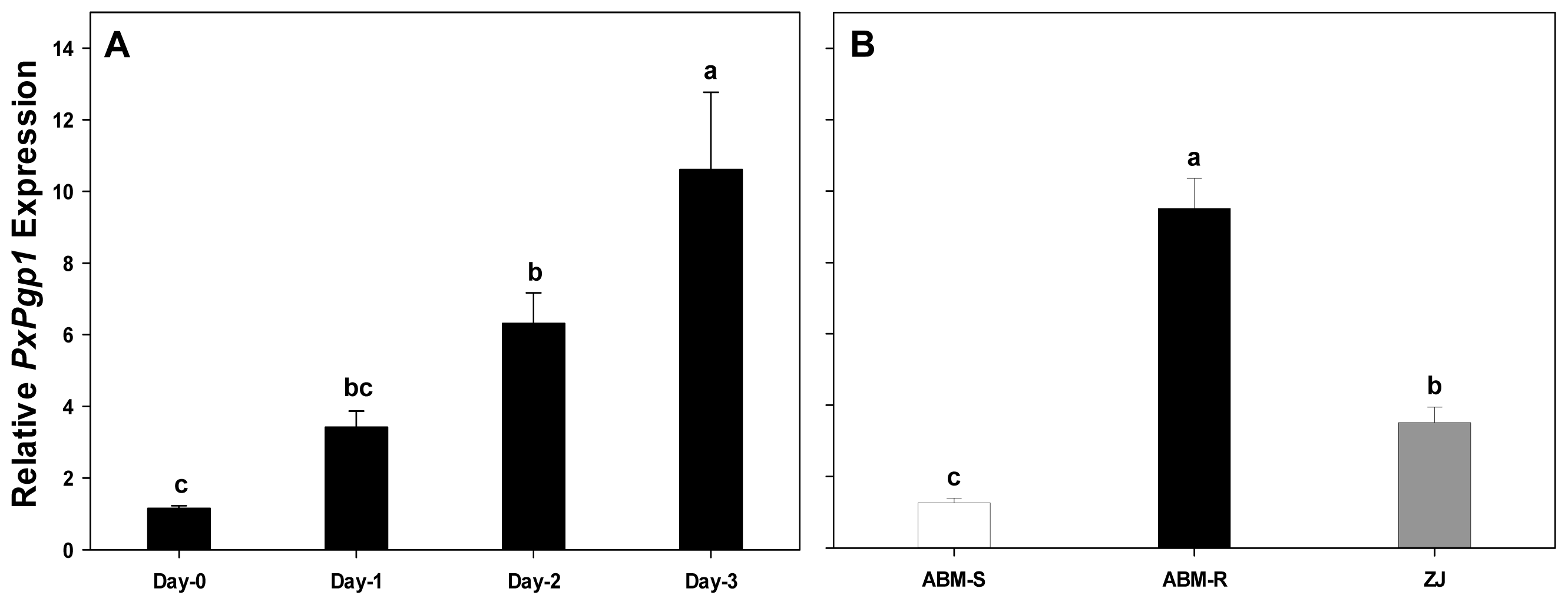
2.4. Transcriptional Response of PxPgp1 to Abamectin Exposure

2.4.1. Acute Response of PxPgp1 to Abamectin Treatment

To investigate the acute transcriptional response of PxPgp1, third-instar larvae of P. xylostella were treated with abamectin at a concentration of LC50 of 25 μg/L (ABM-S, Table 1). The relative expression levels of PxPgp1 over a 3-day period (Day-1, -2 and -3) were 3.14-, 6.31- and 10.61-fold higher than Day-0 (Figure 4A).

2.4.2. Constitutive Expression of PxPgp1 in Abamectin-Resistant and -Susceptible Plutella xylostella

Two laboratory strains, ABM-R and ABM-S, and one field population, ZJ, were used to investigate the expression profiles of PxPgp1 in abamectin-resistant and -susceptible P. xylostella. In comparison to the susceptible ABM-S strain, the resistance ratio of ABM-R and ZJ were 217.7 and 23.1, respectively (Table 1). PxPgp1 expression levels in the first filial generation (F1) of ABM-R and ZJ were 9.51- and 3.51-fold higher than that in the susceptible strain, respectively (Figure 4B).
Drug transporters play an important role in drug resistance, usually showing elevated expression levels. In this study, mRNA expression levels in the susceptible strain were increased after exposure to abamectin in a dose (day)-dependent manner. The expression levels of PxPgp1 in susceptible individuals significantly increased when treated with abamectin at LC50. Furthermore, both laboratory-selected and field-collected abamectin-resistant and -susceptible strains showed distinct differences in their expression levels of PxPgp1. PxPgp1 was consistently expressed at higher levels in the abamectin-resistant strains, with or without exposure to the insecticide. This suggests that elevated expression of PxPgp1 is not only an instantaneous response of P. xylostella to abamectin exposure, but also an existing mechanism to cope with the insecticide challenge in the field. Elevated expression of Pgps in avermectin-resistant strains has been documented in many parasitic nematodes and insects. For example, increased expression of Pgp was found in an ivermectin-resistant strain of Haemonchus contortus [59]. James and Davey also reported that ivermectin resistance is associated with elevated expression of Pgps in Caenorhabditis elegans [45]. In thiodicarb-resistant tobacco budworm, H. virescens, Pgp expression was substantially higher than their susceptible counterpart [42]. The expression of a Pgp (mdr49) mRNA can be induced by colchicine, a toxic natural product and secondary metabolite, in D. melanogaster [56]. In recent years, Pgp-mediated multidrug resistance has been reversed by RNA interference (RNAi), a functional genomics tool, both in vivo and in vitro [6062].

3. Experimental Section

3.1. Plutella Xylostella Strains

Plutella xylostella strains, ABM-S and ABM-R, were originally collected in 1990 from a cabbage (Brassica sp.) field in Guangdong Province, China. The susceptible ABM-S strain had never been exposed to abamectin, while the resistant ABM-R strain was selected continuously with insecticide treatment. The larvae of P. xylostella were reared on Chinese cabbage leaves at 25 ± 1 °C, a relative humidity (RH) of 60%–70% and a photoperiod of 16:8 h (L:D). Adults were provisioned with a 10% honey solution [15]. ZJ strain was originally collected from Zhejiang Province, China.

3.2. Chemicals and Bioassay

Abamectin (containing 93% avermectin B1a and 7% avermectin B1b) was obtained from Department of Applied Chemistry, the China Agricultural University (CAU). Leaf residue bioassays were carried out on glass plates using the third-instar larvae. To establish the dose-response curve, pesticide-free cabbage leaves and different doses of abamectin were applied. Bioassays ran for 3 days, and the cumulative mortality was documented at the endpoint [63].

3.3. Molecular Cloning of Pgp

Total RNA was isolated from the fourth-instar larvae of P. xylostella using a Trizol kit (Invitrogen, Carlsbad, CA, USA) according to the manufacturer’s instruction. First-strand cDNA was synthesized using a PrimeScript II 1st strand cDNA synthesis kit with oligo dT primers (Takara Biotechnology, Dalian, China). Cloning was carried out in three steps integrating primer walking with the rapid amplification of cDNA ends (RACE) PCR. First, two sets of degenerate primers (primers 1 and 2 in Table 2) were used to generate the two conserved fragments, S1 and S2 (Figure S1). Then, a pair of gene specific primers was designed to amplify the gap (G1). The 3′- and 5′-terminus (fragments I and II) were obtained using a SMARTer RACE cDNA Amplification kit following the manufacturer’s protocol (Takara Biotechnology, Dalian, China). Finally, to validate the cloning result, a pair of gene specific primers covering the entire full-length cDNA sequence of Pgp was used to amplify a long fragment (PCR III) containing the putative open reading frame (Figure S1). Primer Premier 5.0 (Premier Biosoft International, Palo Alto, CA, USA) were used to design the above mentioned primers (Table 2). Amplified PCR products were cloned into a pEASY-T5 vector (Trans Gene Biotech, Beijing, China) and sequenced by Tsingke (Beijing, China). Full-length cDNAs of ten individuals including five clones from each P. xylostella were sequenced.

3.4. Bioinformatics Analysis

Sequences alignment was performed with DNAMAN 7.0 (Lynnon Biosoft, Quebec, Canada). The isoelectric point and molecular weight of the deduced protein were estimated using the ExPASy Proteomics Server [64,65]. SignalP 4.1 Server [66] was used to predict the signal peptide. The primary and secondary structures of PxPgp1 were resolved by TMpred [67] and conserved domains were detected by the NCBI sequence analysis tools [68]. The tertiary structure of PxPgp1 was simulated by SWISS-MODEL [69,70] using a readily available template from RCSB Protein Data Bank (PDB). PyMOL-v1.3r1 [71] was used to visualize the 3-D structure and to label important structural features. To investigate the phylogenetic relationship of PxPgp1 with Pgps from other insect species, a neighbor-joining tree was constructed [72]. The peptide sequences of ABC-B orthologs were extracted from the GenBank and P. xylostella genome [73]. Although 17 transcripts from P. xylostella genome were categorized into ABCB1, only 6 genes (Px007221, Px005591, Px013728, Px013729, Px008679, Px000163) containing complete ORF were selected for the subsequent phylogenetic analysis. Amino acid sequences were aligned using MEGA 5.05 [53] and ClustalW [74], and bootstrap values were calculated after 1000 replications [52].

3.5. Quantitative Real-Time RT-PCR

Relative expression levels of PxPgp1 in different developmental stages, in various tissues and in larvae treated with abamectin were examined using quantitative real-time reverse transcription polymerase chain reaction (qRT-PCR). For different developmental stages, eggs, first- to fourth-instar larvae, pre-pupa, pupae and 1-day-old adult female and males were collected. For tissue parts, the head, integument, midgut, malpighian tubules, testes and carcass of fourth-instar larvae were dissected in cold phosphate buffered saline (PBS; 137 mmol/L NaCl, 2.7 mmol/L KCl, 10 mmol/L Na2HPO4, 2 mmol/L KH2PO4) and washed three times in PBS. Tissues were sampled from 50 individuals for each biological replicate. All samples were snap frozen in liquid nitrogen before stored at −80 °C for the subsequent total RNA isolation. To investigate transcriptional response of PxPgp1, third-instar larvae from a susceptible strain were exposed to the median lethal concentration (LC50) of abamectin for 1, 2 or 3 days. Moreover, constitutive expression profiles of PxPgp1 in the abamectin-resistant and susceptible strains were compared using qRT-PCR analysis.
qRT-PCR was conducted using an ABI PRISM 7500 Real-time PCR System (Applied Biosystems, Foster, CA, USA). According to Fu (2013) [75], elongation factor 1 (EF1), ribosomal protein L32 (RPL32), ribosomal protein S23 (RPS23), ribosomal protein S13 (RPS13), and β-actin (ACTB) were selected as reference genes (Table 3). Specifically, EF1, RPL32 and RPS23 were used to normalize transcripts among developmental stages and various tissues. EF1, RPS13 and RPL32 were suited for evaluation of target gene expression after exposure to abamectin. Finally, relative gene expression levels between abamectin-resistant and susceptible strains were evaluated using EF1, ACTB and RPL32 as references. All qRT-PCR analyses were run in triplicate for both technical and biological replicates. The qRT-PCR was carried out in a 25 μL reaction containing 1.0 μL cDNA (200 ng/μL), 12.5 μL 2 × SuperReal PreMix Plus, 0.5 μL 50 × ROX Reference Dye, 0.75 μL forward primer (10 μM), 0.75 μL reverse primer (10 μM) and 9.5 μL RNase-free ddH2O, following the instructions of the SuperReal PreMix Plus (SYBR Green) kit (Tiangen, Beijing, China). The thermal cycling conditions were 15 min of polymerase activation at 95 °C, followed by 40 cycles of denaturation at 95 °C for 30 s, annealing at 58 °C for 30 s and elongation at 72 °C for 35 s. The amplification efficiency was estimated using the equation: E = [10^(−1/slope)−1] × 100%, where slope was derived from plotting the cycle threshold (Ct) value versus six serially diluted template concentrations. Quantification of transcript levels of the Pgp gene was conducted according to the 2−ΔΔCt method [76].

3.6. Statistical Analysis

The gene expression data were analyzed with ANOVA, and the means were separated by Tukey’s test for significance (p < 0.05) using SPSS 19.0 for Windows (SPSS Inc.: Chicago, IL, USA). The LC50 value was calculated using a Probit analysis software [77].

4. Conclusions

In this study, the full length cDNA of PxPgp1, a P-glycoprotein in P. xylostella, a devastating vegetable insect pest worldwide, has been cloned and characterized. As a full ABC transporter, phylogenetic analysis places PxPgp1 with other insect Pgps from the ABCB1 subgroup. Spatial and temporal mRNA expression profiling among different tissues and developmental stages suggest PxPgp1 is most abundant in midgut and is highly expressed in adult males. Exposure to abamectin, a macrocyclic lactone derivative with potent anthelmintic and insecticidal properties, significantly induced the expression of PxPgp1 in P. xylostella, implicating the involvement of PxPgp1 in the acute response to insecticide treatment. More importantly, the constitutive overexpression of PxPgp1 in the abamectin-resistant P. xylostella suggests the potential connection of PxPgp1 and abamectin resistance. However, future research involving RNAi-based functional characterization is warranted to establish a causal link between PxPgp1 and abamectin resistance.

Supplementary Information

ijms-14-22891-s001.pdf
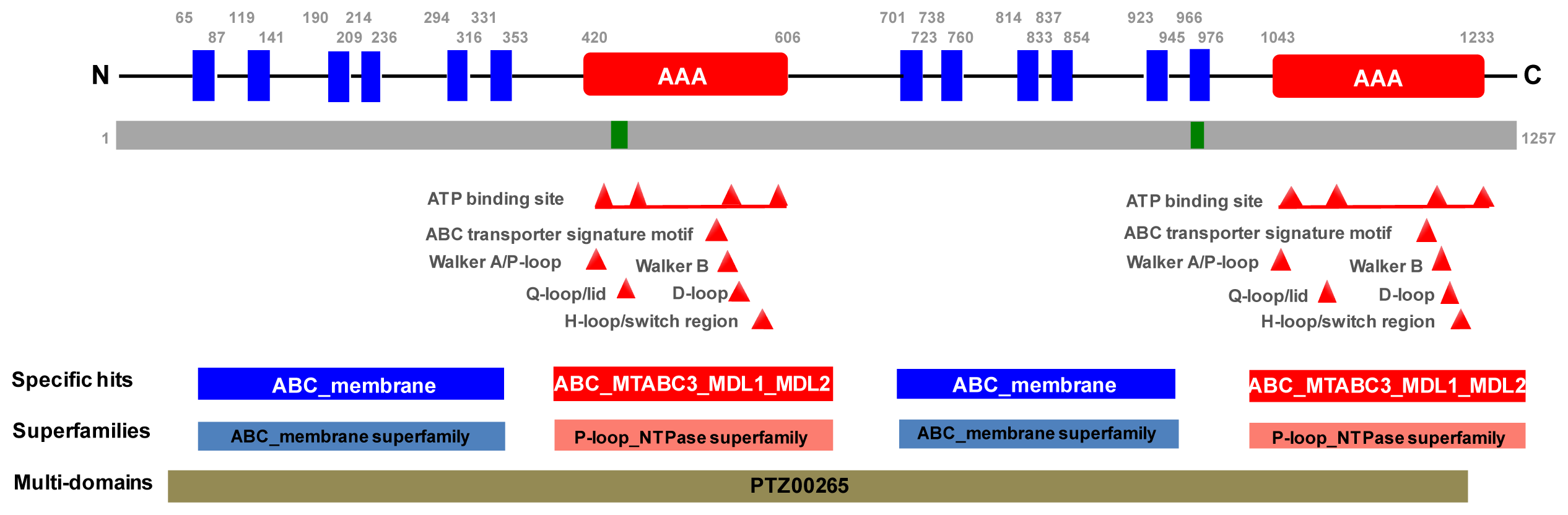
Figure 1. Schematic drawing of the primary and secondary structures of PxPgp1. TMpred and the conserved domain database from NCBI were used to construct this map. Signature motifs of the ABC superfamily are color-coded, including transmembrane domains (blue), a nucleotide domain (red) and regions with low complexity (green). PTZ00265 in grey denotes a multidrug-resistance protein (mdr1).
Ijms 14 22891f1
Figure 2. Phylogenetic relationship of PxPgp1 with other insect Pgps. The percentage of replicate trees in which the associated taxa clustered together in the bootstrap test (1000 replicates) is shown next to the branches [52]. Evolutionary analyses were conducted in MEGA5.05 [53].
Ijms 14 22891f2
Figure 3. Expression profiles of PxPgp1 in different developmental stages and tissues. Distribution of PxPgp1 in different developmental stages of P. xylostella is depicted in (A). The mRNA quantity is expressed relative to the egg stage. L1 to L4 denote the first- to fourth-instar larvae, respectively. The relative expression levels of Pgp in various tissues of fourth-instar larvae are shown in (B). Data are presented as mean ± SE for three independent replicates. Letters denote levels of statistical significance, according to Tukey’s test (p < 0.05).
Ijms 14 22891f3
Figure 4. Transcriptional response of PxPgp1 to abamectin. Relative gene expressions of PxPgp1 after treatment with abamectin are shown in (A). The constitutive expressions of PxPgp1 in abamectin-resistant and -susceptible Plutella xylostella is shown in (B). ABM-R and ABM-S represent laboratory-selected abamectin resistant and susceptible P. xylostella strains, respectively, and ZJ is field strain collected from Zhejiang Province, China. Data are presented as means ± SE. Letters denote levels of statistical significance in expression levels according to Tukey’s test (p < 0.05).
Ijms 14 22891f4
Table 1. Susceptibility of Plutella xylostella strains to abamectin treatment.
StrainLC50 (95% FL) (mg/L−1)Slope (±SE)RR a
ABM-S0.0025 (0.0015–0.042)1.651 (±0.218)1
ABM-R5.442 (3.487–8.493)1.882 (±0.252)217.68
ZJ0.557 (0.242–0.915)1.682 (±0.221)23.08
aRR = LC50 (strain)/LC50 (ABM-S);
RR, resistance ratio; LC50, median lethal concentration; FL, 95% Fiducial limits; SE, standard error.
Table 2. Location and sequence of primers used for the molecular cloning of PxPgp1.
PrimerPositionSequence (5′-3′)Length (bp)
11060
1566
F: TAYGCTCTKGCMTTCTGG
R: GGYTCCTGACYCACSASRCC
506
23289
3722
F: AGYGGCTGYGGVAAGAGYAC
R: CTTGVACMACCTTTTCACTTTC
433
31429
3309
F: CGGCAAGTCGACCATCATAC
R: GTACTCTTCCCGCAGCCGC
1880
GSP13645GCTGAGACGACCGAAGATGCTCCTAC520
GSP21273TAGCAGGGGGTTGATGGACGGCAC1273
Table 3. Primers used for qRT-PCR.
GeneAccession numberPrimer sequence (5′-3′)
ACTBAB282645F: TGGGTATGGAATCTTGCGG
R: GGACATGACGGTGTTGGCG
EF1EF417849F: GCCTCCCTACAGCGAATC
R: CCTTGAACCAGGGCATCT
GAPDHAJ489521F: GCCACCACTGCCACTC
R: CGGGACGGGAACACG
RPL32AB180441F: CCAATTTACCGCCCTACC
R: TACCCTGTTGTCAATACCTCT
RPS13AY174891F: TCAGGCTTATTCTCGTCG
R: GCTGTGCTGGATTCGTAC
RPS23AB180672F: ATGGGCTGACAAGGATTAC
R: TGCGGATGGCAGAGTT
PxPgp1F: GGAATAGTGGCACAAGATGG
R: TCTGGAGTCCTGGAGTTATCG

Acknowledgments

This work was supported by grants from the Natural Science Foundation of China (31171876 and 31071709) and the Special Fund for Agro-scientific Research in the Public Interest (201103021).

Conflicts of Interest

The authors declare no conflict of interest.

References

  1. Talekar, N.S.; Shelton, A.M. Biology, ecology, and management of the diamondback moth. Annu. Rev. Entomol 1993, 38, 275–301. [Google Scholar]
  2. Zalucki, M.P.; Shabbir, A.; Silva, R.; Adamson, D.; Liu, S.S.; Furlong, M.J. Estimating the economic cost of one of the world’s major insect pests, Plutella xylostella: Just how long is a piece of string? J. Econ. Entomol 2012, 105, 1115–1129. [Google Scholar]
  3. Furlong, M.J.; Wright, D.J.; Dosdall, L.M. Diamondback moth ecology and management: Problems, progress and prospects. Annu. Rev. Entomol 2013, 58, 517–541. [Google Scholar]
  4. Iqbal, M.; Verkerk, R.H.J.; Furlong, M.J.; Ong, P.C.; Rahman, S.A.; Wright, D.J. Evidence for resistance to Bacillus thuringiensis (Bt) subsp. kurstaki HD-1, Bt subsp. aizawai and abamectin in field populations of Plutella xylostella from Malaysia. Pestic. Sci 1996, 48, 89–97. [Google Scholar]
  5. Pu, X.; Yang, Y.H.; Wu, S.W.; Wu, Y.D. Characterisation of abamectin resistance in a field-evolved multiresistant population of Plutella xylostella. Pest Manag. Sci 2010, 66, 371–378. [Google Scholar]
  6. Liang, P.; Gao, X.W.; Zheng, B. Genetic basis of resistance and studies on cross-resistance in a population of diamondback moth, Plutella xylostella (Lepidoptera: Plutellidae). Pest Manag. Sci 2003, 59, 1232–1236. [Google Scholar]
  7. Qian, L.; Cao, G.C.; Song, J.X.; Yin, Q.; Han, Z. Biochemical mechanisms conferring cross-resistance between tebufenozide and abamectin in Plutella xylostella. Pestic. Biochem. Physiol 2008, 91, 175–179. [Google Scholar]
  8. Sarfraz, M.; Keddie, B.A. Conserving the efficacy of insecticides against Plutella xylostella (L.) (Lep., Plutellidae). J. Appl. Enotomol 2005, 129, 149–157. [Google Scholar]
  9. Huang, H.S.; Hu, N.T.; Yao, Y.E.; Wu, C.Y.; Chiang, S.W.; Sun, C.N. Molecular cloning and heterologous expression of a glutathione S-transferase involved in insecticide resistance from the diamondback moth Plutella xylostella. Insect Biochem. Mol. Biol 1998, 28, 651–658. [Google Scholar]
  10. Wu, Q.J.; Zhang, W.J.; Zhang, Y.J.; Xu, B.Y.; Zhu, G.R. Cuticular penetration and desensitivity of GABAA receptor in abamectin resistant Plutella xylostella. L. Acta Entomol. Sin 2002, 45, 336–340. (in Chinese). [Google Scholar]
  11. Perry, T.; Batterham, P.; Daborn, P.J. The biology of insecticidal activity and resistance. Insect Biochem. Mol. Biol 2011, 41, 411–422. [Google Scholar]
  12. Ludmerer, S.W.; Warren, V.A.; Williams, B.S.; Zheng, Y.C.; Hunt, D.C.; Ayer, M.B.; Wallace, M.A.; Chaudhary, A.G.; Egan, M.A.; Meinke, P.T.; et al. Ivermectin and nodulisporic acid receptors in Drosophila melanogaster contain both γ-aminobutyric acid-gated rdl and glutamate-gated GluClα chloride channel subunits. Biochemistry 2002, 41, 6548–6560. [Google Scholar]
  13. Ffrench-Constant, R.H.; Daborn, P.J.; Goff, G.L. The genetics and genomics of insecticide resistance. Trends Genet 2004, 20, 163–170. [Google Scholar]
  14. Wolstenholme, A.J.; Rogers, A.T. Glutamate-gated chloride channels and the mode of action of the abamectin/milbemycin anthelmintics. Parasitology 2005, 131, S85–S95. [Google Scholar]
  15. Zhou, X.M.; Wu, Q.J.; Zhang, Y.J.; Bai, L.Y.; Huang, X.Y. Cloning and characterization of a GABA receptor from Plutella xylostella (Lepidoptera: Plutellidae). J. Econ. Entomol 2008, 101, 1888–1896. [Google Scholar]
  16. Casida, J.E.; Durkin, K.A. Neuroactive insecticides: Targets, selectivity, resistance and secondary effects. Annu. Rev. Entomol 2013, 58, 99–117. [Google Scholar]
  17. Liu, S.M.; Zhou, S.; Tian, L.; Guo, E.E.; Luan, Y.X.; Zhang, J.Z.; Li, S. Genome-wide identification and characterization of ATP-binding cassette transporters in the silkworm Bombyx mori. BMC Genomics 2011, 12, 1471–2164. [Google Scholar]
  18. Xie, X.D.; Cheng, T.C.; Wang, G.H.; Duan, J.; Niu, W.H.; Xia, Q.Y. Genome-wide analysis of the ATP-binding cassette (ABC) transporter gene family in the silkworm Bombyx mori. Mol. Biol. Rep 2012, 39, 7281–7291. [Google Scholar]
  19. Labbé, R.; Caveney, S.; Donly, C. Genetic analysis of the xenobiotic resistance- associated ABC gene subfamilies of the Lepidoptera. Insect Mol. Biol 2011, 20, 243–256. [Google Scholar]
  20. Xie, W.; Lei, Y.Y.; Fu, W.; Yang, Z.X.; Zhu, X.; Guo, Z.J.; Wu, Q.J.; Wang, S.L.; Xu, B.Y.; Zhou, X.G.; et al. Tissue-specific transcriptome profiling of Plutella Xylostella third instar larval midgut. Int. J. Biol. Sci 2012, 8, 1142–1155. [Google Scholar]
  21. Baxter, S.W.; Badenes-Pérez, F.R.; Morrison, A.; Vogel, H.; Crickmore, N.; Kain, W.; Wang, P.; Heckel, D.G.; Jiggins, C.D. Parallel evolution of Bacillus thuringiensis toxin resistance in Lepidoptera. Genetics 2011, 189, 675–679. [Google Scholar]
  22. Dassa, E.; Bouige, P. The ABC of ABCs: A phylogenetic and functional classification of ABC systems in living organisms. Res. Microbiol 2001, 152, 211–229. [Google Scholar]
  23. Dean, M.; Hamon, Y.; Chimini, G. The human ATP-binding cassette (ABC) transporter superfamily. J. Lipid Res 2001, 42, 1007–1017. [Google Scholar]
  24. Holland, I.B.; Cole, S.P.C.; Kuchler, K.; Higgins, C.F. ABC Proteins: From Bacteria to Man; Academic Press: London, UK, 2003. [Google Scholar]
  25. Dean, M.; Annilo, T. Evolution of the ATP-binding cassette (ABC) transporter superfamily in vertebrates. Annu. Rev. Genomics Hum. Genet 2005, 6, 123–142. [Google Scholar]
  26. Sturm, A.; Cunningham, P.; Dean, M. The ABC transporter gene family of Daphnia pulex. BMC Genomics 2009, 10, 170–188. [Google Scholar]
  27. Sheps, J.A.; Ralph, S.; Zhao, Z.Y.; Baillie, D.L.; Ling, V. The ABC transporter gene family of Caenorhabditis elegans has implications for the evolutionary dynamics of the ABC transporter gene family multidrug resistance in eukaryotes. Genome Biol 2004, 5, R15:1–R15:17. [Google Scholar]
  28. Dermauw, W.; Osborne, E.J.; Clark, R.M.; Grbić, M.; Tirry, L.; Leeuwen, T.V. A burst of ABC genes in the genome of the polyphagous spider mite Tetranychus urticae. BMC Genomics 2013, 14, 317. [Google Scholar]
  29. Lespine, A.; Alvinerie, M.; Vercruysse, J.; Prichard, R.K.; Geldhof, P. ABC transporter modulation: A strategy to enhance the activity of macrocyclic lactone anthelmintics. Trends Parasitol 2008, 24, 293–298. [Google Scholar]
  30. Juliano, R.L.; Ling, V. A surface glycoprotein modulating drug permeability in Chinese hamster ovary cell mutants. BBA-Biomembranes 1976, 455, 152–162. [Google Scholar]
  31. Sharom, F.J. The P-glycoprotein multidrug transporter. Essays Biochem 2011, 50, 161–178. [Google Scholar]
  32. Gottesman, M.M.; Ling, V. The molecular basis of multidrug resistance in cancer: The early years of P-glycoprotein research. FEBS Lett 2006, 580, 998–1009. [Google Scholar]
  33. Aller, S.G.; Yu, J.; Ward, A.; Weng, Y.; Chittaboina, S.; Zhuo, R.; Harrell, P.M.; Trinh, Y.T.; Zhang, Q.H.; Urbatsch, I.L.; et al. Structure of P-glycoprotein reveals a molecular basis for poly-specific drug binding. Science 2009, 323, 1718–1722. [Google Scholar]
  34. Didier, A.; Loor, F. The abamectin derivative ivermectin is a potent P-glycoprotein inhibitor. Anti-Cancer Drugs 1996, 7, 745–751. [Google Scholar]
  35. Lespine, A.; Dupuy, J.; Orlowski, S.; Nagy, T.; Glavinas, H.; Krajcsi, P.; Alvinerie, M. Interaction of ivermectin with multidrug resistance proteins (MRP1, 2 and 3). Chem. Biol. Interact 2006, 159, 169–179. [Google Scholar]
  36. Lespine, A.; Martin, S.; Dupuy, J.; Roulet, A.; Pineay, T.; Orlowski, S.; Alvinerie, M. Interaction of macrocyclic lactones with P-glycoprotein: Structure–affinity relationship. Eur. J. Pharm. Sci 2007, 30, 84–94. [Google Scholar]
  37. Lespine, A.; Dupuy, J.; Alvinerie, M.; Comera, C.; Nagy, T.; Krajcsi, P.; Orlowski, S. Interaction of macrocyclic lactones with the multidrug transporters: The bases of the pharmacokinetics of lipid-like drugs. Curr. Drug Metab 2009, 10, 272–288. [Google Scholar]
  38. Aurade, R.; Jayalakshmi, S.K.; Sreeramulu, K. Stimulatory effect of insecticides on partially purified P-glycoprotein ATPase from the resistant pest Helicoverpa armigera. Biochem. Cell Biol 2006, 84, 1045–1050. [Google Scholar]
  39. Aurade, R.; Jayalakshmi, S.K.; Sreeramulu, K. P-glycoprotein ATPase from the resistant pest, Helicoverpa armigera: Purification, characterization and effect of various insecticides on its transport function. BBA-Biomembranes 2010, 1798, 1135–1143. [Google Scholar]
  40. Aurade, R.; Jayalakshmi, S.K.; Sreeramulu, K. Modulation of P-glycoprotein ATPase of Helicoverpa armigera by cholesterol: Effects on ATPase activity and interaction of insecticides. Arch. Insect Biochem 2012, 79, 47–60. [Google Scholar]
  41. Heumann, J.; Carmichael, S.; Bron, J.E.; Tildesley, A.; Sturm, A. Molecular cloning and characterisation of a novel P-glycoprotein in the salmon louse Lepeophtheirus salmonis. Comp. Biochem. Phys. C 2012, 155, 198–205. [Google Scholar]
  42. Lanning, C.L.; Fine, R.L.; Corcoran, J.J.; Ayad, H.M.; Rose, R.L.; Abou-Donia, M.B. Tobacco budworm P-glycoprotein: Biochemical characterization and its involvement in pesticide resistance. BBA-Gen. Subj 1996, 1291, 155–162. [Google Scholar]
  43. Srinivas, R.; Udikeri, S.S.; Jayalakshmi, S.K.; Sreeramulu, K. Identification of factors reponsible for insecticide resistance in Helicoverpa armigera. Comp. Biochem. Phys. C 2004, 137, 261–269. [Google Scholar]
  44. Huang, Y.J.; Prichard, R.K. Identification and stage-specific expression of two putative P-glycoprotein coding genes in Onchocerca volvulus. Mol. Biochem. Parasitol 1999, 102, 273–281. [Google Scholar]
  45. James, C.E.; Davey, M.W. Increased expression of ABC transport proteins is associated with ivermectin resistance in the model nematode Caenorhabditis elegans. Int. J. Parasitol 2009, 39, 213–220. [Google Scholar]
  46. Luo, L.; Sun, Y.J.; Wu, Y.J. Abamectin resistance in Drosophila is related to increased expression of P-glycoprotein via the dEGFR and dAkt pathways. Insect Biochem. Mol 2013, 43, 627–634. [Google Scholar]
  47. Proudfoot, N.J.; Brownlee, G.G. 3′ non-coding region sequences in eukaryotic messenger RNA. Nature 1976, 263, 211–214. [Google Scholar]
  48. Senior, A.E.; Bhagat, S. P-glycoprotein shows strong catalytic cooperativity between the two nucleotide sites. Biochemistry 1998, 37, 831–836. [Google Scholar]
  49. Locher, K.P. Structure and mechanism of ATP-binding cassette transporters. Philos. Trans. R. Soc. B 2009, 364, 239–245. [Google Scholar]
  50. Sauna, Z.E.; Ambudkar, S.V. About a switch: How P-glycoprotein (ABCB1) harnesses the energy of ATP binding and hydrolysis to do mechanical work. Mol. Cancer Ther 2007, 6, 13–23. [Google Scholar]
  51. Jin, M.S.; Oldham, M.L.; Zhang, Q.J.; Chen, J. Crystal structure of the multidrug transporter P-glycoprotein from Caenorhabditis elegans. Nature 2012, 490, 566–569. [Google Scholar]
  52. Felsenstein, J. Confidence limits on phylogenies: An approach using the bootstrap. Evolution 1985, 39, 783–791. [Google Scholar]
  53. Tamura, K.; Peterson, D.; Peterson, N.; Stecher, G.; Nei, M.; Kumar, S. MEGA5: Molecular evolutionary genetics analysis using maximum likelihood, evolutionary distance, and maximum parsimony methods. Mol. Biol. Evol 2011, 28, 2731–2739. [Google Scholar]
  54. Simmons, J.; D’Souza, O.; Rheault, M.; Donly, C. Multidrug resistance protein gene expression in Trichoplusia ni caterpillars. Insect Mol. Biol 2013, 22, 62–71. [Google Scholar]
  55. Gaertner, L.S.; Murray, C.L.; Morris, C.E. Transepithelial transport of nicotine and vinblastine in isolated malpighian tubules of the tobacco hornworm (Manduca sexta) suggests a P-glycoprotein-like mechanism. J. Exp. Biol 1998, 201, 2637–2645. [Google Scholar]
  56. Tapadia, M.G.; Lakhotia, S.C. Expression of mdr49 and mdr65 multidrug resistance genes in larval tissues of Drosophila melanogaster under normal and stress conditions. Cell Stress Chaperones 2005, 10, 7–11. [Google Scholar]
  57. Fromm, M.F. Importance of P-glycoprotein at blood-tissue barriers. Trends Pharmacol. Sci 2004, 25, 423–429. [Google Scholar]
  58. Hennessy, M.; Spiers, J.P. A primer on the mechanics of P-glycoprotein the multidrug transporter. Pharmacol. Res 2007, 55, 1–15. [Google Scholar]
  59. Xu, M.; Molento, M.; Blackhall, W.; Ribeiro, P.; Beech, R.; Prichard, R. Ivermectin resistance in nematodes may be caused by alteration of P-glycoprotein homolog. Mol. Biochem. Parasitol 1998, 91, 327–335. [Google Scholar]
  60. Stege, A.; Priebsch, A.; Nieth, C.; Lage, H. Stable and complete overcoming of MDR1/P-glycoprotein-mediated multidrug resistance in human gastric carcinoma cells by RNA interference. Cancer Gene Ther 2004, 11, 699–706. [Google Scholar]
  61. Pichler, A.; Zelcer, N.; Prior, J.L.; Kuil, A.J.; Piwnica-Worms, D. In vivo RNA interference-mediated ablation of MDR1 P-glycoprotein. Clin. Cancer Res 2005, 11, 4487–4494. [Google Scholar]
  62. Shi, Z.; Liang, Y.J.; Chen, Z.S.; Wang, X.W.; Wang, X.H.; Ding, Y.; Chen, L.M.; Yang, X.P.; Fu, L.W. Reversal of MDR1/P-glycoprotein-mediated multidrug resistance by vector-based RNA interference in vitro and in vivo. Cancer Biol. Ther. 2006, 5, 39–47. [Google Scholar]
  63. Tabashnik, B.E.; Cushing, N.L. Leaf residue vs. topical bioassays for assessing insecticide resistance in the diamondback moth, Plutella xylostella L. FAO Plant Prot. Bull 1987, 35, 11–14. [Google Scholar]
  64. Bjellqvist, B.; Hughes, G.J.; Pasquali, C.; Paquet, N.; Ravier, F.; Sanchez, J.-C.; Frutiger, S.; Hochstrasser, D.F. The focusing positions of polypeptides in immobilized pH gradients can be predicted from their amino acid sequences. Electrophoresis 1993, 14, 1023–1031. [Google Scholar]
  65. Bjellqvist, B.; Basse, B.; Olsen, E.; Celis, J.E. Reference points for comparisons of two-dimensional maps of proteins from different human cell types defined in a pH scale where isoelectric points correlate with polypeptide compositions. Electrophoresis 1994, 15, 529–539. [Google Scholar]
  66. Petersen, T.N.; Brunak, S.; Heijne, G.V.; Nielsen, H. SignalP 4.0: Discriminating signal peptides from transmembrane regions. Nat. Methods 2011, 8, 785–786. [Google Scholar]
  67. Hofmann, K.; Stoffel, W. Tmbase—A database of membrane spanning proteins segments. Biol. Chem. Hoppe-Seyler 1993, 374, 166. [Google Scholar]
  68. Marchler-Bauer, A.; Lu, S.; Anderson, J.B.; Chitsaz, F.; Derbyshire, M.K.; DeWeese-Scott, C.; Fong, J.H.; Geer, L.Y.; Geer, R.C.; Gonzales, N.R.; et al. CDD: A Conserved Domain Database for the functional annotation of proteins. Nucleic Acids Res 2011, 39, 225–229. [Google Scholar]
  69. Arnold, K.; Bordoli, L.; Kopp, J.; Schwede, T. The SWISS-MODEL Workspace: A web-based environment for protein structure homology modeling. Bioinformatics 2006, 22, 195–201. [Google Scholar]
  70. Kiefer, F.; Arnold, K.; Künzli, M.; Bordoli, L.; Schwede, T. The SWISS-MODEL Repository and associated resources. Nucleic Acids Res 2009, 37, D387–D392. [Google Scholar]
  71. The PyMOL Molecular Graphics System, Version 1.5.0.4; Schrödinger, LLC: New York, NY, USA, 2011.
  72. Saitou, N.; Nei, M. The neighbor-joining method: A new method for reconstructing phylogenetic trees. Mol. Biol. Evol 1987, 4, 406–425. [Google Scholar]
  73. You, M.; Yue, Z.; He, W.; Yang, X.; Yang, G.; Xie, M.; Zhan, D.; Baxter, S.W.; Vasseur, L.; Gurr, G.M.; et al. A heterozygous moth genome provides insights into herbivory and detoxification. Nat. Genet 2013, 45, 220–225. [Google Scholar] [Green Version]
  74. Thompson, J.D.; Higgins, D.G.; Gibson, T.J. CLUSTAL W: Improving the sensitivity of progressive multiple sequence alignment through sequence weighting, position-specific gap penalties and weight matrix choice. Nucleic Acids Res 1994, 22, 4673–4680. [Google Scholar]
  75. Fu, W.; Xie, W.; Zhang, Z.; Wang, S.L.; Wu, Q.J.; Liu, Y.; Zhou, X.M.; Zhou, X.G.; Zhang, Y.J. Exploring valid reference genes for quantitative real-time PCR analysis in Plutella xylostella (Lepidoptera: Plutellidae). Int. J. Biol. Sci 2013, 9, 792–802. [Google Scholar]
  76. Pfaffl, M.W. A new mathematical model for relative quantification in real-time RT-PCR. Nucleic Acids Res 2001, 29, e45. [Google Scholar]
  77. Finney, D.J. Probit Analysis, 3d ed; Cambridge University Press: London, UK, 1971. [Google Scholar]

