Article

Acidic Residue Glu199 Increases SUMOylation Level of Nuclear Hormone Receptor NR5A1

1
Department of Biomedical Sciences, Mercer University School of Medicine, Savannah, GA 31404, USA
2
Department of Genetics and Comprehensive Cancer Center, University of Alabama at Birmingham, Birmingham, AL 35294, USA
*
Author to whom correspondence should be addressed.
Int. J. Mol. Sci. 2013, 14(11), 22331-22345; https://doi.org/10.3390/ijms141122331
Submission received: 9 October 2013 / Revised: 1 November 2013 / Accepted: 5 November 2013 / Published: 13 November 2013
(This article belongs to the Special Issue Proteins and Protein-Ligand Interactions)

Abstract

:
Steroidogenic factor 1 (NR5A1/SF1) is a well-known master regulator in controlling adrenal and sexual development, as well as regulating numerous genes involved in adrenal and gonadal steroidogenesis. Several studies including ours have demonstrated that NR5A1 can be SUMOylated on lysine 194 (K194, the major site) and lysine 119 (K119, the minor site), and the cycle of SUMOylation regulates NR5A1’s transcriptional activity. An extended consensus negatively charged amino acid-dependent SUMOylation motif (NDSM) enhances the specificity of substrate modification by SUMO has been reported; however, the mechanism of NDSM for NR5A1 remains to be clarified. In this study, we investigated the functional significance of the acidic residue located downstream from the core consensus SUMO site of NR5A1. Here we report that E199A (glutamic acid was replaced with alanine) of NR5A1 reduced, but not completely abolished, its SUMOylation level. We next characterized the functional role of NR5A1 E199A on target gene expression and protein levels. We found that E199A alone, as well as combination with K194R, increased Mc2r and Cyp19a1 reporter activities. Moreover, E199A alone as well as combination with K194R enhanced NR5A1-mediated STAR protein levels in mouse adrenocortical cancer Y1 cells. We also observed that E199A increased interaction of NR5A1 with CDK7 and SRC1. Overall, we provide the evidence that the acidic residue (E199) located downstream from the core consensus SUMO site of NR5A1 is, at least in part, required for SUMOylation of NR5A1 and for its mediated target gene and protein expression.

1. Introduction

Steroidogenic factor 1 (NR5A1/SF1) is a nuclear receptor and plays a crucial role in the regulation of steroid hormone biosynthesis, as well as in the endocrine development and differentiation of both the gonads and adrenal glands [14]. NR5A1 targets a variety of genes, including AMH [5], CYP17A1 [6], CYP19A1 [7], DGKθ [8], GnRHR [9], GSTA3 [10], MC2R [11], and STAR [12] in humans. Regulation of these NR5A1-dependent genes mostly involves the arranged effort of NR5A1 with multiple transcription factors and cofactors with which it can coordinate and synergize, such as PITX1 [13], GATA4 [14], EGR1 [15], SOX9 [16], SREBP1 [17], and WT1 [18]. Several transcriptional co-activators, such as nuclear receptor coactivator 1 (SRC1/NCOA1) [19], cyclic AMP response element-binding protein (CREB)-binding protein/p300 [20], transcriptional intermediary factor 2 (TIF2) [21], and CTNNB1 (β-catenin) [22], have been reported to interact with NR5A1 and probably participate in NR5A1-mediated gene activation. On the other hand, transcriptional co-factors, such as DDX20 [23], nuclear receptor corepressor 1 (NCOR1) [24], and NR0B1 [25], appear to play an inhibitory role by restraining NR5A1 function. Recently, structural and functional analyses have revealed that phospholipids (such as phosphatidic acid) can functionally serve as NR5A1 ligands [26]. Recent extensive clinical studies have also found that NR5A1 is associated with birth defects and developmental disorders, such as adrenal agenesis and aplasia [27], androgen insensitivity syndrome [28], gonadal dysgenesis [29], hypospadias [30], anorchia with microphallus [31], and infertility [32]. Therefore, NR5A1 is not only critical for regulation of steroid hormone biosynthesis but also essential for endocrine organ/tissue development in adrenal glands and gonads.
Nuclear receptors are functionally regulated by post-translational modifications which are crucial for normal physiological functions in cells and efficient ways for the cells to respond to intra- and extra-cellular signals. Among post-translational modifications, the modification by small ubiquitin-related modifier (SUMO) family, a reversible modification used extensively as a regulatory mechanism in eukaryotic cells, has major effects on regulating and influencing diverse cellular pathways and processes, mainly in regulation of transcriptional activity [3337]. Four SUMO family members (SUMO1 to −4, ranging from 90 to 110 amino acids) are encoded by distinct genes in mammals. Functional heterogeneity study has shown that the closely-related (85% identity) SUMO2 and SUMO3 have approximately 46% identity to SUMO1 [38]. In contrast to SUMO1, SUMO2, and SUMO3 possess a clear consensus SUMOylation site in their N-terminal regions, suggesting that SUMO2 and SUMO3 are allowed to form poly-SUMO chains [39]. Functional studies have shown that cellular stress favors SUMO2 and SUMO3 conjugations, but not SUMO1 conjugation [38]. Currently, though SUMO4 polymorphism is associated with the susceptibility of type 1 and type 2 diabetes mellitus [40], the biological role of SUMO4 remains poorly understood. In spite of limited sequence identity, SUMO proteins share with ubiquitin a common three-dimensional structure and use a similar conjugation mechanism, an enzyme-controlled pathway. Importantly, covalent conjugation of proteins by SUMO is highly transient and reversible through action of the SENP family of proteases. Even though the three-dimensional structure and conjugation pathway of SUMO share similarities to those of ubiquitin, the biological functions of SUMOylation are much different from those of ubiquitination [41]. SUMOylation mainly prevents their ubiquitin-mediated proteasomal degradation [42]. Most importantly, SUMO modification of transcription factors and nuclear receptors has a strong impact on their regulation of transcription of genes [37,4346]. Several components of the SUMO pathway are also involved in regulation of transcription. Collectively, understanding the regulation of protein SUMOylation is important for various biological processes such as regulation of transcription and development of disorders.
Accumulated evidence indicates that post-translational modifications regulate NR5A1 functions. For example, SUMOylation at K194 inhibits and phosphorylation at S203 activates NR5A1 [47,48]. We, and others, have demonstrated that SUMOylation inhibits NR5A1 activity by reducing phosphorylation at S203 [37,49], suggesting the importance of interplay between SUMOylation and phosphorylation. Recent studies on SUMO substrates also suggest that SUMOylation is regulated by the acidic residues located downstream from the core consensus SUMO site of the target proteins [50]. However, the mechanism of the consensus negatively charged amino acid-dependent SUMOylation motif (NDSM) on NR5A1 remains to be clarified. In the present work, we demonstrate that the acidic residue (E199) located downstream from the core consensus SUMO site of NR5A1 is, at least in part, required for SUMOylation of NR5A1 and for NR5A1-mediated target gene and protein expression.

2. Results

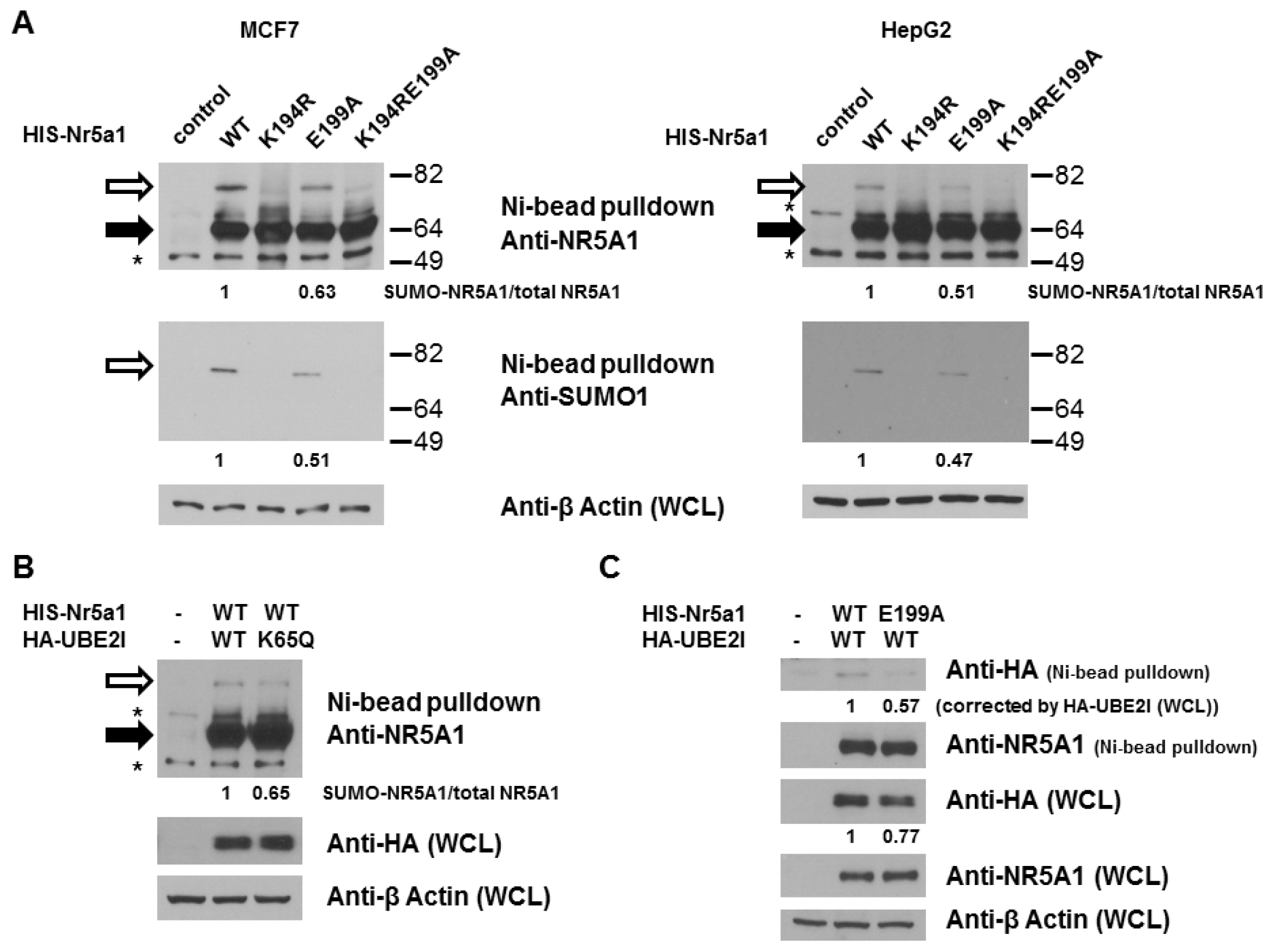
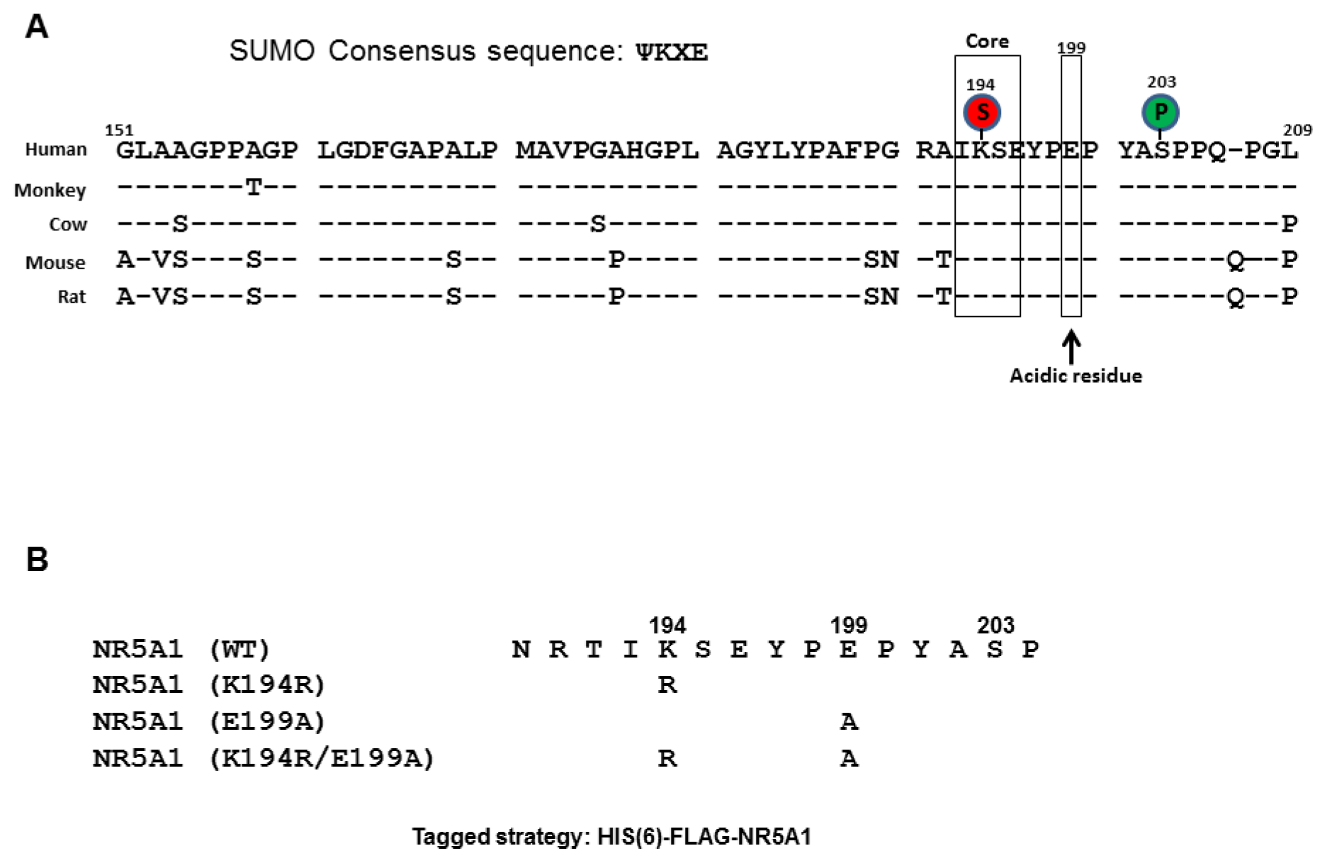
2.1. E199A Reduces NR5A1 SUMOylation

NR5A1 harbors two evolutionarily conserved sequences that conform to the SUMOylation consensus: K194, the major SUMO site, and K119, the minor SUMO site. NR5A1 activity is mainly dependent on its phosphorylation at S203. We, and others, have demonstrated that SUMOylation inhibits NR5A1 activity by reducing phosphorylation at S203 [37,49], suggesting the importance of interplay between phosphorylation and SUMOylation. As NDSM has been shown to play an important role in regulation of SUMOylation and NR5A1 has one acidic residue, E199 (conserved among mammalian species), downstream from the consensus SUMO (K194) site (Figure 1A), we decided to examine the role of E199 on SUMO modification of NR5A1. To facilitate the analysis of NR5A1 SUMOylation, we created HIS-FLAG-tagged mutant forms of NR5A1 in which the acceptor K194 was replaced with arginine and E199 was replaced with alanine (Figure 1B). Importantly, these mutant forms of NR5A1 can be readily isolated and distinguished by virtue of the associated HIS and FLAG tags. To determine the role of E199 on NR5A1 SUMOylation, we probed NR5A1 preparations isolated from MCF7 and HepG2 cells by Ni2+ chelate chromatography under denaturing condition. As shown in Figure 2A, a slowly migrating species (about 75 kDa) was detected in cells expressing WT NR5A1. We interpret this form as being NR5A1 modified by endogenous SUMO1. Moreover, this species is not observed in cells expressing an NR5A1 form with a mutation in SUMOylation motif (K194R). This result confirms the previous studies that K194 is the major SUMO site for NR5A1 [37,47,49,51]. Interestingly, disruption of the downstream acidic residue (E199A) produced a significant reduction (37%–53% reduction compared to WT NR5A1) in NR5A1 SUMOylation. As expected, mutations of both K194 and E1991 led to a complete loss of detectable SUMOylation. Taken together, these results indicate that the acidic residue, E199, downstream from the core consensus K194 SUMO site is important in enhancing the efficiency of NR5A1 SUMOylation. A previous report has shown that UBE2I acetylation attenuates UBE2I binding to NDSM substrates, causing a reduction in NDSM substrate SUMOylation [52]. Thus, we next examined whether UBE2I acetylation is involved in NR5A1 SUMOylation. As shown in Figure 2B, a reduced SUMOylation level of NR5A1 was observed in cells expressing UBE2I K65Q, an UBE2I acetylation-mimic mutant. As the interaction between UBE2I and SUMO substrate is important for substrate SUMOylation, we next tested whether E199A mutant attenuates UBE2I binding. As shown in Figure 2C, a reduced UBE2I interaction was observed in cells expressing NR5A1 E199A mutant. These data suggest that acetylation of UBE2I at K65 reduces its interaction with NR5A1 and E199 residue is important in enhancing the efficiency of NR5A1 SUMOylation by binding to UBE2I.

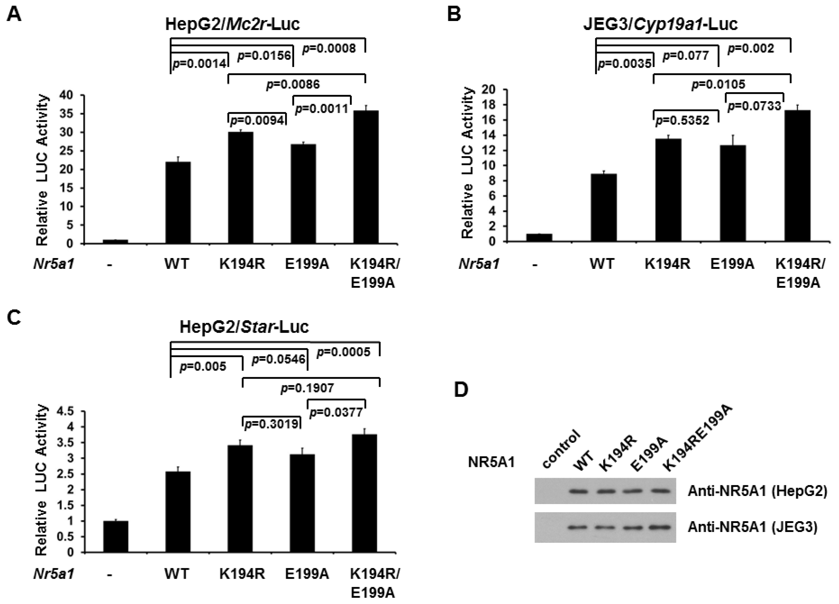
2.2. E199A Increases NR5A1 Transcriptional Activity

The robust steroid hormone biosynthesis depends upon the expression of a battery of genes encoding multiple enzymes involved in steroidogenesis. As NR5A1 is a master regulator of the expression of many such genes involved in steroidogenesis, we next investigated the role of E199A NR5A1 mutant on Mc2r and Cyp19a1 gene transcription by reporter gene assays. As shown in Figure 3A (HepG2 cells) and Figure 3B (JEG3 cells), expression of WT NR5A1 leads to a robust increase in the activity of a Mc2r and Cyp19a1 promoter-driven luciferase reporter, respectively. As expected, disruption SUMO modification of NR5A1 (K194R) further increased the activity of Mc2r and Cyp19a1 promoter-driven luciferase reporter, suggesting that SUMOylation of NR5A1 reduces its transcriptional activity. Interestingly, we observed that removal of acidic residue (E199A) downstream from the K194 SUMO site of NR5A1 also increased the activity of Mc2r and Cyp19a1 promoter-driven luciferase reporter, suggesting that acidic residue located downstream from the SUMO site is essential for determining the efficiency of NR5A1 SUMOylation and hence transcriptional regulatory properties. We also observed the similar results on Star promoter activity in HepG2 cells (Figure 3C). The expression levels of WT NR5A1 and NR5A1 mutants in HepG2 and JEG3 cells from the reporter assays were validated in Figure 3D. These findings indicate that the acidic residue (E199) located downstream from the core consensus K194 SUMO site of NR5A1 is, at least in part, required for SUMOylation of NR5A1 and for NR5A1-mediated target gene expression.

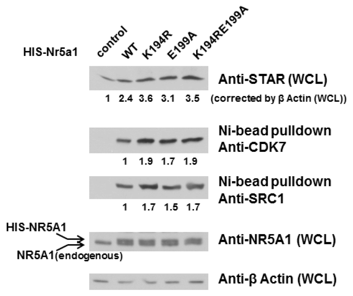
2.3. E199A Increases NR5A1’s Interaction with CDK7 and SRC1 as well as STAR Protein Level

We and others have previously shown that NR5A1 S203 phosphorylation is mediated by CDK7. Since CDK7 interacts with NR5A1 and CDK7 inhibitors block both phosphorylation of NR5A1 and its transactivation capacity, we next investigated whether E199A alters the interaction of NR5A1 and CDK7. We therefore used Y1 cells expressing WT or mutant NR5A1 to examine NR5A1-associated proteins. As shown in Figure 4, NR5A1 preparations isolated via Ni2+ chelate chromatography under nondenaturing condition contained larger amount of associated CDK7 in the case of the mutant NR5A1 than in that of WT NR5A1 (1.9- and 1.7-fold for the K194R and E199A forms of NR5A1, respectively). However, the combination of K194R and E199A did not further increase the interaction of NR5A1 and CDK7. We also detected increased recovery of the coactivator SRC1 related to WT NR5A1 (1.7- and 1.5-fold for the K194R and E199A forms of NR5A1, respectively). However, again, the combination of K194R and E199A did not further increase the interaction of NR5A1 and SRC1. This indicates that loss of acidic residue downstream from the K194 SUMO site of NR5A1 facilitates its interaction with CDK7 and SRC1. As the expression of STAR protein, the rate-limiting factor in the steroidogenesis pathway, is directly regulated by NR5A1 in adrenal glands and gonads, we next examined whether loss of acidic residue resulted in greater expression of STAR proteins. As depicted in Figure 4, as expected and consistent with the previous reports [37], STAR protein levels were indeed increased in K194R NR5A1 expressing Y1 cells. Interestingly, STAR protein levels were also increased in E199A NR5A1 expressing Y1 cells, suggesting that loss of acidic residue not only reduces NR5A1 SUMOylation but also enhances the expression levels (promoter activities and protein levels) of the enzymes involved in steroidogenesis. Taken together, these results provide evidence that the acidic residue downstream from the core K194 SUMO site of NR5A1 is essential, at least in part, for NR5A1 SUMOylation and interaction with others.

3. Discussion

Regulation of protein function by reversible post-translational modifications is the core principle in cell biology and biochemistry. Modifications with SUMO family of proteins have emerged as essential and critical events in a variety of biological processes, including apoptosis, cell cycle regulation, cell growth and differentiation, genomic instability, transcriptional regulation, tumorigensis, and protein-protein interactions. SUMO modification of NR5A1 has a strong influence on its transcriptional activity as evidenced by that NR5A1 transcriptional activity is intensively inhibited by SUMOylation [37,47,51]. Though an extended NDSM enhances the specificity of substrate modification by SUMO has been reported previously [50]; however, the functional significance of NDSM for NR5A1 remains to be clarified. In the present work, we demonstrate that the acidic residue downstream from the core SUMO consensus motif of NR5A1 is essential, at least in part, for NR5A1 SUMOylation and NR5A1-mediated transcriptional activity.
While hundreds of SUMO-conjugated substrates have been identified, the precise mechanisms that differentially regulate substrate SUMOylation are still largely unknown. We also know little about the mechanisms how a specific lysine residue in a SUMO-conjugated substrate which has more than one SUMO site is being preferentially attached by SUMO. The majority of discovered SUMO substrates are recognized by SUMO E2 conjugase UBE2I (also called UBC9) to a consensus motif ΨKxE (where Ψ stands for a large hydrophobic amino acid, such as V, I, L, and F). A recent study on site-specific identification of SUMO2 targets revealed that 76 out of 103 SUMO2 acceptor lysines in endogenous target proteins are situated in the SUMOylation consensus site (approximately 76%) [53]. Moreover, several lines of evidence recently suggest that amino acids upstream or downstream from the SUMOylation consensus tetrapeptide also facilitate substrate SUMOylation. For example, phosphorylation-dependent SUMOylation motif (PDSM) and negatively charged amino acid-dependent SUMOylation motif (NDSM) contain additional phosphorylated and negatively charged amino acids sequences downstream to the consensus motif, respectively. Another example is that a hydrophobic cluster SUMOylation motif (HCSM) contains a series of hydrophobic amino acids (such as V or I) upstream of the consensus motif [53]. In NDSM’s case, the negatively charged amino acids (acidic residues) downstream from the consensus motif are thought to dramatically enhance UBE2I interaction (by binding to a basic patch on UBE2I) and subsequently increase SUMOylation process. We have previously shown that while phosphorylation does not influences its SUMOylation, NR5A1 SUMOylation reduces it phosphorylation by reducing CDK7 interaction [37], suggesting that PDSM is not participated in NR5A1 SUMOylation. Several lines of evidence have demonstrated that numerous UBE2I-interacting proteins are SUMOylated including NR5A1 [54,55], suggesting NDSM and/or HCSM might be present in NR5A1. While we could not find HCSM in NR5A1 based on its protein sequence, we indeed found E199 downstream from the core K194 SUMO site in NR5A1. In the present study, we demonstrated that E199 is essential for NR5A1 SUMOylation because mutation of E199 reduces NR5A1’s SUMOylation (Figure 2), suggesting its role in enhancing UBE2I interaction and subsequently facilitating NR5A1 SUMOylation (Figure 2B,C). Since SUMOylation of NR5A1 reduces its transcriptional activity on target genes, the results of the reporter gene assays from the current study (Figure 3) further support that E199 is involved and essential, at least in part, for NR5A1 SUMOylation, demonstrating that the importance of acidic residues downstream the core consensus SUMO site on NR5A1.
NR5A1-mediated gene transcription is essential for steroidogenesis as well as adrenal and gonadal development and differentiation. For example, binding of cofactors such as SRC1 and CBP to NR5A1 is necessary to fully activate NR5A1-mediated CYP17 transcription [56]. Previously, we have shown that CDK7 and SRC1 bind preferentially to the SUMOylation-deficient form of NR5A1 and that CDK inhibition reduces NR5A1 phosphorylation [37], suggesting that SUMOylation inhibits NR5A1 activity by reducing CDK7-mediated phosphorylation of NR5A1. In current study, we observed that removal of acidic residue (E199A) not only reduces its SUMOylation but also increases NR5A1’s binding to SRC1 and CDK7, suggesting that E199 plays an important role in facilitating NR5A1 SUMOylation. Collectively, our results not only extend the conclusion with the previous NDSM report that acidic residues located downstream from the consensus SUMO conjugation site increase substrate SUMOylation possibly by enhancing UBE2I interaction but also provide the novel mechanism that how NDSM regulates NR5A1 activity by influencing its SUMOylation.

4. Experimental Section

4.1. Reagents

All cell culture reagents were purchased from Invitrogen (Carlsbad, CA, USA). Protein A and protein G magnetic beads were purchased from Fisher Scientific (Pittsburgh, PA, USA). Antibodies against NR5A1 (Upstate Biochemistry Inc., Charlottesville, VA, USA), STAR, CDK7, HA, and SRC1 (Santa Cruz Biotechnology Inc., Santa Cruz, CA, USA), SUMO1 (Active motif, Carsbad, CA, USA), β-Actin (Sigma, St. Louis, MO, USA) were purchased commercially. Luciferase activity was measured using the Dual Luciferase Assay System (Promega, Madison, WI, USA). Ni-NTA agarose was purchased from QIAGEN (Valencia, CA, USA).

4.2. DNA Constructs

Mouse Nr5a1-pcDNA3.1(+) expression plasmid, mouse mc2r promoter-LUC plasmid, and mouse Star promoter-LUC plasmid were previously established in our laboratory as described in Yang et al. [57]. K194R Nr5a1, E199A Nr5a1, and K194RE199A Nr5a1 expression plasmids were generated by PCR-based mutagenesis (QuikChange Lightning site-directed mutagenesis kit, Strategene, La Jolla, CA, USA). The mouse Cyp19a1 promoter (~600-bp) was PCR amplified by using forward primer 5′-ACGAGGTACCGTCCTAGTTCTAG-3′ and reverse primer 5′-TCGTAAGCTTGTTCTATGGGAAG-3′, digested with KpnI and HindIII and then ligated into the KpnI and HindIII sites of pGL3 to create Cyp19a1 LUC plasmid. Human HA-tagged UBE2I-pcDNA3.1(+) expression plasmid was previously established in our laboratory as described in Wang et al. [35]. K65Q HA-UBE2I expression plasmid was generated by PCR-based mutagenesis (QuikChange Lightning site-directed mutagenesis kit, Strategene, La Jolla, CA, USA). All constructs were verified by nucleotide sequencing.

4.3. Cell Culture and Transfection

HepG2, JEG3, MCF7, and Y1 cells were purchased from the American Type Culture Collection. HepG2, MCF7, and JEG3 cells were maintained in Dulbecco’s modified Eagle’s medium (DMEM) in the presence of 10% fetal bovine serum and antibiotics (GIBCO/Life Technologies, Grand Island, NY, USA) in humidified air containing 5% CO2, at 37 °C. Y1 cells were maintained in DMEM supplemented with 7.5% horse serum and 2.5% fetal bovine serum and antibiotics in humidified air containing 5% CO2, at 37 °C. After incubation, the cells were transfected using Fugene HD Transfection Reagent (Roche, Madison, WI, USA). Approximately 45−48 h after transfection, the cells were harvested. Luciferase activity was measured and normalized with Renilla activity. All experiments were performed three times in triplicate.

4.4. Immunoprecipitation Assay

MCF7, HepG2 or Y1 cells (2 × 10⁶) were seeded onto 10-cm plates. Twenty-four hours after transient transfection, cells were harvested and lysed in lysis buffer (40 mM HEPES, 120 mM sodium chloride, 10 mM sodium pyrophosphate, 10 mM sodium glycerophosphate, 1 mM EDTA, 50 mM sodium fluoride, 0.5 mM sodium orthovanadate, 1% Triton X-100) containing protease inhibitor cocktail (Sigma), followed by rotation for 1 h at 4 °C to solubilize proteins. Soluble protein was collected and immunoprecipitated with the indicated antibody overnight. Protein A or G magnetic beads were added to protein lysates for 2 h in the cold room. Beads were separated from lysate solution by magnetic force (in a magnetic separation rack) and washed at least three times with lysis buffer. For Ni2+-bead pull-down assays, Ni2+-NTA agarose was used to precipitate HIS-tagged NR5A1 from cell lysates. Proteins were eluted by boiling in 50 μL of 2× Laemmli sample buffer, resolved by 8% SDS-PAGE, and processed for immunoblotting as described below.

4.5. Immunoblotting

Protein lysates were allowed to rotate at 4 °C for 30 min, and protein contents of the high-speed supernatant were determined using the BCA™ Protein Assay kit assay (Pierce/Thermo Scientific, Rockford, IL, USA). Equivalent quantities of protein (20–40 μg) were resolved on polyacrylamide-SDS gels, transferred to nitrocellulose membrane (Bio-Rad, Hercules, CA, USA), and immunoblotted with specific antibodies. Results were visualized using the Supersignal West Dura Extended Duration Substrate kit (Pierce Chemical Co., Rockford, IL, USA). Band intensity was quantified by ImageJ program.

4.6. In Vivo SUMOylation Assays

The in vivo SUMOylation assay was carried out as previously described [37]. Briefly, MCF7 or HepG2 cells (2 × 10⁶) were seeded in 10 cm plates and 24 h later were transfected with indicated HIS-FLAG-Nr5a1 expression vectors. After 48 h, cells were harvested in 700 μL lysis buffer (500 mM NaCl, 10 mM imidazole, 45 mM Na2HPO4, 5 mM Na2H2PO4, 8 M urea, pH 8.0) containing complete protease inhibitors without EDTA (1 tablet/10 mL; Roche, Madison, WI, USA) and sonicated. Lysates were cleared and incubated with 100 μL of 50% Ni2+-NTA agarose (QIAGEN, Valencia, CA, USA) at room temperature for 60 min on a rotator. The resin was washed 3 times in wash buffer 1 (400 mM NaCl, 10 mM imidazole, 17.6 mM Na2HPO4, 32.4 mM Na2H2PO4, 8 M urea, pH 6.75), washed 3 times in wash buffer 2 (150 mM NaCl, 10 mM imidazole, 17.6 mM Na2HPO4, 32.4 mM Na2H2PO4, pH 6.75). Samples were resuspended in 2X EDTA SDS-PAGE sample buffer. Samples (20 μL) were resolved by 8% SDS-PAGE and processed for immunoblotting using anti-NR5A1 or anti-SUMO1 primary antibody. Images were captured in a Kodak Image Station 440 CF using Super Signal West Femto substrates (Thermo scientific/Pierce, Rockford, IL, USA).

4.7. Statistical Analysis

Statistical analyses were performed using the Student’s t test or a one-way ANOVA when more than two groups were compared. After the ANOVA analysis, the post hoc multiple comparisons were performed by using Tukey honestly significant difference (HSD) test to determine the statistical difference from each other among subgroups. For each test, p values less than 0.05 were considered significant.

5. Conclusions

In summary, this investigation has demonstrated that NDSM serves an important additional determinant of SUMO modification on NR5A1. Our study also adds a new layer to the previous understanding of how NR5A1 functions to regulate adrenal and gonadal development and differentiation as well as steroid hormone biosynthesis.
Figure 1. Protein sequence alignment of NR5A1 and experimental strategy. (A) Sequence alignment of the human, monkey, cow, mouse, and rat NR5A1 proteins showing the regions that contain the potential SUMO site (S) followed by 12–13 downstream amino acids including phosphorylation site (P); (B) Schematic representation of the mouse NR5A1 protein with the lysine-to-arginine and/or glutamic acid-to-alanine NR5A1 mutants generated in this study to determine potential SUMOylation sites and activities on NR5A1.
Ijms 14 22331f1
Figure 2. E199A reduces NR5A1 SUMOylation. (A) MCF7 and HepG2 cells were transiently transfected with 3 μg HIS-FLAG-tagged WT Nr5a1 or Nr5a1 in which lysine 194 (K194R), glutamic acid 199 (E199A), or both lysine 194 and glutamic acid 199 (K194RE199A) were mutated to arginine or alanine. Forty-eight hours later, cell lysates were subjected to Ni2+ bead pulldown under denaturing condition, followed by anti-NR5A1 or anti-SUMO1 immunoblotting; (B) HepG2 cells were transfected with 2 μg HIS-FLAG-tagged WT Nr5a1 with or without 1μg HA-tagged UBE2I (WT or K65Q). Forty-eight hours later, cell lysates were subjected to Ni2+ bead pulldown under denaturing condition, followed by anti-NR5A1 immunoblotting; (C) MCF7 cells were transfected with 2 μg HIS-FLAG-tagged Nr5a1 (WT or E199A) with or without 1μg HA-tagged UBE2I. Forty-eight hours later, cell lysates were subjected to Ni2+ bead pulldown under non-denaturing condition, followed by anti-HA or anti-NR5A1 immunoblotting. The empty arrows indicate SUMOylated NR5A1. The solid arrows indicate non-SUMOylated NR5A1. WCL indicates whole cell lysates. * indicates non-specific band. Experiments were performed three times with similar results.
Ijms 14 22331f2
Figure 3. E199A increases NR5A1 transcriptional activity. (A) HepG2 cells were transfected, where indicated, with WT, K194R, E199A, or K194RE199A Nr5a1 expression plasmid (0.1 μg), and a reporter plasmid with Mc2r natural promoter (0.1 μg); (B) JEG3 cells were transfected, where indicated, with WT, K194R, E199A, or K194RE199A Nr5a1 expression plasmid (0.1 μg), and a reporter plasmid with Cyp19a1 natural promoter (0.1 μg); (C) HepG2 cells were transfected, where indicated, with WT, K194R, E199A, or K194RE199A Nr5a1 expression plasmid (0.1 μg), and a reporter plasmid with Star natural promoter (0.15 μg). Luciferase activities were measured at 48 h after transfection. Luciferase activity was measured and normalized with Renilla activity. Relative LUC activity (fold activation) was calculated and plotted. Experiments were performed three times in triplicate. Error bars indicate standard errors; (D) The expression levels of WT NR5A1 and NR5A1 mutants in HepG2 and JEG3 cells from the reporter assays (A and B, respectively) were validated using anti-NR5A1 immunoblotting.
Ijms 14 22331f3
Figure 4. E199A increases NR5A1’s interaction with CDK7 and SRC1 as well as STAR protein level. Mouse adrenocortical cancer Y1 cells were transfected with WT, K194R, E199A, or K194RE199A Nr5a1 expression plasmid (1 μg). After 48 h, whole cell lysates (WCLs) were collected and then subjected to Ni2+ bead pulldown, followed by anti-CDK7 or anti-SRC1 immunoblotting. WCLs were validated using anti-STAR immunoblotting to measure STAR protein levels. The expression levels of NR5A1 and β-Actin in WCLs were also validated using anti-NR5A1 and β-Actin immunoblotting, respectively. Experiments were performed three times with similar results.
Ijms 14 22331f4

Acknowledgments

This work was supported by a Mercer University Seed Grant (W.H.Y.) and a Larsen Research Endowment Fellowship Program Grant (W.H.Y.).

Abbreviations

AMH
muellerian-inhibiting factor
CDK7
cyclin-dependent kinase 7
CYP17A1
steroid 17-alpha-hydroxylase/17,20-lyase
CYP19A1
cytochrome p450 19A1
DDX20
ATP-dependent RNA helicase DDX20
DGKθ
diacylglycerol kinase theta
EGR1
early growth response protein 1
GATA4
transcription factor GATA-4
GnRHR
gonadotropin-releasing hormone receptor
GSTA3
glutathione S-tranferase A3
MC2R
adrenocorticotropic hormone receptor
NCOR1
nuclear receptor corepressor 1
NDSM
negatively charged amino acid-dependent SUMOylation motif
NR0B1/DAX1
nuclear receptor subfamily 0 group B member 1
NR5A1
steroidogenic factor-1
PDSM
phosphorylation-dependent SUMOylation motif
PITX1
pituitary homeobox 1
SOX9
transcription factor SOX-9
SRC1/NCOA1
nuclear receptor coactivator 1
SREBP1
sterol regulatory element-binding protein 1
STAR
steroidogenic acute regulatory protein, mitochondrial
SUMO
small ubiquitin-like modifier
TIF2
nuclear receptor coactivator 2
UBE2I
SUMO-conjugating enzyme UBC9
WT1
Wilms tumor protein.

Conflicts of Interest

The authors declare no conflict of interest.

References

  1. Shen, W.H.; Moore, C.C.; Ikeda, Y.; Parker, K.L.; Ingraham, H.A. Nuclear receptor steroidogenic factor 1 regulates the müllerian inhibiting substance gene: A link to the sex determination cascade. Cell 1994, 77, 651–661. [Google Scholar]
  2. Luo, X.; Ikeda, Y.; Parker, K.L. A cell-specific nuclear receptor is essential for adrenal and gonadal development and sexual differentiation. Cell 1994, 77, 481–490. [Google Scholar]
  3. Hammer, G.D.; Ingraham, H.A. Steroidogenic factor-1: Its role in endocrine organ development and differentiation. Front. Neuroendocrinol 1999, 20, 199–223. [Google Scholar]
  4. Val, P.; Lefrançois-Martinez, A.M.; Veyssière, G.; Martinez, A. SF-1 a key player in the development and differentiation of steroidogenic tissues. Nucl. Recept 2003, 1, 8. [Google Scholar]
  5. De Santa Barbara, P.; Bonneaud, N.; Boizet, B.; Desclozeaux, M.; Moniot, B.; Sudbeck, P.; Scherer, G.; Poulat, F.; Berta, P. Direct interaction of SRY-related protein SOX9 and steroidogenic factor 1 regulates transcription of the human anti-Müllerian hormone gene. Mol. Cell. Biol 1998, 18, 6653–6665. [Google Scholar]
  6. Dammer, E.B.; Leon, A.; Sewer, M.B. Coregulator exchange and sphingosine-sensitive cooperativity of steroidogenic factor-1, general control nonderepressed 5, p54, and p160 coactivators regulate cyclic adenosine 3′,5′-monophosphate-dependent cytochrome P450c17 transcription rate. Mol. Endocrinol 2007, 21, 415–438. [Google Scholar]
  7. Carlone, D.L.; Richards, J.S. Functional interactions, phosphorylation, and levels of 3′,5′-cyclic adenosine monophosphate-regulatory element binding protein and steroidogenic factor-1 mediate hormone-regulated and constitutive expression of aromatase in gonadal cells. Mol. Endocrinol 1997, 11, 292–304. [Google Scholar]
  8. Li, D.; Urs, A.N.; Allegood, J.; Leon, A.; Merrill, A.H., Jr.; Sewer, M.B. Cyclic AMP-stimulated interaction between steroidogenic factor 1 and diacylglycerol kinase theta facilitates induction of CYP17. Mol. Cell. Biol 2007, 27, 6669–6685. [Google Scholar]
  9. Ngan, E.S.; Cheng, P.K.; Leung, P.C.; Chow, B.K. Steroidogenic factor-1 interacts with a gonadotrope-specific element within the first exon of the human gonadotropin-releasing hormone receptor gene to mediate gonadotrope-specific expression. Endocrinology 1999, 140, 2452–2462. [Google Scholar]
  10. Matsumura, T.; Imamichi, Y.; Mizutani, T.; Ju, Y.; Yazawa, T.; Kawabe, S.; Kanno, M.; Ayabe, T.; Katsumata, N.; Fukami, M.; et al. Human glutathione S-transferase A (GSTA) family genes are regulated by steroidogenic factor 1 (SF-1) and are involved in steroidogenesis. FASEB J 2013, 27, 3198–3208. [Google Scholar]
  11. Winnay, J.N.; Hammer, G.D. Adrenocorticotropic hormone-mediated signaling cascades coordinate a cyclic pattern of steroidogenic factor 1-dependent transcriptional activation. Mol. Endocrinol 2006, 20, 147–166. [Google Scholar]
  12. Sugawara, T.; Kiriakidou, M.; McAllister, J.M.; Holt, J.A.; Arakane, F.; Strauss, J.F., III. Regulation of expression of the steroidogenic acute regulatory protein (StAR) gene: A central role for steroidogenic factor 1. Steroids 1997, 62, 5–9. [Google Scholar]
  13. Tremblay, J.J.; Marcil, A.; Gauthier, Y.; Drouin, J. Ptx1 regulates SF-1 activity by an interaction that mimics the role of the ligand-binding domain. EMBO J 1999, 18, 3431–3441. [Google Scholar]
  14. Tremblay, J.J.; Viger, R.S. Transcription factor GATA-4 enhances Müllerian inhibiting substance gene transcription through a direct interaction with the nuclear receptor SF-1. Mol. Endocrinol 1999, 13, 1388–1401. [Google Scholar]
  15. Dorn, C.; Ou, Q.; Svaren, J.; Crawford, P.A.; Sadovsky, Y. Activation of luteinizing hormone beta gene by gonadotropin-releasing hormone requires the synergy of early growth response-1 and steroidogenic factor-1. J. Biol. Chem 1999, 274, 13870–13876. [Google Scholar]
  16. Wilson, M.J.; Jeyasuria, P.; Parker, K.L.; Koopman, P. The transcription factors steroidogenic factor-1 and SOX9 regulate expression of Vanin-1 during mouse testis development. J. Biol. Chem 2005, 280, 5917–5923. [Google Scholar]
  17. Cai, K.; Sewer, M.B. cAMP-Stimulated transcription of diacylglycerol kinase theta (DGKθ) requires steroidogenic factor-1 (SF-1) and sterol responsive element binding protein 1 (SREBP1). J. Lipid Res 2013, 54, 2121–2132. [Google Scholar]
  18. Gurates, B.; Amsterdam, A.; Tamura, M.; Yang, S.; Zhou, J.; Fang, Z.; Amin, S.; Sebastian, S.; Bulun, S.E. WT1 and DAX-1 regulate SF-1-mediated human P450arom gene expression in gonadal cells. Mol. Cell. Endocrinol 2003, 208, 61–75. [Google Scholar]
  19. Ito, M.; Yu, R.N.; Jameson, J.L. Steroidogenic factor-1 contains a carboxy-terminal transcriptional activation domain that interacts with steroid receptor coactivator-1. Mol. Endocrinol 1998, 12, 290–301. [Google Scholar]
  20. Monté, D.; DeWitte, F.; Hum, D.W. Regulation of the human P450scc gene by steroidogenic factor 1 is mediated by CBP/p300. J. Biol. Chem 1998, 273, 4585–4591. [Google Scholar]
  21. Børud, B.; Hoang, T.; Bakke, M.; Jacob, A.L.; Lund, J.; Mellgren, G. The nuclear receptor coactivators p300/CBP/cointegrator-associated protein (p/CIP) and transcription intermediary factor 2 (TIF2) differentially regulate PKA-stimulated transcriptional activity of steroidogenic factor 1. Mol. Endocrinol 2002, 16, 757–773. [Google Scholar]
  22. Jordan, B.K.; Shen, J.H.; Olaso, R.; Ingraham, H.A.; Vilain, E. Wnt4 overexpression disrupts normal testicular vasculature and inhibits testosterone synthesis by repressing steroidogenic factor 1/beta-catenin synergy. Proc. Natl. Acad. Sci. USA 2003, 100, 10866–10871. [Google Scholar]
  23. Ou, Q.; Mouillet, J.F.; Yan, X.; Dorn, C.; Crawford, P.A.; Sadovsky, Y. The DEAD box protein DP103 is a regulator of steroidogenic factor-1. Mol. Endocrinol 2001, 15, 69–79. [Google Scholar]
  24. Crawford, P.A.; Dorn, C.; Sadovsky, Y.; Milbrandt, J. Nuclear receptor DAX-1 recruits nuclear receptor corepressor N-CoR to steroidogenic factor 1. Mol. Cell. Biol 1998, 18, 2949–2956. [Google Scholar]
  25. Xu, B.; Yang, W.H.; Gerin, I.; Hu, C.D.; Hammer, G.D.; Koenig, R.J. Dax-1 and steroid receptor RNA activator (SRA) function as transcriptional coactivators for steroidogenic factor 1 in steroidogenesis. Mol. Cell. Biol 2009, 29, 1719–1734. [Google Scholar]
  26. Urs, A.N.; Dammer, E.; Kelly, S.; Wang, E.; Merrill, A.H., Jr.; Sewer, M.B. Steroidogenic factor-1 is a sphingolipid binding protein. Mol. Cell. Endocrinol 2007, 265–266, 174–178. [Google Scholar]
  27. Shalitin, S.; Josefsberg, Z.; Vilain, E.; Shomrat, R.; Weintrob, N. Adrenal hypoplasia congenita with multiple pituitary hormone deficiency without documented mutation in DAX1 or SF1 gene. Mol. Genet. Metab 2002, 76, 157–161. [Google Scholar]
  28. Coutant, R.; Mallet, D.; Lahlou, N.; Bouhours-Nouet, N.; Guichet, A.; Coupris, L.; Croué, A.; Morel, Y. Heterozygous mutation of steroidogenic factor-1 in 46,XY subjects may mimic partial androgen insensitivity syndrome. J. Clin. Endocrinol. Metab 2007, 92, 2868–2873. [Google Scholar]
  29. Soardi, F.C.; Coeli, F.B.; Maciel-Guerra, A.T.; Guerra-Júnior, G.; Mello, M.P. Complete XY gonadal dysgenesis due to p.D293N homozygous mutation in the NR5A1 gene: A case study. J. Appl. Genet 2010, 51, 223–224. [Google Scholar]
  30. Köhler, B.; Lin, L.; Mazen, I.; Cetindag, C.; Biebermann, H.; Akkurt, I.; Rossi, R.; Hiort, O.; Grüters, A.; Achermann, J.C. The spectrum of phenotypes associated with mutations in steroidogenic factor 1 (SF-1, NR5A1, Ad4BP) includes severe penoscrotal hypospadias in 46,XY males without adrenal insufficiency. Eur. J. Endocrinol 2009, 161, 237–242. [Google Scholar]
  31. Philibert, P.; Zenaty, D.; Lin, L.; Soskin, S.; Audran, F.; Léger, J.; Achermann, J.C.; Sultan, C. Mutational analysis of steroidogenic factor 1 (NR5a1) in 24 boys with bilateral anorchia: A French collaborative study. Hum. Reprod 2007, 22, 3255–3261. [Google Scholar]
  32. Bashamboo, A.; Ferraz-de-Souza, B.; Lourenço, D.; Lin, L.; Sebire, N.J.; Montjean, D.; Bignon-Topalovic, J.; Mandelbaum, J.; Siffroi, J.P.; Christin-Maitre, S.; et al. Human male infertility associated with mutations in NR5A1 encoding steroidogenic factor 1. Am. J. Hum. Genet 2010, 87, 505–512. [Google Scholar]
  33. Druker, J.; Liberman, A.C.; Antunica-Noguerol, M.; Gerez, J.; Paez-Pereda, M.; Rein, T.; Iñiguez-Lluhí, J.A.; Holsboer, F.; Arzt, E. RSUME enhances glucocorticoid receptor SUMOylation and transcriptional activity. Mol. Cell. Biol 2013, 33, 2116–2127. [Google Scholar]
  34. Yang, F.; Yao, Y.; Jiang, Y.; Lu, L.; Ma, Y.; Dai, W. Sumoylation is important for stability, subcellular localization, and transcriptional activity of SALL4, an essential stem cell transcription factor. J. Biol. Chem 2012, 287, 38600–38608. [Google Scholar]
  35. Wang, C.M.; Brennan, V.C.; Gutierrez, N.M.; Wang, X.; Wang, L.; Yang, W.H. SUMOylation of ATF3 alters its transcriptional activity on regulation of TP53 gene. J. Cell. Biochem 2013, 114, 589–598. [Google Scholar]
  36. Lee, P.C.; Taylor-Jaffe, K.M.; Nordin, K.M.; Prasad, M.S.; Lander, R.M.; LaBonne, C. SUMOylated SoxE factors recruit Grg4 and function as transcriptional repressors in the neural crest. J. Cell. Biol 2012, 198, 799–813. [Google Scholar]
  37. Yang, W.H.; Heaton, J.H.; Brevig, H.; Mukherjee, S.; Iñiguez-Lluhí, J.A.; Hammer, G.D. SUMOylation inhibits SF-1 activity by reducing CDK7-mediated serine 203 phosphorylation. Mol. Cell. Biol 2009, 29, 613–625. [Google Scholar]
  38. Saitoh, H.; Hinchey, J. Functional heterogeneity of small ubiquitin-related protein modifiers SUMO-1 versus SUMO-2/3. J. Biol. Chem 2000, 275, 6252–6258. [Google Scholar]
  39. Tatham, M.H.; Jaffray, E.; Vaughan, O.A.; Desterro, J.M.; Botting, C.H.; Naismith, J.H.; Hay, R.T. Polymeric chains of SUMO-2 and SUMO-3 are conjugated to protein substrates by SAE1/SAE2 and Ubc9. J. Biol. Chem 2001, 276, 35368–35374. [Google Scholar]
  40. Noso, S.; Ikegami, H.; Fujisawa, T.; Kawabata, Y.; Asano, K.; Hiromine, Y.; Tsurumaru, M.; Sugihara, S.; Lee, I.; Kawasaki, E.; et al. Genetic heterogeneity in association of the SUMO4 M55V variant with susceptibility to type 1 diabetes. Diabetes 2005, 54, 3582–3586. [Google Scholar]
  41. Gareau, J.R.; Lima, C.D. The SUMO pathway: Emerging mechanisms that shape specificity, conjugation and recognition. Nat. Rev. Mol. Cell. Biol 2010, 11, 861–871. [Google Scholar]
  42. Wilkinson, K.A.; Henley, J.M. Mechanisms, regulation and consequences of protein SUMOylation. Biochem. J 2010, 428, 133–145. [Google Scholar]
  43. Abed, M.; Barry, K.C.; Kenyagin, D.; Koltun, B.; Phippen, T.M.; Delrow, J.J.; Parkhurst, S.M.; Orian, A. Degringolade, a SUMO-targeted ubiquitin ligase, inhibits Hairy/Groucho-mediated repression. EMBO J 2011, 30, 1289–1301. [Google Scholar]
  44. Gong, Z.; Brackertz, M.; Renkawitz, R. SUMO modification enhances p66-mediated transcriptional repression of the Mi-2/NuRD complex. Mol. Cell. Biol 2006, 26, 4519–4528. [Google Scholar]
  45. Rytinki, M.M.; Palvimo, J.J. SUMOylation modulates the transcription repressor function of RIP140. J. Biol. Chem 2008, 283, 11586–11595. [Google Scholar]
  46. Duverger, O.; Chen, S.X.; Lee, D.; Li, T.; Chock, P.B.; Morasso, M.I. SUMOylation of DLX3 by SUMO1 promotes its transcriptional activity. J. Cell. Biochem 2011, 112, 445–452. [Google Scholar]
  47. Chen, W.Y.; Lee, W.C.; Hsu, N.C.; Huang, F.; Chung, B.C. SUMO modification of repression domains modulates function of nuclear receptor 5A1 (steroidogenic factor-1). J. Biol. Chem 2004, 279, 38730–38735. [Google Scholar]
  48. Hammer, G.D.; Krylova, I.; Zhang, Y.; Darimont, B.D.; Simpson, K.; Weigel, N.L.; Ingraham, H.A. Phosphorylation of the nuclear receptor SF-1 modulates cofactor recruitment: Integration of hormone signaling in reproduction and stress. Mol. Cell 1999, 3, 521–526. [Google Scholar]
  49. Campbell, L.A.; Faivre, E.J.; Show, M.D.; Ingraham, J.G.; Flinders, J.; Gross, J.D.; Ingraham, H.A. Decreased recognition of SUMO-sensitive target genes following modification of SF-1 (NR5A1). Mol. Cell. Biol 2008, 28, 7476–7486. [Google Scholar]
  50. Yang, S.H.; Galanis, A.; Witty, J.; Sharrocks, A.D. An extended consensus motif enhances the specificity of substrate modification by SUMO. EMBO J 2006, 25, 5083–5093. [Google Scholar]
  51. Komatsu, T.; Mizusaki, H.; Mukai, T.; Ogawa, H.; Baba, D.; Shirakawa, M.; Hatakeyama, S.; Nakayama, K.I.; Yamamoto, H.; Kikuchi, A.; et al. Small ubiquitin-like modifier 1 (SUMO-1) modification of the synergy control motif of Ad4 binding protein/steroidogenic factor 1 (Ad4BP/SF-1) regulates synergistic transcription between Ad4BP/SF-1 and Sox9. Mol. Endocrinol 2004, 18, 2451–2462. [Google Scholar]
  52. Hsieh, Y.L.; Kuo, H.Y.; Chang, C.C.; Naik, M.T.; Liao, P.H.; Ho, C.C.; Huang, T.C.; Jeng, J.C.; Hsu, P.H.; Tsai, M.D.; et al. Ubc9 acetylation modulates distinct SUMO target modification and hypoxia response. EMBO J 2013, 32, 791–804. [Google Scholar]
  53. Matic, I.; Schimmel, J.; Hendriks, I.A.; van Santen, M.A.; van de Rijke, F.; van Dam, H.; Gnad, F.; Mann, M.; Vertegaal, A.C. Site-specific identification of SUMO-2 targets in cells reveals an inverted SUMOylation motif and a hydrophobic cluster SUMOylation motif. Mol. Cell 2010, 39, 641–652. [Google Scholar]
  54. Melchior, F. SUMO—Nonclassical ubiquitin. Annu. Rev. Cell. Dev. Biol 2000, 16, 591–626. [Google Scholar]
  55. Suda, N.; Shibata, H.; Kurihara, I.; Ikeda, Y.; Kobayashi, S.; Yokota, K.; Murai-Takeda, A.; Nakagawa, K.; Oya, M.; Murai, M.; et al. Coactivation of SF-1-mediated transcription of steroidogenic enzymes by Ubc9 and PIAS1. Endocrinology 2011, 152, 2266–2277. [Google Scholar]
  56. Sirianni, R.; Nogueira, E.; Bassett, M.H.; Carr, B.R.; Suzuki, T.; Pezzi, V.; Andò, S.; Rainey, W.E. The AP-1 family member FOS blocks transcriptional activity of the nuclear receptor steroidogenic factor 1. J. Cell. Sci 2010, 123, 3956–3965. [Google Scholar]
  57. Yang, W.H.; Gutierrez, N.M.; Wang, L.; Ellsworth, B.S.; Wang, C.M. Synergistic activation of the Mc2r promoter by FOXL2 and NR5A1 in mice. Biol. Reprod 2010, 83, 842–851. [Google Scholar]

