Article

Stable Isolation of Phycocyanin from Spirulina platensis Associated with High-Pressure Extraction Process

1
Department of Medical Biomaterials Engineering, Kangwon National University, Chuncheon 200-701, Korea
2
DAEBONG LS. Ltd., 122-9, Gojan-dong, Namdong-gu, Incheon 405-820, Korea
3
Department of Biotechnology, Korea National University of Transportation, Chungcheongbuk-do 368-701, Korea
4
Department of Teaics, Seowon University, Cheongju, Chungbuk 361-742, Korea
*
Author to whom correspondence should be addressed.
Int. J. Mol. Sci. 2013, 14(1), 1778-1787; https://doi.org/10.3390/ijms14011778
Submission received: 18 October 2012 / Revised: 17 December 2012 / Accepted: 7 January 2013 / Published: 16 January 2013

Abstract

:
A method for stably purifying a functional dye, phycocyanin from Spirulina platensis was developed by a hexane extraction process combined with high pressure. This was necessary because this dye is known to be very unstable during normal extraction processes. The purification yield of this method was estimated as 10.2%, whose value is 3%–5% higher than is the case from another conventional separation method using phosphate buffer. The isolated phycocyanin from this process also showed the highest purity of 0.909 based on absorbance of 2.104 at 280 nm and 1.912 at 620 nm. Two subunits of phycocyanin namely α-phycocyanin (18.4 kDa) and β-phycocyanin (21.3 kDa) were found to remain from the original mixtures after being extracted, based on SDS-PAGE analysis, clearly demonstrating that this process can stably extract phycocyanin and is not affected by extraction solvent, temperature, etc. The stability of the extracted phycocyanin was also confirmed by comparing its DPPH (α,α-diphenyl-β-picrylhydrazyl) scavenging activity, showing 83% removal of oxygen free radicals. This activity was about 15% higher than that of commercially available standard phycocyanin, which implies that the combined extraction method can yield relatively intact chromoprotein through absence of degradation. The results were achieved because the low temperature and high pressure extraction effectively disrupted the cell membrane of Spirulina platensis and degraded less the polypeptide subunits of phycocyanin (which is a temperature/pH-sensitive chromoprotein) as well as increasing the extraction yield.

1. Introduction

Most microalgae found in marine and fresh water are green algae or blue-green algae. These algae use solar energy, carbon dioxide, and minerals within the water to grow, and their growth rate is very high [1]. Additionally, microalgae can produce various products by photosynthesis during their growth. Spirulina platensis, one of the blue-green algae among microalgae, is larger than other species of microalgae and is easily digested and absorbed in the human body because its cell membrane does not contain cellulose. Spirulina platensis contains a relatively low nucleic acid content; is composed of 55%–70% protein, 6%–9% fat, and 15%–20% carbohydrate, and is rich in minerals, vitamins, fibers, and pigments [2]. Spirulina platensis is an alkaliphilic Halobacteria that lives in tropical and subtropical lakes in Africa and in Central and South America. It has many unique chromoproteins known as phycobiliproteins [3]. In some countries, phycobiliprotein, a fluorescent chromoprotein, has been used as an additive in foods, cosmetic products, and medical diagnostic reagents [4].
The types of phycobiliprotein include red phycoerythrin and blue phycocyanin, which are composed of distinct α and β polypeptide subunits [5]. Phycocyanin, a blue natural pigment, is purported to be of high value, as it has been reported to have anti-aging, antioxidant, and anti-inflammatory activities as well as to suppress cancer metastases [69]. However, phycocyanin is very sensitive to temperature and pH changes in the environment because of its polypeptide subunits. Thus, the current method for isolating phycocyanin from phycobiliprotein in Spirulina platensis has limitations, such as inactivation by denaturation and disruption of phycocyanin, a long isolation time, and a high cost [10]. Accordingly, in this study, we applied a more effective separation method in terms of time and cost than the conventional separation method; we were able to isolate phycocyanin from Spirulina platensis in a short time at low temperature using a high-pressure process, which resulted in a high yield and less damage by denaturation and disruption of the components.
The high-pressure process was reported to make the cell membrane disassemble irreversibly due to the separation of weak bonds such as hydrogen bonds, coupling, and van der Waals bonds and to induce morphological and structural changes in macromolecules, while small molecules like amino acids and vitamins are known to suffer to a lesser extent [11]. Low-temperature and high-pressure treatments have the advantage of isolating the chromoprotein phycocyanin from Spirulina platensis with greater stability and at a higher level of purity. Currently, with the higher living standards and longer life expectancy due to the advancements in science and economic development, people have increasing interest in maintaining their health and youth. Skin care, in particular, has been the focus of much attention. For these reasons, the cosmetic business has been growing rapidly, and research in cosmetics and beauty is actively being conducted. Accordingly, in this study, we examined the possibility of phycocyanin as a new material in the cosmetic and beauty industry by establishing an isolation process for phycocyanin that is contained in Spirulina platensis using a high-pressure process and by verifying its reported antioxidant capabilities.

2. Results and Discussion

2.1. The Yield of Phycocyanin Isolated Using the High-Pressure Process

Table 1 shows the yields of phycocyanin isolated from Spirulina platensis. The yield using the conventional separation method was 7.2%, and the yield using the hexane separation method without the high-pressure process was 8.4%. This difference in yield was attributed to the lower phycocyanin loss due to the use of fewer chemical components and the simpler separation process. Moreover, we found that the yield of the isolated phycocyanin using both the high-pressure process and the hexane separation method increased up to 10.2%. This increase is attributable to the increased elution of biological active materials from Spirulina platensis as a result of the high pressure destroying the cell membrane. Furthermore, although the cell membrane of Spirulina platensis was destroyed by the high pressure, the temperature-sensitive polypeptide subunits (α and β) that make up phycocyanin were not damaged, thus increasing the yield [12].

2.2. The Purity of Phycocyanin Isolated Using the High-Pressure Process

We then compared the purities of the isolated phycocyanins obtained using the three different methods, the high-pressure process and hexane separation method, the hexane separation method alone, and the conventional separation method, by measuring the absorbances of these isolated phycocyanins at 620 nm and 280 nm and comparing, as shown in Table 2. The results showed that the purity of the phycocyanin isolated using the conventional separation method (A) was 0.884 and that the purity of the isolated phycocyanin using the hexane separation method without the high-pressure process (C) was 0.882, which was similar to (A). Compared to the results from the two processes of (A) and (C), the data from the combined process (B) showed both a better extraction yield and a higher purity. These data suggest that this high pressure extraction can maintain optimium stability of phycocyanin during the purification process due to less denaturation and broken bonds of the proteins in the complex structure. Also, the purity of the phycocyanin isolated using the hexane separation method was calculated as 0.881 using the absorbance values of 2.125 at 280 nm and 1.873 at 620 nm. The phycocyanin isolated using the high-pressure process and hexane separation method had a purity of 0.909 based on absorbances of 2.104 at 280 nm and 1.912 at 620 nm. Thus, the purity of the phycocyanin isolated using the hexane separation method and high-pressure process was slightly better than that of the phycocyanin standard, 0.904. The higher purity is due to the decrease in the absorbance of the total protein at 280 nm while the absorbance of the specific phycocyanin at 620 nm increased. The phycocyanin isolated using the high-pressure process is thought to be stable because it was isolated under low-temperature and high-pressure conditions; these conditions destroyed the cell membrane of Spirulina platensis without denaturing phycocyanin, which is a chromoprotein that is composed of temperature-sensitive α and β polypeptide subunits [13]. The hexane separation method can be used as a new separation method because it requires fewer chemical components, a simpler separation process, and a shorter separation time. Thus, the high-pressure process with the hexane separation method used in this study is considered to be the best method to isolate under stable coditions a highly purified phycocyanin and is expected to solve the issues associated with the current method, such as the denaturation and destruction of phycocyanin, the long separation time, and the high cost.
Thus, if the phycocyanin is used in the cosmetic industry in the future, the hexane separation method is expected to establish itself as a more effective and stable method due to its use of fewer chemical components and its shorter and simpler process.

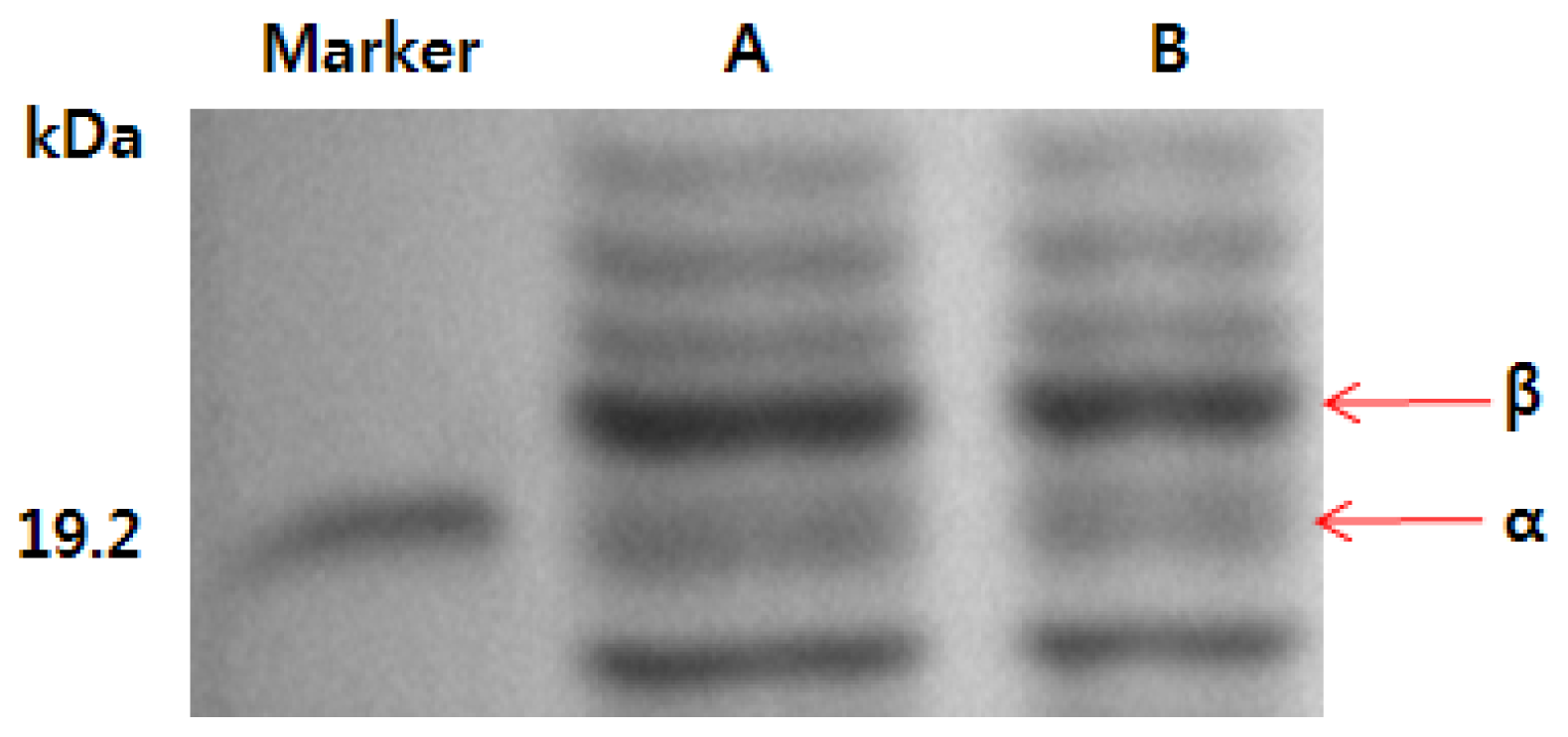
2.3. SDS-PAGE Analysis of Phycocyanin

The molecular weights of the phycocyanin isolated using the high-pressure process and hexane separation method described in this study and those of the phycocyanin standard are shown in Figure 1. The α and β polypeptide subunits of the phycocyanin standard were detected, and their molecular weights were 18.4 kDa and 21.3 kDa, respectively. These weights are consistent with those of previous reports [14]. Moreover, the phycocyanin isolated using the high-pressure process and hexane separation method had two α and β polypeptide subunits that migrated to the same location on the gel and had the same molecular weights as the phycocyanin standard. Furthermore, the bands were clearer than those of the phycocyanin standard. This finding suggests that the high-pressure process and hexane separation method enabledd isolation of a stable, highly purified phycocyanin with a short treatment time using fewer chemical components and increasing the structural stability of the α and β polypeptide subunits.

2.4. Stability of Phycocyanin Extract

To measure the stability of the phycocyanin isolated using the high-pressure process and hexane separation method, the changes of the absorbance of the phycocyanin solution were measured over time, as shown in Figure 2. The phycocyanin from the conventional extraction process had the poorest stability because the absorbance of the phycocyanin from the conventional separation method decreased to 0.808 after 14 days while the phycocyanin standard decreased to 0.841 after 14 days. However, the absorbance of the phycocyanin isolated using the high-pressure process and hexane separation method remained over 0.884 for 14 days. These data suggest that the phycocyanin isolated using the high-pressure process and hexane separation method, compared to two other samples, was more stable because the low temperature and high pressure extraction process effectively destroyed the cell membrane of Spirulina platensis while minimizing the damage to the polypeptide subunits of phycocyanin and increasing the elution of phycobiliprotein. Furthermore, the reduced chemical usage, simple separation process, and short process time of the hexane separation method contributed to the attainment of stable phycocyanin containing two chromophore subunit proteins intact along with a relatively pure form. Phycocyanin with high stability and purity is expected to have an application as a new material in the cosmetic and beauty industry.

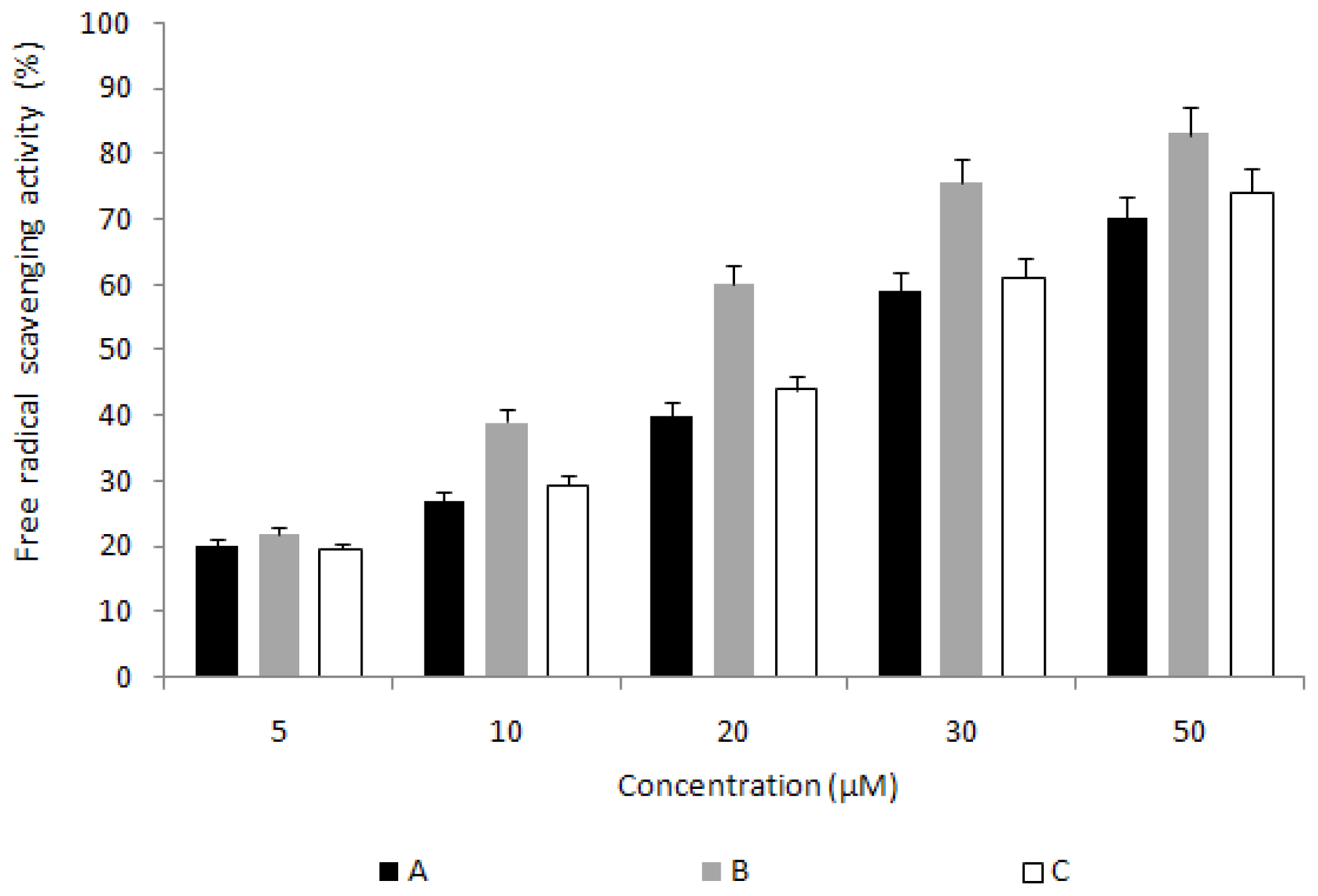
2.5. Measurement of DPPH (α,α-diphenyl-β-picrylhydrazyl) Scavenging Activity

To verify the antioxidant activity of phycocyanin, the electron donating ability of the phycocyanin standard and of the phycocyanin isolated using the high-pressure process and hexane separation method were measured. The results are shown in Figure 3. All of the phycocyanins showed increased activity as their concentrations were increased. Phycocyanin isolated using the high-pressure process and hexane separation method showed a high removal rate of oxygen free radicals (83%) at a concentration 50 μM and showed 9% higher activity than that of the phycocyanin standard. These data imply that the purified sample from our process contained more stable chromophore phycocyanin than the samples from conventional processes, which results in better anti oxidant activity than the others.

2.6. Measurement of Reducing Power

Reducing power is defined as the ability to donate electrons to free oxygen radicals and other free radicals and is one of the various mechanisms of antioxidant activities; thus, a measurement of reducing power can be used to verify antioxidant activity. All of the phycocyanins showed increased reducing power with increased concentration. Moreover, in the reducing power test, phycocyanin isolated using the described method showed a peak absorbance value of 0.38 at a concentration of 50 μM, which was 0.1 higher than the absorbance of the phycocyanin standard and phycocyanin isolated using the conventional separation method (Table 3). This result, along with the measurement of DPPH scavenging activity, implies that the isolation of stable and highly purified phycocyanin with enhanced antioxidant activity is possible with the high-pressure process and hexane separation method. Furthermore, phycocyanin could be used as a material with antioxidant and anti-aging activities in the cosmetic and beauty industry.

3. Experimental Section

3.1. Extraction from Spirulina platensis

In this study, S. platensis from a culture aquarium was filtered using a filter membrane and washed with water to prepare a dried powder. Washed S. platensis was powdered using a freeze-dryer, and it was used in the experiment after grinding in a grinder. Ten volumes of distilled water were added to 100 g of S. platensis powder and sealed tightly in a special plastic bag, and then hydrostatically pressurized in a 1 L volume high-pressure extraction vessel (Ilshin autoclave, Daejeon, Korea) at 5000 bar and 20 °C for 15 min [15]. After the high-pressure treatment, the sample was immersed in water at 4 °C for 12 h, and filtered by vacuum filtration using filter paper. Then, it was incubated at 4 °C for 12 h, and the water layer was isolated using fractionation. The phycocyanin in the water layer was collected and powdered by freeze-drying as a sample.

3.2. Isolation of Phycocyanin

To remove beta-carotene and chlorophyll, which have low light stability, and to isolate phycocyanin from the high pressurized sample, the Spirulina platensis extract from the high-pressure treatment was mixed with hexane (Sigma, St. Louis, MO, USA) in a 1:1 volume ratio and shaken using a vortex mixer (VM-96B, JEIO TECH, Daejeon, Korea) for 5 min. Then, it was incubated at 4 °C for 12 h, and the water layer was isolated using fractionation. The phycocyanin in the water layer was collected and powdered by freeze-drying as a sample.
For comparison with the separation method of this study, phycocyanin powder was also extracted by a conventional separation method: In the first, approximately 5.0 g of S. platensis was suspended in 200 mL of 0.1 M Na-phosphate buffer pH 7.0 containing 100 g/mL lyzozyme and 10 mM EDTA. The enzymatic disintegration of the cell-wall was brought about by placing the S. platensis powder in a shaking bath at 30 °C for 24 h. The slurry was then centrifuged for 1 h at 40,000 × g to remove cell debris, yielding a clear supernatant. In the second, the crude phycocyanin was precipitated in 50% ammonium sulfate and was then recovered by centrifugation at 10,000× g for 10 min. The colourless, clear supernatant was discarded and the blue precipitate was dissolved in a small volume of 0.0025 M Na-phosphate buffer pH 7.0 and dialyzed against the same buffer. The dialyzed phycocyanin was then placed in a 2.5 × 30 cm hydroxylapatite column and the two main fractions were pooled following a stepwise elution with phosphate buffers of increasing ionic strength at pH 7.0, as follows: The first fraction was eluted between 2.5 mM and 70 mM and represented c-phycocyanin. The fraction containing c-phycocyanin was purified by chromatography on a 1.5 × 20 cm DEAE Sephadex A-50, according to Binder et al.[16,17].

3.3. Measurement of Extraction Yield and the Purity of Phycocyanin

The extraction yield of the phycocyanin from the three different methods was estimated by the procedure below. The phycocyanin extract in the above method was prepared using three different methods:by the hexane separation method and the high-pressure process used in this study, by the hexane separation method only without the high-pressure process, and by the conventional separation method.
Weight of phycocyanin extract  ( g ) / Weight of spirulina platensis powder  ( g ) × 100 %
The purity of isolated phycocyanin was checked by the procedure below by the ratio of 280 nm for absorbance of total proteins in phycocyanin to 620 nm of specific protein in the complex [18].
PP  ( phycocyanin purity ) = OD 620 nm / OD 280 nm
The purity of the standard, a commercially available phycocyanin (Anaspec, Inc., CA, USA) was also measured by the above method [18].

3.4. SDS-PAGE Pattern of Phycocyanin

In addition to the measurement of purity, for a closer examination of the phycocyanin isolated using the high-pressure process and the hexane separation method, the molecular weight was confirmed using sodium dodecyl sulfate-polyacrylamide gel electrophoresis (SDS-PAGE, Mini-Protean, Bio-rad Ltd., Hercules, CA, USA) and compared with a phycocyanin standard (Anaspec, Inc., CA, USA). SDS-PAGE was performed using a 12% polyacrylamide slab gel and a 4.5% stacking gel and was confirmed after staining with Coomassie blue R25 (Sigma, St. Louis, MO, USA) and destaining. The molecular weights of the phycocyanin subunits were calculated by comparison with a standard ladder (pre-stained SDS-PAGE standard; Bio-Rad, Seoul, Korea) [14].

3.5. Stability of Phycocyanin

To measure the stability of the phycocyanin pigment isolated from Spirulina platensis at a given temperature, an aqueous solution of phycocyanin was incubated at 40 °C under UV exposure. Then, changes in phycocyanin purity were calculated from the absorbance values at 620 nm and 280 nm with an ELISA reader [19].

3.6. Measurement of DPPH (α,α-diphenyl-β-picrylhydrazyl) Scavenging Activity

DPPH scavenging activity was measured using Kang’s method [20]. One milliliter of a 200-μM DPPH solution was added to a series of phycocyanin standards made by solubilizing in ethanol and 2 mL of the phycocyanin isolated using the high-pressure process and hexane separation method. The mixture was vortexed for 10 s to mix completely and incubated at room temperature for 30 min. To measure the remaining DPPH, the absorbance was measured at 517 nm by a UV-vis spectrophotometer (852A Diode Array Spectrophotometer, Agilent Technologies, Seoul, Korea).

3.7. Measurement of Reducing Power

The reducing power was measured as follows: 2.5 mL of 200 mM sodium phosphate buffer (pH 6.6) and 2.5 mL of 1% potassium ferricyanide were added to a series of phycocyanin standard concentrations and 2.5 mL of the phycocyanin isolated using the high-pressure process and hexane separation method. The mixture was incubated at 50 °C for 20 min. Then, 2.5 mL of trichloroacetic acid (10%, w/v) was added and centrifuged at 650× g for 10 min. Five milliliters of centrifuged supernatant was mixed with 5 mL of deionized water and 1 mL of 1% ferric chloride, and its absorbance was measured at 700 nm using a UV-vis Spectrophotometer (852A Diode Array Spectrophotometer, Agilent Technologies, Seoul, Korea) [21].

3.8. Statistics

All of the analyzed values were presented as the mean ± S.D., and all of the experimental data were analyzed using the statistics software SPSS 16.0. Tests of the significance of differences were analyzed by ANOVA (analysis of variance) and DMRT (Duncan’s multiple range test) at the level of α = 0.05.

4. Conclusions

In this study, a high-pressure extraction process associated with hexane obtained relatively stable phycocyanin from S. platensis as well as in high purity. This process also has the advantage of using fewer steps and having a shorter process time, which may have contributed to the improvement in the stability and the purity of the phycocyanin [18]. Therefore, these results suggest application of this combined process for expanding the uses of phycocyanin in the cosmetic and food industries, etc.

Acknowledgments

This study was carried out with the support of Korea Institute of Ocean Science and Technology (Project No. PE98592), Korea.

References

  1. Becker, E.W. Microalgae: Biotechnology and Microbiology; Cambridge University Press: Cambridge, UK, 1994. [Google Scholar]
  2. Kay, R.A. Microalgae as food and supplement. Crit. Rev. Food Sci 1991, 30, 555–573. [Google Scholar]
  3. Glazer, A.N. Light harvesting by phycobilisomes. Ann. Rev. Biophys. Chem 1985, 14, 47–77. [Google Scholar]
  4. Sautier, C.; Tremolieres, J. Food value in Spirulina algae in humans. J. Ann. Nutr. Aliment 1975, 30, 517–522. [Google Scholar]
  5. Abalde, J.; Betancourt, L.; Torres, E.; Cid, A.; Barwell, C. Purification and characterization of phycocyanin from the marine cyanobacterium Synechococcus sp. IO9201. Plant Sci 1998, 136, 109–120. [Google Scholar]
  6. Diego, J.M.; Gomez, C.; Ibanez, E.; Ruperez, F.J.; Barbas, C. Tocopherol measurement in edible products of vegetable origin. J. Chromatogr. A 2004, 1054, 227–233. [Google Scholar]
  7. Grinstead, G.S.; Tokach, S.S.; Goodband, R.D.; Nelssen, J.L. Effects of Spirulina platensis in growth performance of weanling pigs. Anim. Feed Sci. Tech 2000, 83, 237–247. [Google Scholar]
  8. Kaji, T.; Fujiwara, Y.; Inomata, Y.; Hamada, C.; Yamamoto, C.; Shimada, S.; Lee, J.B.; Hayashi, T. Repair of wounded monolayers of cultured bovine aortic endothelial cells is inhibited by calcium spirulan, a novel sulfated polysaccharide isolated form Spirulina platensis. Life Sci 2002, 70, 1841–1848. [Google Scholar]
  9. Lee, H.S.; Lee, S.H.; Mun, H.C.; Lee, H.Y. Screening of the immuno-stimulatory activity of the marine alga Chlorella capsulate. Kor. J. Biotechnol. Bioeng 2003, 18, 19–24. [Google Scholar]
  10. Sarada, R.; Pillai, M.G.; Ravishankar, G.A. Phycocyanin from Spirulina sp: Influence of processing of biomass on phycocyanin yield, analysis of efficacy of extraction methods and stability studies on phycocyanin. Process Biochem 1999, 34, 795–801. [Google Scholar]
  11. Zhang, S.; Zhu, J.; Wang, C. Novel high pressure extraction technology. Int. J. Pharm 2004, 278, 471–474. [Google Scholar]
  12. Kim, Y.S.; Choi, J.M. Physiochemical properties and dyeability of safflower colorants extracted by ultrasonic treatment. J. Korean Soc. Cloth. Ind 2009, 11, 337–343. [Google Scholar]
  13. Antelo, F.S.; Anschau, A.; Costa, J.A.V.; Kalil, S.J. Extraction and purification of C-phycocyanin from Spirulina platensis in conventional and integrated aqueous two-phase systems. J. Braz. Chem. Soc 2010, 21, 921–926. [Google Scholar]
  14. Chaiklahan, R.; Chirasuwan, N.; Loha, V.; Tia, S.; Bunnag, B. Separation and purification of phycocyanin from Spirulina sp. using a membrane process. Bioresour. Technol 2011, 102, 7159–7164. [Google Scholar]
  15. Kim, S.S.; Jeong, M.H.; Seo, Y.C.; Kim, J.S.; Kim, N.S.; Woon, W.B.; Ahn, J.H.; Hwang, B.; Park, D.S.; Park, S.J.; Lee, H.Y. Comparison of antioxidation activities by high pressure extraction of Codonopsis lanceolata from different production Areas. Korean J. Med. Crop Sci 2010, 18, 248–254. [Google Scholar]
  16. Boussiba, S.; Richmond, A. Isolation and characterization of phycocyanins from the blue-green alga Spirulina platensis. Arch. Microbiol 1979, 120, 155–159. [Google Scholar]
  17. Binder, A.; Wilson, K.; Zuber, H. C-phycocyanin from the thermophilic blue-green alga Mastigocladus laminousus. FEBS Lett 1972, 20, 111–116. [Google Scholar]
  18. Liu, L.N.; Chen, X.L.; Zhang, X.Y.; Zhang, Y.Z.; Zhou, B.C. One-step chromatography method for efficient separation and purification of R-phycoerythrin from Polysiphonia urceolata. J. Biotechnol 2005, 116, 91–100. [Google Scholar]
  19. Joo, D.S.; Cho, S.Y. Stability of phycocyanin and spectral characteristic of phycobilins from Spirulina platensis. J. Korean Fish. Soc 2000, 33, 482–488. [Google Scholar]
  20. Kang, M.Y.; Lee, Y.R.; Koh, H.J.; Nam, S.H. Antioxidative and antimutagenic activity of ethanolic extracts from Glant embroynic rices. J. Korean Soc. Appl. Biol. Chem 2004, 47, 61–66. [Google Scholar]
  21. Oyaizu, M. Studies on products of browning reactions: Antioxidative activities of products of browning reaction prepared from glucosamine. Jpn. J. Nutr 1986, 44, 307–315. [Google Scholar]
Figure 1. SDS-PAGE of phycocyanin. (A) phycocyanin isolated using the hexane separation method and high-pressure process; (B) phycocyanin standard (a commercial product).
Ijms 14 01778f1
Figure 2. The absorbance ratios of phycocyanin isolated from two different extraction processes such as a conventional hexane separation method and high-pressure process over time. (A) phycocyanin isolated using the conventional separation method; (B) phycocyanin isolated using the hexane separation method and high-pressure process; (C) phycocyanin standard (a commercial product).
Ijms 14 01778f2
Figure 3. Comparison of free radical scavenging activity of phycocyanins from two different extraction processes. (A) phycocyanin isolated using the conventional separation method; (B) phycocyanin isolated using the hexane separation method and high-pressure process; (C) phycocyanin standard (a commercial product).
Ijms 14 01778f3
Table 1. Yields of phycocyanin isolated from Spirulina.
SampleYields (%)
A7.2 ± 0.5
B10.2 ± 0.2
C8.4 ± 0.4
A: phycocyanin isolated using the conventional separation method; B: phycocyanin isolated using the hexane separation method and high-pressure process; C: phycocyanin isolated using the hexane separation method.
Table 2. Absorbance ratios of phycocyanin isolated from Spirulina.
SampleAbsorbance ratios (nm)

280620620/280
A2.114 ± 0.0051.868 ± 0.0120.884 ± 0.004
B2.104 ± 0.0071.912 ± 0.0040.909 ± 0.003
C2.125 ± 0.0081.873 ± 0.0090.882 ± 0.006
D2.105 ± 0.0061.903 ± 0.0040.904 ± 0.005
A: phycocyanin isolated using the conventional separation method; B: phycocyanin isolated using the hexane separation method and high-pressure process; C: phycocyanin isolated using the hexane separation method. D: phycocyanin standard (a commercial product).
Table 3. Comparison of reducing power of phycocyanins from two different extraction processes.
Reducing power (O.D.)

SampleConcentration (μM)

510203050
A0.07 ± 0.130.15 ± 0.120.15 ± 0.080.18 ± 0.040.19 ± 0.06
B0.08 ± 0.040.16 ± 0.090.25 ± 0.130.31 ± 0.060.38 ± 0.05
C0.09 ± 0.040.15 ± 0.180.18 ± 0.040.20 ± 0.060.21 ± 0.08
A: phycocyanin isolated using the conventional separation method; B: phycocyanin isolated using the hexane separation method and high-pressure process; C: phycocyanin standard (a commercial product).

