<?xml version="1.0" encoding="UTF-8"?>
<!DOCTYPE article PUBLIC "-//NLM//DTD Journal Publishing DTD v2.3 20070202//EN" "journalpublishing.dtd">
<article xmlns:xlink="http://www.w3.org/1999/xlink" xml:lang="en" article-type="review-article">
<front>
<journal-meta>
<journal-id journal-id-type="publisher-id">ijms</journal-id>
<journal-title>International Journal of Molecular Sciences</journal-title>
<abbrev-journal-title>Int. J. Mol. Sci.</abbrev-journal-title>
<issn pub-type="epub">1422-0067</issn>
<publisher>
<publisher-name>Molecular Diversity Preservation International (MDPI)</publisher-name></publisher></journal-meta>
<article-meta>
<article-id pub-id-type="doi">10.3390/ijms130910771</article-id>
<article-id pub-id-type="publisher-id">ijms-13-10771</article-id>
<article-categories>
<subj-group>
<subject>Review</subject></subj-group></article-categories>
<title-group>
<article-title>Adaptive Response, Evidence of Cross-Resistance and Its Potential Clinical Use</article-title></title-group>
<contrib-group>
<contrib contrib-type="author">
<name><surname>Milisav</surname><given-names>Irina</given-names></name><xref ref-type="aff" rid="af1-ijms-13-10771">1</xref><xref ref-type="aff" rid="af2-ijms-13-10771">2</xref><xref ref-type="corresp" rid="c1-ijms-13-10771">*</xref></contrib>
<contrib contrib-type="author">
<name><surname>Poljsak</surname><given-names>Borut</given-names></name><xref ref-type="aff" rid="af2-ijms-13-10771">2</xref></contrib>
<contrib contrib-type="author">
<name><surname>Šuput</surname><given-names>Dušan</given-names></name><xref ref-type="aff" rid="af1-ijms-13-10771">1</xref></contrib></contrib-group>
<aff id="af1-ijms-13-10771">
<label>1</label>Institute of Pathophysiology, Faculty of Medicine, University of Ljubljana, Zaloska 4, Ljubljana SI-1000, Slovenia; E-Mail: <email>dusan.suput@mf.uni-lj.si</email></aff>
<aff id="af2-ijms-13-10771">
<label>2</label>Faculty of Health Sciences, University of Ljubljana, Zdravstvena pot 5, Ljubljana SI-1000, Slovenia; E-Mail: <email>borut.poljsak@zf.uni-lj.si</email></aff>
<author-notes>
<corresp id="c1-ijms-13-10771">
<label>*</label>Author to whom correspondence should be addressed; E-Mail: <email>irina.milisav@mf.uni-lj.si</email>; Tel.: +386-1-543-7020; Fax: +386-1-543-7021.</corresp></author-notes>
<pub-date pub-type="collection">
<year>2012</year></pub-date>
<pub-date pub-type="epub">
<day>29</day>
<month>08</month>
<year>2012</year></pub-date>
<volume>13</volume>
<issue>9</issue>
<fpage>10771</fpage>
<lpage>10806</lpage>
<history>
<date date-type="received">
<day>02</day>
<month>07</month>
<year>2012</year></date>
<date date-type="rev-recd">
<day>07</day>
<month>08</month>
<year>2012</year></date>
<date date-type="accepted">
<day>13</day>
<month>08</month>
<year>2012</year></date></history>
<permissions>
<copyright-statement>© 2012 by the authors; licensee Molecular Diversity Preservation International, Basel, Switzerland.</copyright-statement>
<copyright-year>2012</copyright-year>
<license license-type="open-access" xlink:href="http://creativecommons.org/licenses/by/3.0">
<p>This article is an open-access article distributed under the terms and conditions of the Creative Commons Attribution license (http://creativecommons.org/licenses/by/3.0/).</p></license></permissions>
<abstract>
<p>Organisms and their cells are constantly exposed to environmental fluctuations. Among them are stressors, which can induce macromolecular damage that exceeds a set threshold, independent of the underlying cause. Stress responses are mechanisms used by organisms to adapt to and overcome stress stimuli. Different stressors or different intensities of stress trigger different cellular responses, namely induce cell repair mechanisms, induce cell responses that result in temporary adaptation to some stressors, induce autophagy or trigger cell death. Studies have reported life-prolonging effects of a wide variety of so-called stressors, such as oxidants, heat shock, some phytochemicals, ischemia, exercise and dietary energy restriction, hypergravity, <italic>etc</italic>. These stress responses, which result in enhanced defense and repair and even cross-resistance against multiple stressors, may have clinical use and will be discussed, while the emphasis will be on the effects/cross-effects of oxidants.</p></abstract>
<kwd-group>
<kwd>oxidative stress</kwd>
<kwd>stress</kwd>
<kwd>adaptation</kwd>
<kwd>adaptive response</kwd>
<kwd>preconditioning</kwd>
<kwd>pretreatment</kwd>
<kwd>hormesis</kwd>
<kwd>autophagy</kwd>
<kwd>apoptosis</kwd>
<kwd>anti-apoptosis</kwd></kwd-group></article-meta></front>
<body>
<sec sec-type="intro">
<title>1. Introduction</title>
<p>Stress is any physical, chemical, or biological factor that adversely affects cellular physiology, growth or survival. Stress response is a mechanism used by organisms to adapt to and to overcome a stress stimulus. Different stressors trigger different cellular responses, namely induce cell repair mechanisms, temporary adaptation to some stressors, induce autophagy or trigger cell death. Redox regulation is an important mechanism for the regulation of cellular processes. There is a delicate balance between the production of reactive oxygen species (ROS) and the antioxidant defense. Having too many ROS in relation to the available antioxidants is often considered as a state of oxidative stress [<xref ref-type="bibr" rid="b1-ijms-13-10771">1</xref>]. Oxidative stress can thus result from diminished antioxidants, like mutations decreasing the levels of glutathione (GSH) or manganese superoxide dismutase (MnSOD), or increased production of ROS, e.g., by exposure to toxins, due to mitochondrial damage or in chronic inflammatory diseases. Like in the case of other stressors, oxidative stress can result in various responses from increased proliferation [<xref ref-type="bibr" rid="b1-ijms-13-10771">1</xref>], adaptation, cell injury and cell death, or a combination of these [<xref ref-type="bibr" rid="b2-ijms-13-10771">2</xref>,<xref ref-type="bibr" rid="b3-ijms-13-10771">3</xref>]. The stress response depends on the type of stressor and on its severity. For example, mild oxidative stress increases proliferation, moderate oxidative stress alters cell physiology to increase the level of protective systems that render the cell more resistant to subsequent insults. The mechanisms of stress responses include the activation of transcription factors and cell cycle arrest to allow repair of DNA damage. Severe oxidative stress results in severe oxidative damage and cell injury, cell senescence and/or cell death [<xref ref-type="bibr" rid="b2-ijms-13-10771">2</xref>–<xref ref-type="bibr" rid="b4-ijms-13-10771">4</xref>]. Here we shall focus on the adaptive responses.</p></sec>
<sec>
<title>
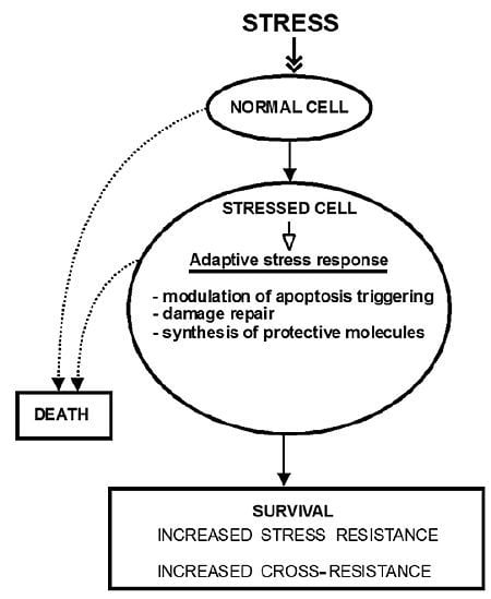
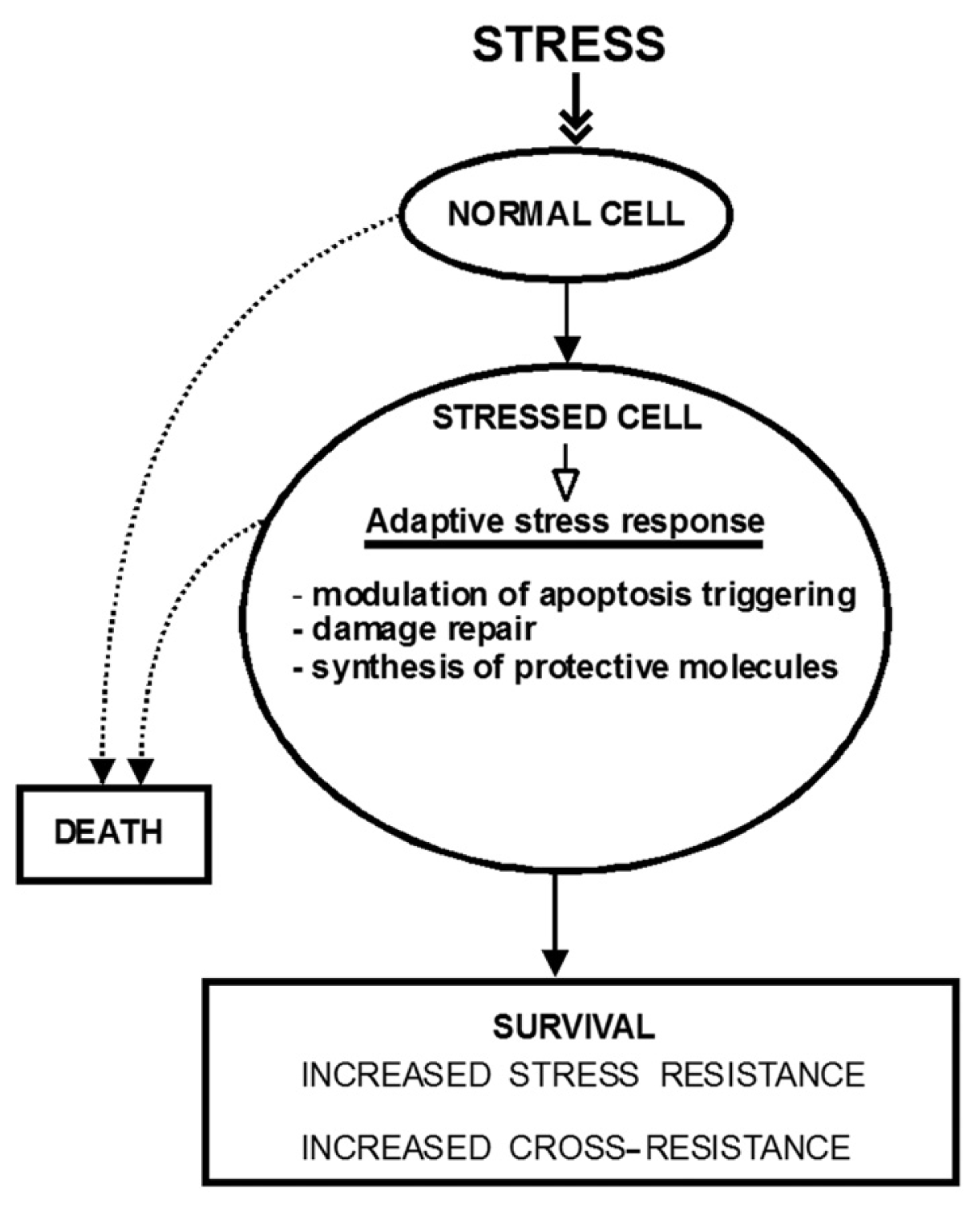
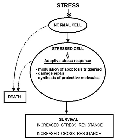
2. The Molecular Mechanisms of Stress-Induced Adaptive Responses</title>
<p>The mechanisms of adaptive stress responses are largely unknown. Milisav and coworkers discovered that adaptation to stress involves the inhibition of apoptosis triggering through the intrinsic pathway in primary hepatocytes [<xref ref-type="bibr" rid="b5-ijms-13-10771">5</xref>,<xref ref-type="bibr" rid="b6-ijms-13-10771">6</xref>]. The main apoptosis initiator, caspase-9 is translocated from the cytoplasm to the nucleus as the consequence of moderate stress. Caspase-9 can be activated only after the activation of the main apoptosis execution caspase, caspase-3, in the stress-adapted cells [<xref ref-type="bibr" rid="b5-ijms-13-10771">5</xref>]. This is in contrast to the situation in the unstressed cells, in which apoptosis is triggered by activation of the caspase-9 first. In other words, at least one of the main apoptotic triggering pathways is inactivated as the consequence of adaptation to stress. Nevertheless, apoptosis can be triggered even then by a more intense trigger than it is needed for the same effect in normal cells.</p>
<p>Cellular adaptations to stress can evade apoptosis by activating the so-called anti-apoptotic genes and proteins, which can temporarily block cell death [<xref ref-type="bibr" rid="b2-ijms-13-10771">2</xref>,<xref ref-type="bibr" rid="b3-ijms-13-10771">3</xref>,<xref ref-type="bibr" rid="b7-ijms-13-10771">7</xref>]. The workable definition of the anti-apoptotic gene is that its product decreases or increases cell survival in response to a given dose of stress if its levels are decreased or increased (reviewed in [<xref ref-type="bibr" rid="b2-ijms-13-10771">2</xref>]). The list of such genes and proteins is huge already and is expanding. Examples include anti-apoptotic proteins, FLICE-inhibitory proteins (FLIPs), B cell lymphoma-2 family members (BCL2), Inhibitors of apoptosis proteins (IAPs), ROS scavengers, like peroxiredoxin, glutaredoxins, superoxide dismutase, glutathione peroxidase, the ceramide utilizing Sphingomyelin Synthase 1 (SMS1), sphingomyelinase, <italic>etc</italic>. For more detailed information please refer to excellent reviews on the topics [<xref ref-type="bibr" rid="b2-ijms-13-10771">2</xref>,<xref ref-type="bibr" rid="b3-ijms-13-10771">3</xref>,<xref ref-type="bibr" rid="b7-ijms-13-10771">7</xref>–<xref ref-type="bibr" rid="b10-ijms-13-10771">10</xref>].</p>
<p>The exposure to stress releases intracellular second messengers, including ROS, calcium, 2′-Deoxyuridine 5′-Triphosphate (dUTP) and a sphingolipid ceramide [<xref ref-type="bibr" rid="b2-ijms-13-10771">2</xref>,<xref ref-type="bibr" rid="b3-ijms-13-10771">3</xref>,<xref ref-type="bibr" rid="b9-ijms-13-10771">9</xref>–<xref ref-type="bibr" rid="b11-ijms-13-10771">11</xref>], as well as currently unknown extracellular factors that enable stress adaptations at sites distant to the site experiencing moderate stress. This is seen in remote ischemic conditioning, where the limb ischemia induced by inflation of a blood pressure cuff can reduce myocardial ischemic injury in animal models (Section 4) [<xref ref-type="bibr" rid="b12-ijms-13-10771">12</xref>]. The above-mentioned intracellular messengers participate in the propagation of survival and death signals and are covered in several reviews [<xref ref-type="bibr" rid="b2-ijms-13-10771">2</xref>,<xref ref-type="bibr" rid="b3-ijms-13-10771">3</xref>,<xref ref-type="bibr" rid="b9-ijms-13-10771">9</xref>–<xref ref-type="bibr" rid="b11-ijms-13-10771">11</xref>,<xref ref-type="bibr" rid="b13-ijms-13-10771">13</xref>].</p>
<p>Many proteins involved in decisions of life and death are likely to play a part in adaptation to stress, since it is often the intensity and duration of the stress trigger that determines whether the cells can adapt to the stress or whether they will die as the result of it [<xref ref-type="bibr" rid="b3-ijms-13-10771">3</xref>,<xref ref-type="bibr" rid="b14-ijms-13-10771">14</xref>]. Also, any stressor will likely cause some damage to cellular structures, which will be removed and repaired. The cells may increase the synthesis of protective molecules, as is well established during oxidative stress (<xref ref-type="fig" rid="f1-ijms-13-10771">Figure 1</xref>).</p>
<sec>
<title>
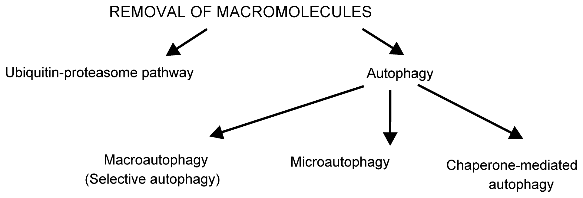
2.1. Removal of Damaged Macromolecules
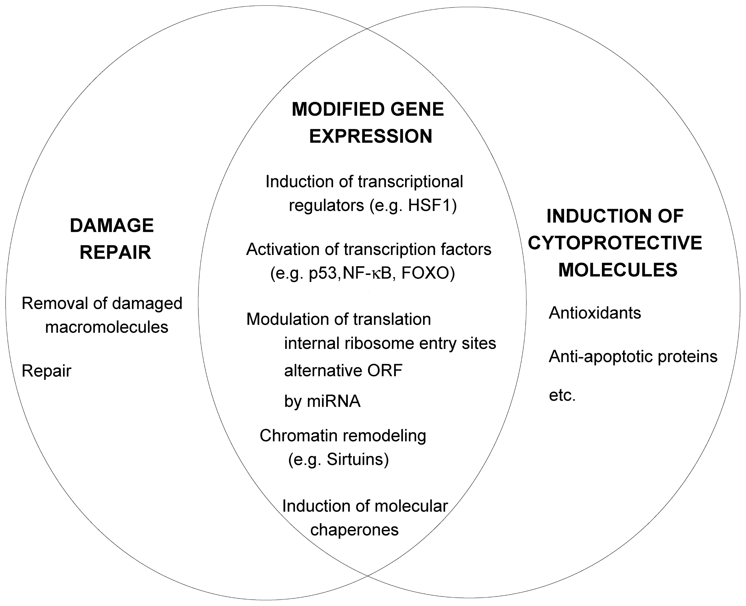
</title>
<p>There are two major degradation pathways in eukaryotic cells for removal of stress-damaged components: the ubiquitin-proteasome pathway (UPP) and autophagy (<xref ref-type="fig" rid="f2-ijms-13-10771">Figure 2</xref>). The UPP is the main pathway for degradation of soluble proteins, mainly in the cytosol. The UPP has been recognized recently to respond to oxidative stress and has a role in the degradation of oxidized proteins [<xref ref-type="bibr" rid="b15-ijms-13-10771">15</xref>]. In the simplest form, UPP consists of labeling the protein target for degradation by covalent attachment of several molecules of a peptide ubiquitin and its degradation by cytoplasmic complex 26S proteasome. The 26S proteasome consists of a barrel-shaped catalytic core, also called 20S [<xref ref-type="bibr" rid="b16-ijms-13-10771">16</xref>]. Two 19S regulatory particles (PA700) bind at either ends of the 20S cylinder and act as a gatekeeper for substrate entry. The ATPase subunits of PA700 contribute to unfolding of the protein substrates and delivery into the proteolytic chamber of 20S [<xref ref-type="bibr" rid="b17-ijms-13-10771">17</xref>]. Other subunits of PA700 deubiquitinate the target proteins, or recruit polyubiquitinated substrates to the proteasome. Some proteins, including oxidized proteins, are degraded by the 20S proteasome in an ATP-independent and ubiquitin-independent manner [<xref ref-type="bibr" rid="b18-ijms-13-10771">18</xref>–<xref ref-type="bibr" rid="b22-ijms-13-10771">22</xref>]. Although many papers report the degradation of oxidized proteins in an ubiquitin-independent manner [<xref ref-type="bibr" rid="b18-ijms-13-10771">18</xref>,<xref ref-type="bibr" rid="b20-ijms-13-10771">20</xref>,<xref ref-type="bibr" rid="b23-ijms-13-10771">23</xref>–<xref ref-type="bibr" rid="b29-ijms-13-10771">29</xref>], there are also reports of degradation of ubiquitinated oxidized proteins [<xref ref-type="bibr" rid="b30-ijms-13-10771">30</xref>–<xref ref-type="bibr" rid="b36-ijms-13-10771">36</xref>]. Proteasome has an important role in selective degradation of oxidized proteins, however how the proteasome distinguishes oxidized proteins from the native ones remains to be determined. Sometimes the oxidation of specific residues results in changes in secondary, tertiary even quaternary structures of the proteins or even results in partial unfolding of the protein [<xref ref-type="bibr" rid="b28-ijms-13-10771">28</xref>,<xref ref-type="bibr" rid="b37-ijms-13-10771">37</xref>–<xref ref-type="bibr" rid="b40-ijms-13-10771">40</xref>]. Such proteins may be recognized by molecular chaperones in a similar manner to other missfolded proteins [<xref ref-type="bibr" rid="b37-ijms-13-10771">37</xref>]. The capacity of UPP to degrade proteins is altered by the cellular redox status. A mild to moderate oxidative stress promotes intracellular protein degradation by increasing susceptibility of proteins to degradation and enhancing the proteolytic capacity. Sustained oxidative stress inactivates proteasome without inhibiting the ubiquitination and results in accumulation of ubiquitin conjugates in the cells. Extensive oxidative stress inactivates the proteasome and inhibits the ubiquitination, resulting in accumulation of oxidatively damaged proteins [<xref ref-type="bibr" rid="b15-ijms-13-10771">15</xref>].</p>
<p>UPP also has a role in the regulation of the intracellular redox status, as it degrades nuclear factor-E2-related factor 2 (Nrf2), which is a transcription factor involved in the transcriptional regulation of antioxidant enzymes [<xref ref-type="bibr" rid="b41-ijms-13-10771">41</xref>,<xref ref-type="bibr" rid="b42-ijms-13-10771">42</xref>]. Under normal cellular redox conditions, Nrf2 is ubiquitinated through binding the adapter protein Kelch-like ECH/associated protein (Keap1), which is an adaptor for one of the ubiquitin ligases (cullin 3); Nrf2 is subsequently degraded by a proteasome. When a cell is exposed to oxidative stress, Nrf2 dissociates from the Keap1/Cul3 complex, shifts into the nucleus and binds to an antioxidant response element (ARE) [<xref ref-type="bibr" rid="b49-ijms-13-10771">49</xref>]. In this manner it regulates the ARE-mediated expression of antioxidant enzymes, like catalase, superoxide dismutase, NAD(P)H: quinone oxidoreductase, some glutathione <italic>S</italic>-transferases and γ-glutamate cysteine ligase regulatory subunit [<xref ref-type="bibr" rid="b50-ijms-13-10771">50</xref>–<xref ref-type="bibr" rid="b52-ijms-13-10771">52</xref>].</p>
<p>Autophagy, the other major degradation pathway, or rather pathways, degrades large cellular cargos that are firstly enclosed into double membrane vesicles; these vesicles are delivered to lysosomes for degradation. Almost any type of cellular material can be enclosed by a double membrane called phagophore, including whole organelles, protein complexes or aggregated proteins. The enclosed vesicles are called autophagosomes. These need to fuse with endosomes to become amphisomes [<xref ref-type="bibr" rid="b43-ijms-13-10771">43</xref>] or with lysosomes to become autolysosomes [<xref ref-type="bibr" rid="b44-ijms-13-10771">44</xref>]. Amphisomes can fuse sequentially with late endosomes or lysosomes. The process described above is macroautophagy, which is a non-selective process of engulfment. It is the best-studied form of autophagy and is often referred to as autophagy in the literature.</p>
<p>Another form of autophagy is selective autophagy with selective cargo recognition by adaptor proteins [<xref ref-type="bibr" rid="b45-ijms-13-10771">45</xref>]. Ubiquitination, described above, has an important role in targeting the cargo for selective autophagy. The types of selective autophagy can be divided into three groups, those dependent on ubiquitin, on the autophagy Atg8 recognition motif, or on both [<xref ref-type="bibr" rid="b53-ijms-13-10771">53</xref>]. Selective autophagy has a role in the case of large quantities of ubiquitinated proteins as in inclusion bodies and protein aggregates. The attachment of ubiquitins (polyubiquitination) is the most common to the lysine residue 48 (Lys<sup>48</sup>) of ubiquitin in UPP; however, the attachment to other lysine residues is also possible. Protein aggregates modified by ubiquitin chains linked to other lysines, including Lys<sup>63</sup>, Lys<sup>27</sup>, Lys<sup>11</sup> are often recognized by selective autophagy. The autophagy adaptor proteins p62 and NBR1 (neighbour of BRCA1) recognize polyubiquitin chains [<xref ref-type="bibr" rid="b45-ijms-13-10771">45</xref>] and expand the phagophore through Atg8. Protein p62 also regulates the stability of Nrf2 (see above) and nuclear factor κB pathway (NF-κB) [<xref ref-type="bibr" rid="b54-ijms-13-10771">54</xref>]. Aggregates containing p62 are seen in neurodegenerative diseases, like Alzheimer’s, Parkinson’s and Huntington’s diseases, in myopathies and in liver diseases.</p>
<p>Macroautophagy also has an important role in the adaptation of organism to stress, like calorie restriction [<xref ref-type="bibr" rid="b55-ijms-13-10771">55</xref>]. Oxidative stress is linked to mitochondrial dysfunction, as mitochondria generate and are targets for reactive oxygen species [<xref ref-type="bibr" rid="b56-ijms-13-10771">56</xref>]. Defective mitochondria are degraded by autophagy, the activity of this pathway declines with age and in neurodegenerative diseases [<xref ref-type="bibr" rid="b57-ijms-13-10771">57</xref>]. The cross talk between the redox signaling, mitochondrial dysfunction and autophagy is not well understood [<xref ref-type="bibr" rid="b58-ijms-13-10771">58</xref>]. Autophagy is regulated by a protein complex mammalian target of rapamycin complex 1 (mTORC1). Protein mTOR (mammalian target of rapamycin) is a serine/threonine protein kinase and is the catalytic subunit of the complex mTORC1 [<xref ref-type="bibr" rid="b59-ijms-13-10771">59</xref>]. mTOR inhibits autophagy by phosphorylating autophagy specific proteins Atg1 and Atg13; the phosphorylation inhibits the kinase activities of these autophagy proteins (Atg) and inhibits the autophagosome initiation [<xref ref-type="bibr" rid="b55-ijms-13-10771">55</xref>].</p>
<p>The term microautophagy was proposed in 1966 when it was suggested that tiny portions of cytoplasm could be sequestered and subsequently engulfed by lysosomes [<xref ref-type="bibr" rid="b60-ijms-13-10771">60</xref>]. Indeed, the lysosome-like organelles with multiple vesicles trapped in their lumens (multivesicular lysosomes) are often observed in mammalian cells; so are the lysosomes containing the organelles, like mitochondria or peroxisomes. Besides the images obtained by electron microscopy, other methods to detect microautophagy in mammalian cells are needed [<xref ref-type="bibr" rid="b46-ijms-13-10771">46</xref>,<xref ref-type="bibr" rid="b47-ijms-13-10771">47</xref>]. On the other hand, a lot of knowledge on microautophagy has been recently obtained from yeasts. Non-selective microautophagy is involved in the degradation of randomly sequestered portions of yeast cytosol. It starts by tubular invaginations of vacuolar membrane from which the small autophagic vesicles are released (pinched off). The selective forms of microautophagy are involved in the degradation of specific organelles, like mitochondria (micromitophagy) [<xref ref-type="bibr" rid="b61-ijms-13-10771">61</xref>], parts of nuclei (piecemeal microautophagy) [<xref ref-type="bibr" rid="b62-ijms-13-10771">62</xref>,<xref ref-type="bibr" rid="b63-ijms-13-10771">63</xref>] and peroxisomes (micropexophagy) [<xref ref-type="bibr" rid="b64-ijms-13-10771">64</xref>,<xref ref-type="bibr" rid="b65-ijms-13-10771">65</xref>]. Yeast microautophagy is regulated by the target of rapamycin complex 1 (TORC1) [<xref ref-type="bibr" rid="b66-ijms-13-10771">66</xref>], a conserved kinase complex, whose mammalian homologue is one of the key metabolic regulators (see below). As so little is known about the mechanisms and physiological relevance of mammalian microautophagy, there can be no definite judgement on the relevance of the results obtained in yeast for these processes in mammals as yet [<xref ref-type="bibr" rid="b46-ijms-13-10771">46</xref>].</p>
<p>Chaperone-mediated autophagy (CMA) targets and delivers some proteins directly across the lysosomal membranes through a specific receptor [<xref ref-type="bibr" rid="b48-ijms-13-10771">48</xref>]. This pathway has been described only in mammals. The basic machinery is composed of at least three types of proteins: (1) the chaperones that recognize the substrates, <italic>i.e.</italic>, the chaperone Hsc70 located in the cytosol and in the lumen of lysosomes [<xref ref-type="bibr" rid="b67-ijms-13-10771">67</xref>,<xref ref-type="bibr" rid="b68-ijms-13-10771">68</xref>]; (2) the receptors that bind and transport the substrates across the lysosomal membranes (Lgp96, human Lamp2a) [<xref ref-type="bibr" rid="b69-ijms-13-10771">69</xref>]; and (3) the substrate proteins with specific motif KFERQ [<xref ref-type="bibr" rid="b70-ijms-13-10771">70</xref>,<xref ref-type="bibr" rid="b71-ijms-13-10771">71</xref>]. About 30% of cytosolic proteins carry this motif [<xref ref-type="bibr" rid="b71-ijms-13-10771">71</xref>]. The signal transduction pathways involved in regulation may involve p38 mitogen-activated protein kinases (p38 MAPK) and are largely unknown [<xref ref-type="bibr" rid="b72-ijms-13-10771">72</xref>]. CMA was initially identified as an inducible pathway in response to stress, there is, however, also a basal level of CMA activity [<xref ref-type="bibr" rid="b73-ijms-13-10771">73</xref>]. The physiological roles of CMA include protein recycling and quality control, delivering antigens for antigen presentation by major histocompatibility complex class II, protein recycling in response to starvation and the removal of oxidized proteins during oxidative stress [<xref ref-type="bibr" rid="b48-ijms-13-10771">48</xref>]. The activity of CMA increases progressively in the first 10 hours of starvation and, unlike macroautophagy, remains activated for the entire duration of starvation [<xref ref-type="bibr" rid="b48-ijms-13-10771">48</xref>,<xref ref-type="bibr" rid="b74-ijms-13-10771">74</xref>]. In this way, the essential proteins can be selectively maintained in the cytosol. CMA activation during nutrient deprivation is tissue- and celltype-specific and depends on age [<xref ref-type="bibr" rid="b74-ijms-13-10771">74</xref>,<xref ref-type="bibr" rid="b75-ijms-13-10771">75</xref>]. The upregulation of CMA was observed during oxidative stress <italic>in vitro</italic>; antioxidants could at least partly prevent the increased degradation of some proteins by CMA [<xref ref-type="bibr" rid="b75-ijms-13-10771">75</xref>]. CMA declines with age, which may contribute to the accumulation of oxidized proteins characteristic in old organisms [<xref ref-type="bibr" rid="b76-ijms-13-10771">76</xref>].</p></sec>
<sec>
<title>2.2. Damage Repair</title>
<p>In addition to clearance of damaged macromolecules (described above), cellular repair is mediated through changed gene expression patterns [<xref ref-type="bibr" rid="b77-ijms-13-10771">77</xref>], induction of molecular chaperones [<xref ref-type="bibr" rid="b78-ijms-13-10771">78</xref>], growth arrest, <italic>etc</italic>. (<xref ref-type="fig" rid="f3-ijms-13-10771">Figure 3</xref>). The alterations of transcription during stress are often mediated by micro ribonucleic acids (miRNAs) (reviewed in [<xref ref-type="bibr" rid="b14-ijms-13-10771">14</xref>]). These are short, noncoding, RNAs of about 22 nucleotides that bind to mRNAs and either accelerate their degradation or inhibit the translation of mRNA, <italic>i.e.</italic>, modulate the stability and/or translational potential of their targets. The stress responses modify the synthesis of miRNAs. The level of target gene repression depends on the relative concentrations of the genes inhibited by a miRNA and of a particular miRNA [<xref ref-type="bibr" rid="b79-ijms-13-10771">79</xref>]. The outcome of miRNA repression depends also on the interactions with stress proteins that can modulate the activity of miRNA protein complexes, e.g., by inhibiting the access to target mRNA. The proteins transformation-related protein 53 (p53) and NF-κB (section 2.4) regulate the transcription and processing of miRNA. The subcellular location of miRNA can change as a consequence of stress. Most miRNA are diffused in the cytoplasm; they have to associate with the member of the Argonaute protein family for activity. After a maturation process at the Argonaute protein, mature miRNAs guide the Argonaute-containing complexes to target sites at mRNAs that are partially complementary to the miRNA sequence, and induce repression of gene expression at the level of mRNA stability or translation. miRNA are synthesized in about two hours, while the mature miRNA can peak in about 24 h. Therefore, the action of miRNA is delayed and can sometimes time the stress response. This is important in acute stress responses, such as during inflammation. Nutrient stress, temperature shock, DNA damage and hypoxia can lead to changes in gene expression by shutdown and reprogramming of protein synthesis through selective recruitment of ribosomes to mRNAs [<xref ref-type="bibr" rid="b77-ijms-13-10771">77</xref>]. This is regulated by elements in untranslated regions of mRNAs, like internal ribosome entry segments, upstream open reading frames and miRNA target sites [<xref ref-type="bibr" rid="b14-ijms-13-10771">14</xref>].</p>
<p>Heat shock factors (HSF) are inducible transcriptional regulators of genes encoding stress proteins, like molecular chaperones and others [<xref ref-type="bibr" rid="b14-ijms-13-10771">14</xref>]. In vertebrates, HSF1 is the most important regulator of expression of heat shock proteins (Hsp) [<xref ref-type="bibr" rid="b80-ijms-13-10771">80</xref>]. Hsp are molecular chaperones and among others assist proteins in folding or prevent and reverse protein missfolding and aggregation. In response to temperature shock or oxidative stress the HSF1 is converted from a monomer to a trimer. Monomeric HSF1 is a phosphorylated protein and interacts with Hsp90. Under stress, HSF1 dissociates from Hsp90, which enables its trimerization and binding to heat shock elements of Hsp genes. These processes are complex to enable a versatile regulation. HSF1 interacts with different Hsp at different phases of its activation cycle and is subjected to post-translational modifications, like phosphorylation, sumoylation and acetilation, many of them upon stress (reviewed in [<xref ref-type="bibr" rid="b80-ijms-13-10771">80</xref>]). Both, HSF1 and HSF2 accumulate into nuclear stress bodies upon their stress-induced transcription. Nuclear stress bodies are thought to participate in rapid, transient, and global reprogramming of gene expression through different types of mechanisms including chromatin remodeling and trapping of transcription and splicing factors [<xref ref-type="bibr" rid="b81-ijms-13-10771">81</xref>].</p>
<p>Molecular chaperones are proteins that assist the non-covalent folding or unfolding of proteins and the assembly or disassembly of multimeric macromolecular structures, but do not occur in these structures during their normal biological functions when these are in correctly folded states. Different classes of chaperones have different functions, including folding of newly made proteins, the repair of the damage caused by misfolding, membrane transport, keeping the protein precursor in translocation-competent state, assistance in protein degradation, <italic>etc</italic>. Chaperons function as intracellular quality control components for proteins. Many chaperons were discovered as their expression was elevated after heat shock, thus they were named heat shock proteins. Chaperons of the Hsp70 family interact with short extended peptide stretches of hydrophobic and basic amino acid residues of unfolded, natively folded or aggregated proteins [<xref ref-type="bibr" rid="b78-ijms-13-10771">78</xref>]. The binding of Hsp70 prevents the aggregation of these proteins and can even induce conformational changes. Disaggregation of proteins is promoted by repeated Hsp70 binding and release to protein substrates. This is enhanced by cooperation with Hsp100 family chaperones. Substrate specificity of Hsp70, stabilization of Hsp70-substrate interactions and triggering of ATP hydrolysis by Hsp70 is promoted by several Hsp40 chaperones (J domain containing chaperones). Also, nucleotide exchange factors, like Hsp110 are required for ADP release and regeneration of ATP at Hsp70.</p>
<p>Chaperone Hsp90 is thought to bind and stabilize partially folded but inactive conformations of its substrates; some of these substrates may be recognized in extended conformations. Hsp90 hydrolyzes ATP like Hsp70; it interacts with a large number of co-chaperones. Its substrates are proteins involved in signal transduction, like protein kinases and transcription factors [<xref ref-type="bibr" rid="b78-ijms-13-10771">78</xref>]. Co-chaperone Hop enables the cooperation of chaperones Hsp90 and Hsp70 by promoting their coupling.</p>
<p>TRiC (TCP-1 Ring Complex or chaperonin containing TCP-1; CCT) is an Hsp60 family member in the cytosol of eukaryotic cells. Like other Hsp60 family members, TRiC is a barrel-shaped protein complex that encapsulates substrates into a protected folding environment. Subunits of oligomers, with beta sheet secondary structures, are its substrates [<xref ref-type="bibr" rid="b78-ijms-13-10771">78</xref>] and possibly late folding intermediates or misfolded proteins. Several small heat shock protein family members are located in the cytosol. ATP-independent small heat shock proteins (sHsp) bind misfolded proteins to prevent their aggregation and may loosen aggregates by coagreggation therefore facilitating subsequent re-folding by Hsp70. Aggregated proteins are presented to Hsp100 by Hsp70 or Hsp40. The solubilized protein can re-enter chaperone-mediated folding cycles or degradation by an ubiquitin-proteasome system. Irreversibly aggregated proteins are degraded primarily by selective autophagy (see above).</p>
<p>Proteins are often damaged as a consequence of stress in the course of their normal biogenesis, which is an error-prone process. For example, truncated polypeptides that result from incomplete translation, misfolded intermediates, and unassembled subunits of protein complexes have exposed hydrophobic regions, which may facilitate aggregation [<xref ref-type="bibr" rid="b14-ijms-13-10771">14</xref>]. Neurodegenerative diseases may be the effect of cellular stress induced by defective clearance of aggregated proteins. Environmental stress triggers can induce nonnative posttraslational modifications and damage proteins in other ways and consequently induce cell stress as well. Even the normal metabolism in every cell is associated with some degree of protein damage. However, the extent of protein damage increases by adverse intrinsic and environmental conditions, like unbalanced protein synthesis, oxidative stress, metabolic stress, some environmental toxins and pollutants, elevated temperature, high-energy radiation, <italic>etc</italic>. To cope with a significant amount of protein damage, the damaged proteins are either repaired by molecular chaperones or degraded by the ubiquitin proteasome system or autophagy [<xref ref-type="bibr" rid="b78-ijms-13-10771">78</xref>].</p>
<p>The stressed cells attempt to repair or degrade acutely damaged proteins by decreasing global protein translation and increasing the translation of molecular chaperones and proteins of the proteolytic system. The well-known adaptive responses in eukaryotic cells are the heat shock response (HSR) and unfolded protein response (UPR), a response to an accumulation of unfolded or misfolded proteins in the endoplasmic reticulum [<xref ref-type="bibr" rid="b78-ijms-13-10771">78</xref>]. HSF1 modulates the HSR’s ability to up-regulate the expression of genes [<xref ref-type="bibr" rid="b80-ijms-13-10771">80</xref>]. Under non-stress conditions, the monomeric HSF1 is in the cytosol bound by Hsp90, perhaps also Hsp70/40. These chaperones are tugged away under heat stress, which enables trimerization of HSF1, the activation of HSF1 trimer and expression of HSR target genes.</p>
<p>Chronic or unresolved ER stress can lead to apoptosis; therefore the cell’s ability to respond to stress in the endoplasmic reticulum (ER) is critical for its survival (reviewed in [<xref ref-type="bibr" rid="b78-ijms-13-10771">78</xref>]). Several factors contribute to the development of ER stress including increases of protein synthesis or protein missfolding rates that exceed the capacity of chaperones, changes in calcium concentration in the ER lumen, oxidative stress and disturbances of the redox balance [<xref ref-type="bibr" rid="b92-ijms-13-10771">92</xref>]. ER stress in eukaryotic cells is sensed by three upstream signaling proteins that initiate cascades of corrective reactions collectively known as the unfolded protein response. These are inositol-requiring protein-1 (IRE1), protein kinase RNA (PKR)-like ER kinase (PERK) and activating transcription factor-6 (ATF6). Prolonged activation of IRE1 can trigger apoptosis.</p></sec>
<sec>
<title>2.3. Increased Synthesis of Protective Molecules</title>
<p>The protective molecules include antioxidant enzymes, like superoxide dismutase (SOD), glutathione peroxidases, catalase, molecules like glutathione and the compounds derived from diet, like ascorbate, <italic>etc</italic>. (<xref ref-type="fig" rid="f4-ijms-13-10771">Figure 4</xref>). Regarding their function, the antioxidant defences can be divided into (1) enzymes that catalytically remove reactive oxygen species, like SOD, superoxide reductase, catalase and peroxidase; (2) agents that decrease the formation of reactive oxygen species by minimizing the availability of pro-oxidants, like iron and copper ions or heme. Such are transferrins, albumin, heme oxigenases, metallothionein, <italic>etc.</italic>; (3) proteins protecting biomolecules against oxidative damage, like chaperones; (4) physical quenchers, for example of a singlet oxygen by carotenoids; (5) agents that are preferentially oxidized, like glutathione, α-tocopherol, bilirubin, ascorbate, urate, albumin, <italic>etc</italic>. [<xref ref-type="bibr" rid="b93-ijms-13-10771">93</xref>,<xref ref-type="bibr" rid="b94-ijms-13-10771">94</xref>].</p>
<p>Since some free-radical production in cells is inevitable and can be very damaging, a defense system against the deleterious actions of free radicals has evolved. These are known as antioxidant defenses, and the two main categories are those whose role is to prevent the generation of ROS and those that intercept any radicals that are generated [<xref ref-type="bibr" rid="b95-ijms-13-10771">95</xref>]. Beside enzymatic defenses, cells also possess a non-enzymatic defense system for the protection of cellular constituents against free radicals and reactive oxygen species (ROS) and for maintaining the cellular redox state. Glutathione (GSH) is the most abundant intracellular thiol-based antioxidant, prevalent in millimolar concentrations in living aerobic cells [<xref ref-type="bibr" rid="b93-ijms-13-10771">93</xref>]. A second category of defence encompasses repair processes, which remove the damaged biomolecules before they accumulate to cause altered cell metabolism or viability [<xref ref-type="bibr" rid="b95-ijms-13-10771">95</xref>]. Besides, natural antioxidants like vitamin C and E, carotenoids and polyphenols are generally considered to be beneficial components of fruits and vegetables. There are many more cytoprotective molecules in addition to the ones mentioned in this section, including anti-apoptotic proteins (Section 2) and others [<xref ref-type="bibr" rid="b2-ijms-13-10771">2</xref>].</p>
<p>The BCL2 family of proteins consists of proapoptotic (Bax, Bak, Bid, <italic>etc</italic>.) and anti-apoptotic members (including Bcl-2, Bcl-<italic>X</italic><italic><sub>L</sub></italic>, Mcl-1) [<xref ref-type="bibr" rid="b7-ijms-13-10771">7</xref>,<xref ref-type="bibr" rid="b14-ijms-13-10771">14</xref>,<xref ref-type="bibr" rid="b96-ijms-13-10771">96</xref>]. Members of both groups are important regulators of apoptosis; the ratio of anti-apoptotic to proapoptotic BCL2 proteins regulates the cells’ sensitivity to apoptosis. Anti-apoptotic BCL2 proteins are important survival factors for many cancers and their overexpression has been associated with tumor initiation, progression, and resistance to current anticancer therapies [<xref ref-type="bibr" rid="b3-ijms-13-10771">3</xref>,<xref ref-type="bibr" rid="b7-ijms-13-10771">7</xref>,<xref ref-type="bibr" rid="b96-ijms-13-10771">96</xref>–<xref ref-type="bibr" rid="b99-ijms-13-10771">99</xref>]. Anti-apoptotic Bcl-2 is the best-studied member of the family. Among many other functions, it controls the integrity of the mitochondrial outer membrane in mammals, e.g., by inhibiting the pro-apoptotic Bax, it is involved in the regulation of autophagy by binding to Beclin-1 and has been implicated in the regulation of the intracellular redox status [<xref ref-type="bibr" rid="b96-ijms-13-10771">96</xref>,<xref ref-type="bibr" rid="b100-ijms-13-10771">100</xref>].</p></sec>
<sec>
<title>
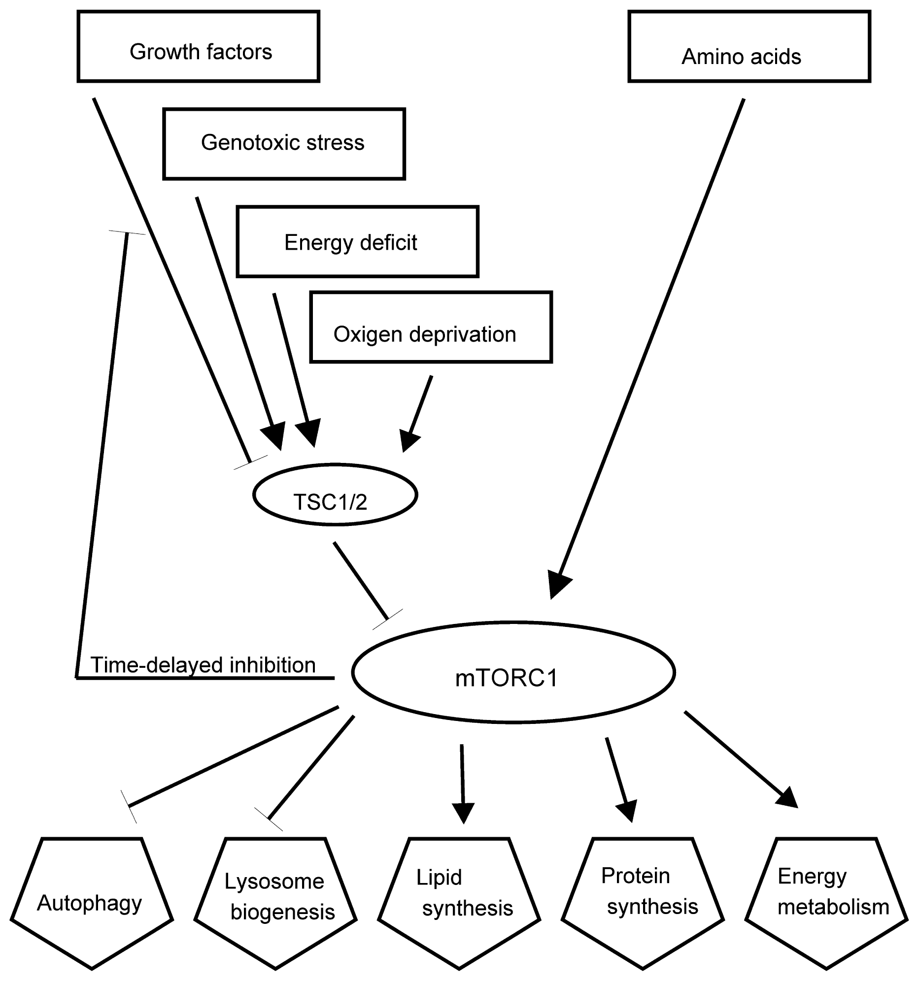
2.4. Stress Signaling Pathways</title>
<p>As the consequences of stress are alterations in cellular metabolism, growth and division, it is no surprise that the main signaling pathways that regulate cell growth, metabolism, senescence and apoptosis also regulate the stress responses. The complex mTORC1 with its catalytic subunit mTOR regulates growth by maintaining the balance between the anabolic processes, like macromolecular synthesis, and catabolic processes, such as autophagy (<xref ref-type="fig" rid="f5-ijms-13-10771">Figure 5</xref>). mTORC1 mediates cellular responses to many types of stress, like DNA damage and drops in the level of energy, oxygen, amino acids and glucose. mTORC1 is at the bottom of PI3K and AKT signaling pathways, which receive signals from growth factor receptors at the plasma membrane and other inputs [<xref ref-type="bibr" rid="b55-ijms-13-10771">55</xref>]. mTORC1 consists of mTOR and regulatory-associated protein of mTOR (raptor), mammalian lethal with Sec13 protein 8 (mLST8), proline-rich AKT substrate 40 kDa (PRAS40), and DEP-domain-containing mTOR-interacting protein (Deptor). The small G protein Rheb in the GTP-bound form stimulates the activity of mTORC1. Rheb is inhibited by the heterodimer of tuberous sclerosis proteins TSC1 and TSC2 (TSC1/2), which convert Rheb into an inactive GDP-bound state. Growth factors promote activities of several kinases, such as AKT, Erk and Rsk, which phosphorylate TSC1/2 and inhibit its activities, therefore enabling the mTOR signaling pathway. Sustained activation of mTORC1 blocks growth factor signaling through the activation of negative loops that inhibit PI3-kinase (PI3K) signaling [<xref ref-type="bibr" rid="b101-ijms-13-10771">101</xref>]. The activation of mTORC1 promotes protein synthesis mainly by phosphorylating the kinase S6K and through the regulator of translation 4E-BP1 [<xref ref-type="bibr" rid="b102-ijms-13-10771">102</xref>]. The biogenesis of lipids is induced through the activation of transcription factors SREBP1 and PPARγ [<xref ref-type="bibr" rid="b103-ijms-13-10771">103</xref>]. The active complex mTORC1 promotes anabolism and inhibits catabolism by blocking autophagy through the phosphorylation of a complex that consists of ULK1-Atg13-FIP200 [<xref ref-type="bibr" rid="b104-ijms-13-10771">104</xref>].</p>
<p>Stress, like hypoxia, energy deprivation, DNA damage and inflammation, activates TSC1/2, thus inhibiting mTORC1 activity. Amino-acid activation of mTORC1 is regulated by Rag GTPase and is independent of TSC1/2 [<xref ref-type="bibr" rid="b105-ijms-13-10771">105</xref>]. A possible mechanism of mTOR regulation could involve the targeting of mTORC1 to different locations within the cells. The availability of amino acids stimulates the translocation of mTORC1 to the lysosomal surface, where it associates with Rheb resulting in activation of mTOR kinase [<xref ref-type="bibr" rid="b106-ijms-13-10771">106</xref>,<xref ref-type="bibr" rid="b107-ijms-13-10771">107</xref>]. In cells exposed to moderate starvation lysosomes relocate from close to the plasma membrane to the perinuclear microtubule organizing center [<xref ref-type="bibr" rid="b55-ijms-13-10771">55</xref>]. During starvation, mTOR co-localizes with protein LC3 after the formation of autophagosomes [<xref ref-type="bibr" rid="b58-ijms-13-10771">58</xref>]. This step may be sensitive to ROS, as the oxidation of mTOR results in the inhibition of its activity [<xref ref-type="bibr" rid="b108-ijms-13-10771">108</xref>]. The association of mTORC1 with autophagosomes may facilitate autophagosome-lysosome fusion. The changes in lysosomal positioning in response to nutrients are mediated by intracellular pH, which is increased during starvation [<xref ref-type="bibr" rid="b55-ijms-13-10771">55</xref>].</p>
<p>The changes in concentration of reactive oxygen species (ROS) and reactive nitrogen species (RNS) influence the regulators of autophagy [<xref ref-type="bibr" rid="b58-ijms-13-10771">58</xref>]. Starvation induced autophagy is associated with increased oxidative stress in mammalian cells. Starvation probably triggers the accumulation of (hydrogen peroxide) H<sub>2</sub>O<sub>2</sub>, which was shown to be necessary for autophagosome formation in permanent cell cultures, mainly CHO cells [<xref ref-type="bibr" rid="b109-ijms-13-10771">109</xref>]. The starvation induces formation of the complex of PI3 kinase and Beclin 1 with other signals and results in a local rise of H<sub>2</sub>O<sub>2</sub> near mitochondria. This inactivates Atg4 and promotes lipidation of Atg8. Subsequently around the time of fusion with the lysosome, the vesicles are shifted to an H<sub>2</sub>O<sub>2</sub> poor environment, where the active Atg4 can de-lipidate and recycle Atg8 [<xref ref-type="bibr" rid="b109-ijms-13-10771">109</xref>].</p>
<p>H<sub>2</sub>O<sub>2</sub> has a role in the response to nutrient starvation, rapamycin, TNF-α and nerve growth factor deprivation [<xref ref-type="bibr" rid="b110-ijms-13-10771">110</xref>,<xref ref-type="bibr" rid="b111-ijms-13-10771">111</xref>]. Both H<sub>2</sub>O<sub>2</sub> and superoxide (O<sub>2</sub> <sup>•−</sup>) can induce autophagy [<xref ref-type="bibr" rid="b109-ijms-13-10771">109</xref>,<xref ref-type="bibr" rid="b112-ijms-13-10771">112</xref>]. Chen and coworkers found that the amount of autophagy induced by starvation, mitochondrial electron transport chain inhibition and exogenous H<sub>2</sub>O<sub>2</sub> correlated with increased intracellular levels of O<sub>2</sub> <sup>•−</sup> and decreased levels of H<sub>2</sub>O<sub>2</sub> [<xref ref-type="bibr" rid="b112-ijms-13-10771">112</xref>]. Cell death induced by starvation was increased by the inhibition of autophagy with autophagy inhibitors and superoxide scavengers and decreased by the autophagy by knocking down the expression of superoxide dismutase 2 (SOD2) [<xref ref-type="bibr" rid="b112-ijms-13-10771">112</xref>]. The same authors reported the enhancement of autophagy induced by the inhibitors of mitochondrial electron chain transport with SOD2 inhibition that resulted in enhanced cell death. These data imply that autophagy is a cell survival mechanism that generates amino acids and fatty acids for growth or for the conversion into energy during starvation, however, if autophagy is prolonged it leads to cell death, since many proteins and organelles required for homeostasis and cell survival are degraded. The regulatory role of ROS on autophagy, its regulator and the regulator of homeostasis mTORC1 and other metabolic pathways is complex and more exciting discoveries will pave the way to improve the understanding of these processes.</p>
<p>p53 is a transcription factor, with transcription dependent and transcription independent activities [<xref ref-type="bibr" rid="b84-ijms-13-10771">84</xref>]. It responds to many types of cellular stress, such as DNA damage, hypoxia and oncogene activation to regulate target genes that induce cell cycle arrest, apoptosis, senescence, DNA repair or changes in metabolism. p53 can interact with and modulate the activity of some members of the BCL2 family, the regulators of apoptosis [<xref ref-type="bibr" rid="b84-ijms-13-10771">84</xref>]. Both, transcription dependent and transcription independent activities of p53 may lead to apoptosis; there are accounts when transcription independent mechanisms are essential for the full apoptotic response [<xref ref-type="bibr" rid="b84-ijms-13-10771">84</xref>].</p>
<p>The concentration and activity of p53 are regulated mostly on the posttranslational level. The half-life of p53 protein in normal, unstressed cells is about six to 20 min, while it can last for hours under stress. Post-translational modifications are selectively activated by different stress signals. p53 is then modified by phosphorylation, methylation, monoubiquination, sumoylation, neddylation and glycosylation [<xref ref-type="bibr" rid="b14-ijms-13-10771">14</xref>,<xref ref-type="bibr" rid="b85-ijms-13-10771">85</xref>]. Different stress signals result in different post-translational modifications of p53 and transcription of different sets of genes.</p>
<p>In addition to the initiation of cell cycle arrest, senescence or apoptosis, p53 regulates cell growth and metabolism through down-regulation of IGF-1/AKT-1 and mTOR pathways. Insulin-like growth factor-1 (IGF-1) is secreted when insulin is secreted in response to high levels of nutrients. The binding of IGF-1 to a tyrosine kinase receptor recruits PI3 kinase and leads to its activation, which phosphorylates phosphoinositides, increasing their concentration at the plasma membrane [<xref ref-type="bibr" rid="b113-ijms-13-10771">113</xref>]. Then lipid kinases are activated, like 3-phosphoinositide-dependent protein kinase 1 (PDK-1), which activates AKT by phosphorylation [<xref ref-type="bibr" rid="b114-ijms-13-10771">114</xref>]. AKT is fully activated after the additional phosphorylation by mTORC2 [<xref ref-type="bibr" rid="b113-ijms-13-10771">113</xref>]. AKT phosphorylates and inactivates proapoptotic protein BAD and activates antiapoptotic MDM2 [<xref ref-type="bibr" rid="b115-ijms-13-10771">115</xref>]. Activated AKT translocates from the plasma membrane to the nucleus, phosphorylates the O subclass of the forkhead family of transcription factors (FOXO) [<xref ref-type="bibr" rid="b86-ijms-13-10771">86</xref>], which leaves the nucleus. This results in transcriptional enhancement of proteins for oxidative phosphorylation, chaperones and the production of antioxidant proteins that decrease the levels of ROS [<xref ref-type="bibr" rid="b113-ijms-13-10771">113</xref>]. The removal of FOXO from the nucleus turns off the production of p27, an inhibitor of cyclin D-cdk-4/6 kinases, activating the pathway for driving the cell into the cell cycle [<xref ref-type="bibr" rid="b113-ijms-13-10771">113</xref>]. AKT also regulates mTORC1 by inhibiting TSC2, thus activating Rheb and mTORC1. mTOR regulates a translational control over cell growth, division and energy metabolism, whereas IGF-1/AKT regulate the transcriptional regulators of these processes. The result of the activated IGF/AKT pathway is therefore the promotion of growth and division and the inhibition of apoptosis.</p>
<p>Sirtuins are NAD<sup>+</sup>-dependent histone deacetylases whose activity can prolong the lifespan of organisms, such as yeast, worms and flies [<xref ref-type="bibr" rid="b116-ijms-13-10771">116</xref>] and are important for aging regulation and caloric restriction-extended lifespan in <italic>in vitro</italic> cell models, animals and humans [<xref ref-type="bibr" rid="b82-ijms-13-10771">82</xref>,<xref ref-type="bibr" rid="b83-ijms-13-10771">83</xref>,<xref ref-type="bibr" rid="b117-ijms-13-10771">117</xref>–<xref ref-type="bibr" rid="b121-ijms-13-10771">121</xref>]. This is especially true for the mammalian sirtuin 1 (SIRT1) whose activity depends on NAD<sup>+</sup>/NADH ratio, which is the key indicator of oxygen consumption, thus SIRT1 may be responsive to the metabolic state of cells. SIRT1 increases stress resistance by negative regulation of p53 and FOXO [<xref ref-type="bibr" rid="b87-ijms-13-10771">87</xref>–<xref ref-type="bibr" rid="b90-ijms-13-10771">90</xref>]. It also initiates endocrine responses, including inhibition of adipogenesis and insulin secretion of pancreatic β cells by regulation of proliferator-activated receptor g coactivator 1α (PGC-1α) [<xref ref-type="bibr" rid="b122-ijms-13-10771">122</xref>,<xref ref-type="bibr" rid="b123-ijms-13-10771">123</xref>]. PGC-1α is also a regulator of mitochondrial biogenesis and function and is responsive to metabolic stress, therefore the number and capacity of mitochondria are adapted to the energetic demands of tissues [<xref ref-type="bibr" rid="b124-ijms-13-10771">124</xref>].</p>
<p>Protein p53 also selectively regulates the transcription of genes that initiate cell-cycle arrest, DNA repair, senescence and apoptosis. It senses stress signals, like hypoxia, heat and cold shocks, DNA damage, nutrition starvation and oncogene activation [<xref ref-type="bibr" rid="b125-ijms-13-10771">125</xref>,<xref ref-type="bibr" rid="b126-ijms-13-10771">126</xref>]. In response to stress, p53 modulates IGF-1/AKT and mTOR pathways by the induction of expression of genes of negative regulators of these two pathways, like IGF-BP3 and PTEN, TSC2, AMPKβ1, Sestrin1 and Sestrin2 [<xref ref-type="bibr" rid="b126-ijms-13-10771">126</xref>–<xref ref-type="bibr" rid="b129-ijms-13-10771">129</xref>]. This results in the shutdown of cell growth and division and induction of autophagy. On the other hand mTORC1 phosphorylates and activates phosphatase PP2A, which dephosphorylates p53 at the site of its activation. Also, AMPK can directly and indirectly phosphorylate p53 in response to nutrient starvation [<xref ref-type="bibr" rid="b129-ijms-13-10771">129</xref>]. More regulatory loops are known between these core pathways to balance the commitment to cell growth and division and stress responses. p53 responds to the intrinsic stress. Another transcription factor, NF-κB, responds to extrinsic stress [<xref ref-type="bibr" rid="b14-ijms-13-10771">14</xref>,<xref ref-type="bibr" rid="b85-ijms-13-10771">85</xref>]. NF-κB is a protein complex. In the absence of stress signals it is in the cytosol associated with the inhibitor IκB. IκB is phosphorylated upon stress by IκB kinase (IKK), then ubiquinated and degraded by a proteasome. The translocation of NF-κB to the nucleus enables the transcription of genes with NF-κB response elements. The activation of these genes results in cellular replication, inflammatory responses mediated by tumor necrosis factor, and cell survival signals. NF-κB also transcribes the gene of its regulator IκB-α resulting in a negative autoregulatory loop. The proteins p53 and NF-κB cannot function at the same time in the same cell; on activation of one, the other one is inactivated.</p>
<p>Ataxia-telangiectasia mutated (ATM) protein kinase has been recently recognized to have a role as redox sensor that controls the levels of reactive oxygen species in human cells [<xref ref-type="bibr" rid="b130-ijms-13-10771">130</xref>]. The cells deficient in ATM have a reduced antioxidative response [<xref ref-type="bibr" rid="b131-ijms-13-10771">131</xref>] and are more sensitive to treatment with oxidizing agents [<xref ref-type="bibr" rid="b132-ijms-13-10771">132</xref>–<xref ref-type="bibr" rid="b134-ijms-13-10771">134</xref>]. It is well established, that ATM is recruited to the sites of double strand DNA breaks and is among the kinases, which activate DNA repair and signaling pathways [<xref ref-type="bibr" rid="b130-ijms-13-10771">130</xref>]. In addition, a novel mechanism was identified, where ATM was activated directly through oxidation. This was shown by studies <italic>in vitro</italic> in the presence of H<sub>2</sub>O<sub>2</sub> [<xref ref-type="bibr" rid="b135-ijms-13-10771">135</xref>]; oxidative stress may also inhibit ATM activation by DNA damage, as it disrupts the binding of the regulator proteins required for ATM activation through this pathway. Nevertheless, it is likely that ATM is exposed to the increase of ROS and DNA damage simultaneously, as ROS production also induces DNA damage. ATM interacts with all main signaling pathways mentioned above. ATM is required for NF-κB activation following exposure to ionizing radiation and reactive oxygen species [<xref ref-type="bibr" rid="b136-ijms-13-10771">136</xref>]. ATM is linked to the expression of IGF1 receptor by an unknown mechanism, as there are reduced levels of this receptor in ATM-deficient cells [<xref ref-type="bibr" rid="b137-ijms-13-10771">137</xref>,<xref ref-type="bibr" rid="b138-ijms-13-10771">138</xref>]; the effects of ATM on insulin function and glucose metabolism may be mediated through phosphorylation of p53 [<xref ref-type="bibr" rid="b139-ijms-13-10771">139</xref>]. The signaling between ATM and mTORC1 was identified where ATM phosphorylates LKB1 tumor suppressor proteins lead to the activation of AMPK in response to elevated levels of ROS [<xref ref-type="bibr" rid="b140-ijms-13-10771">140</xref>]. ATM directly phosphorylates the subunit of hypoxia-inducible factor (HIF), HIF1α, which promotes the activity of TSC2, therefore inhibiting mTORC1 [<xref ref-type="bibr" rid="b141-ijms-13-10771">141</xref>]. HIF is a protein complex that plays an integral role in the body’s response to low oxygen concentrations. The insight into the interconnections of the above-mentioned and other metabolic pathways are starting to shed light on the relationships between stress, longevity, control over metabolic networks and some disease pathways, and pave the way to many new discoveries needed to understand these core regulatory pathways.</p></sec></sec>
<sec>
<title>3. Adaptive Response and Cross-Resistance</title>
<p>The adaptive response induced by low- or moderate-intensity stressor(s) could induce beneficial responses of an organism. Toxicologists sometimes use the term hormesis to refer to a biphasic dose response to an environmental agent that has a beneficial effect in low doses and a toxic one at high doses [<xref ref-type="bibr" rid="b142-ijms-13-10771">142</xref>]. Therefore, hormesis can be defined as an adaptive response of cells and organisms to a moderate, usually intermittent, stress. Such stress inducers include oxidants, UV-radiation, heat shock, some phytochemicals, ischemia, exercise and dietary energy restriction as well as hypergravity. There are many reports of cross-resistance to the above-mentioned stressors in organisms from bacteria to humans; some examples will be mentioned below (<xref ref-type="table" rid="t1-ijms-13-10771">Table 1</xref>).</p>
<sec>
<title>
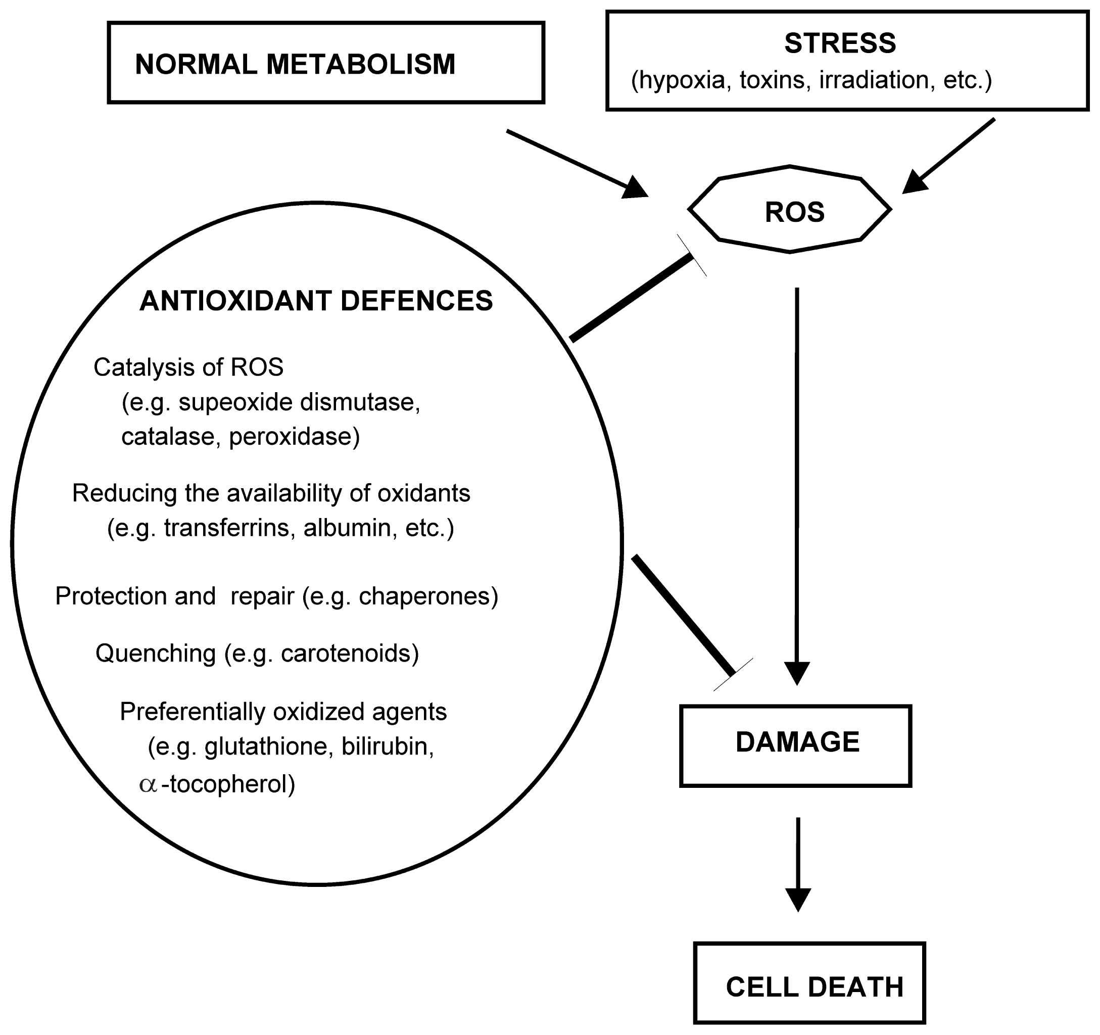
3.1. Oxidative Stress Inducers (Oxidants)</title>
<p>Reactive oxygen species (ROS) are beneficial in moderate amounts and harmful in excess, leading to the state of oxidative stress. Human cells generate some hydrogen peroxide and other ROS molecules deliberately to use them as chemical signals to regulate glucose metabolism, cell growth and proliferation [<xref ref-type="bibr" rid="b172-ijms-13-10771">172</xref>].</p>
<p>Adaptive responses after the exposure to sublethal levels of oxidants were observed in eukaryotic cells [<xref ref-type="bibr" rid="b143-ijms-13-10771">143</xref>,<xref ref-type="bibr" rid="b144-ijms-13-10771">144</xref>]. The protective responses to oxidants were observed upon the subsequent exposure to the same and related oxidants. Spitz and coworkers [<xref ref-type="bibr" rid="b143-ijms-13-10771">143</xref>] observed resistance to normally lethal dose of H<sub>2</sub>O<sub>2</sub> when CHO fibroblasts were preconditioned with the same agent. These cells were slightly resistant to heat shock damage as well. Also, pretreatment with heat shock somewhat protected the cells from the subsequent oxidative stress. Bovine vascular endothelial cells were also more resistant to oxidative damage after they had been pre-exposed to low levels of H<sub>2</sub>O<sub>2</sub> [<xref ref-type="bibr" rid="b173-ijms-13-10771">173</xref>]. Also, xanthine and xanthine oxidase pretreatment for one hour, which produces O<sub>2</sub> <sup>•−</sup> and H<sub>2</sub>O<sub>2</sub> resulted in resistance of CHO and rat hepatoma cells to subsequent H<sub>2</sub>O<sub>2</sub> and γ-irradiation [<xref ref-type="bibr" rid="b144-ijms-13-10771">144</xref>]. Even ten minutes after the pretreatment of CHO cells with H<sub>2</sub>O<sub>2</sub> a cross-resistance to <italic>N-</italic>methyl<italic>-N</italic>′<italic>-</italic>nitro<italic>-N-</italic>nitrosoguanidine (MNNG) and γ-irradiation was reported; however, the cells were not cross-resistant to UV light [<xref ref-type="bibr" rid="b174-ijms-13-10771">174</xref>]. The adaptive response induced by H<sub>2</sub>O<sub>2</sub> was observed in human lymphocytes to protect these cells against otherwise lethal X-ray irradiation [<xref ref-type="bibr" rid="b145-ijms-13-10771">145</xref>]. Some molecular changes were observed during the stress adaptation. In H<sub>2</sub>O<sub>2</sub>-pretreated bovine vascular endothelial cells there were increased activities of SOD, catalase and glutathione peroxidase [<xref ref-type="bibr" rid="b173-ijms-13-10771">173</xref>]. Some increase in SOD was observed in xantine and xantine-oxidase treated cells; however, there was no increased SOD expression when the same cells were treated with H<sub>2</sub>O<sub>2</sub> [<xref ref-type="bibr" rid="b144-ijms-13-10771">144</xref>]. Also, the expression of the Hsp70 family of proteins was increased upon the pretreatment with xantine and xantine-oxidase [<xref ref-type="bibr" rid="b144-ijms-13-10771">144</xref>], in pretreatment with H<sub>2</sub>O<sub>2</sub> in chinese hamster fibroblasts [<xref ref-type="bibr" rid="b143-ijms-13-10771">143</xref>], in reoxygenation of CHO cells after anoxia [<xref ref-type="bibr" rid="b175-ijms-13-10771">175</xref>] and after the reperfusion injury in rat hearts [<xref ref-type="bibr" rid="b176-ijms-13-10771">176</xref>].</p>
<p>The organisms can gradually adapt to a stressor. If adult rats are acclimated to elevated concentrations of O<sub>2</sub>, they can tolerate pure O<sub>2</sub> longer than control rats, possibly due to increased synthesis of SOD in lungs, although a co-ordinated increase in antioxidant defences is essential for maximum protection [<xref ref-type="bibr" rid="b177-ijms-13-10771">177</xref>]. For example, injection of liposomes containing catalase and SOD is more protective than SOD on its own. Like the rats, mice overexpressing MnSOD or CuSOD are more tolerant to hyperoxia [<xref ref-type="bibr" rid="b177-ijms-13-10771">177</xref>]. Culturing HeLa cells at gradually increasing O<sub>2</sub> concentrations over 21 months enabled the cells to grow at 80% O<sub>2</sub>; this was otherwise lethal [<xref ref-type="bibr" rid="b178-ijms-13-10771">178</xref>].</p></sec>
<sec>
<title>3.2. Temperature Shock</title>
<p>The heat-induced adaptations were investigated by challenging cells and organisms with moderate stress that often resulted in anti-aging and life-prolonging effects [<xref ref-type="bibr" rid="b146-ijms-13-10771">146</xref>]. It was demonstrated in a series of experimental studies that repeated moderate heat stress had anti-aging effects in human skin fibroblasts <italic>in vitro</italic>. The exposure to moderate heat stress could protect the cells from being damaged by oxidative stress or toxins such as cyanide [<xref ref-type="bibr" rid="b147-ijms-13-10771">147</xref>]. In <italic>C. elegans</italic>, brief thermal stress was sufficient to induce thermotolerance and caused small but statistically significant increases in lifespan [<xref ref-type="bibr" rid="b148-ijms-13-10771">148</xref>]. Overexpression of certain HSPs significantly extended the longevity of normal-lived animals [<xref ref-type="bibr" rid="b179-ijms-13-10771">179</xref>,<xref ref-type="bibr" rid="b180-ijms-13-10771">180</xref>]. HSP induced increase in glutathione (GSH) may offer protection against subsequent oxidative stress (e.g., induced by H<sub>2</sub>O<sub>2</sub>), since small Hsp protect against oxidative stress through a glucose-6-phosphate dehydrogenase (G6PD)-dependent ability to increase and uphold GSH in its reduced form [<xref ref-type="bibr" rid="b181-ijms-13-10771">181</xref>]. The long-lived insulin/IGF-1 mutants of <italic>C. elegans</italic> are resistant to both oxidative and thermal stresses, possibly through increased expression of heat shock proteins and antioxidant enzymes [<xref ref-type="bibr" rid="b182-ijms-13-10771">182</xref>].</p>
<p>Cold stress also increases resistance to other stresses, like heat and fungal infection in aged <italic>Drosophila melanogaster</italic> flies [<xref ref-type="bibr" rid="b149-ijms-13-10771">149</xref>]. Hormetic effects of repeated exposures to cold at young age reflected on longevity, aging and resistance to heat or cold shocks in <italic>Drosophila melanogaster</italic> [<xref ref-type="bibr" rid="b150-ijms-13-10771">150</xref>].</p></sec>
<sec>
<title>3.3. Exposure to Chemicals and Toxins</title>
<p>As it was mentioned above, the exposure of cells to moderate heat stress can protect them from being damaged by oxidative stress or toxins such as cyanide [<xref ref-type="bibr" rid="b147-ijms-13-10771">147</xref>]. Similarly, when cells are exposed to a low dose of the mitochondrial uncoupling agent 2,4-dinitrophenol they are less vulnerable to ischemia [<xref ref-type="bibr" rid="b151-ijms-13-10771">151</xref>]. Recent findings imply that health benefits of many phytochemicals may be the consequence of cross-resistance in which a phytochemical activates one or more adaptive stress response pathways [<xref ref-type="bibr" rid="b183-ijms-13-10771">183</xref>].</p>
<p>Beneficial effects for health have been demonstrated for many chemicals isolated from plants [<xref ref-type="bibr" rid="b184-ijms-13-10771">184</xref>,<xref ref-type="bibr" rid="b185-ijms-13-10771">185</xref>]. Initially it was thought that the antioxidant activity of phytochemicals is responsible for their health benefits [<xref ref-type="bibr" rid="b186-ijms-13-10771">186</xref>]. It still stands that the oxidative stress contributes to the pathogenesis of many diseases and to the ageing process itself [<xref ref-type="bibr" rid="b187-ijms-13-10771">187</xref>,<xref ref-type="bibr" rid="b188-ijms-13-10771">188</xref>], and that fruits and vegetables contain the substances that exhibit direct free radical-scavenging properties at high concentrations. Micromolar concentrations of vitamin E and numerous polyphenols can protect a variety of cells against oxidative stress in cell culture models of cancer, atherosclerosis and neurodegenerative disorders [<xref ref-type="bibr" rid="b189-ijms-13-10771">189</xref>–<xref ref-type="bibr" rid="b191-ijms-13-10771">191</xref>]. However, the clinical trials and primary prevention studies of high doses of antioxidants in humans have not fulfilled the expectations [<xref ref-type="bibr" rid="b192-ijms-13-10771">192</xref>]. Low levels of ROS function as signaling molecules and may induce adaptive responses, therefore over-consumption of exogenous antioxidants can lead to “anti-oxidative” stress when antioxidants attenuate or block adaptive stress responses [<xref ref-type="bibr" rid="b193-ijms-13-10771">193</xref>]. Through the consumption of fruits and vegetables we receive relatively low amounts of phytochemicals. At least some of them may induce adaptive responses through the mechanisms described in Chapter 2. This notion is supported, among others, by the studies of experimental models of cancer [<xref ref-type="bibr" rid="b194-ijms-13-10771">194</xref>], cardiovascular disease [<xref ref-type="bibr" rid="b195-ijms-13-10771">195</xref>] and neurodegenerative disorders [<xref ref-type="bibr" rid="b183-ijms-13-10771">183</xref>]. Examples of the effect of phytochemicals on the activation of stress induced pathways include the activation of the transcription factor Nrf-2 and its target, the antioxidant response element (ARE) by sulforaphane and curcumin [<xref ref-type="bibr" rid="b196-ijms-13-10771">196</xref>]; activation of histone deacetylases and their target FOXO transcription factors by resveratrol [<xref ref-type="bibr" rid="b91-ijms-13-10771">91</xref>]; and the activation of the transient receptor potential calcium channels by capsaicin and allicin [<xref ref-type="bibr" rid="b197-ijms-13-10771">197</xref>]. While these phytochemicals may deter microorganisms and insects from eating a particular plant, they are resorbed in relatively low doses by humans and may induce cellular stress response pathways and improve the survival of cells.</p></sec>
<sec>
<title>3.4. Ischemia</title>
<p>Preconditioning ischemia occurs when an organ, often the heart or brain, is exposed to a brief period of moderate ischemia. The cells subsequently become more resistant to ischemia at a full-blown heart attack or stroke [<xref ref-type="bibr" rid="b198-ijms-13-10771">198</xref>,<xref ref-type="bibr" rid="b199-ijms-13-10771">199</xref>]. A brief period of ischemia followed by reperfusion in pig hearts resulted in prolonged depression of contractile function. This effect was attenuated by antioxidants [<xref ref-type="bibr" rid="b200-ijms-13-10771">200</xref>]. Repeated brief periods of ischemia/reperfusion led to a quicker return of contractile function on reperfusion; this adaptive response was not seen in the antioxidant treated animals. The question arises whether such an adaptive response could be induced by other triggers?</p>
<p>Environmental factors that may improve the performance of the cardiovascular system upon stress are caloric restriction and exercise. Both can induce oxidative and metabolic stresses and activate stress resistance pathways in cells of different organs [<xref ref-type="bibr" rid="b201-ijms-13-10771">201</xref>]. In addition to ischemia, the protective response can be initiated by multiple other stressors including heat stress, exercise, adrenergic drugs and even noise [<xref ref-type="bibr" rid="b202-ijms-13-10771">202</xref>]. Clinical application of subjecting patients and their organs to transient stresses may broaden the therapeutic potential for the future clinical settings.</p></sec>
<sec>
<title>3.5. Physical Activity</title>
<p>A single bout of exercise can induce oxidative damage in an untrained person [<xref ref-type="bibr" rid="b203-ijms-13-10771">203</xref>], while moderate daily exercise appears to be beneficial; for example, it reduced the damage to rat skeletal muscle [<xref ref-type="bibr" rid="b153-ijms-13-10771">153</xref>]. Endurance training adaptation causes increased efficiency in ATP synthesis at the expense of a potential increase in oxidative stress that is likely to be compensated by enhanced activities of antioxidant enzymes [<xref ref-type="bibr" rid="b204-ijms-13-10771">204</xref>] and proteasome [<xref ref-type="bibr" rid="b205-ijms-13-10771">205</xref>]. Increased O<sub>2</sub> consumption during sporting activity also increases free radical defense systems for example by increasing muscle levels of (SOD), glutathione peroxidase and reduced glutathione (GSH) [<xref ref-type="bibr" rid="b206-ijms-13-10771">206</xref>]. Regular or repeated bouts of exercise are associated with lower resting metabolic rate, higher antioxidant activity, and lower oxidation of LDLs and more protection against oxidation of proteins and DNA [<xref ref-type="bibr" rid="b207-ijms-13-10771">207</xref>,<xref ref-type="bibr" rid="b208-ijms-13-10771">208</xref>]. Radak and coworkers suggest that the beneficial effects of regular exercise are partly based on the ROS generating capability of exercise [<xref ref-type="bibr" rid="b209-ijms-13-10771">209</xref>]. Single bouts of exercise increase, while regular exercise decreases the oxidative challenge to the body by regulating oxidative stress-induced adaptations such as induction of antioxidants, DNA repair and protein degrading enzymes.</p>
<p>Cross protection between physical activity and aging was also noticed. Exercise improves the average lifespan of rats given access to voluntary running wheels compared to their sedentary counterparts [<xref ref-type="bibr" rid="b155-ijms-13-10771">155</xref>]. Taking up a moderate sports activity is associated with lower death rates from all causes among middle-aged and older men [<xref ref-type="bibr" rid="b158-ijms-13-10771">158</xref>].</p>
<p>There are reports of cross protection between endurance training and lower susceptibility to ischemia-reperfusion insults in animals <italic>in vitro</italic> (e.g., isolated perfused model), <italic>in situ</italic> (e.g., open heart surgery model) or <italic>in vivo</italic> [<xref ref-type="bibr" rid="b154-ijms-13-10771">154</xref>–<xref ref-type="bibr" rid="b157-ijms-13-10771">157</xref>].</p></sec>
<sec>
<title>3.6. Caloric Restriction</title>
<p>Caloric restriction (CR) refers to limiting the dietary energy intake while maintaining all essential nutrients; it is the most effective procedure to increase the average and maximum life spans and lower the risk of age-related diseases in many organisms [<xref ref-type="bibr" rid="b164-ijms-13-10771">164</xref>] (prokaryotes [<xref ref-type="bibr" rid="b160-ijms-13-10771">160</xref>–<xref ref-type="bibr" rid="b162-ijms-13-10771">162</xref>] and eukaryotes [<xref ref-type="bibr" rid="b163-ijms-13-10771">163</xref>,<xref ref-type="bibr" rid="b164-ijms-13-10771">164</xref>,<xref ref-type="bibr" rid="b210-ijms-13-10771">210</xref>]). Carbohydrate-starved cultures of <italic>Lactococcus lactis subsp. lactis</italic> IL1403 had enhanced resistance to heat, ethanol, acid, osmotic, and oxidative stresses [<xref ref-type="bibr" rid="b162-ijms-13-10771">162</xref>]. Starved <italic>Aeromonas hydrophila</italic> developed increased resistance to lower temperature and ethanol stresses [<xref ref-type="bibr" rid="b160-ijms-13-10771">160</xref>]. Starvation induced cross protection against heat or H<sub>2</sub>O<sub>2</sub> in <italic>Escherichia coli</italic> [<xref ref-type="bibr" rid="b161-ijms-13-10771">161</xref>]. The examples of the CR studies on primates include a 20-year longitudinal adult onset CR study in rhesus monkeys, where the baseline intake of calories was reduced progressively by 10% per month and maintained reduced by 30% from unrestricted intake for the duration of the experiment. Such CR significantly reduced the incidence of diabetes, cancer cardiovascular disease and brain atrophy [<xref ref-type="bibr" rid="b163-ijms-13-10771">163</xref>]. The effects of the CR on the human body were studied in a limited number of experiments (reviewed in [<xref ref-type="bibr" rid="b211-ijms-13-10771">211</xref>]). Globally, the effects of CR are achieved through modulating the IGF1/insulin and mTOR signaling pathways, gene expression regulation, including through DNA methylation and histone acetylation/deacetylation.</p></sec>
<sec>
<title>3.7. Hypergravity</title>
<p>Hypergravity is defined as the condition when the force of gravity exceeds the gravity level on the surface of the earth. Hypergravity can be regarded as stress since the subjects exposed to high <italic>g</italic>-loads have to adapt to a higher weight, which increases the metabolic demand on the organism. The higher metabolic rate may increase the production of free radicals and oxidative stress, thus inducing a higher activity of the antioxidant systems. The beneficial effects of hypergravity on longevity and aging were observed in male flies of <italic>Drosophyla</italic> sp. that lived in hypergravity at a young age [<xref ref-type="bibr" rid="b150-ijms-13-10771">150</xref>,<xref ref-type="bibr" rid="b165-ijms-13-10771">165</xref>]. Chronic exposure to hypergravity increased the thermotolerance in young and middle-aged <italic>Drosophila</italic> [<xref ref-type="bibr" rid="b150-ijms-13-10771">150</xref>]. The exposure to hypergravity can induce an adaptive response. This mechanism of adaptation does not seem to include the increased antioxidant response [<xref ref-type="bibr" rid="b165-ijms-13-10771">165</xref>,<xref ref-type="bibr" rid="b212-ijms-13-10771">212</xref>]. Interestingly, hypergravity did not increase the synthesis of heat shock proteins, although there was an increase in total protein synthesis after a heat shock when the model organisms were exposed to hypergravity at a young age [<xref ref-type="bibr" rid="b213-ijms-13-10771">213</xref>]. To clarify these observations, the mechanisms of adaptation to hypergravity will need to be investigated further.</p>
<p>The challenge with hydrostatic pressure improved post-thaw survival of cryopreserved mouse blastocysts [<xref ref-type="bibr" rid="b166-ijms-13-10771">166</xref>] and increased <italic>in vitro</italic> developmental competence of pig oocytes [<xref ref-type="bibr" rid="b167-ijms-13-10771">167</xref>] and vitrified bovine embryos [<xref ref-type="bibr" rid="b168-ijms-13-10771">168</xref>]. The pre-treated porcine oocytes led to the birth of healthy piglets [<xref ref-type="bibr" rid="b167-ijms-13-10771">167</xref>]. High hydrostatic pressure pretreatment also improved the survival rates, motility and fertility of bull and boar semen [<xref ref-type="bibr" rid="b169-ijms-13-10771">169</xref>,<xref ref-type="bibr" rid="b170-ijms-13-10771">170</xref>]. Therefore, induction of an adaptive response by high hydrostatic pressure has been proven in animal reproductive cells and needs validation in clinical practice.</p></sec></sec>
<sec>
<title>4. Clinical Applications of Adaptive Stress Response</title>
<p>Understanding the adaptive response is of clinical interest. There are many examples where the adaptive response to stress is beneficial, however, the stress adaptations are potentially harmful in the case of prolonged stress. It has recently been established that the presence of stress phenotypes is one of the hallmarks of cancer [<xref ref-type="bibr" rid="b214-ijms-13-10771">214</xref>]. Prolonged stress is connected to development or deterioration of various pathologies: For example, the accumulation of unfolded proteins contributes to many neurodegenerative diseases [<xref ref-type="bibr" rid="b78-ijms-13-10771">78</xref>]. It needs further investigation to find out whether the pathologies are due to lack of adaptive response, the adaptive response being not efficient enough or because of the prolongation of this process.</p>
<p>Procedures to induce the adaptive response are used in clinical practice already. Ischemic reperfusion injury can be decreased by ischemic preconditioning [<xref ref-type="bibr" rid="b215-ijms-13-10771">215</xref>]. Since it has been described in heart [<xref ref-type="bibr" rid="b216-ijms-13-10771">216</xref>] and in brain slices there have been numerous reports of ischemic preconditioning, mainly of heart, brain, kidney and liver (for a review consult [<xref ref-type="bibr" rid="b217-ijms-13-10771">217</xref>–<xref ref-type="bibr" rid="b220-ijms-13-10771">220</xref>]). Procedures with great clinical potential include post-conditioning and remote ischemic conditioning (Section 2). The principles of ischemic preconditioning are applied after the ischemic event in post-conditioning [<xref ref-type="bibr" rid="b12-ijms-13-10771">12</xref>,<xref ref-type="bibr" rid="b221-ijms-13-10771">221</xref>,<xref ref-type="bibr" rid="b222-ijms-13-10771">222</xref>]. Clinical investigations of this procedure were initiated within two years of the first report of the post-conditioning to reduce the myocardial infarct size on animals in 2003 [<xref ref-type="bibr" rid="b12-ijms-13-10771">12</xref>,<xref ref-type="bibr" rid="b223-ijms-13-10771">223</xref>]. The results of these clinical trials are promising; the improvements of the early markers of infarct size and the myocardial function for up to one year after the infarction were reported. The protective roles of post-conditioning are being investigated also in other organs of model organisms, for example to reduce the brain damage [<xref ref-type="bibr" rid="b221-ijms-13-10771">221</xref>].</p>
<p>Remote ischemic conditioning is of practical importance, as limb ischemia induced by inflation of a blood pressure cuff can reduce myocardial ischemic injury [<xref ref-type="bibr" rid="b12-ijms-13-10771">12</xref>]. The cardioprotective role of this procedure was reported in various animal models, when ischemia was applied as remote preconditioning or remote post-conditioning. The meta-analysis of 17 randomized controlled trials on the effects of remote ischemic preconditioning on myocardial and renal injuries pointed out the heterogeneity of results and no relevant publication bias [<xref ref-type="bibr" rid="b224-ijms-13-10771">224</xref>]. The authors concluded that remote ischemic preconditioning has potential beneficial effects on serological markers of myocardial and renal injury during cardiovascular interventions.</p>
<p>Modulation of stress mechanisms may improve the outcome of transplantations. For example, cold ischemia pretreatment correlates with increased regeneration of epithelial cells immediately after transplantation of kidney allografts [<xref ref-type="bibr" rid="b14-ijms-13-10771">14</xref>,<xref ref-type="bibr" rid="b225-ijms-13-10771">225</xref>]. The adaptations to increased hydrostatic pressure improve the survival of murine and bovine blastocysts after freezing or in suboptimal culture conditions [<xref ref-type="bibr" rid="b226-ijms-13-10771">226</xref>]. On the other hand, the deterioration of mechanisms of cellular adaptation to stress results in reduced survival of kidney grafts. For example, the chances of successful kidney transplantation are reduced in old donors [<xref ref-type="bibr" rid="b225-ijms-13-10771">225</xref>]. Namely, many processes associated with aging are general pathways involved in tissue damage and stress responses. Modulation of adaptive response may delay aging; the adaptive response triggered by 1% DMSO substantially extends the lifetime of primary hepatocytes in the culture [<xref ref-type="bibr" rid="b6-ijms-13-10771">6</xref>,<xref ref-type="bibr" rid="b152-ijms-13-10771">152</xref>].</p>
<p>Pretreatments of cells to manipulate the stress response pathways can reduce cellular damage and improve transplantation outcome. Preconditioning of model cells of retinal pigment epithelium with non-lethal oxidative stress protects these cells from cell death induced by oxidative-stress [<xref ref-type="bibr" rid="b227-ijms-13-10771">227</xref>]. The application of moderate shear stress on liver tissue slices was better than no shear or high shear stress for the survival of cells. Therefore, perioperative flow management is needed to regulate shear stress, <italic>i.e.</italic>, to avoid the excessive shear stress on liver tissue upon a massive liver resection [<xref ref-type="bibr" rid="b171-ijms-13-10771">171</xref>]. Like excessive shear stress, the absence of shear stress was also damaging for the tissue; it resulted in destruction of sinusoidal structures.</p>
<p>Targeting the expression of proteins involved in stress response increased the stress resistance of grafts. Examples are increased synthesis of heme oxygenase-1 that improved the viability of liver graft in Lewis rats, presumably by increasing the cellular resistance against oxidative injury [<xref ref-type="bibr" rid="b228-ijms-13-10771">228</xref>]. Heat shock proteins were protective in models of transplantation; however, there is a need to develop strategies for their upregulation in clinical practice. Overexpression of Hsp90-binding agent geldanamycin and some of its analogs protected renal cells from oxidative stress and reduced kidney ischemia-reperfusion injury in a mouse model [<xref ref-type="bibr" rid="b229-ijms-13-10771">229</xref>]. Rat mesenchymal stem cells overexpressing Hsp20 were resistant to oxidative stress and had about a twofold higher survival rate after transplantation into infarcted hearts [<xref ref-type="bibr" rid="b230-ijms-13-10771">230</xref>]. The evidence that the adaptive response may improve the survival of grafts is important, as the cells are often transplanted into an environment that is damaging to the original tissue. Temporary adaptation to stressors may be crucial for the survival of grafted cells; this can be achieved by inducing the adaptive response.</p></sec>
<sec sec-type="conclusions">
<title>5. Conclusions</title>
<p>Adaptive response is an adaptation of healthy cells to moderate environmental stress. It improves their chances of survival under stress conditions, <italic>i.e.</italic>, in the presence of the stressor that initially induced the stress response or another stressor; cross-resistance is achieved in the latter case. Manipulating the cellular mechanisms to adapt or cross-adapt to stress is important in clinical settings, for example, to improve the outcomes of damaging events, such as transient ischemias, for improvement of the success rate of transplantations and perhaps even of surgical procedures. The prolonged adaptation to stress with inefficient damage repair may lead to cancerogenesis, thus there is a need for manipulation of this as well. Finally, the cellular adaptation to stress is important to improve the quality of life, as the stress-adaptation mechanisms can increase the longevity and delay the onset of age-related diseases.</p></sec></body>
<back>
<ack>
<title>Acknowledgements</title>
<p>We thank Samo Ribarič for assistance with the graphic 1. D. Š. and I. M. are supported by a Grant P3-0019 from the Ministry of Education, Science, Culture and Sport of Republic of Slovenia.</p></ack>
<fn-group><fn id="fn1-ijms-13-10771">
<p><bold>Conflict of Interest</bold></p>
<p>The authors declare no conflict of interest.</p></fn></fn-group>
<ref-list>
<title>References</title>
<ref id="b1-ijms-13-10771"><label>1</label><citation citation-type="book"><person-group person-group-type="author"><name><surname>Halliwell</surname><given-names>B.</given-names></name><name><surname>Gutteridge</surname><given-names>J.M.C.</given-names></name></person-group><article-title>Cellular Responses to Oxidative Stress: Adaptation, Damage, Repair, Senescence and Death</article-title><source>Free Radicals in Biology and Medicine</source><edition>4th ed</edition><publisher-name>Oxford University Press</publisher-name><publisher-loc>Oxford, UK</publisher-loc><year>2007</year><fpage>187</fpage><lpage>267</lpage></citation></ref>
<ref id="b2-ijms-13-10771"><label>2</label><citation citation-type="journal"><person-group person-group-type="author"><name><surname>Portt</surname><given-names>L.</given-names></name><name><surname>Norman</surname><given-names>G.</given-names></name><name><surname>Clapp</surname><given-names>C.</given-names></name><name><surname>Greenwood</surname><given-names>M.</given-names></name><name><surname>Greenwood</surname><given-names>M.T.</given-names></name></person-group><article-title>Anti-Apoptosis and cell survival: A review</article-title><source>Biochim. Biophys. Acta</source><year>2011</year><volume>1813</volume><fpage>238</fpage><lpage>259</lpage><pub-id pub-id-type="doi">10.1016/j.bbamcr.2010.10.010</pub-id><pub-id pub-id-type="pmid">20969895</pub-id></citation></ref>
<ref id="b3-ijms-13-10771"><label>3</label><citation citation-type="journal"><person-group person-group-type="author"><name><surname>Fulda</surname><given-names>S.</given-names></name><name><surname>Gorman</surname><given-names>A.M.</given-names></name><name><surname>Hori</surname><given-names>O.</given-names></name><name><surname>Samali</surname><given-names>A</given-names></name></person-group><article-title>Cellular stress responses: Cell survival and cell death</article-title><source>Int. J. Cell Biol</source><year>2010</year><volume>2010</volume><pub-id pub-id-type="doi">10.1155/2010/214074</pub-id></citation></ref>
<ref id="b4-ijms-13-10771"><label>4</label><citation citation-type="journal"><person-group person-group-type="author"><name><surname>Halliwell</surname><given-names>B.</given-names></name></person-group><article-title>The antioxidant paradox</article-title><source>Lancet</source><year>2000</year><volume>355</volume><fpage>1179</fpage><lpage>1180</lpage><pub-id pub-id-type="doi">10.1016/S0140-6736(00)02075-4</pub-id><pub-id pub-id-type="pmid">10791396</pub-id></citation></ref>
<ref id="b5-ijms-13-10771"><label>5</label><citation citation-type="journal"><person-group person-group-type="author"><name><surname>Nipic</surname><given-names>D.</given-names></name><name><surname>Pirc</surname><given-names>A.</given-names></name><name><surname>Banic</surname><given-names>B.</given-names></name><name><surname>Suput</surname><given-names>D.</given-names></name><name><surname>Milisav</surname><given-names>I.</given-names></name></person-group><article-title>Preapoptotic cell stress response of primary hepatocytes</article-title><source>Hepatology</source><year>2010</year><volume>51</volume><fpage>2140</fpage><lpage>2151</lpage><pub-id pub-id-type="doi">10.1002/hep.23598</pub-id><pub-id pub-id-type="pmid">20513000</pub-id></citation></ref>
<ref id="b6-ijms-13-10771"><label>6</label><citation citation-type="journal"><person-group person-group-type="author"><name><surname>Banič</surname><given-names>B.</given-names></name><name><surname>Nipič</surname><given-names>D.</given-names></name><name><surname>Suput</surname><given-names>D.</given-names></name><name><surname>Milisav</surname><given-names>I.</given-names></name></person-group><article-title>DMSO modulates the pathway of apoptosis triggering</article-title><source>Cell Mol. Biol. Lett</source><year>2011</year><volume>16</volume><fpage>328</fpage><lpage>341</lpage><pub-id pub-id-type="doi">10.2478/s11658-011-0007-y</pub-id><pub-id pub-id-type="pmid">21442445</pub-id></citation></ref>
<ref id="b7-ijms-13-10771"><label>7</label><citation citation-type="journal"><person-group person-group-type="author"><name><surname>Fulda</surname><given-names>S</given-names></name></person-group><article-title>Evasion of apoptosis as a cellular stress response in cancer</article-title><source>Int. J. Cell Biol</source><year>2010</year><volume>2010</volume><pub-id pub-id-type="doi">10.1155/2010/370835</pub-id></citation></ref>
<ref id="b8-ijms-13-10771"><label>8</label><citation citation-type="journal"><person-group person-group-type="author"><name><surname>Kourtis</surname><given-names>N.</given-names></name><name><surname>Tavernarakis</surname><given-names>N.</given-names></name></person-group><article-title>Cellular stress response pathways and ageing: Intricate molecular relationships</article-title><source>EMBO J</source><year>2011</year><volume>30</volume><fpage>2520</fpage><lpage>2531</lpage><pub-id pub-id-type="doi">10.1038/emboj.2011.162</pub-id><pub-id pub-id-type="pmid">21587205</pub-id></citation></ref>
<ref id="b9-ijms-13-10771"><label>9</label><citation citation-type="journal"><person-group person-group-type="author"><name><surname>Stancevic</surname><given-names>B.</given-names></name><name><surname>Kolesnick</surname><given-names>R.</given-names></name></person-group><article-title>Ceramide-Rich platforms in transmembrane signaling</article-title><source>FEBS Lett</source><year>2010</year><volume>584</volume><fpage>1728</fpage><lpage>1740</lpage><pub-id pub-id-type="doi">10.1016/j.febslet.2010.02.026</pub-id><pub-id pub-id-type="pmid">20178791</pub-id></citation></ref>
<ref id="b10-ijms-13-10771"><label>10</label><citation citation-type="journal"><person-group person-group-type="author"><name><surname>Nikolova-Karakashian</surname><given-names>M.N.</given-names></name><name><surname>Rozenova</surname><given-names>K.A.</given-names></name></person-group><article-title>Ceramide in stress response</article-title><source>Adv. Exp. Med. Biol</source><year>2010</year><volume>688</volume><fpage>86</fpage><lpage>108</lpage><pub-id pub-id-type="pmid">20919648</pub-id></citation></ref>
<ref id="b11-ijms-13-10771"><label>11</label><citation citation-type="journal"><person-group person-group-type="author"><name><surname>Van Brocklyn</surname><given-names>J.R.</given-names></name><name><surname>Williams</surname><given-names>J.B.</given-names></name></person-group><article-title>The control of the balance between ceramide and sphingosine-1-phosphate by sphingosine kinase: Oxidative stress and the seesaw of cell survival and death</article-title><source>Comp. Biochem. Physiol. Biochem. Mol. Biol</source><year>2012</year><volume>163</volume><fpage>26</fpage><lpage>36</lpage><pub-id pub-id-type="doi">10.1016/j.cbpb.2012.05.006</pub-id></citation></ref>
<ref id="b12-ijms-13-10771"><label>12</label><citation citation-type="journal"><person-group person-group-type="author"><name><surname>Gerczuk</surname><given-names>P.Z.</given-names></name><name><surname>Kloner</surname><given-names>R.A.</given-names></name></person-group><article-title>An update on cardioprotection: A review of the latest adjunctive therapies to limit myocardial infarction size in clinical trials</article-title><source>J. Am. Coll. Cardiol</source><year>2012</year><volume>59</volume><fpage>969</fpage><lpage>978</lpage><pub-id pub-id-type="doi">10.1016/j.jacc.2011.07.054</pub-id><pub-id pub-id-type="pmid">22402067</pub-id></citation></ref>
<ref id="b13-ijms-13-10771"><label>13</label><citation citation-type="journal"><person-group person-group-type="author"><name><surname>Ray</surname><given-names>P.D.</given-names></name><name><surname>Huang</surname><given-names>B.W.</given-names></name><name><surname>Tsuji</surname><given-names>Y.</given-names></name></person-group><article-title>Reactive oxygen species (ROS) homeostasis and redox regulation in cellular signaling</article-title><source>Cell Signal</source><year>2012</year><volume>24</volume><fpage>981</fpage><lpage>990</lpage><pub-id pub-id-type="doi">10.1016/j.cellsig.2012.01.008</pub-id><pub-id pub-id-type="pmid">22286106</pub-id></citation></ref>
<ref id="b14-ijms-13-10771"><label>14</label><citation citation-type="book"><person-group person-group-type="author"><name><surname>Milisav</surname><given-names>I</given-names></name></person-group><article-title>Cellular Stress Responses</article-title><source>Advances in Regenerative Medicine</source><person-group person-group-type="editor"><name><surname>Wislet-Gendebien</surname><given-names>Sabine</given-names></name></person-group><publisher-name>InTech</publisher-name><publisher-loc>Rijeka, Croatia</publisher-loc><year>2011</year><fpage>215</fpage><lpage>232</lpage></citation></ref>
<ref id="b15-ijms-13-10771"><label>15</label><citation citation-type="journal"><person-group person-group-type="author"><name><surname>Shang</surname><given-names>F.</given-names></name><name><surname>Taylor</surname><given-names>A.</given-names></name></person-group><article-title>Ubiquitin-Proteasome pathway and cellular responses to oxidative stress</article-title><source>Free Radic. Biol. Med</source><year>2011</year><volume>51</volume><fpage>5</fpage><lpage>16</lpage><pub-id pub-id-type="doi">10.1016/j.freeradbiomed.2011.03.031</pub-id><pub-id pub-id-type="pmid">21530648</pub-id></citation></ref>
<ref id="b16-ijms-13-10771"><label>16</label><citation citation-type="journal"><person-group person-group-type="author"><name><surname>Baumeister</surname><given-names>W.</given-names></name><name><surname>Walz</surname><given-names>J.</given-names></name><name><surname>Zühl</surname><given-names>F.</given-names></name><name><surname>Seemüller</surname><given-names>E.</given-names></name></person-group><article-title>The proteasome: Paradigm of a self-compartmentalizing protease</article-title><source>Cell</source><year>1998</year><volume>92</volume><fpage>367</fpage><lpage>380</lpage><pub-id pub-id-type="doi">10.1016/S0092-8674(00)80929-0</pub-id><pub-id pub-id-type="pmid">9476896</pub-id></citation></ref>
<ref id="b17-ijms-13-10771"><label>17</label><citation citation-type="journal"><person-group person-group-type="author"><name><surname>Strickland</surname><given-names>E.</given-names></name><name><surname>Hakala</surname><given-names>K.</given-names></name><name><surname>Thomas</surname><given-names>P.J.</given-names></name><name><surname>DeMartino</surname><given-names>G.N.</given-names></name></person-group><article-title>Recognition of misfolding proteins by PA700, the regulatory subcomplex of the 26S proteasome</article-title><source>J. Biol. Chem</source><year>2000</year><volume>275</volume><fpage>5565</fpage><lpage>5572</lpage><pub-id pub-id-type="doi">10.1074/jbc.275.8.5565</pub-id><pub-id pub-id-type="pmid">10681537</pub-id></citation></ref>
<ref id="b18-ijms-13-10771"><label>18</label><citation citation-type="journal"><person-group person-group-type="author"><name><surname>Pacifici</surname><given-names>R.E.</given-names></name><name><surname>Salo</surname><given-names>D.C.</given-names></name><name><surname>Davies</surname><given-names>K.J.</given-names></name></person-group><article-title>Macroxyproteinase (M.O.P.): A 670 kDa proteinase complex that degrades oxidatively denatured proteins in red blood cells</article-title><source>Free Radic. Biol. Med</source><year>1989</year><volume>7</volume><fpage>521</fpage><lpage>536</lpage><pub-id pub-id-type="doi">10.1016/0891-5849(89)90028-2</pub-id><pub-id pub-id-type="pmid">2558981</pub-id></citation></ref>
<ref id="b19-ijms-13-10771"><label>19</label><citation citation-type="journal"><person-group person-group-type="author"><name><surname>Salo</surname><given-names>D.C.</given-names></name><name><surname>Pacifici</surname><given-names>R.E.</given-names></name><name><surname>Lin</surname><given-names>S.W.</given-names></name><name><surname>Giulivi</surname><given-names>C.</given-names></name><name><surname>Davies</surname><given-names>K.J.</given-names></name></person-group><article-title>Superoxide dismutase undergoes proteolysis and fragmentation following oxidative modification and inactivation</article-title><source>J. Biol. Chem</source><year>1990</year><volume>265</volume><fpage>11919</fpage><lpage>11927</lpage><pub-id pub-id-type="pmid">2195028</pub-id></citation></ref>
<ref id="b20-ijms-13-10771"><label>20</label><citation citation-type="journal"><person-group person-group-type="author"><name><surname>Shringarpure</surname><given-names>R.</given-names></name><name><surname>Grune</surname><given-names>T.</given-names></name><name><surname>Mehlhase</surname><given-names>J.</given-names></name><name><surname>Davies</surname><given-names>K.J.</given-names></name></person-group><article-title>Ubiquitin conjugation is not required for the degradation of oxidized proteins by proteasome</article-title><source>J. Biol. Chem</source><year>2003</year><volume>278</volume><fpage>311</fpage><lpage>318</lpage><pub-id pub-id-type="pmid">12401807</pub-id></citation></ref>
<ref id="b21-ijms-13-10771"><label>21</label><citation citation-type="journal"><person-group person-group-type="author"><name><surname>Ferrington</surname><given-names>D.A.</given-names></name><name><surname>Sun</surname><given-names>H.</given-names></name><name><surname>Murray</surname><given-names>K.K.</given-names></name><name><surname>Costa</surname><given-names>J.</given-names></name><name><surname>Williams</surname><given-names>T.D.</given-names></name><name><surname>Bigelow</surname><given-names>D.J.</given-names></name><name><surname>Squier</surname><given-names>T.C.</given-names></name></person-group><article-title>Selective degradation of oxidized calmodulin by the 20S proteasome</article-title><source>J. Biol. Chem</source><year>2001</year><volume>276</volume><fpage>937</fpage><lpage>943</lpage><pub-id pub-id-type="doi">10.1074/jbc.M005356200</pub-id><pub-id pub-id-type="pmid">11010965</pub-id></citation></ref>
<ref id="b22-ijms-13-10771"><label>22</label><citation citation-type="journal"><person-group person-group-type="author"><name><surname>Balog</surname><given-names>E.M.</given-names></name><name><surname>Lockamy</surname><given-names>E.L.</given-names></name><name><surname>Thomas</surname><given-names>D.D.</given-names></name><name><surname>Ferrington</surname><given-names>D.A.</given-names></name></person-group><article-title>Site-Specific methionine oxidation initiates calmodulin degradation by the 20S proteasome</article-title><source>Biochemistry</source><year>2009</year><volume>48</volume><fpage>3005</fpage><lpage>3016</lpage><pub-id pub-id-type="doi">10.1021/bi802117k</pub-id><pub-id pub-id-type="pmid">19231837</pub-id></citation></ref>
<ref id="b23-ijms-13-10771"><label>23</label><citation citation-type="journal"><person-group person-group-type="author"><name><surname>Grune</surname><given-names>T.</given-names></name><name><surname>Reinheckel</surname><given-names>T.</given-names></name><name><surname>Davies</surname><given-names>K.J.</given-names></name></person-group><article-title>Degradation of oxidized proteins in K562 human hematopoietic cells by proteasome</article-title><source>J. Biol. Chem</source><year>1996</year><volume>271</volume><fpage>15504</fpage><lpage>15509</lpage><pub-id pub-id-type="doi">10.1074/jbc.271.26.15504</pub-id><pub-id pub-id-type="pmid">8663134</pub-id></citation></ref>
<ref id="b24-ijms-13-10771"><label>24</label><citation citation-type="journal"><person-group person-group-type="author"><name><surname>Grune</surname><given-names>T.</given-names></name><name><surname>Merker</surname><given-names>K.</given-names></name><name><surname>Jung</surname><given-names>T.</given-names></name><name><surname>Sitte</surname><given-names>N.</given-names></name><name><surname>Davies</surname><given-names>K.J.</given-names></name></person-group><article-title>Protein oxidation and degradation during postmitotic senescence</article-title><source>Free Radic. Biol. Med</source><year>2005</year><volume>39</volume><fpage>1208</fpage><lpage>1215</lpage><pub-id pub-id-type="doi">10.1016/j.freeradbiomed.2005.06.009</pub-id><pub-id pub-id-type="pmid">16214036</pub-id></citation></ref>
<ref id="b25-ijms-13-10771"><label>25</label><citation citation-type="journal"><person-group person-group-type="author"><name><surname>Grune</surname><given-names>T.</given-names></name><name><surname>Reinheckel</surname><given-names>T.</given-names></name><name><surname>Joshi</surname><given-names>M.</given-names></name><name><surname>Davies</surname><given-names>K.J.</given-names></name></person-group><article-title>Proteolysis in cultured liver epithelial cells during oxidative stress. Role of the multicatalytic proteinase complex, proteasome</article-title><source>J. Biol. Chem</source><year>1995</year><volume>270</volume><fpage>2344</fpage><lpage>2351</lpage><pub-id pub-id-type="doi">10.1074/jbc.270.5.2344</pub-id><pub-id pub-id-type="pmid">7836468</pub-id></citation></ref>
<ref id="b26-ijms-13-10771"><label>26</label><citation citation-type="journal"><person-group person-group-type="author"><name><surname>Grune</surname><given-names>T.</given-names></name><name><surname>Blasig</surname><given-names>I.E.</given-names></name><name><surname>Sitte</surname><given-names>N.</given-names></name><name><surname>Roloff</surname><given-names>B.</given-names></name><name><surname>Haseloff</surname><given-names>R.</given-names></name><name><surname>Davies</surname><given-names>K.J.</given-names></name></person-group><article-title>Peroxynitrite increases the degradation of aconitase and other cellular proteins by proteasome</article-title><source>J. Biol. Chem</source><year>1998</year><volume>273</volume><fpage>10857</fpage><lpage>10862</lpage><pub-id pub-id-type="doi">10.1074/jbc.273.18.10857</pub-id><pub-id pub-id-type="pmid">9556559</pub-id></citation></ref>
<ref id="b27-ijms-13-10771"><label>27</label><citation citation-type="journal"><person-group person-group-type="author"><name><surname>Sitte</surname><given-names>N.</given-names></name><name><surname>Merker</surname><given-names>K.</given-names></name><name><surname>von Zglinicki</surname><given-names>T.</given-names></name><name><surname>Grune</surname><given-names>T.</given-names></name><name><surname>Davies</surname><given-names>K.J.</given-names></name></person-group><article-title>Protein oxidation and degradation during cellular senescence of human BJ fibroblasts: Part I—Effects of proliferative senescence</article-title><source>FASEB J</source><year>2000</year><volume>14</volume><fpage>2495</fpage><lpage>2502</lpage><pub-id pub-id-type="doi">10.1096/fj.00-0209com</pub-id><pub-id pub-id-type="pmid">11099467</pub-id></citation></ref>
<ref id="b28-ijms-13-10771"><label>28</label><citation citation-type="journal"><person-group person-group-type="author"><name><surname>Giulivi</surname><given-names>C.</given-names></name><name><surname>Pacifici</surname><given-names>R.E.</given-names></name><name><surname>Davies</surname><given-names>K.J.</given-names></name></person-group><article-title>Exposure of hydrophobic moieties promotes the selective degradation of hydrogen peroxide-modified hemoglobin by the multicatalytic proteinase complex, proteasome</article-title><source>Arch. Biochem. Biophys</source><year>1994</year><volume>311</volume><fpage>329</fpage><lpage>341</lpage><pub-id pub-id-type="doi">10.1006/abbi.1994.1245</pub-id><pub-id pub-id-type="pmid">8203895</pub-id></citation></ref>
<ref id="b29-ijms-13-10771"><label>29</label><citation citation-type="journal"><person-group person-group-type="author"><name><surname>Davies</surname><given-names>K.J.</given-names></name><name><surname>Shringarpure</surname><given-names>R.</given-names></name></person-group><article-title>Preferential degradation of oxidized proteins by the 20S proteasome may be inhibited in aging and in inflammatory neuromuscular diseases</article-title><source>Neurology</source><year>2006</year><volume>66</volume><fpage>S93</fpage><lpage>S96</lpage><pub-id pub-id-type="doi">10.1212/01.wnl.0000192308.43151.63</pub-id><pub-id pub-id-type="pmid">16432154</pub-id></citation></ref>
<ref id="b30-ijms-13-10771"><label>30</label><citation citation-type="journal"><person-group person-group-type="author"><name><surname>Hershko</surname><given-names>A.</given-names></name><name><surname>Heller</surname><given-names>H.</given-names></name><name><surname>Eytan</surname><given-names>E.</given-names></name><name><surname>Reiss</surname><given-names>Y.</given-names></name></person-group><article-title>The protein substrate binding site of the ubiquitin-protein ligase system</article-title><source>J. Biol. Chem</source><year>1986</year><volume>261</volume><fpage>11992</fpage><lpage>11999</lpage><pub-id pub-id-type="pmid">3017957</pub-id></citation></ref>
<ref id="b31-ijms-13-10771"><label>31</label><citation citation-type="journal"><person-group person-group-type="author"><name><surname>Shang</surname><given-names>F.</given-names></name><name><surname>Nowell</surname><given-names>T.R.</given-names><suffix>Jr</suffix></name><name><surname>Taylor</surname><given-names>A.</given-names></name></person-group><article-title>Removal of oxidatively damaged proteins from lens cells by the ubiquitin-proteasome pathway</article-title><source>Exp. Eye Res.</source><year>2001</year><volume>73</volume><fpage>229</fpage><lpage>238</lpage><pub-id pub-id-type="doi">10.1006/exer.2001.1029</pub-id><pub-id pub-id-type="pmid">11446773</pub-id></citation></ref>
<ref id="b32-ijms-13-10771"><label>32</label><citation citation-type="journal"><person-group person-group-type="author"><name><surname>Dudek</surname><given-names>E.J.</given-names></name><name><surname>Shang</surname><given-names>F.</given-names></name><name><surname>Valverde</surname><given-names>P.</given-names></name><name><surname>Liu</surname><given-names>Q.</given-names></name><name><surname>Hobbs</surname><given-names>M.</given-names></name><name><surname>Taylor</surname><given-names>A.</given-names></name></person-group><article-title>Selectivity of the ubiquitin pathway for oxidatively modified proteins: Relevance to protein precipitation diseases</article-title><source>FASEB J</source><year>2005</year><volume>19</volume><fpage>1707</fpage><lpage>1709</lpage><pub-id pub-id-type="pmid">16099947</pub-id></citation></ref>
<ref id="b33-ijms-13-10771"><label>33</label><citation citation-type="journal"><person-group person-group-type="author"><name><surname>Iwai</surname><given-names>K.</given-names></name><name><surname>Drake</surname><given-names>S.K.</given-names></name><name><surname>Wehr</surname><given-names>N.B.</given-names></name><name><surname>Weissman</surname><given-names>A.M.</given-names></name><name><surname>LaVaute</surname><given-names>T.</given-names></name><name><surname>Minato</surname><given-names>N.</given-names></name><name><surname>Klausner</surname><given-names>R.D.</given-names></name><name><surname>Levine</surname><given-names>R.L.</given-names></name><name><surname>Rouault</surname><given-names>T.A.</given-names></name></person-group><article-title>Iron-Dependent oxidation, ubiquitination, and degradation of iron regulatory protein 2: Implications for degradation of oxidized proteins</article-title><source>Proc. Natl. Acad. Sci. USA</source><year>1998</year><volume>95</volume><fpage>4924</fpage><lpage>4928</lpage><pub-id pub-id-type="doi">10.1073/pnas.95.9.4924</pub-id><pub-id pub-id-type="pmid">9560204</pub-id></citation></ref>
<ref id="b34-ijms-13-10771"><label>34</label><citation citation-type="journal"><person-group person-group-type="author"><name><surname>Huang</surname><given-names>L.L.</given-names></name><name><surname>Shang</surname><given-names>F.</given-names></name><name><surname>Nowell</surname><given-names>T.R.</given-names><suffix>Jr</suffix></name><name><surname>Taylor</surname><given-names>A.</given-names></name></person-group><article-title>Degradation of differentially oxidized alpha-crystallins in bovine lens epithelial cells</article-title><source>Exp. Eye Res.</source><year>1995</year><volume>61</volume><fpage>45</fpage><lpage>54</lpage><pub-id pub-id-type="doi">10.1016/S0014-4835(95)80057-3</pub-id><pub-id pub-id-type="pmid">7556469</pub-id></citation></ref>
<ref id="b35-ijms-13-10771"><label>35</label><citation citation-type="journal"><person-group person-group-type="author"><name><surname>Yamanaka</surname><given-names>K.</given-names></name><name><surname>Ishikawa</surname><given-names>H.</given-names></name><name><surname>Megumi</surname><given-names>Y.</given-names></name><name><surname>Tokunaga</surname><given-names>F.</given-names></name><name><surname>Kanie</surname><given-names>M.</given-names></name><name><surname>Rouault</surname><given-names>T.A.</given-names></name><name><surname>Morishima</surname><given-names>I.</given-names></name><name><surname>Minato</surname><given-names>N.</given-names></name><name><surname>Ishimori</surname><given-names>K.</given-names></name><name><surname>Iwai</surname><given-names>K.</given-names></name></person-group><article-title>Identification of the ubiquitin-protein ligase that recognizes oxidized IRP2</article-title><source>Nat. Cell Biol</source><year>2003</year><volume>5</volume><fpage>336</fpage><lpage>340</lpage><pub-id pub-id-type="doi">10.1038/ncb952</pub-id><pub-id pub-id-type="pmid">12629548</pub-id></citation></ref>
<ref id="b36-ijms-13-10771"><label>36</label><citation citation-type="journal"><person-group person-group-type="author"><name><surname>Lee</surname><given-names>B.H.</given-names></name><name><surname>Lee</surname><given-names>M.J.</given-names></name><name><surname>Park</surname><given-names>S.</given-names></name><name><surname>Oh</surname><given-names>D.C.</given-names></name><name><surname>Elsasser</surname><given-names>S.</given-names></name><name><surname>Chen</surname><given-names>P.C.</given-names></name><name><surname>Gartner</surname><given-names>C.</given-names></name><name><surname>Dimova</surname><given-names>N.</given-names></name><name><surname>Hanna</surname><given-names>J.</given-names></name><name><surname>Gygi</surname><given-names>S.P.</given-names></name><etal/></person-group><article-title>Enhancement of proteasome activity by a small-molecule inhibitor of USP14</article-title><source>Nature</source><year>2010</year><volume>467</volume><fpage>179</fpage><lpage>184</lpage><pub-id pub-id-type="doi">10.1038/nature09299</pub-id><pub-id pub-id-type="pmid">20829789</pub-id></citation></ref>
<ref id="b37-ijms-13-10771"><label>37</label><citation citation-type="journal"><person-group person-group-type="author"><name><surname>Dunten</surname><given-names>R.L.</given-names></name><name><surname>Cohen</surname><given-names>R.E.</given-names></name></person-group><article-title>Recognition of modified forms of ribonuclease a by the ubiquitin system</article-title><source>J. Biol. Chem</source><year>1989</year><volume>264</volume><fpage>16739</fpage><lpage>16747</lpage><pub-id pub-id-type="pmid">2550456</pub-id></citation></ref>
<ref id="b38-ijms-13-10771"><label>38</label><citation citation-type="journal"><person-group person-group-type="author"><name><surname>Pacifici</surname><given-names>R.E.</given-names></name><name><surname>Kono</surname><given-names>Y.</given-names></name><name><surname>Davies</surname><given-names>K.J.</given-names></name></person-group><article-title>Hydrophobicity as the signal for selective degradation of hydroxyl radical-modified hemoglobin by the multicatalytic proteinase complex, proteasome</article-title><source>J. Biol. Chem</source><year>1993</year><volume>268</volume><fpage>15405</fpage><lpage>15411</lpage><pub-id pub-id-type="pmid">8393440</pub-id></citation></ref>
<ref id="b39-ijms-13-10771"><label>39</label><citation citation-type="journal"><person-group person-group-type="author"><name><surname>Zetterberg</surname><given-names>M.</given-names></name><name><surname>Zhang</surname><given-names>X.</given-names></name><name><surname>Taylor</surname><given-names>A.</given-names></name><name><surname>Liu</surname><given-names>B.</given-names></name><name><surname>Liang</surname><given-names>J.J.</given-names></name><name><surname>Shang</surname><given-names>F.</given-names></name></person-group><article-title>Glutathiolation enhances the degradation of gammaC-crystallin in lens and reticulocyte lysates, partially via the ubiquitin-proteasome pathway</article-title><source>Invest. Ophthalmol. Vis. Sci</source><year>2006</year><volume>47</volume><fpage>3467</fpage><lpage>3473</lpage><pub-id pub-id-type="doi">10.1167/iovs.05-1664</pub-id><pub-id pub-id-type="pmid">16877417</pub-id></citation></ref>
<ref id="b40-ijms-13-10771"><label>40</label><citation citation-type="journal"><person-group person-group-type="author"><name><surname>Lasch</surname><given-names>P.</given-names></name><name><surname>Petras</surname><given-names>T.</given-names></name><name><surname>Ullrich</surname><given-names>O.</given-names></name><name><surname>Backmann</surname><given-names>J.</given-names></name><name><surname>Naumann</surname><given-names>D.</given-names></name><name><surname>Grune</surname><given-names>T.</given-names></name></person-group><article-title>Hydrogen peroxide-induced structural alterations of RNAse A</article-title><source>J. Biol. Chem</source><year>2001</year><volume>276</volume><fpage>9492</fpage><lpage>9502</lpage><pub-id pub-id-type="doi">10.1074/jbc.M008528200</pub-id><pub-id pub-id-type="pmid">11115501</pub-id></citation></ref>
<ref id="b41-ijms-13-10771"><label>41</label><citation citation-type="journal"><person-group person-group-type="author"><name><surname>Villeneuve</surname><given-names>N.F.</given-names></name><name><surname>Lau</surname><given-names>A.</given-names></name><name><surname>Zhang</surname><given-names>D.D.</given-names></name></person-group><article-title>Regulation of the Nrf2-Keap1 antioxidant response by the ubiquitin proteasome system: An insight into cullin-ring ubiquitin ligases</article-title><source>Antioxid. Redox Signal</source><year>2010</year><volume>13</volume><fpage>1699</fpage><lpage>1712</lpage><pub-id pub-id-type="doi">10.1089/ars.2010.3211</pub-id><pub-id pub-id-type="pmid">20486766</pub-id></citation></ref>
<ref id="b42-ijms-13-10771"><label>42</label><citation citation-type="journal"><person-group person-group-type="author"><name><surname>Nguyen</surname><given-names>T.</given-names></name><name><surname>Sherratt</surname><given-names>P.J.</given-names></name><name><surname>Huang</surname><given-names>H.C.</given-names></name><name><surname>Yang</surname><given-names>C.S.</given-names></name><name><surname>Pickett</surname><given-names>C.B.</given-names></name></person-group><article-title>Increased protein stability as a mechanism that enhances Nrf2-mediated transcriptional activation of the antioxidant response element. Degradation of Nrf2 by the 26 S proteasome</article-title><source>J. Biol. Chem</source><year>2003</year><volume>278</volume><fpage>4536</fpage><lpage>4541</lpage><pub-id pub-id-type="doi">10.1074/jbc.M207293200</pub-id><pub-id pub-id-type="pmid">12446695</pub-id></citation></ref>
<ref id="b43-ijms-13-10771"><label>43</label><citation citation-type="journal"><person-group person-group-type="author"><name><surname>Fengsrud</surname><given-names>M.</given-names></name><name><surname>Roos</surname><given-names>N.</given-names></name><name><surname>Berg</surname><given-names>T.</given-names></name><name><surname>Liou</surname><given-names>W.</given-names></name><name><surname>Slot</surname><given-names>J.W.</given-names></name><name><surname>Seglen</surname><given-names>P.O.</given-names></name></person-group><article-title>Ultrastructural and immunocytochemical characterization of autophagic vacuoles in isolated hepatocytes: Effects of vinblastine and asparagine on vacuole distributions</article-title><source>Exp. Cell Res</source><year>1995</year><volume>221</volume><fpage>504</fpage><lpage>519</lpage><pub-id pub-id-type="doi">10.1006/excr.1995.1402</pub-id><pub-id pub-id-type="pmid">7493651</pub-id></citation></ref>
<ref id="b44-ijms-13-10771"><label>44</label><citation citation-type="journal"><person-group person-group-type="author"><name><surname>Orsi</surname><given-names>A.</given-names></name><name><surname>Polson</surname><given-names>H.E.</given-names></name><name><surname>Tooze</surname><given-names>S.A.</given-names></name></person-group><article-title>Membrane trafficking events that partake in autophagy</article-title><source>Curr. Opin. Cell Biol</source><year>2010</year><volume>22</volume><fpage>150</fpage><lpage>156</lpage><pub-id pub-id-type="doi">10.1016/j.ceb.2009.11.013</pub-id><pub-id pub-id-type="pmid">20036114</pub-id></citation></ref>
<ref id="b45-ijms-13-10771"><label>45</label><citation citation-type="journal"><person-group person-group-type="author"><name><surname>Kirkin</surname><given-names>V.</given-names></name><name><surname>Dikic</surname><given-names>I.</given-names></name></person-group><article-title>Ubiquitin networks in cancer</article-title><source>Curr. Opin. Genet. Dev</source><year>2011</year><volume>21</volume><fpage>21</fpage><lpage>28</lpage><pub-id pub-id-type="doi">10.1016/j.gde.2010.10.004</pub-id><pub-id pub-id-type="pmid">21071203</pub-id></citation></ref>
<ref id="b46-ijms-13-10771"><label>46</label><citation citation-type="journal"><person-group person-group-type="author"><name><surname>Mijaljica</surname><given-names>D.</given-names></name><name><surname>Prescott</surname><given-names>M.</given-names></name><name><surname>Devenish</surname><given-names>R.J.</given-names></name></person-group><article-title>Microautophagy in mammalian cells: Revisiting a 40-year-old conundrum</article-title><source>Autophagy</source><year>2011</year><volume>7</volume><fpage>673</fpage><lpage>682</lpage><pub-id pub-id-type="doi">10.4161/auto.7.7.14733</pub-id><pub-id pub-id-type="pmid">21646866</pub-id></citation></ref>
<ref id="b47-ijms-13-10771"><label>47</label><citation citation-type="journal"><person-group person-group-type="author"><name><surname>Li</surname><given-names>W.W.</given-names></name><name><surname>Li</surname><given-names>J.</given-names></name><name><surname>Bao</surname><given-names>J.K.</given-names></name></person-group><article-title>Microautophagy: Lesser-Known self-eating</article-title><source>Cell Mol. Life Sci</source><year>2012</year><volume>69</volume><fpage>1125</fpage><lpage>1136</lpage><pub-id pub-id-type="doi">10.1007/s00018-011-0865-5</pub-id><pub-id pub-id-type="pmid">22080117</pub-id></citation></ref>
<ref id="b48-ijms-13-10771"><label>48</label><citation citation-type="journal"><person-group person-group-type="author"><name><surname>Li</surname><given-names>W.</given-names></name><name><surname>Yang</surname><given-names>Q.</given-names></name><name><surname>Mao</surname><given-names>Z.</given-names></name></person-group><article-title>Chaperone-Mediated autophagy: Machinery, regulation and biological consequences</article-title><source>Cell Mol. Life Sci</source><year>2011</year><volume>68</volume><fpage>749</fpage><lpage>763</lpage><pub-id pub-id-type="doi">10.1007/s00018-010-0565-6</pub-id><pub-id pub-id-type="pmid">20976518</pub-id></citation></ref>
<ref id="b49-ijms-13-10771"><label>49</label><citation citation-type="journal"><person-group person-group-type="author"><name><surname>Kaspar</surname><given-names>J.W.</given-names></name><name><surname>Niture</surname><given-names>S.K.</given-names></name><name><surname>Jaiswal</surname><given-names>A.K.</given-names></name></person-group><article-title>Nrf2:INrf2 (Keap1) signaling in oxidative stress</article-title><source>Free Radic. Biol. Med</source><year>2009</year><volume>47</volume><fpage>1304</fpage><lpage>1309</lpage><pub-id pub-id-type="doi">10.1016/j.freeradbiomed.2009.07.035</pub-id><pub-id pub-id-type="pmid">19666107</pub-id></citation></ref>
<ref id="b50-ijms-13-10771"><label>50</label><citation citation-type="journal"><person-group person-group-type="author"><name><surname>Venugopal</surname><given-names>R.</given-names></name><name><surname>Jaiswal</surname><given-names>A.K.</given-names></name></person-group><article-title>Nrf1 and Nrf2 positively and c-Fos and fra1 negatively regulate the human antioxidant response element-mediated expression of NAD(P)H: quinone oxidoreductase1 gene</article-title><source>Proc. Natl. Acad. Sci. USA</source><year>1996</year><volume>93</volume><fpage>14960</fpage><lpage>14965</lpage><pub-id pub-id-type="doi">10.1073/pnas.93.25.14960</pub-id><pub-id pub-id-type="pmid">8962164</pub-id></citation></ref>
<ref id="b51-ijms-13-10771"><label>51</label><citation citation-type="journal"><person-group person-group-type="author"><name><surname>Wild</surname><given-names>A.C.</given-names></name><name><surname>Moinova</surname><given-names>H.R.</given-names></name><name><surname>Mulcahy</surname><given-names>R.T.</given-names></name></person-group><article-title>Regulation of gamma-glutamylcysteine synthetase subunit gene expression by the transcription factor Nrf2</article-title><source>J. Biol. Chem</source><year>1999</year><volume>274</volume><fpage>33627</fpage><lpage>33636</lpage><pub-id pub-id-type="doi">10.1074/jbc.274.47.33627</pub-id><pub-id pub-id-type="pmid">10559251</pub-id></citation></ref>
<ref id="b52-ijms-13-10771"><label>52</label><citation citation-type="journal"><person-group person-group-type="author"><name><surname>Nguyen</surname><given-names>T.</given-names></name><name><surname>Huang</surname><given-names>H.C.</given-names></name><name><surname>Pickett</surname><given-names>C.B.</given-names></name></person-group><article-title>Transcriptional regulation of the antioxidant response element. Activation by Nrf2 and repression by MafK</article-title><source>J. Biol. Chem</source><year>2000</year><volume>275</volume><fpage>15466</fpage><lpage>15473</lpage><pub-id pub-id-type="doi">10.1074/jbc.M000361200</pub-id><pub-id pub-id-type="pmid">10747902</pub-id></citation></ref>
<ref id="b53-ijms-13-10771"><label>53</label><citation citation-type="journal"><person-group person-group-type="author"><name><surname>Dikic</surname><given-names>I.</given-names></name><name><surname>Wakatsuki</surname><given-names>S.</given-names></name><name><surname>Walters</surname><given-names>K.J.</given-names></name></person-group><article-title>Ubiquitin-binding domains—From structures to functions</article-title><source>Nat. Rev. Mol. Cell Biol</source><year>2009</year><volume>10</volume><fpage>659</fpage><lpage>671</lpage><pub-id pub-id-type="pmid">19773779</pub-id></citation></ref>
<ref id="b54-ijms-13-10771"><label>54</label><citation citation-type="journal"><person-group person-group-type="author"><name><surname>Moscat</surname><given-names>J.</given-names></name><name><surname>Diaz-Meco</surname><given-names>M.T.</given-names></name><name><surname>Wooten</surname><given-names>M.W.</given-names></name></person-group><article-title>Signal integration and diversification through the p62 scaffold protein</article-title><source>Trends Biochem. Sci</source><year>2007</year><volume>32</volume><fpage>95</fpage><lpage>100</lpage><pub-id pub-id-type="doi">10.1016/j.tibs.2006.12.002</pub-id><pub-id pub-id-type="pmid">17174552</pub-id></citation></ref>
<ref id="b55-ijms-13-10771"><label>55</label><citation citation-type="journal"><person-group person-group-type="author"><name><surname>Korolchuk</surname><given-names>V.I.</given-names></name><name><surname>Saiki</surname><given-names>S.</given-names></name><name><surname>Lichtenberg</surname><given-names>M.</given-names></name><name><surname>Siddiqi</surname><given-names>F.H.</given-names></name><name><surname>Roberts</surname><given-names>E.A.</given-names></name><name><surname>Imarisio</surname><given-names>S.</given-names></name><name><surname>Jahreiss</surname><given-names>L.</given-names></name><name><surname>Sarkar</surname><given-names>S.</given-names></name><name><surname>Futter</surname><given-names>M.</given-names></name><name><surname>Menzies</surname><given-names>F.M.</given-names></name><etal/></person-group><article-title>Lysosomal positioning coordinates cellular nutrient responses</article-title><source>Nat. Cell Biol</source><year>2011</year><volume>13</volume><fpage>453</fpage><lpage>460</lpage><pub-id pub-id-type="doi">10.1038/ncb2204</pub-id><pub-id pub-id-type="pmid">21394080</pub-id></citation></ref>
<ref id="b56-ijms-13-10771"><label>56</label><citation citation-type="journal"><person-group person-group-type="author"><name><surname>Murphy</surname><given-names>M.P.</given-names></name></person-group><article-title>How mitochondria produce reactive oxygen species</article-title><source>Biochem. J</source><year>2009</year><volume>417</volume><fpage>1</fpage><lpage>13</lpage><pub-id pub-id-type="doi">10.1042/BJ20081386</pub-id><pub-id pub-id-type="pmid">19061483</pub-id></citation></ref>
<ref id="b57-ijms-13-10771"><label>57</label><citation citation-type="journal"><person-group person-group-type="author"><name><surname>Shacka</surname><given-names>J.J.</given-names></name><name><surname>Roth</surname><given-names>K.A.</given-names></name><name><surname>Zhang</surname><given-names>J.</given-names></name></person-group><article-title>The autophagy-lysosomal degradation pathway: Role in neurodegenerative disease and therapy</article-title><source>Front. Biosci</source><year>2008</year><volume>13</volume><fpage>718</fpage><lpage>736</lpage><pub-id pub-id-type="doi">10.2741/2714</pub-id><pub-id pub-id-type="pmid">17981582</pub-id></citation></ref>
<ref id="b58-ijms-13-10771"><label>58</label><citation citation-type="journal"><person-group person-group-type="author"><name><surname>Lee</surname><given-names>J.</given-names></name><name><surname>Giordano</surname><given-names>S.</given-names></name><name><surname>Zhang</surname><given-names>J.</given-names></name></person-group><article-title>Autophagy, mitochondria and oxidative stress: Cross-Talk and redox signalling</article-title><source>Biochem. J</source><year>2012</year><volume>441</volume><fpage>523</fpage><lpage>540</lpage><pub-id pub-id-type="doi">10.1042/BJ20111451</pub-id><pub-id pub-id-type="pmid">22187934</pub-id></citation></ref>
<ref id="b59-ijms-13-10771"><label>59</label><citation citation-type="journal"><person-group person-group-type="author"><name><surname>Sengupta</surname><given-names>S.</given-names></name><name><surname>Peterson</surname><given-names>T.R.</given-names></name><name><surname>Sabatini</surname><given-names>D.M.</given-names></name></person-group><article-title>Regulation of the mTOR complex 1 pathway by nutrients, growth factors, and stress</article-title><source>Mol. Cell</source><year>2010</year><volume>40</volume><fpage>310</fpage><lpage>322</lpage><pub-id pub-id-type="doi">10.1016/j.molcel.2010.09.026</pub-id><pub-id pub-id-type="pmid">20965424</pub-id></citation></ref>
<ref id="b60-ijms-13-10771"><label>60</label><citation citation-type="journal"><person-group person-group-type="author"><name><surname>De Duve</surname><given-names>C.</given-names></name><name><surname>Wattiaux</surname><given-names>R.</given-names></name></person-group><article-title>Functions of lysosomes</article-title><source>Annu. Rev. Physiol</source><year>1966</year><volume>28</volume><fpage>435</fpage><lpage>492</lpage><pub-id pub-id-type="doi">10.1146/annurev.ph.28.030166.002251</pub-id><pub-id pub-id-type="pmid">5322983</pub-id></citation></ref>
<ref id="b61-ijms-13-10771"><label>61</label><citation citation-type="journal"><person-group person-group-type="author"><name><surname>Nowikovsky</surname><given-names>K.</given-names></name><name><surname>Reipert</surname><given-names>S.</given-names></name><name><surname>Devenish</surname><given-names>R.J.</given-names></name><name><surname>Schweyen</surname><given-names>R.J.</given-names></name></person-group><article-title>Mdm38 protein depletion causes loss of mitochondrial K<sup>+</sup>/H<sup>+</sup> exchange activity, osmotic swelling and mitophagy</article-title><source>Cell Death Differ</source><year>2007</year><volume>14</volume><fpage>1647</fpage><lpage>1656</lpage><pub-id pub-id-type="doi">10.1038/sj.cdd.4402167</pub-id><pub-id pub-id-type="pmid">17541427</pub-id></citation></ref>
<ref id="b62-ijms-13-10771"><label>62</label><citation citation-type="journal"><person-group person-group-type="author"><name><surname>Roberts</surname><given-names>P.</given-names></name><name><surname>Moshitch-Moshkovitz</surname><given-names>S.</given-names></name><name><surname>Kvam</surname><given-names>E.</given-names></name><name><surname>O’Toole</surname><given-names>E.</given-names></name><name><surname>Winey</surname><given-names>M.</given-names></name><name><surname>Goldfarb</surname><given-names>D.S.</given-names></name></person-group><article-title>Piecemeal microautophagy of nucleus in Saccharomyces cerevisiae</article-title><source>Mol. Biol. Cell</source><year>2003</year><volume>14</volume><fpage>129</fpage><lpage>141</lpage><pub-id pub-id-type="doi">10.1091/mbc.E02-08-0483</pub-id><pub-id pub-id-type="pmid">12529432</pub-id></citation></ref>
<ref id="b63-ijms-13-10771"><label>63</label><citation citation-type="journal"><person-group person-group-type="author"><name><surname>Krick</surname><given-names>R.</given-names></name><name><surname>Muehe</surname><given-names>Y.</given-names></name><name><surname>Prick</surname><given-names>T.</given-names></name><name><surname>Bremer</surname><given-names>S.</given-names></name><name><surname>Schlotterhose</surname><given-names>P.</given-names></name><name><surname>Eskelinen</surname><given-names>E.L.</given-names></name><name><surname>Millen</surname><given-names>J.</given-names></name><name><surname>Goldfarb</surname><given-names>D.S.</given-names></name><name><surname>Thumm</surname><given-names>M.</given-names></name></person-group><article-title>Piecemeal microautophagy of the nucleus requires the core macroautophagy genes</article-title><source>Mol. Biol. Cell</source><year>2008</year><volume>19</volume><fpage>4492</fpage><lpage>4505</lpage><pub-id pub-id-type="doi">10.1091/mbc.E08-04-0363</pub-id><pub-id pub-id-type="pmid">18701704</pub-id></citation></ref>
<ref id="b64-ijms-13-10771"><label>64</label><citation citation-type="journal"><person-group person-group-type="author"><name><surname>Sakai</surname><given-names>Y.</given-names></name><name><surname>Koller</surname><given-names>A.</given-names></name><name><surname>Rangell</surname><given-names>L.K.</given-names></name><name><surname>Keller</surname><given-names>G.A.</given-names></name><name><surname>Subramani</surname><given-names>S.</given-names></name></person-group><article-title>Peroxisome degradation by microautophagy in Pichia pastoris: Identification of specific steps and morphological intermediates</article-title><source>J. Cell Biol</source><year>1998</year><volume>141</volume><fpage>625</fpage><lpage>636</lpage><pub-id pub-id-type="doi">10.1083/jcb.141.3.625</pub-id><pub-id pub-id-type="pmid">9566964</pub-id></citation></ref>
<ref id="b65-ijms-13-10771"><label>65</label><citation citation-type="journal"><person-group person-group-type="author"><name><surname>Dunn</surname><given-names>W.A.</given-names><suffix>Jr</suffix></name><name><surname>Cregg</surname><given-names>J.M.</given-names></name><name><surname>Kiel</surname><given-names>J.A.</given-names></name><name><surname>van der Klei</surname><given-names>I.J.</given-names></name><name><surname>Oku</surname><given-names>M.</given-names></name><name><surname>Sakai</surname><given-names>Y.</given-names></name><name><surname>Sibirny</surname><given-names>A.A.</given-names></name><name><surname>Stasyk</surname><given-names>O.V.</given-names></name><name><surname>Veenhuis</surname><given-names>M.</given-names></name></person-group><article-title>Pexophagy: The selective autophagy of peroxisomes</article-title><source>Autophagy</source><year>2005</year><volume>1</volume><fpage>75</fpage><lpage>83</lpage><pub-id pub-id-type="doi">10.4161/auto.1.2.1737</pub-id><pub-id pub-id-type="pmid">16874024</pub-id></citation></ref>
<ref id="b66-ijms-13-10771"><label>66</label><citation citation-type="journal"><person-group person-group-type="author"><name><surname>Dubouloz</surname><given-names>F.</given-names></name><name><surname>Deloche</surname><given-names>O.</given-names></name><name><surname>Wanke</surname><given-names>V.</given-names></name><name><surname>Cameroni</surname><given-names>E.</given-names></name><name><surname>de Virgilio</surname><given-names>C.</given-names></name></person-group><article-title>The TOR and EGO protein complexes orchestrate microautophagy in yeast</article-title><source>Mol. Cell</source><year>2005</year><volume>19</volume><fpage>15</fpage><lpage>26</lpage><pub-id pub-id-type="doi">10.1016/j.molcel.2005.05.020</pub-id><pub-id pub-id-type="pmid">15989961</pub-id></citation></ref>
<ref id="b67-ijms-13-10771"><label>67</label><citation citation-type="journal"><person-group person-group-type="author"><name><surname>Agarraberes</surname><given-names>F.A.</given-names></name><name><surname>Dice</surname><given-names>J.F.</given-names></name></person-group><article-title>A molecular chaperone complex at the lysosomal membrane is required for protein translocation</article-title><source>J. Cell Sci</source><year>2001</year><volume>114</volume><fpage>2491</fpage><lpage>2499</lpage><pub-id pub-id-type="pmid">11559757</pub-id></citation></ref>
<ref id="b68-ijms-13-10771"><label>68</label><citation citation-type="journal"><person-group person-group-type="author"><name><surname>Chiang</surname><given-names>H.L.</given-names></name><name><surname>Terlecky</surname><given-names>S.R.</given-names></name><name><surname>Plant</surname><given-names>C.P.</given-names></name><name><surname>Dice</surname><given-names>J.F.</given-names></name></person-group><article-title>A role for a 70-kilodalton heat shock protein in lysosomal degradation of intracellular proteins</article-title><source>Science</source><year>1989</year><volume>246</volume><fpage>382</fpage><lpage>385</lpage><pub-id pub-id-type="doi">10.1126/science.2799391</pub-id><pub-id pub-id-type="pmid">2799391</pub-id></citation></ref>
<ref id="b69-ijms-13-10771"><label>69</label><citation citation-type="journal"><person-group person-group-type="author"><name><surname>Cuervo</surname><given-names>A.M.</given-names></name><name><surname>Dice</surname><given-names>J.F.</given-names></name></person-group><article-title>A receptor for the selective uptake and degradation of proteins by lysosomes</article-title><source>Science</source><year>1996</year><volume>273</volume><fpage>501</fpage><lpage>503</lpage><pub-id pub-id-type="doi">10.1126/science.273.5274.501</pub-id><pub-id pub-id-type="pmid">8662539</pub-id></citation></ref>
<ref id="b70-ijms-13-10771"><label>70</label><citation citation-type="journal"><person-group person-group-type="author"><name><surname>Dice</surname><given-names>J.F.</given-names></name><name><surname>Chiang</surname><given-names>H.L.</given-names></name><name><surname>Spencer</surname><given-names>E.P.</given-names></name><name><surname>Backer</surname><given-names>J.M.</given-names></name></person-group><article-title>Regulation of catabolism of microinjected ribonuclease A. Identification of residues 7–11 as the essential pentapeptide</article-title><source>J. Biol. Chem</source><year>1986</year><volume>261</volume><fpage>6853</fpage><lpage>6859</lpage><pub-id pub-id-type="pmid">3700419</pub-id></citation></ref>
<ref id="b71-ijms-13-10771"><label>71</label><citation citation-type="journal"><person-group person-group-type="author"><name><surname>Chiang</surname><given-names>H.L.</given-names></name><name><surname>Dice</surname><given-names>J.F.</given-names></name></person-group><article-title>Peptide sequences that target proteins for enhanced degradation during serum withdrawal</article-title><source>J. Biol. Chem</source><year>1988</year><volume>263</volume><fpage>6797</fpage><lpage>6805</lpage><pub-id pub-id-type="pmid">3360807</pub-id></citation></ref>
<ref id="b72-ijms-13-10771"><label>72</label><citation citation-type="journal"><person-group person-group-type="author"><name><surname>Finn</surname><given-names>P.F.</given-names></name><name><surname>Mesires</surname><given-names>N.T.</given-names></name><name><surname>Vine</surname><given-names>M.</given-names></name><name><surname>Dice</surname><given-names>J.F.</given-names></name></person-group><article-title>Effects of small molecules on chaperone-mediated autophagy</article-title><source>Autophagy</source><year>2005</year><volume>1</volume><fpage>141</fpage><lpage>145</lpage><pub-id pub-id-type="doi">10.4161/auto.1.3.2000</pub-id><pub-id pub-id-type="pmid">16874031</pub-id></citation></ref>
<ref id="b73-ijms-13-10771"><label>73</label><citation citation-type="journal"><person-group person-group-type="author"><name><surname>Kaushik</surname><given-names>S.</given-names></name><name><surname>Massey</surname><given-names>A.C.</given-names></name><name><surname>Mizushima</surname><given-names>N.</given-names></name><name><surname>Cuervo</surname><given-names>A.M.</given-names></name></person-group><article-title>Constitutive activation of chaperone-mediated autophagy in cells with impaired macroautophagy</article-title><source>Mol. Biol. Cell</source><year>2008</year><volume>19</volume><fpage>2179</fpage><lpage>2192</lpage><pub-id pub-id-type="doi">10.1091/mbc.E07-11-1155</pub-id><pub-id pub-id-type="pmid">18337468</pub-id></citation></ref>
<ref id="b74-ijms-13-10771"><label>74</label><citation citation-type="journal"><person-group person-group-type="author"><name><surname>Wing</surname><given-names>S.S.</given-names></name><name><surname>Chiang</surname><given-names>H.L.</given-names></name><name><surname>Goldberg</surname><given-names>A.L.</given-names></name><name><surname>Dice</surname><given-names>J.F.</given-names></name></person-group><article-title>Proteins containing peptide sequences related to Lys-Phe-Glu-Arg-Gln are selectively depleted in liver and heart, but not skeletal muscle, of fasted rats</article-title><source>Biochem. J</source><year>1991</year><volume>275</volume><fpage>165</fpage><lpage>169</lpage><pub-id pub-id-type="pmid">2018472</pub-id></citation></ref>
<ref id="b75-ijms-13-10771"><label>75</label><citation citation-type="journal"><person-group person-group-type="author"><name><surname>Kiffin</surname><given-names>R.</given-names></name><name><surname>Christian</surname><given-names>C.</given-names></name><name><surname>Knecht</surname><given-names>E.</given-names></name><name><surname>Cuervo</surname><given-names>A.M.</given-names></name></person-group><article-title>Activation of chaperone-mediated autophagy during oxidative stress</article-title><source>Mol. Biol. Cell</source><year>2004</year><volume>15</volume><fpage>4829</fpage><lpage>4840</lpage><pub-id pub-id-type="doi">10.1091/mbc.E04-06-0477</pub-id><pub-id pub-id-type="pmid">15331765</pub-id></citation></ref>
<ref id="b76-ijms-13-10771"><label>76</label><citation citation-type="journal"><person-group person-group-type="author"><name><surname>Cuervo</surname><given-names>A.M.</given-names></name><name><surname>Dice</surname><given-names>J.F.</given-names></name></person-group><article-title>Age-Related decline in chaperone-mediated autophagy</article-title><source>J. Biol. Chem</source><year>2000</year><volume>275</volume><fpage>31505</fpage><lpage>31513</lpage><pub-id pub-id-type="doi">10.1074/jbc.M002102200</pub-id><pub-id pub-id-type="pmid">10806201</pub-id></citation></ref>
<ref id="b77-ijms-13-10771"><label>77</label><citation citation-type="journal"><person-group person-group-type="author"><name><surname>Spriggs</surname><given-names>K.A.</given-names></name><name><surname>Bushell</surname><given-names>M.</given-names></name><name><surname>Willis</surname><given-names>A.E.</given-names></name></person-group><article-title>Translational regulation of gene expression during conditions of cell stress</article-title><source>Mol. Cell</source><year>2010</year><volume>40</volume><fpage>228</fpage><lpage>237</lpage><pub-id pub-id-type="doi">10.1016/j.molcel.2010.09.028</pub-id><pub-id pub-id-type="pmid">20965418</pub-id></citation></ref>
<ref id="b78-ijms-13-10771"><label>78</label><citation citation-type="journal"><person-group person-group-type="author"><name><surname>Buchberger</surname><given-names>A.</given-names></name><name><surname>Bukau</surname><given-names>B.</given-names></name><name><surname>Sommer</surname><given-names>T.</given-names></name></person-group><article-title>Protein quality control in the cytosol and the endoplasmic reticulum: Brothers in arms</article-title><source>Mol. Cell</source><year>2010</year><volume>40</volume><fpage>238</fpage><lpage>252</lpage><pub-id pub-id-type="doi">10.1016/j.molcel.2010.10.001</pub-id><pub-id pub-id-type="pmid">20965419</pub-id></citation></ref>
<ref id="b79-ijms-13-10771"><label>79</label><citation citation-type="journal"><person-group person-group-type="author"><name><surname>Leung</surname><given-names>A.K.</given-names></name><name><surname>Sharp</surname><given-names>P.A.</given-names></name></person-group><article-title>MicroRNA functions in stress responses</article-title><source>Mol. Cell</source><year>2010</year><volume>40</volume><fpage>205</fpage><lpage>215</lpage><pub-id pub-id-type="doi">10.1016/j.molcel.2010.09.027</pub-id><pub-id pub-id-type="pmid">20965416</pub-id></citation></ref>
<ref id="b80-ijms-13-10771"><label>80</label><citation citation-type="journal"><person-group person-group-type="author"><name><surname>Akerfelt</surname><given-names>M.</given-names></name><name><surname>Morimoto</surname><given-names>R.I.</given-names></name><name><surname>Sistonen</surname><given-names>L.</given-names></name></person-group><article-title>Heat shock factors: Integrators of cell stress, development and lifespan</article-title><source>Nat. Rev. Mol. Cell Biol</source><year>2010</year><volume>11</volume><fpage>545</fpage><lpage>555</lpage><pub-id pub-id-type="doi">10.1038/nrm2938</pub-id><pub-id pub-id-type="pmid">20628411</pub-id></citation></ref>
<ref id="b81-ijms-13-10771"><label>81</label><citation citation-type="journal"><person-group person-group-type="author"><name><surname>Biamonti</surname><given-names>G.</given-names></name><name><surname>Vourc’h</surname><given-names>C</given-names></name></person-group><article-title>Nuclear stress bodies</article-title><source>Cold Spring Harb. Perspect. Biol</source><year>2010</year><volume>2</volume><pub-id pub-id-type="doi">10.1101/cshperspect.a000695</pub-id></citation></ref>
<ref id="b82-ijms-13-10771"><label>82</label><citation citation-type="journal"><person-group person-group-type="author"><name><surname>Li</surname><given-names>Y.</given-names></name><name><surname>Liu</surname><given-names>L.</given-names></name><name><surname>Tollefsbol</surname><given-names>T.O.</given-names></name></person-group><article-title>Glucose restriction can extend normal cell lifespan and impair precancerous cell growth through epigenetic control of hTERT and p16 expression</article-title><source>FASEB J</source><year>2010</year><volume>24</volume><fpage>1442</fpage><lpage>1453</lpage><pub-id pub-id-type="doi">10.1096/fj.09-149328</pub-id><pub-id pub-id-type="pmid">20019239</pub-id></citation></ref>
<ref id="b83-ijms-13-10771"><label>83</label><citation citation-type="journal"><person-group person-group-type="author"><name><surname>Cohen</surname><given-names>H.Y.</given-names></name><name><surname>Miller</surname><given-names>C.</given-names></name><name><surname>Bitterman</surname><given-names>K.J.</given-names></name><name><surname>Wall</surname><given-names>N.R.</given-names></name><name><surname>Hekking</surname><given-names>B.</given-names></name><name><surname>Kessler</surname><given-names>B.</given-names></name><name><surname>Howitz</surname><given-names>K.T.</given-names></name><name><surname>Gorospe</surname><given-names>M.</given-names></name><name><surname>de Cabo</surname><given-names>R.</given-names></name><name><surname>Sinclair</surname><given-names>D.A.</given-names></name></person-group><article-title>Calorie restriction promotes mammalian cell survival by inducing the SIRT1 deacetylase</article-title><source>Science</source><year>2004</year><volume>305</volume><fpage>390</fpage><lpage>392</lpage><pub-id pub-id-type="doi">10.1126/science.1099196</pub-id><pub-id pub-id-type="pmid">15205477</pub-id></citation></ref>
<ref id="b84-ijms-13-10771"><label>84</label><citation citation-type="journal"><person-group person-group-type="author"><name><surname>Speidel</surname><given-names>D.</given-names></name></person-group><article-title>Transcription-Independent p53 apoptosis: An alternative route to death</article-title><source>Trends Cell Biol</source><year>2010</year><volume>20</volume><fpage>14</fpage><lpage>24</lpage><pub-id pub-id-type="doi">10.1016/j.tcb.2009.10.002</pub-id><pub-id pub-id-type="pmid">19879762</pub-id></citation></ref>
<ref id="b85-ijms-13-10771"><label>85</label><citation citation-type="journal"><person-group person-group-type="author"><name><surname>Ak</surname><given-names>P.</given-names></name><name><surname>Levine</surname><given-names>A.J.</given-names></name></person-group><article-title>p53 and NF-κB: Different strategies for responding to stress lead to a functional antagonism</article-title><source>FASEB J</source><year>2010</year><volume>24</volume><fpage>3643</fpage><lpage>3652</lpage><pub-id pub-id-type="doi">10.1096/fj.10-160549</pub-id><pub-id pub-id-type="pmid">20530750</pub-id></citation></ref>
<ref id="b86-ijms-13-10771"><label>86</label><citation citation-type="journal"><person-group person-group-type="author"><name><surname>Brunet</surname><given-names>A.</given-names></name><name><surname>Bonni</surname><given-names>A.</given-names></name><name><surname>Zigmond</surname><given-names>M.J.</given-names></name><name><surname>Lin</surname><given-names>M.Z.</given-names></name><name><surname>Juo</surname><given-names>P.</given-names></name><name><surname>Hu</surname><given-names>L.S.</given-names></name><name><surname>Anderson</surname><given-names>M.J.</given-names></name><name><surname>Arden</surname><given-names>K.C.</given-names></name><name><surname>Blenis</surname><given-names>J.</given-names></name><name><surname>Greenberg</surname><given-names>M.E.</given-names></name></person-group><article-title>Akt promotes cell survival by phosphorylating and inhibiting a Forkhead transcription factor</article-title><source>Cell</source><year>1999</year><volume>96</volume><fpage>857</fpage><lpage>868</lpage><pub-id pub-id-type="doi">10.1016/S0092-8674(00)80595-4</pub-id><pub-id pub-id-type="pmid">10102273</pub-id></citation></ref>
<ref id="b87-ijms-13-10771"><label>87</label><citation citation-type="journal"><person-group person-group-type="author"><name><surname>Luo</surname><given-names>J.</given-names></name><name><surname>Nikolaev</surname><given-names>A.Y.</given-names></name><name><surname>Imai</surname><given-names>S.</given-names></name><name><surname>Chen</surname><given-names>D.</given-names></name><name><surname>Su</surname><given-names>F.</given-names></name><name><surname>Shiloh</surname><given-names>A.</given-names></name><name><surname>Guarente</surname><given-names>L.</given-names></name><name><surname>Gu</surname><given-names>W.</given-names></name></person-group><article-title>Negative control of p53 by Sir2alpha promotes cell survival under stress</article-title><source>Cell</source><year>2001</year><volume>107</volume><fpage>137</fpage><lpage>148</lpage><pub-id pub-id-type="doi">10.1016/S0092-8674(01)00524-4</pub-id><pub-id pub-id-type="pmid">11672522</pub-id></citation></ref>
<ref id="b88-ijms-13-10771"><label>88</label><citation citation-type="journal"><person-group person-group-type="author"><name><surname>Langley</surname><given-names>E.</given-names></name><name><surname>Pearson</surname><given-names>M.</given-names></name><name><surname>Faretta</surname><given-names>M.</given-names></name><name><surname>Bauer</surname><given-names>U.M.</given-names></name><name><surname>Frye</surname><given-names>R.A.</given-names></name><name><surname>Minucci</surname><given-names>S.</given-names></name><name><surname>Pelicci</surname><given-names>P.G.</given-names></name><name><surname>Kouzarides</surname><given-names>T.</given-names></name></person-group><article-title>Human SIR2 deacetylates p53 and antagonizes PML/p53-induced cellular senescence</article-title><source>EMBO J</source><year>2002</year><volume>21</volume><fpage>2383</fpage><lpage>2396</lpage><pub-id pub-id-type="doi">10.1093/emboj/21.10.2383</pub-id><pub-id pub-id-type="pmid">12006491</pub-id></citation></ref>
<ref id="b89-ijms-13-10771"><label>89</label><citation citation-type="journal"><person-group person-group-type="author"><name><surname>Vaziri</surname><given-names>H.</given-names></name><name><surname>Dessain</surname><given-names>S.K.</given-names></name><name><surname>Ng Eaton</surname><given-names>E.</given-names></name><name><surname>Imai</surname><given-names>S.I.</given-names></name><name><surname>Frye</surname><given-names>R.A.</given-names></name><name><surname>Pandita</surname><given-names>T.K.</given-names></name><name><surname>Guarente</surname><given-names>L.</given-names></name><name><surname>Weinberg</surname><given-names>R.A.</given-names></name></person-group><article-title>hSIR2(SIRT1) functions as an NAD-dependent p53 deacetylase</article-title><source>Cell</source><year>2001</year><volume>107</volume><fpage>149</fpage><lpage>159</lpage><pub-id pub-id-type="doi">10.1016/S0092-8674(01)00527-X</pub-id><pub-id pub-id-type="pmid">11672523</pub-id></citation></ref>
<ref id="b90-ijms-13-10771"><label>90</label><citation citation-type="journal"><person-group person-group-type="author"><name><surname>Motta</surname><given-names>M.C.</given-names></name><name><surname>Divecha</surname><given-names>N.</given-names></name><name><surname>Lemieux</surname><given-names>M.</given-names></name><name><surname>Kamel</surname><given-names>C.</given-names></name><name><surname>Chen</surname><given-names>D.</given-names></name><name><surname>Gu</surname><given-names>W.</given-names></name><name><surname>Bultsma</surname><given-names>Y.</given-names></name><name><surname>McBurney</surname><given-names>M.</given-names></name><name><surname>Guarente</surname><given-names>L.</given-names></name></person-group><article-title>Mammalian SIRT1 represses forkhead transcription factors</article-title><source>Cell</source><year>2004</year><volume>116</volume><fpage>551</fpage><lpage>563</lpage><pub-id pub-id-type="doi">10.1016/S0092-8674(04)00126-6</pub-id><pub-id pub-id-type="pmid">14980222</pub-id></citation></ref>
<ref id="b91-ijms-13-10771"><label>91</label><citation citation-type="journal"><person-group person-group-type="author"><name><surname>Frescas</surname><given-names>D.</given-names></name><name><surname>Valenti</surname><given-names>L.</given-names></name><name><surname>Accili</surname><given-names>D.</given-names></name></person-group><article-title>Nuclear trapping of the forkhead transcription factor FoxO1 via Sirt-dependent deacetylation promotes expression of glucogenetic genes</article-title><source>J. Biol. Chem</source><year>2005</year><volume>280</volume><fpage>20589</fpage><lpage>20595</lpage><pub-id pub-id-type="doi">10.1074/jbc.M412357200</pub-id><pub-id pub-id-type="pmid">15788402</pub-id></citation></ref>
<ref id="b92-ijms-13-10771"><label>92</label><citation citation-type="journal"><person-group person-group-type="author"><name><surname>Tabas</surname><given-names>I.</given-names></name><name><surname>Ron</surname><given-names>D.</given-names></name></person-group><article-title>Integrating the mechanisms of apoptosis induced by endoplasmic reticulum stress</article-title><source>Nat. Cell Biol</source><year>2011</year><volume>13</volume><fpage>184</fpage><lpage>190</lpage><pub-id pub-id-type="doi">10.1038/ncb0311-184</pub-id><pub-id pub-id-type="pmid">21364565</pub-id></citation></ref>
<ref id="b93-ijms-13-10771"><label>93</label><citation citation-type="book"><person-group person-group-type="author"><name><surname>Halliwell</surname><given-names>B.</given-names></name><name><surname>Gutteridge</surname><given-names>J.M.C.</given-names></name></person-group><article-title>Antioxidant Defenses: Endogenous and Diet Derived</article-title><source>Free Radicals in Biology and Medicine</source><edition>4th ed</edition><publisher-name>Oxford University Press</publisher-name><publisher-loc>Oxford, UK</publisher-loc><year>2007</year><fpage>79</fpage><lpage>186</lpage></citation></ref>
<ref id="b94-ijms-13-10771"><label>94</label><citation citation-type="journal"><person-group person-group-type="author"><name><surname>Poljsak</surname><given-names>B</given-names></name></person-group><article-title>Strategies for reducing or preventing the generation of oxidative stress</article-title><source>Oxid. Med. Cell Longev</source><year>2011</year><volume>2011</volume><fpage>194586:1</fpage><lpage>194586:15</lpage></citation></ref>
<ref id="b95-ijms-13-10771"><label>95</label><citation citation-type="journal"><person-group person-group-type="author"><name><surname>Cheeseman</surname><given-names>K.H.</given-names></name><name><surname>Slater</surname><given-names>T.F.</given-names></name></person-group><article-title>An introduction to free radical biochemistry</article-title><source>Br. Med. Bull</source><year>1993</year><volume>49</volume><fpage>481</fpage><lpage>493</lpage><pub-id pub-id-type="pmid">8221017</pub-id></citation></ref>
<ref id="b96-ijms-13-10771"><label>96</label><citation citation-type="journal"><person-group person-group-type="author"><name><surname>Chipuk</surname><given-names>J.E.</given-names></name><name><surname>Moldoveanu</surname><given-names>T.</given-names></name><name><surname>Llambi</surname><given-names>F.</given-names></name><name><surname>Parsons</surname><given-names>M.J.</given-names></name><name><surname>Green</surname><given-names>D.R.</given-names></name></person-group><article-title>The BCL-2 family reunion</article-title><source>Mol. Cell</source><year>2010</year><volume>37</volume><fpage>299</fpage><lpage>310</lpage><pub-id pub-id-type="doi">10.1016/j.molcel.2010.01.025</pub-id><pub-id pub-id-type="pmid">20159550</pub-id></citation></ref>
<ref id="b97-ijms-13-10771"><label>97</label><citation citation-type="journal"><person-group person-group-type="author"><name><surname>Llambi</surname><given-names>F.</given-names></name><name><surname>Green</surname><given-names>D.R.</given-names></name></person-group><article-title>Apoptosis and oncogenesis: Give and take in the BCL-2 family</article-title><source>Curr. Opin. Genet. Dev</source><year>2011</year><volume>21</volume><fpage>12</fpage><lpage>20</lpage><pub-id pub-id-type="doi">10.1016/j.gde.2010.12.001</pub-id><pub-id pub-id-type="pmid">21236661</pub-id></citation></ref>
<ref id="b98-ijms-13-10771"><label>98</label><citation citation-type="journal"><person-group person-group-type="author"><name><surname>Martinou</surname><given-names>J.C.</given-names></name><name><surname>Youle</surname><given-names>R.J.</given-names></name></person-group><article-title>Mitochondria in apoptosis: Bcl-2 family members and mitochondrial dynamics</article-title><source>Dev. Cell</source><year>2011</year><volume>21</volume><fpage>92</fpage><lpage>101</lpage><pub-id pub-id-type="doi">10.1016/j.devcel.2011.06.017</pub-id><pub-id pub-id-type="pmid">21763611</pub-id></citation></ref>
<ref id="b99-ijms-13-10771"><label>99</label><citation citation-type="journal"><person-group person-group-type="author"><name><surname>Bajwa</surname><given-names>N.</given-names></name><name><surname>Liao</surname><given-names>C.</given-names></name><name><surname>Nikolovska-Coleska</surname><given-names>Z.</given-names></name></person-group><article-title>Inhibitors of the anti-apoptotic Bcl-2 proteins: A patent review</article-title><source>Expert Opin. Ther. Pat</source><year>2012</year><volume>22</volume><fpage>37</fpage><lpage>55</lpage><pub-id pub-id-type="doi">10.1517/13543776.2012.644274</pub-id><pub-id pub-id-type="pmid">22195752</pub-id></citation></ref>
<ref id="b100-ijms-13-10771"><label>100</label><citation citation-type="journal"><person-group person-group-type="author"><name><surname>Chen</surname><given-names>Z.X.</given-names></name><name><surname>Pervaiz</surname><given-names>S.</given-names></name></person-group><article-title>BCL-2: Pro-Or anti-oxidant?</article-title><source>Front. Biosci. (Elite Ed)</source><year>2009</year><volume>1</volume><fpage>263</fpage><lpage>268</lpage></citation></ref>
<ref id="b101-ijms-13-10771"><label>101</label><citation citation-type="journal"><person-group person-group-type="author"><name><surname>Laplante</surname><given-names>M.</given-names></name><name><surname>Sabatini</surname><given-names>D.M.</given-names></name></person-group><article-title>mTOR Signaling</article-title><source>Cold Spring Harb. Perspect. Biol</source><year>2012</year><volume>4</volume><pub-id pub-id-type="doi">10.1101/cshperspect.a011593</pub-id></citation></ref>
<ref id="b102-ijms-13-10771"><label>102</label><citation citation-type="journal"><person-group person-group-type="author"><name><surname>Ma</surname><given-names>X.M.</given-names></name><name><surname>Blenis</surname><given-names>J.</given-names></name></person-group><article-title>Molecular mechanisms of mTOR-mediated translational control</article-title><source>Nat. Rev. Mol. Cell Biol</source><year>2009</year><volume>10</volume><fpage>307</fpage><lpage>318</lpage><pub-id pub-id-type="doi">10.1038/nrm2672</pub-id><pub-id pub-id-type="pmid">19339977</pub-id></citation></ref>
<ref id="b103-ijms-13-10771"><label>103</label><citation citation-type="journal"><person-group person-group-type="author"><name><surname>Laplante</surname><given-names>M.</given-names></name><name><surname>Sabatini</surname><given-names>D.M.</given-names></name></person-group><article-title>An emerging role of mTOR in lipid biosynthesis</article-title><source>Curr. Biol</source><year>2009</year><volume>19</volume><fpage>R1046</fpage><lpage>R1052</lpage><pub-id pub-id-type="doi">10.1016/j.cub.2009.09.058</pub-id><pub-id pub-id-type="pmid">19948145</pub-id></citation></ref>
<ref id="b104-ijms-13-10771"><label>104</label><citation citation-type="journal"><person-group person-group-type="author"><name><surname>Laplante</surname><given-names>M.</given-names></name><name><surname>Sabatini</surname><given-names>D.M.</given-names></name></person-group><article-title>mTOR signaling in growth control and disease</article-title><source>Cell</source><year>2012</year><volume>149</volume><fpage>274</fpage><lpage>293</lpage><pub-id pub-id-type="doi">10.1016/j.cell.2012.03.017</pub-id><pub-id pub-id-type="pmid">22500797</pub-id></citation></ref>
<ref id="b105-ijms-13-10771"><label>105</label><citation citation-type="journal"><person-group person-group-type="author"><name><surname>McCormick</surname><given-names>M.A.</given-names></name><name><surname>Tsai</surname><given-names>S.Y.</given-names></name><name><surname>Kennedy</surname><given-names>B.K.</given-names></name></person-group><article-title>TOR and ageing: A complex pathway for a complex process</article-title><source>Philos. Trans. R. Soc. Lond Biol. Sci</source><year>2011</year><volume>366</volume><fpage>17</fpage><lpage>27</lpage><pub-id pub-id-type="doi">10.1098/rstb.2010.0198</pub-id></citation></ref>
<ref id="b106-ijms-13-10771"><label>106</label><citation citation-type="journal"><person-group person-group-type="author"><name><surname>Yu</surname><given-names>L.</given-names></name><name><surname>McPhee</surname><given-names>C.K.</given-names></name><name><surname>Zheng</surname><given-names>L.</given-names></name><name><surname>Mardones</surname><given-names>G.A.</given-names></name><name><surname>Rong</surname><given-names>Y.</given-names></name><name><surname>Peng</surname><given-names>J.</given-names></name><name><surname>Mi</surname><given-names>N.</given-names></name><name><surname>Zhao</surname><given-names>Y.</given-names></name><name><surname>Liu</surname><given-names>Z.</given-names></name><name><surname>Wan</surname><given-names>F.</given-names></name><etal/></person-group><article-title>Termination of autophagy and reformation of lysosomes regulated by mTOR</article-title><source>Nature</source><year>2010</year><volume>465</volume><fpage>942</fpage><lpage>946</lpage><pub-id pub-id-type="doi">10.1038/nature09076</pub-id><pub-id pub-id-type="pmid">20526321</pub-id></citation></ref>
<ref id="b107-ijms-13-10771"><label>107</label><citation citation-type="journal"><person-group person-group-type="author"><name><surname>Sancak</surname><given-names>Y.</given-names></name><name><surname>Bar-Peled</surname><given-names>L.</given-names></name><name><surname>Zoncu</surname><given-names>R.</given-names></name><name><surname>Markhard</surname><given-names>A.L.</given-names></name><name><surname>Nada</surname><given-names>S.</given-names></name><name><surname>Sabatini</surname><given-names>D.M.</given-names></name></person-group><article-title>Ragulator-rag complex targets mTORC1 to the lysosomal surface and is necessary for its activation by amino acids</article-title><source>Cell</source><year>2010</year><volume>141</volume><fpage>290</fpage><lpage>303</lpage><pub-id pub-id-type="doi">10.1016/j.cell.2010.02.024</pub-id><pub-id pub-id-type="pmid">20381137</pub-id></citation></ref>
<ref id="b108-ijms-13-10771"><label>108</label><citation citation-type="journal"><person-group person-group-type="author"><name><surname>Dames</surname><given-names>S.A.</given-names></name><name><surname>Mulet</surname><given-names>J.M.</given-names></name><name><surname>Rathgeb-Szabo</surname><given-names>K.</given-names></name><name><surname>Hall</surname><given-names>M.N.</given-names></name><name><surname>Grzesiek</surname><given-names>S.</given-names></name></person-group><article-title>The solution structure of the FATC domain of the protein kinase target of rapamycin suggests a role for redox-dependent structural and cellular stability</article-title><source>J. Biol. Chem</source><year>2005</year><volume>280</volume><fpage>20558</fpage><lpage>20564</lpage><pub-id pub-id-type="doi">10.1074/jbc.M501116200</pub-id><pub-id pub-id-type="pmid">15772072</pub-id></citation></ref>
<ref id="b109-ijms-13-10771"><label>109</label><citation citation-type="journal"><person-group person-group-type="author"><name><surname>Scherz-Shouval</surname><given-names>R.</given-names></name><name><surname>Shvets</surname><given-names>E.</given-names></name><name><surname>Fass</surname><given-names>E.</given-names></name><name><surname>Shorer</surname><given-names>H.</given-names></name><name><surname>Gil</surname><given-names>L.</given-names></name><name><surname>Elazar</surname><given-names>Z.</given-names></name></person-group><article-title>Reactive oxygen species are essential for autophagy and specifically regulate the activity of Atg4</article-title><source>EMBO J</source><year>2007</year><volume>26</volume><fpage>1749</fpage><lpage>1760</lpage><pub-id pub-id-type="doi">10.1038/sj.emboj.7601623</pub-id><pub-id pub-id-type="pmid">17347651</pub-id></citation></ref>
<ref id="b110-ijms-13-10771"><label>110</label><citation citation-type="journal"><person-group person-group-type="author"><name><surname>Kirkland</surname><given-names>R.A.</given-names></name><name><surname>Saavedra</surname><given-names>G.M.</given-names></name><name><surname>Franklin</surname><given-names>J.L.</given-names></name></person-group><article-title>Rapid activation of antioxidant defenses by nerve growth factor suppresses reactive oxygen species during neuronal apoptosis: Evidence for a role in cytochrome c redistribution</article-title><source>J. Neurosci</source><year>2007</year><volume>27</volume><fpage>11315</fpage><lpage>11326</lpage><pub-id pub-id-type="doi">10.1523/JNEUROSCI.3590-07.2007</pub-id><pub-id pub-id-type="pmid">17942726</pub-id></citation></ref>
<ref id="b111-ijms-13-10771"><label>111</label><citation citation-type="journal"><person-group person-group-type="author"><name><surname>Kirkland</surname><given-names>R.A.</given-names></name><name><surname>Adibhatla</surname><given-names>R.M.</given-names></name><name><surname>Hatcher</surname><given-names>J.F.</given-names></name><name><surname>Franklin</surname><given-names>J.L.</given-names></name></person-group><article-title>Loss of cardiolipin and mitochondria during programmed neuronal death: Evidence of a role for lipid peroxidation and autophagy</article-title><source>Neuroscience</source><year>2002</year><volume>115</volume><fpage>587</fpage><lpage>602</lpage><pub-id pub-id-type="doi">10.1016/S0306-4522(02)00512-2</pub-id><pub-id pub-id-type="pmid">12421624</pub-id></citation></ref>
<ref id="b112-ijms-13-10771"><label>112</label><citation citation-type="journal"><person-group person-group-type="author"><name><surname>Chen</surname><given-names>Y.</given-names></name><name><surname>Azad</surname><given-names>M.B.</given-names></name><name><surname>Gibson</surname><given-names>S.B.</given-names></name></person-group><article-title>Superoxide is the major reactive oxygen species regulating autophagy</article-title><source>Cell Death Differ</source><year>2009</year><volume>16</volume><fpage>1040</fpage><lpage>1052</lpage><pub-id pub-id-type="doi">10.1038/cdd.2009.49</pub-id><pub-id pub-id-type="pmid">19407826</pub-id></citation></ref>
<ref id="b113-ijms-13-10771"><label>113</label><citation citation-type="journal"><person-group person-group-type="author"><name><surname>Feng</surname><given-names>Z</given-names></name></person-group><article-title>p53 regulation of the IGF-1/AKT/mTOR pathways and the endosomal compartment</article-title><source>Cold Spring Harb. Perspect. Biol</source><year>2010</year><volume>2</volume><pub-id pub-id-type="doi">10.1101/cshperspect.a001057</pub-id></citation></ref>
<ref id="b114-ijms-13-10771"><label>114</label><citation citation-type="journal"><person-group person-group-type="author"><name><surname>Toker</surname><given-names>A.</given-names></name><name><surname>Newton</surname><given-names>A.C.</given-names></name></person-group><article-title>Cellular signaling: Pivoting around PDK-1</article-title><source>Cell</source><year>2000</year><volume>103</volume><fpage>185</fpage><lpage>188</lpage><pub-id pub-id-type="doi">10.1016/S0092-8674(00)00110-0</pub-id><pub-id pub-id-type="pmid">11057891</pub-id></citation></ref>
<ref id="b115-ijms-13-10771"><label>115</label><citation citation-type="journal"><person-group person-group-type="author"><name><surname>Datta</surname><given-names>S.R.</given-names></name><name><surname>Dudek</surname><given-names>H.</given-names></name><name><surname>Tao</surname><given-names>X.</given-names></name><name><surname>Masters</surname><given-names>S.</given-names></name><name><surname>Fu</surname><given-names>H.</given-names></name><name><surname>Gotoh</surname><given-names>Y.</given-names></name><name><surname>Greenberg</surname><given-names>M.E.</given-names></name></person-group><article-title>Akt phosphorylation of BAD couples survival signals to the cell-intrinsic death machinery</article-title><source>Cell</source><year>1997</year><volume>91</volume><fpage>231</fpage><lpage>241</lpage><pub-id pub-id-type="doi">10.1016/S0092-8674(00)80405-5</pub-id><pub-id pub-id-type="pmid">9346240</pub-id></citation></ref>
<ref id="b116-ijms-13-10771"><label>116</label><citation citation-type="journal"><person-group person-group-type="author"><name><surname>Verdin</surname><given-names>E.</given-names></name><name><surname>Hirschey</surname><given-names>M.D.</given-names></name><name><surname>Finley</surname><given-names>L.W.</given-names></name><name><surname>Haigis</surname><given-names>M.C.</given-names></name></person-group><article-title>Sirtuin regulation of mitochondria: Energy production, apoptosis, and signaling</article-title><source>Trends Biochem. Sci</source><year>2010</year><volume>35</volume><fpage>669</fpage><lpage>675</lpage><pub-id pub-id-type="doi">10.1016/j.tibs.2010.07.003</pub-id><pub-id pub-id-type="pmid">20863707</pub-id></citation></ref>
<ref id="b117-ijms-13-10771"><label>117</label><citation citation-type="journal"><person-group person-group-type="author"><name><surname>Lin</surname><given-names>S.J.</given-names></name><name><surname>Defossez</surname><given-names>P.A.</given-names></name><name><surname>Guarente</surname><given-names>L.</given-names></name></person-group><article-title>Requirement of NAD and SIR2 for life-span extension by calorie restriction in Saccharomyces cerevisiae</article-title><source>Science</source><year>2000</year><volume>289</volume><fpage>2126</fpage><lpage>2128</lpage><pub-id pub-id-type="doi">10.1126/science.289.5487.2126</pub-id><pub-id pub-id-type="pmid">11000115</pub-id></citation></ref>
<ref id="b118-ijms-13-10771"><label>118</label><citation citation-type="journal"><person-group person-group-type="author"><name><surname>Bordone</surname><given-names>L.</given-names></name><name><surname>Cohen</surname><given-names>D.</given-names></name><name><surname>Robinson</surname><given-names>A.</given-names></name><name><surname>Motta</surname><given-names>M.C.</given-names></name><name><surname>van Veen</surname><given-names>E.</given-names></name><name><surname>Czopik</surname><given-names>A.</given-names></name><name><surname>Steele</surname><given-names>A.D.</given-names></name><name><surname>Crowe</surname><given-names>H.</given-names></name><name><surname>Marmor</surname><given-names>S.</given-names></name><name><surname>Luo</surname><given-names>J.</given-names></name><etal/></person-group><article-title>SIRT1 transgenic mice show phenotypes resembling calorie restriction</article-title><source>Aging Cell</source><year>2007</year><volume>6</volume><fpage>759</fpage><lpage>767</lpage><pub-id pub-id-type="doi">10.1111/j.1474-9726.2007.00335.x</pub-id><pub-id pub-id-type="pmid">17877786</pub-id></citation></ref>
<ref id="b119-ijms-13-10771"><label>119</label><citation citation-type="journal"><person-group person-group-type="author"><name><surname>Kanfi</surname><given-names>Y.</given-names></name><name><surname>Peshti</surname><given-names>V.</given-names></name><name><surname>Gozlan</surname><given-names>Y.M.</given-names></name><name><surname>Rathaus</surname><given-names>M.</given-names></name><name><surname>Gil</surname><given-names>R.</given-names></name><name><surname>Cohen</surname><given-names>H.Y.</given-names></name></person-group><article-title>Regulation of SIRT1 protein levels by nutrient availability</article-title><source>FEBS Lett</source><year>2008</year><volume>582</volume><fpage>2417</fpage><lpage>2423</lpage><pub-id pub-id-type="doi">10.1016/j.febslet.2008.06.005</pub-id><pub-id pub-id-type="pmid">18544345</pub-id></citation></ref>
<ref id="b120-ijms-13-10771"><label>120</label><citation citation-type="journal"><person-group person-group-type="author"><name><surname>Crujeiras</surname><given-names>A.B.</given-names></name><name><surname>Parra</surname><given-names>D.</given-names></name><name><surname>Goyenechea</surname><given-names>E.</given-names></name><name><surname>Martínez</surname><given-names>J.A.</given-names></name></person-group><article-title>Sirtuin gene expression in human mononuclear cells is modulated by caloric restriction</article-title><source>Eur. J. Clin. Invest</source><year>2008</year><volume>38</volume><fpage>672</fpage><lpage>678</lpage><pub-id pub-id-type="doi">10.1111/j.1365-2362.2008.01998.x</pub-id><pub-id pub-id-type="pmid">18837744</pub-id></citation></ref>
<ref id="b121-ijms-13-10771"><label>121</label><citation citation-type="journal"><person-group person-group-type="author"><name><surname>Wakeling</surname><given-names>L.A.</given-names></name><name><surname>Ions</surname><given-names>L.J.</given-names></name><name><surname>Ford</surname><given-names>D.</given-names></name></person-group><article-title>Could Sirt1-mediated epigenetic effects contribute to the longevity response to dietary restriction and be mimicked by other dietary interventions?</article-title><source>Age (Dordr)</source><year>2009</year><volume>31</volume><fpage>327</fpage><lpage>341</lpage><pub-id pub-id-type="doi">10.1007/s11357-009-9104-5</pub-id></citation></ref>
<ref id="b122-ijms-13-10771"><label>122</label><citation citation-type="journal"><person-group person-group-type="author"><name><surname>Schilling</surname><given-names>M.M.</given-names></name><name><surname>Oeser</surname><given-names>J.K.</given-names></name><name><surname>Boustead</surname><given-names>J.N.</given-names></name><name><surname>Flemming</surname><given-names>B.P.</given-names></name><name><surname>O’Brien</surname><given-names>R.M.</given-names></name></person-group><article-title>Gluconeogenesis: Re-evaluating the FOXO1-PGC-1alpha connection</article-title><source>Nature</source><year>2006</year><volume>443</volume><fpage>E10</fpage><lpage>E11</lpage><pub-id pub-id-type="doi">10.1038/nature05288</pub-id><pub-id pub-id-type="pmid">17024043</pub-id></citation></ref>
<ref id="b123-ijms-13-10771"><label>123</label><citation citation-type="journal"><person-group person-group-type="author"><name><surname>Vega</surname><given-names>R.B.</given-names></name><name><surname>Huss</surname><given-names>J.M.</given-names></name><name><surname>Kelly</surname><given-names>D.P.</given-names></name></person-group><article-title>The coactivator PGC-1 cooperates with peroxisome proliferator-activated receptor alpha in transcriptional control of nuclear genes encoding mitochondrial fatty acid oxidation enzymes</article-title><source>Mol. Cell Biol</source><year>2000</year><volume>20</volume><fpage>1868</fpage><lpage>1876</lpage><pub-id pub-id-type="doi">10.1128/MCB.20.5.1868-1876.2000</pub-id><pub-id pub-id-type="pmid">10669761</pub-id></citation></ref>
<ref id="b124-ijms-13-10771"><label>124</label><citation citation-type="journal"><person-group person-group-type="author"><name><surname>Kelly</surname><given-names>D.P.</given-names></name><name><surname>Scarpulla</surname><given-names>R.C.</given-names></name></person-group><article-title>Transcriptional regulatory circuits controlling mitochondrial biogenesis and function</article-title><source>Genes Dev</source><year>2004</year><volume>18</volume><fpage>357</fpage><lpage>368</lpage><pub-id pub-id-type="doi">10.1101/gad.1177604</pub-id><pub-id pub-id-type="pmid">15004004</pub-id></citation></ref>
<ref id="b125-ijms-13-10771"><label>125</label><citation citation-type="journal"><person-group person-group-type="author"><name><surname>Vogelstein</surname><given-names>B.</given-names></name><name><surname>Lane</surname><given-names>D.</given-names></name><name><surname>Levine</surname><given-names>A.J.</given-names></name></person-group><article-title>Surfing the p53 network</article-title><source>Nature</source><year>2000</year><volume>408</volume><fpage>307</fpage><lpage>310</lpage><pub-id pub-id-type="doi">10.1038/35042675</pub-id><pub-id pub-id-type="pmid">11099028</pub-id></citation></ref>
<ref id="b126-ijms-13-10771"><label>126</label><citation citation-type="journal"><person-group person-group-type="author"><name><surname>Levine</surname><given-names>A.J.</given-names></name><name><surname>Feng</surname><given-names>Z.</given-names></name><name><surname>Mak</surname><given-names>T.W.</given-names></name><name><surname>You</surname><given-names>H.</given-names></name><name><surname>Jin</surname><given-names>S.</given-names></name></person-group><article-title>Coordination and communication between the p53 and IGF-1-AKT-TOR signal transduction pathways</article-title><source>Genes Dev</source><year>2006</year><volume>20</volume><fpage>267</fpage><lpage>275</lpage><pub-id pub-id-type="doi">10.1101/gad.1363206</pub-id><pub-id pub-id-type="pmid">16452501</pub-id></citation></ref>
<ref id="b127-ijms-13-10771"><label>127</label><citation citation-type="journal"><person-group person-group-type="author"><name><surname>Buckbinder</surname><given-names>L.</given-names></name><name><surname>Talbott</surname><given-names>R.</given-names></name><name><surname>Velasco-Miguel</surname><given-names>S.</given-names></name><name><surname>Takenaka</surname><given-names>I.</given-names></name><name><surname>Faha</surname><given-names>B.</given-names></name><name><surname>Seizinger</surname><given-names>B.R.</given-names></name><name><surname>Kley</surname><given-names>N.</given-names></name></person-group><article-title>Induction of the growth inhibitor IGF-binding protein 3 by p53</article-title><source>Nature</source><year>1995</year><volume>377</volume><fpage>646</fpage><lpage>649</lpage><pub-id pub-id-type="doi">10.1038/377646a0</pub-id><pub-id pub-id-type="pmid">7566179</pub-id></citation></ref>
<ref id="b128-ijms-13-10771"><label>128</label><citation citation-type="journal"><person-group person-group-type="author"><name><surname>Stambolic</surname><given-names>V.</given-names></name><name><surname>MacPherson</surname><given-names>D.</given-names></name><name><surname>Sas</surname><given-names>D.</given-names></name><name><surname>Lin</surname><given-names>Y.</given-names></name><name><surname>Snow</surname><given-names>B.</given-names></name><name><surname>Jang</surname><given-names>Y.</given-names></name><name><surname>Benchimol</surname><given-names>S.</given-names></name><name><surname>Mak</surname><given-names>T.W.</given-names></name></person-group><article-title>Regulation of PTEN transcription by p53</article-title><source>Mol. Cell</source><year>2001</year><volume>8</volume><fpage>317</fpage><lpage>325</lpage><pub-id pub-id-type="doi">10.1016/S1097-2765(01)00323-9</pub-id><pub-id pub-id-type="pmid">11545734</pub-id></citation></ref>
<ref id="b129-ijms-13-10771"><label>129</label><citation citation-type="journal"><person-group person-group-type="author"><name><surname>Feng</surname><given-names>Z.</given-names></name><name><surname>Zhang</surname><given-names>H.</given-names></name><name><surname>Levine</surname><given-names>A.J.</given-names></name><name><surname>Jin</surname><given-names>S.</given-names></name></person-group><article-title>The coordinate regulation of the p53 and mTOR pathways in cells</article-title><source>Proc. Natl. Acad. Sci. USA</source><year>2005</year><volume>102</volume><fpage>8204</fpage><lpage>8209</lpage><pub-id pub-id-type="doi">10.1073/pnas.0502857102</pub-id><pub-id pub-id-type="pmid">15928081</pub-id></citation></ref>
<ref id="b130-ijms-13-10771"><label>130</label><citation citation-type="journal"><person-group person-group-type="author"><name><surname>Ditch</surname><given-names>S.</given-names></name><name><surname>Paull</surname><given-names>T.T.</given-names></name></person-group><article-title>The ATM protein kinase and cellular redox signaling: Beyond the DNA damage response</article-title><source>Trends Biochem. Sci</source><year>2012</year><volume>37</volume><fpage>15</fpage><lpage>22</lpage><pub-id pub-id-type="doi">10.1016/j.tibs.2011.10.002</pub-id><pub-id pub-id-type="pmid">22079189</pub-id></citation></ref>
<ref id="b131-ijms-13-10771"><label>131</label><citation citation-type="journal"><person-group person-group-type="author"><name><surname>Barzilai</surname><given-names>A.</given-names></name><name><surname>Rotman</surname><given-names>G.</given-names></name><name><surname>Shiloh</surname><given-names>Y.</given-names></name></person-group><article-title>ATM deficiency and oxidative stress: A new dimension of defective response to DNA damage</article-title><source>DNA Repair. (Amst)</source><year>2002</year><volume>1</volume><fpage>3</fpage><lpage>25</lpage><pub-id pub-id-type="doi">10.1016/S1568-7864(01)00007-6</pub-id></citation></ref>
<ref id="b132-ijms-13-10771"><label>132</label><citation citation-type="journal"><person-group person-group-type="author"><name><surname>Yi</surname><given-names>M.</given-names></name><name><surname>Rosin</surname><given-names>M.P.</given-names></name><name><surname>Anderson</surname><given-names>C.K.</given-names></name></person-group><article-title>Response of fibroblast cultures from ataxia-telangiectasia patients to oxidative stress</article-title><source>Cancer Lett</source><year>1990</year><volume>54</volume><fpage>43</fpage><lpage>50</lpage><pub-id pub-id-type="doi">10.1016/0304-3835(90)90089-G</pub-id><pub-id pub-id-type="pmid">2208088</pub-id></citation></ref>
<ref id="b133-ijms-13-10771"><label>133</label><citation citation-type="journal"><person-group person-group-type="author"><name><surname>Ward</surname><given-names>A.J.</given-names></name><name><surname>Olive</surname><given-names>P.L.</given-names></name><name><surname>Burr</surname><given-names>A.H.</given-names></name><name><surname>Rosin</surname><given-names>M.P.</given-names></name></person-group><article-title>Response of fibroblast cultures from ataxia-telangiectasia patients to reactive oxygen species generated during inflammatory reactions</article-title><source>Environ. Mol. Mutagen</source><year>1994</year><volume>24</volume><fpage>103</fpage><lpage>111</lpage><pub-id pub-id-type="doi">10.1002/em.2850240205</pub-id><pub-id pub-id-type="pmid">7925323</pub-id></citation></ref>
<ref id="b134-ijms-13-10771"><label>134</label><citation citation-type="journal"><person-group person-group-type="author"><name><surname>Shackelford</surname><given-names>R.E.</given-names></name><name><surname>Innes</surname><given-names>C.L.</given-names></name><name><surname>Sieber</surname><given-names>S.O.</given-names></name><name><surname>Heinloth</surname><given-names>A.N.</given-names></name><name><surname>Leadon</surname><given-names>S.A.</given-names></name><name><surname>Paules</surname><given-names>R.S.</given-names></name></person-group><article-title>The ataxia telangiectasia gene product is required for oxidative stress-induced G1 and G2 checkpoint function in human fibroblasts</article-title><source>J. Biol. Chem</source><year>2001</year><volume>276</volume><fpage>21951</fpage><lpage>21959</lpage><pub-id pub-id-type="doi">10.1074/jbc.M011303200</pub-id><pub-id pub-id-type="pmid">11290740</pub-id></citation></ref>
<ref id="b135-ijms-13-10771"><label>135</label><citation citation-type="journal"><person-group person-group-type="author"><name><surname>Guo</surname><given-names>Z.</given-names></name><name><surname>Kozlov</surname><given-names>S.</given-names></name><name><surname>Lavin</surname><given-names>M.F.</given-names></name><name><surname>Person</surname><given-names>M.D.</given-names></name><name><surname>Paull</surname><given-names>T.T.</given-names></name></person-group><article-title>ATM activation by oxidative stress</article-title><source>Science</source><year>2010</year><volume>330</volume><fpage>517</fpage><lpage>521</lpage><pub-id pub-id-type="doi">10.1126/science.1192912</pub-id><pub-id pub-id-type="pmid">20966255</pub-id></citation></ref>
<ref id="b136-ijms-13-10771"><label>136</label><citation citation-type="journal"><person-group person-group-type="author"><name><surname>Shaw</surname><given-names>R.J.</given-names></name></person-group><article-title>LKB1 and AMP-activated protein kinase control of mTOR signalling and growth</article-title><source>Acta Physiol. (Oxf)</source><year>2009</year><volume>196</volume><fpage>65</fpage><lpage>80</lpage><pub-id pub-id-type="doi">10.1111/j.1748-1716.2009.01972.x</pub-id></citation></ref>
<ref id="b137-ijms-13-10771"><label>137</label><citation citation-type="journal"><person-group person-group-type="author"><name><surname>Peretz</surname><given-names>S.</given-names></name><name><surname>Jensen</surname><given-names>R.</given-names></name><name><surname>Baserga</surname><given-names>R.</given-names></name><name><surname>Glazer</surname><given-names>P.M.</given-names></name></person-group><article-title>ATM-Dependent expression of the insulin-like growth factor-I receptor in a pathway regulating radiation response</article-title><source>Proc. Natl. Acad. Sci. USA</source><year>2001</year><volume>98</volume><fpage>1676</fpage><lpage>1681</lpage><pub-id pub-id-type="doi">10.1073/pnas.98.4.1676</pub-id><pub-id pub-id-type="pmid">11172010</pub-id></citation></ref>
<ref id="b138-ijms-13-10771"><label>138</label><citation citation-type="journal"><person-group person-group-type="author"><name><surname>Shahrabani-Gargir</surname><given-names>L.</given-names></name><name><surname>Pandita</surname><given-names>T.K.</given-names></name><name><surname>Werner</surname><given-names>H.</given-names></name></person-group><article-title>Ataxia-Telangiectasia mutated gene controls insulin-like growth factor I receptor gene expression in a deoxyribonucleic acid damage response pathway via mechanisms involving zinc-finger transcription factors Sp1 and WT1</article-title><source>Endocrinology</source><year>2004</year><volume>145</volume><fpage>5679</fpage><lpage>5687</lpage><pub-id pub-id-type="doi">10.1210/en.2004-0613</pub-id><pub-id pub-id-type="pmid">15345673</pub-id></citation></ref>
<ref id="b139-ijms-13-10771"><label>139</label><citation citation-type="journal"><person-group person-group-type="author"><name><surname>Armata</surname><given-names>H.L.</given-names></name><name><surname>Golebiowski</surname><given-names>D.</given-names></name><name><surname>Jung</surname><given-names>D.Y.</given-names></name><name><surname>Ko</surname><given-names>H.J.</given-names></name><name><surname>Kim</surname><given-names>J.K.</given-names></name><name><surname>Sluss</surname><given-names>H.K.</given-names></name></person-group><article-title>Requirement of the ATM/p53 tumor suppressor pathway for glucose homeostasis</article-title><source>Mol. Cell Biol</source><year>2010</year><volume>30</volume><fpage>5787</fpage><lpage>5794</lpage><pub-id pub-id-type="doi">10.1128/MCB.00347-10</pub-id><pub-id pub-id-type="pmid">20956556</pub-id></citation></ref>
<ref id="b140-ijms-13-10771"><label>140</label><citation citation-type="journal"><person-group person-group-type="author"><name><surname>Alexander</surname><given-names>A.</given-names></name><name><surname>Cai</surname><given-names>S.L.</given-names></name><name><surname>Kim</surname><given-names>J.</given-names></name><name><surname>Nanez</surname><given-names>A.</given-names></name><name><surname>Sahin</surname><given-names>M.</given-names></name><name><surname>MacLean</surname><given-names>K.H.</given-names></name><name><surname>Inoki</surname><given-names>K.</given-names></name><name><surname>Guan</surname><given-names>K.L.</given-names></name><name><surname>Shen</surname><given-names>J.</given-names></name><name><surname>Person</surname><given-names>M.D.</given-names></name><etal/></person-group><article-title>ATM signals to TSC2 in the cytoplasm to regulate mTORC1 in response to ROS</article-title><source>Proc. Natl. Acad. Sci. USA</source><year>2010</year><volume>107</volume><fpage>4153</fpage><lpage>4158</lpage><pub-id pub-id-type="doi">10.1073/pnas.0913860107</pub-id><pub-id pub-id-type="pmid">20160076</pub-id></citation></ref>
<ref id="b141-ijms-13-10771"><label>141</label><citation citation-type="journal"><person-group person-group-type="author"><name><surname>Cam</surname><given-names>H.</given-names></name><name><surname>Easton</surname><given-names>J.B.</given-names></name><name><surname>High</surname><given-names>A.</given-names></name><name><surname>Houghton</surname><given-names>P.J.</given-names></name></person-group><article-title>mTORC1 signaling under hypoxic conditions is controlled by ATM-dependent phosphorylation of HIF-1α</article-title><source>Mol. Cell</source><year>2010</year><volume>40</volume><fpage>509</fpage><lpage>520</lpage><pub-id pub-id-type="doi">10.1016/j.molcel.2010.10.030</pub-id><pub-id pub-id-type="pmid">21095582</pub-id></citation></ref>
<ref id="b142-ijms-13-10771"><label>142</label><citation citation-type="journal"><person-group person-group-type="author"><name><surname>Mattson</surname><given-names>M.P.</given-names></name></person-group><article-title>Hormesis defined</article-title><source>Ageing Res. Rev</source><year>2008</year><volume>7</volume><fpage>1</fpage><lpage>7</lpage><pub-id pub-id-type="doi">10.1016/j.arr.2007.08.007</pub-id><pub-id pub-id-type="pmid">18162444</pub-id></citation></ref>
<ref id="b143-ijms-13-10771"><label>143</label><citation citation-type="journal"><person-group person-group-type="author"><name><surname>Spitz</surname><given-names>D.R.</given-names></name><name><surname>Dewey</surname><given-names>W.C.</given-names></name><name><surname>Li</surname><given-names>G.C.</given-names></name></person-group><article-title>Hydrogen peroxide or heat shock induces resistance to hydrogen peroxide in Chinese hamster fibroblasts</article-title><source>J. Cell Physiol</source><year>1987</year><volume>131</volume><fpage>364</fpage><lpage>373</lpage><pub-id pub-id-type="doi">10.1002/jcp.1041310308</pub-id><pub-id pub-id-type="pmid">3597544</pub-id></citation></ref>
<ref id="b144-ijms-13-10771"><label>144</label><citation citation-type="journal"><person-group person-group-type="author"><name><surname>Laval</surname><given-names>F.</given-names></name></person-group><article-title>Pretreatment with oxygen species increases the resistance of mammalian cells to hydrogen peroxide and gamma-rays</article-title><source>Mutat. Res</source><year>1988</year><volume>201</volume><fpage>73</fpage><lpage>79</lpage><pub-id pub-id-type="doi">10.1016/0027-5107(88)90112-1</pub-id><pub-id pub-id-type="pmid">3419450</pub-id></citation></ref>
<ref id="b145-ijms-13-10771"><label>145</label><citation citation-type="journal"><person-group person-group-type="author"><name><surname>Cortés</surname><given-names>F.</given-names></name><name><surname>Dominguez</surname><given-names>I.</given-names></name><name><surname>Piñero</surname><given-names>J.</given-names></name><name><surname>Mateos</surname><given-names>J.C.</given-names></name></person-group><article-title>Adaptive response in human lymphocytes conditioned with hydrogen peroxide before irradiation with X-rays</article-title><source>Mutagenesis</source><year>1990</year><volume>5</volume><fpage>555</fpage><lpage>557</lpage><pub-id pub-id-type="doi">10.1093/mutage/5.6.555</pub-id><pub-id pub-id-type="pmid">2263213</pub-id></citation></ref>
<ref id="b146-ijms-13-10771"><label>146</label><citation citation-type="journal"><person-group person-group-type="author"><name><surname>Rattan</surname><given-names>S.I.</given-names></name></person-group><article-title>Hormesis in aging</article-title><source>Aging Res. Rev</source><year>2008</year><volume>7</volume><fpage>63</fpage><lpage>78</lpage><pub-id pub-id-type="doi">10.1016/j.arr.2007.03.002</pub-id></citation></ref>
<ref id="b147-ijms-13-10771"><label>147</label><citation citation-type="journal"><person-group person-group-type="author"><name><surname>Li</surname><given-names>F.</given-names></name><name><surname>Mao</surname><given-names>H.P.</given-names></name><name><surname>Ruchalski</surname><given-names>K.L.</given-names></name><name><surname>Wang</surname><given-names>Y.H.</given-names></name><name><surname>Choy</surname><given-names>W.</given-names></name><name><surname>Schwartz</surname><given-names>J.H.</given-names></name><name><surname>Borkan</surname><given-names>S.C.</given-names></name></person-group><article-title>Heat stress prevents mitochondrial injury in ATP-depleted renal epithelial cells</article-title><source>Am. J. Physiol. Cell Physiol</source><year>2002</year><volume>283</volume><fpage>C917</fpage><lpage>C926</lpage><pub-id pub-id-type="pmid">12176748</pub-id></citation></ref>
<ref id="b148-ijms-13-10771"><label>148</label><citation citation-type="journal"><person-group person-group-type="author"><name><surname>Lithgow</surname><given-names>G.J.</given-names></name><name><surname>White</surname><given-names>T.M.</given-names></name><name><surname>Melov</surname><given-names>S.</given-names></name><name><surname>Johnson</surname><given-names>T.E.</given-names></name></person-group><article-title>Thermotolerance and extended life-span conferred by single-gene mutations and induced by thermal stress</article-title><source>Proc. Natl. Acad. Sci. USA</source><year>1995</year><volume>92</volume><fpage>7540</fpage><lpage>7544</lpage><pub-id pub-id-type="doi">10.1073/pnas.92.16.7540</pub-id><pub-id pub-id-type="pmid">7638227</pub-id></citation></ref>
<ref id="b149-ijms-13-10771"><label>149</label><citation citation-type="journal"><person-group person-group-type="author"><name><surname>Le Bourg</surname><given-names>E.</given-names></name></person-group><article-title>A cold stress applied at various ages can increase resistance to heat and fungal infection in aged Drosophila melanogaster flies</article-title><source>Biogerontology</source><year>2011</year><volume>12</volume><fpage>185</fpage><lpage>193</lpage><pub-id pub-id-type="doi">10.1007/s10522-010-9309-0</pub-id><pub-id pub-id-type="pmid">21132363</pub-id></citation></ref>
<ref id="b150-ijms-13-10771"><label>150</label><citation citation-type="journal"><person-group person-group-type="author"><name><surname>Le Bourg</surname><given-names>E.</given-names></name></person-group><article-title>Hormetic effects of repeated exposures to cold at young age on longevity, aging and resistance to heat or cold shocks in <italic>Drosophila melanogaster</italic></article-title><source>Biogerontology</source><year>2007</year><volume>8</volume><fpage>431</fpage><lpage>444</lpage><pub-id pub-id-type="doi">10.1007/s10522-007-9086-6</pub-id><pub-id pub-id-type="pmid">17318365</pub-id></citation></ref>
<ref id="b151-ijms-13-10771"><label>151</label><citation citation-type="journal"><person-group person-group-type="author"><name><surname>Korde</surname><given-names>A.S.</given-names></name><name><surname>Pettigrew</surname><given-names>L.C.</given-names></name><name><surname>Craddock</surname><given-names>S.D.</given-names></name><name><surname>Maragos</surname><given-names>W.F.</given-names></name></person-group><article-title>The mitochondrial uncoupler 2,4-dinitrophenol attenuates tissue damage and improves mitochondrial homeostasis following transient focal cerebral ischemia</article-title><source>J. Neurochem</source><year>2005</year><volume>94</volume><fpage>1676</fpage><lpage>1684</lpage><pub-id pub-id-type="doi">10.1111/j.1471-4159.2005.03328.x</pub-id><pub-id pub-id-type="pmid">16045446</pub-id></citation></ref>
<ref id="b152-ijms-13-10771"><label>152</label><citation citation-type="journal"><person-group person-group-type="author"><name><surname>Liang</surname><given-names>J.F.</given-names></name><name><surname>Akaike</surname><given-names>T.</given-names></name></person-group><article-title>Dimethyl sulfoxide induces multilayer aggregates and prolongs survival of primary cultured hepatocytes</article-title><source>Biotechnol. Technol</source><year>1997</year><volume>11</volume><fpage>869</fpage><lpage>872</lpage><pub-id pub-id-type="doi">10.1023/A:1018402014414</pub-id></citation></ref>
<ref id="b153-ijms-13-10771"><label>153</label><citation citation-type="journal"><person-group person-group-type="author"><name><surname>Smith</surname><given-names>H.K.</given-names></name><name><surname>Plyley</surname><given-names>M.J.</given-names></name><name><surname>Rodgers</surname><given-names>C.D.</given-names></name><name><surname>McKee</surname><given-names>N.H.</given-names></name></person-group><article-title>Skeletal muscle damage in the rat hindlimb following single or repeated daily bouts of downhill exercise</article-title><source>Int. J. Sports Med</source><year>1997</year><volume>18</volume><fpage>94</fpage><lpage>100</lpage><pub-id pub-id-type="doi">10.1055/s-2007-972602</pub-id><pub-id pub-id-type="pmid">9081264</pub-id></citation></ref>
<ref id="b154-ijms-13-10771"><label>154</label><citation citation-type="journal"><person-group person-group-type="author"><name><surname>Bowles</surname><given-names>D.K.</given-names></name><name><surname>Farrar</surname><given-names>R.P.</given-names></name><name><surname>Starnes</surname><given-names>J.W.</given-names></name></person-group><article-title>Exercise training improves cardiac function after ischemia in the isolated, working rat heart</article-title><source>Am. J. Physiol</source><year>1992</year><volume>263</volume><fpage>H804</fpage><lpage>H809</lpage><pub-id pub-id-type="pmid">1415606</pub-id></citation></ref>
<ref id="b155-ijms-13-10771"><label>155</label><citation citation-type="journal"><person-group person-group-type="author"><name><surname>Holloszy</surname><given-names>J.O.</given-names></name></person-group><article-title>Exercise increases average longevity of female rats despite increased food intake and no growth retardation</article-title><source>J. Gerontol</source><year>1993</year><volume>48</volume><fpage>B97</fpage><lpage>B100</lpage><pub-id pub-id-type="doi">10.1093/geronj/48.3.B97</pub-id><pub-id pub-id-type="pmid">8482812</pub-id></citation></ref>
<ref id="b156-ijms-13-10771"><label>156</label><citation citation-type="journal"><person-group person-group-type="author"><name><surname>Lennon</surname><given-names>S.L.</given-names></name><name><surname>Quindry</surname><given-names>J.C.</given-names></name><name><surname>French</surname><given-names>J.P.</given-names></name><name><surname>Kim</surname><given-names>S.</given-names></name><name><surname>Mehta</surname><given-names>J.L.</given-names></name><name><surname>Powers</surname><given-names>S.K.</given-names></name></person-group><article-title>Exercise and myocardial tolerance to ischaemia-reperfusion</article-title><source>Acta Physiol. Scand</source><year>2004</year><volume>182</volume><fpage>161</fpage><lpage>169</lpage><pub-id pub-id-type="doi">10.1111/j.1365-201X.2004.01346.x</pub-id><pub-id pub-id-type="pmid">15450112</pub-id></citation></ref>
<ref id="b157-ijms-13-10771"><label>157</label><citation citation-type="journal"><person-group person-group-type="author"><name><surname>Ramires</surname><given-names>P.R.</given-names></name><name><surname>Ji</surname><given-names>L.L.</given-names></name></person-group><article-title>Glutathione supplementation and training increases myocardial resistance to ischemia-reperfusion <italic>in vivo</italic></article-title><source>Am. J. Physiol. Heart Circ. Physiol</source><year>2001</year><volume>281</volume><fpage>H679</fpage><lpage>H688</lpage><pub-id pub-id-type="pmid">11454572</pub-id></citation></ref>
<ref id="b158-ijms-13-10771"><label>158</label><citation citation-type="journal"><person-group person-group-type="author"><name><surname>Paffenbarger</surname><given-names>R.S.</given-names><suffix>Jr</suffix></name><name><surname>Hyde</surname><given-names>R.T.</given-names></name><name><surname>Wing</surname><given-names>A.L.</given-names></name><name><surname>Lee</surname><given-names>I.M.</given-names></name><name><surname>Jung</surname><given-names>D.L.</given-names></name><name><surname>Kampert</surname><given-names>J.B.</given-names></name></person-group><article-title>The association of changes in physical-activity level and other lifestyle characteristics with mortality among men</article-title><source>N. Engl. J. Med.</source><year>1993</year><volume>328</volume><fpage>538</fpage><lpage>545</lpage><pub-id pub-id-type="doi">10.1056/NEJM199302253280804</pub-id><pub-id pub-id-type="pmid">8426621</pub-id></citation></ref>
<ref id="b159-ijms-13-10771"><label>159</label><citation citation-type="journal"><person-group person-group-type="author"><name><surname>Kavazis</surname><given-names>A.N.</given-names></name></person-group><article-title>Exercise preconditioning of the myocardium</article-title><source>Sports Med</source><year>2009</year><volume>39</volume><fpage>923</fpage><lpage>935</lpage><pub-id pub-id-type="doi">10.2165/11317870-000000000-00000</pub-id><pub-id pub-id-type="pmid">19827860</pub-id></citation></ref>
<ref id="b160-ijms-13-10771"><label>160</label><citation citation-type="journal"><person-group person-group-type="author"><name><surname>Mary</surname><given-names>P.</given-names></name><name><surname>Sautour</surname><given-names>M.</given-names></name><name><surname>Chihib</surname><given-names>N.E.</given-names></name><name><surname>Tierny</surname><given-names>Y.</given-names></name><name><surname>Hornez</surname><given-names>J.P.</given-names></name></person-group><article-title>Tolerance and starvation induced cross-protection against different stresses in <italic>Aeromonas hydrophila</italic></article-title><source>Int. J. Food Microbiol</source><year>2003</year><volume>87</volume><fpage>121</fpage><lpage>130</lpage><pub-id pub-id-type="doi">10.1016/S0168-1605(03)00061-8</pub-id><pub-id pub-id-type="pmid">12927714</pub-id></citation></ref>
<ref id="b161-ijms-13-10771"><label>161</label><citation citation-type="journal"><person-group person-group-type="author"><name><surname>Jenkins</surname><given-names>D.E.</given-names></name><name><surname>Schultz</surname><given-names>J.E.</given-names></name><name><surname>Matin</surname><given-names>A.</given-names></name></person-group><article-title>Starvation-Induced cross protection against heat or H<sub>2</sub>O<sub>2</sub> challenge in <italic>Escherichia coli</italic></article-title><source>J. Bacteriol</source><year>1988</year><volume>170</volume><fpage>3910</fpage><lpage>3914</lpage><pub-id pub-id-type="pmid">3045081</pub-id></citation></ref>
<ref id="b162-ijms-13-10771"><label>162</label><citation citation-type="journal"><person-group person-group-type="author"><name><surname>Hartke</surname><given-names>A.</given-names></name><name><surname>Bouche</surname><given-names>S.</given-names></name><name><surname>Gansel</surname><given-names>X.</given-names></name><name><surname>Boutibonnes</surname><given-names>P.</given-names></name><name><surname>Auffray</surname><given-names>Y.</given-names></name></person-group><article-title>Starvation-Induced stress resistance in lactococcus lactis subsp. lactis IL1403</article-title><source>Appl. Environ. Microbiol</source><year>1994</year><volume>60</volume><fpage>3474</fpage><lpage>3478</lpage><pub-id pub-id-type="pmid">16349399</pub-id></citation></ref>
<ref id="b163-ijms-13-10771"><label>163</label><citation citation-type="journal"><person-group person-group-type="author"><name><surname>Colman</surname><given-names>R.J.</given-names></name><name><surname>Anderson</surname><given-names>R.M.</given-names></name><name><surname>Johnson</surname><given-names>S.C.</given-names></name><name><surname>Kastman</surname><given-names>E.K.</given-names></name><name><surname>Kosmatka</surname><given-names>K.J.</given-names></name><name><surname>Beasley</surname><given-names>T.M.</given-names></name><name><surname>Allison</surname><given-names>D.B.</given-names></name><name><surname>Cruzen</surname><given-names>C.</given-names></name><name><surname>Simmons</surname><given-names>H.A.</given-names></name><name><surname>Kemnitz</surname><given-names>J.W.</given-names></name><etal/></person-group><article-title>Caloric restriction delays disease onset and mortality in rhesus monkeys</article-title><source>Science</source><year>2009</year><volume>325</volume><fpage>201</fpage><lpage>204</lpage><pub-id pub-id-type="doi">10.1126/science.1173635</pub-id><pub-id pub-id-type="pmid">19590001</pub-id></citation></ref>
<ref id="b164-ijms-13-10771"><label>164</label><citation citation-type="journal"><person-group person-group-type="author"><name><surname>Willcox</surname><given-names>D.C.</given-names></name><name><surname>Willcox</surname><given-names>B.J.</given-names></name><name><surname>Todoriki</surname><given-names>H.</given-names></name><name><surname>Curb</surname><given-names>J.D.</given-names></name><name><surname>Suzuki</surname><given-names>M.</given-names></name></person-group><article-title>Caloric restriction and human longevity: What can we learn from the Okinawans?</article-title><source>Biogerontology</source><year>2006</year><volume>7</volume><fpage>173</fpage><lpage>177</lpage><pub-id pub-id-type="doi">10.1007/s10522-006-9008-z</pub-id><pub-id pub-id-type="pmid">16810568</pub-id></citation></ref>
<ref id="b165-ijms-13-10771"><label>165</label><citation citation-type="journal"><person-group person-group-type="author"><name><surname>Minois</surname><given-names>N.</given-names></name></person-group><article-title>The hormetic effects of hypergravity on longevity and aging</article-title><source>Dose Response</source><year>2006</year><volume>4</volume><fpage>145</fpage><lpage>154</lpage><pub-id pub-id-type="doi">10.2203/dose-response.05-008.Minois</pub-id><pub-id pub-id-type="pmid">18648640</pub-id></citation></ref>
<ref id="b166-ijms-13-10771"><label>166</label><citation citation-type="journal"><person-group person-group-type="author"><name><surname>Pribenszky</surname><given-names>C.</given-names></name><name><surname>Molnár</surname><given-names>M.</given-names></name><name><surname>Cseh</surname><given-names>S.</given-names></name><name><surname>Solti</surname><given-names>L.</given-names></name></person-group><article-title>Improving post-thaw survival of cryopreserved mouse blastocysts by hydrostatic pressure challenge</article-title><source>Anim. Reprod. Sci</source><year>2005</year><volume>87</volume><fpage>143</fpage><lpage>150</lpage><pub-id pub-id-type="doi">10.1016/j.anireprosci.2004.09.007</pub-id><pub-id pub-id-type="pmid">15885447</pub-id></citation></ref>
<ref id="b167-ijms-13-10771"><label>167</label><citation citation-type="journal"><person-group person-group-type="author"><name><surname>Du</surname><given-names>Y.</given-names></name><name><surname>Lin</surname><given-names>L.</given-names></name><name><surname>Schmidt</surname><given-names>M.</given-names></name><name><surname>Bøgh</surname><given-names>I.B.</given-names></name><name><surname>Kragh</surname><given-names>P.M.</given-names></name><name><surname>Sørensen</surname><given-names>C.B.</given-names></name><name><surname>Li</surname><given-names>J.</given-names></name><name><surname>Purup</surname><given-names>S.</given-names></name><name><surname>Pribenszky</surname><given-names>C.</given-names></name><name><surname>Molnár</surname><given-names>M.</given-names></name><etal/></person-group><article-title>High hydrostatic pressure treatment of porcine oocytes before handmade cloning improves developmental competence and cryosurvival</article-title><source>Cloning Stem Cells</source><year>2008</year><volume>10</volume><fpage>325</fpage><lpage>330</lpage><pub-id pub-id-type="doi">10.1089/clo.2007.0089</pub-id><pub-id pub-id-type="pmid">18479211</pub-id></citation></ref>
<ref id="b168-ijms-13-10771"><label>168</label><citation citation-type="journal"><person-group person-group-type="author"><name><surname>Siqueira Filho</surname><given-names>E.</given-names></name><name><surname>Caixeta</surname><given-names>E.S.</given-names></name><name><surname>Pribenszky</surname><given-names>C.</given-names></name><name><surname>Molnar</surname><given-names>M.</given-names></name><name><surname>Horvath</surname><given-names>A.</given-names></name><name><surname>Harnos</surname><given-names>A.</given-names></name><name><surname>Franco</surname><given-names>M.M.</given-names></name><name><surname>Rumpf</surname><given-names>R.</given-names></name></person-group><article-title>Vitrification of bovine blastocysts pretreated with sublethal hydrostatic pressure stress: Evaluation of post-thaw <italic>in vitro</italic> development and gene expression</article-title><source>Reprod. Fertil. Dev</source><year>2011</year><volume>23</volume><fpage>585</fpage><lpage>590</lpage><pub-id pub-id-type="doi">10.1071/RD10203</pub-id><pub-id pub-id-type="pmid">21557925</pub-id></citation></ref>
<ref id="b169-ijms-13-10771"><label>169</label><citation citation-type="journal"><person-group person-group-type="author"><name><surname>Huang</surname><given-names>S.Y.</given-names></name><name><surname>Pribenszky</surname><given-names>C.</given-names></name><name><surname>Kuo</surname><given-names>Y.H.</given-names></name><name><surname>Teng</surname><given-names>S.H.</given-names></name><name><surname>Chen</surname><given-names>Y.H.</given-names></name><name><surname>Chung</surname><given-names>M.T.</given-names></name><name><surname>Chiu</surname><given-names>Y.F.</given-names></name></person-group><article-title>Hydrostatic pressure pre-treatment affects the protein profile of boar sperm before and after freezing-thawing</article-title><source>Anim. Reprod. Sci</source><year>2009</year><volume>112</volume><fpage>136</fpage><lpage>149</lpage><pub-id pub-id-type="doi">10.1016/j.anireprosci.2008.04.016</pub-id><pub-id pub-id-type="pmid">18538515</pub-id></citation></ref>
<ref id="b170-ijms-13-10771"><label>170</label><citation citation-type="journal"><person-group person-group-type="author"><name><surname>Pribenszky</surname><given-names>C.</given-names></name><name><surname>Horváth</surname><given-names>A.</given-names></name><name><surname>Végh</surname><given-names>L.</given-names></name><name><surname>Huang</surname><given-names>S.Y.</given-names></name><name><surname>Kuo</surname><given-names>Y.H.</given-names></name><name><surname>Szenci</surname><given-names>O.</given-names></name></person-group><article-title>Stress preconditioning of boar spermatozoa: A new approach to enhance semen quality</article-title><source>Reprod. Domest. Anim</source><year>2011</year><volume>46</volume><fpage>26</fpage><lpage>30</lpage><pub-id pub-id-type="pmid">21639865</pub-id></citation></ref>
<ref id="b171-ijms-13-10771"><label>171</label><citation citation-type="journal"><person-group person-group-type="author"><name><surname>Torii</surname><given-names>T.</given-names></name><name><surname>Miyazawa</surname><given-names>M.</given-names></name><name><surname>Koyama</surname><given-names>I.</given-names></name></person-group><article-title>Effect of continuous application of shear stress on liver tissue: Continuous application of appropriate shear stress has advantage in protection of liver tissue</article-title><source>Transplant. Proc</source><year>2005</year><volume>37</volume><fpage>4575</fpage><lpage>4578</lpage><pub-id pub-id-type="doi">10.1016/j.transproceed.2005.10.118</pub-id><pub-id pub-id-type="pmid">16387174</pub-id></citation></ref>
<ref id="b172-ijms-13-10771"><label>172</label><citation citation-type="journal"><person-group person-group-type="author"><name><surname>Rhee</surname><given-names>S.G.</given-names></name></person-group><article-title>Redox signaling: Hydrogen peroxide as intracellular messenger</article-title><source>Exp. Mol. Med</source><year>1999</year><volume>31</volume><fpage>53</fpage><lpage>59</lpage><pub-id pub-id-type="doi">10.1038/emm.1999.9</pub-id><pub-id pub-id-type="pmid">10410302</pub-id></citation></ref>
<ref id="b173-ijms-13-10771"><label>173</label><citation citation-type="journal"><person-group person-group-type="author"><name><surname>Lu</surname><given-names>D.</given-names></name><name><surname>Maulik</surname><given-names>N.</given-names></name><name><surname>Moraru</surname><given-names>I.I.</given-names></name><name><surname>Kreutzer</surname><given-names>D.L.</given-names></name><name><surname>Das</surname><given-names>D.K.</given-names></name></person-group><article-title>Molecular adaptation of vascular endothelial cells to oxidative stress</article-title><source>Am. J. Physiol</source><year>1993</year><volume>264</volume><fpage>C715</fpage><lpage>7C22</lpage><pub-id pub-id-type="pmid">8460674</pub-id></citation></ref>
<ref id="b174-ijms-13-10771"><label>174</label><citation citation-type="journal"><person-group person-group-type="author"><name><surname>Sen Gupta</surname><given-names>S.</given-names></name><name><surname>Bhattacharjee</surname><given-names>S.B.</given-names></name></person-group><article-title>Induction of repair functions by hydrogen peroxide in Chinese hamster cells</article-title><source>Int. J. Radiat. Biol. Relat. Stud. Phys. Chem. Med</source><year>1988</year><volume>53</volume><fpage>935</fpage><lpage>942</lpage><pub-id pub-id-type="doi">10.1080/09553008814551301</pub-id><pub-id pub-id-type="pmid">3259563</pub-id></citation></ref>
<ref id="b175-ijms-13-10771"><label>175</label><citation citation-type="journal"><person-group person-group-type="author"><name><surname>Sciandra</surname><given-names>J.J.</given-names></name><name><surname>Subjeck</surname><given-names>J.R.</given-names></name><name><surname>Hughes</surname><given-names>C.S.</given-names></name></person-group><article-title>Induction of glucose-regulated proteins during anaerobic exposure and of heat-shock proteins after reoxygenation</article-title><source>Proc. Natl. Acad. Sci. USA</source><year>1984</year><volume>81</volume><fpage>4843</fpage><lpage>4847</lpage><pub-id pub-id-type="doi">10.1073/pnas.81.15.4843</pub-id><pub-id pub-id-type="pmid">6589630</pub-id></citation></ref>
<ref id="b176-ijms-13-10771"><label>176</label><citation citation-type="journal"><person-group person-group-type="author"><name><surname>Currie</surname><given-names>R.W.</given-names></name></person-group><article-title>Effects of ischemia and perfusion temperature on the synthesis of stress-induced (heat shock) proteins in isolated and perfused rat hearts</article-title><source>J. Mol. Cell Cardiol</source><year>1987</year><volume>19</volume><fpage>795</fpage><lpage>808</lpage><pub-id pub-id-type="doi">10.1016/S0022-2828(87)80390-5</pub-id><pub-id pub-id-type="pmid">3694680</pub-id></citation></ref>
<ref id="b177-ijms-13-10771"><label>177</label><citation citation-type="journal"><person-group person-group-type="author"><name><surname>Turrens</surname><given-names>J.F.</given-names></name><name><surname>Crapo</surname><given-names>J.D.</given-names></name><name><surname>Freeman</surname><given-names>B.A.</given-names></name></person-group><article-title>Protection against oxygen toxicity by intravenous injection of liposome-entrapped catalase and superoxide dismutase</article-title><source>J. Clin. Invest</source><year>1984</year><volume>73</volume><fpage>87</fpage><lpage>95</lpage><pub-id pub-id-type="doi">10.1172/JCI111210</pub-id><pub-id pub-id-type="pmid">6690485</pub-id></citation></ref>
<ref id="b178-ijms-13-10771"><label>178</label><citation citation-type="journal"><person-group person-group-type="author"><name><surname>Gille</surname><given-names>J.J.</given-names></name><name><surname>Joenje</surname><given-names>H.</given-names></name></person-group><article-title>Chromosomal instability and progressive loss of chromosomes in HeLa cells during adaptation to hyperoxic growth conditions</article-title><source>Mutat. Res</source><year>1989</year><volume>219</volume><fpage>225</fpage><lpage>230</lpage><pub-id pub-id-type="doi">10.1016/0921-8734(89)90004-0</pub-id><pub-id pub-id-type="pmid">2770770</pub-id></citation></ref>
<ref id="b179-ijms-13-10771"><label>179</label><citation citation-type="journal"><person-group person-group-type="author"><name><surname>Kurapati</surname><given-names>R.</given-names></name><name><surname>Passananti</surname><given-names>H.B.</given-names></name><name><surname>Rose</surname><given-names>M.R.</given-names></name><name><surname>Tower</surname><given-names>J.</given-names></name></person-group><article-title>Increased hsp22 RNA levels in Drosophila lines genetically selected for increased longevity</article-title><source>J. Gerontol. Biol. Sci. Med. Sci</source><year>2000</year><volume>55</volume><fpage>B552</fpage><lpage>B559</lpage><pub-id pub-id-type="doi">10.1093/gerona/55.11.B552</pub-id></citation></ref>
<ref id="b180-ijms-13-10771"><label>180</label><citation citation-type="journal"><person-group person-group-type="author"><name><surname>Morrow</surname><given-names>G.</given-names></name><name><surname>Battistini</surname><given-names>S.</given-names></name><name><surname>Zhang</surname><given-names>P.</given-names></name><name><surname>Tanguay</surname><given-names>R.M.</given-names></name></person-group><article-title>Decreased lifespan in the absence of expression of the mitochondrial small heat shock protein Hsp22 in Drosophila</article-title><source>J. Biol. Chem</source><year>2004</year><volume>279</volume><fpage>43382</fpage><lpage>43385</lpage><pub-id pub-id-type="doi">10.1074/jbc.C400357200</pub-id><pub-id pub-id-type="pmid">15331597</pub-id></citation></ref>
<ref id="b181-ijms-13-10771"><label>181</label><citation citation-type="journal"><person-group person-group-type="author"><name><surname>Préville</surname><given-names>X.</given-names></name><name><surname>Salvemini</surname><given-names>F.</given-names></name><name><surname>Giraud</surname><given-names>S.</given-names></name><name><surname>Chaufour</surname><given-names>S.</given-names></name><name><surname>Paul</surname><given-names>C.</given-names></name><name><surname>Stepien</surname><given-names>G.</given-names></name><name><surname>Ursini</surname><given-names>M.V.</given-names></name><name><surname>Arrigo</surname><given-names>A.P.</given-names></name></person-group><article-title>Mammalian small stress proteins protect against oxidative stress through their ability to increase glucose-6-phosphate dehydrogenase activity and by maintaining optimal cellular detoxifying machinery</article-title><source>Exp. Cell Res</source><year>1999</year><volume>247</volume><fpage>61</fpage><lpage>78</lpage><pub-id pub-id-type="doi">10.1006/excr.1998.4347</pub-id><pub-id pub-id-type="pmid">10047448</pub-id></citation></ref>
<ref id="b182-ijms-13-10771"><label>182</label><citation citation-type="journal"><person-group person-group-type="author"><name><surname>Lithgow</surname><given-names>G.J.</given-names></name><name><surname>Walker</surname><given-names>G.A.</given-names></name></person-group><article-title>Stress resistance as a determinate of C. elegans lifespan</article-title><source>Mech. Ageing Dev</source><year>2002</year><volume>123</volume><fpage>765</fpage><lpage>771</lpage><pub-id pub-id-type="doi">10.1016/S0047-6374(01)00422-5</pub-id><pub-id pub-id-type="pmid">11869734</pub-id></citation></ref>
<ref id="b183-ijms-13-10771"><label>183</label><citation citation-type="journal"><person-group person-group-type="author"><name><surname>Mattson</surname><given-names>M.P.</given-names></name><name><surname>Cheng</surname><given-names>A.</given-names></name></person-group><article-title>Neurohormetic phytochemicals: Low-Dose toxins that induce adaptive neuronal stress responses</article-title><source>Trends Neurosci</source><year>2006</year><volume>29</volume><fpage>632</fpage><lpage>639</lpage><pub-id pub-id-type="doi">10.1016/j.tins.2006.09.001</pub-id><pub-id pub-id-type="pmid">17000014</pub-id></citation></ref>
<ref id="b184-ijms-13-10771"><label>184</label><citation citation-type="journal"><person-group person-group-type="author"><name><surname>Raskin</surname><given-names>I.</given-names></name><name><surname>Ribnicky</surname><given-names>D.M.</given-names></name><name><surname>Komarnytsky</surname><given-names>S.</given-names></name><name><surname>Ilic</surname><given-names>N.</given-names></name><name><surname>Poulev</surname><given-names>A.</given-names></name><name><surname>Borisjuk</surname><given-names>N.</given-names></name><name><surname>Brinker</surname><given-names>A.</given-names></name><name><surname>Moreno</surname><given-names>D.A.</given-names></name><name><surname>Ripoll</surname><given-names>C.</given-names></name><name><surname>Yakoby</surname><given-names>N.</given-names></name><etal/></person-group><article-title>Plants and human health in the twenty-first century</article-title><source>Trends Biotechnol</source><year>2002</year><volume>20</volume><fpage>522</fpage><lpage>531</lpage><pub-id pub-id-type="doi">10.1016/S0167-7799(02)02080-2</pub-id><pub-id pub-id-type="pmid">12443874</pub-id></citation></ref>
<ref id="b185-ijms-13-10771"><label>185</label><citation citation-type="journal"><person-group person-group-type="author"><name><surname>Reddy</surname><given-names>L.</given-names></name><name><surname>Odhav</surname><given-names>B.</given-names></name><name><surname>Bhoola</surname><given-names>K.D.</given-names></name></person-group><article-title>Natural products for cancer prevention: A global perspective</article-title><source>Pharmacol. Ther</source><year>2003</year><volume>99</volume><fpage>1</fpage><lpage>13</lpage><pub-id pub-id-type="doi">10.1016/S0163-7258(03)00042-1</pub-id><pub-id pub-id-type="pmid">12804695</pub-id></citation></ref>
<ref id="b186-ijms-13-10771"><label>186</label><citation citation-type="journal"><person-group person-group-type="author"><name><surname>Bravo</surname><given-names>L.</given-names></name></person-group><article-title>Polyphenols: Chemistry, dietary sources, metabolism, and nutritional significance</article-title><source>Nutr. Rev</source><year>1998</year><volume>56</volume><fpage>317</fpage><lpage>333</lpage><pub-id pub-id-type="pmid">9838798</pub-id></citation></ref>
<ref id="b187-ijms-13-10771"><label>187</label><citation citation-type="journal"><person-group person-group-type="author"><name><surname>Sohal</surname><given-names>R.S.</given-names></name><name><surname>Weindruch</surname><given-names>R.</given-names></name></person-group><article-title>Oxidative stress, caloric restriction, and aging</article-title><source>Science</source><year>1996</year><volume>273</volume><fpage>59</fpage><lpage>63</lpage><pub-id pub-id-type="doi">10.1126/science.273.5271.59</pub-id><pub-id pub-id-type="pmid">8658196</pub-id></citation></ref>
<ref id="b188-ijms-13-10771"><label>188</label><citation citation-type="journal"><person-group person-group-type="author"><name><surname>Valko</surname><given-names>M.</given-names></name><name><surname>Izakovic</surname><given-names>M.</given-names></name><name><surname>Mazur</surname><given-names>M.</given-names></name><name><surname>Rhodes</surname><given-names>C.J.</given-names></name><name><surname>Telser</surname><given-names>J.</given-names></name></person-group><article-title>Role of oxygen radicals in DNA damage and cancer incidence</article-title><source>Mol. Cell Biochem</source><year>2004</year><volume>266</volume><fpage>37</fpage><lpage>56</lpage><pub-id pub-id-type="doi">10.1023/B:MCBI.0000049134.69131.89</pub-id><pub-id pub-id-type="pmid">15646026</pub-id></citation></ref>
<ref id="b189-ijms-13-10771"><label>189</label><citation citation-type="journal"><person-group person-group-type="author"><name><surname>Barbaste</surname><given-names>M.</given-names></name><name><surname>Berké</surname><given-names>B.</given-names></name><name><surname>Dumas</surname><given-names>M.</given-names></name><name><surname>Soulet</surname><given-names>S.</given-names></name><name><surname>Delaunay</surname><given-names>J.C.</given-names></name><name><surname>Castagnino</surname><given-names>C.</given-names></name><name><surname>Arnaudinaud</surname><given-names>V.</given-names></name><name><surname>Chèze</surname><given-names>C.</given-names></name><name><surname>Vercauteren</surname><given-names>J.</given-names></name></person-group><article-title>Dietary antioxidants, peroxidation and cardiovascular risks</article-title><source>J. Nutr. Health Aging</source><year>2002</year><volume>6</volume><fpage>209</fpage><lpage>223</lpage><pub-id pub-id-type="pmid">11887247</pub-id></citation></ref>
<ref id="b190-ijms-13-10771"><label>190</label><citation citation-type="journal"><person-group person-group-type="author"><name><surname>Butterfield</surname><given-names>D.A.</given-names></name><name><surname>Castegna</surname><given-names>A.</given-names></name><name><surname>Drake</surname><given-names>J.</given-names></name><name><surname>Scapagnini</surname><given-names>G.</given-names></name><name><surname>Calabrese</surname><given-names>V.</given-names></name></person-group><article-title>Vitamin E and neurodegenerative disorders associated with oxidative stress</article-title><source>Nutr. Neurosci</source><year>2002</year><volume>5</volume><fpage>229</fpage><lpage>239</lpage><pub-id pub-id-type="doi">10.1080/10284150290028954</pub-id><pub-id pub-id-type="pmid">12168685</pub-id></citation></ref>
<ref id="b191-ijms-13-10771"><label>191</label><citation citation-type="journal"><person-group person-group-type="author"><name><surname>Kline</surname><given-names>K.</given-names></name><name><surname>Lawson</surname><given-names>K.A.</given-names></name><name><surname>Yu</surname><given-names>W.</given-names></name><name><surname>Sanders</surname><given-names>B.G.</given-names></name></person-group><article-title>Vitamin E and cancer</article-title><source>Vitam. Horm</source><year>2007</year><volume>76</volume><fpage>435</fpage><lpage>461</lpage><pub-id pub-id-type="pmid">17628185</pub-id></citation></ref>
<ref id="b192-ijms-13-10771"><label>192</label><citation citation-type="journal"><person-group person-group-type="author"><name><surname>Riccioni</surname><given-names>G.</given-names></name><name><surname>Bucciarelli</surname><given-names>T.</given-names></name><name><surname>Mancini</surname><given-names>B.</given-names></name><name><surname>Di Ilio</surname><given-names>C.</given-names></name><name><surname>Capra</surname><given-names>V.</given-names></name><name><surname>D’Orazio</surname><given-names>N.</given-names></name></person-group><article-title>The role of the antioxidant vitamin supplementation in the prevention of cardiovascular diseases</article-title><source>Expert Opin. Investig. Drugs</source><year>2007</year><volume>16</volume><fpage>25</fpage><lpage>32</lpage><pub-id pub-id-type="doi">10.1517/13543784.16.1.25</pub-id><pub-id pub-id-type="pmid">17155851</pub-id></citation></ref>
<ref id="b193-ijms-13-10771"><label>193</label><citation citation-type="journal"><person-group person-group-type="author"><name><surname>Poljsak</surname><given-names>B.</given-names></name><name><surname>Milisav</surname><given-names>I.</given-names></name></person-group><article-title>The neglected significance of “antioxidative stress”</article-title><source>Oxid. Med. Cell Longev</source><year>2012</year><volume>2012</volume><fpage>1</fpage><lpage>12</lpage></citation></ref>
<ref id="b194-ijms-13-10771"><label>194</label><citation citation-type="journal"><person-group person-group-type="author"><name><surname>Soobrattee</surname><given-names>M.A.</given-names></name><name><surname>Bahorun</surname><given-names>T.</given-names></name><name><surname>Aruoma</surname><given-names>O.I.</given-names></name></person-group><article-title>Chemopreventive actions of polyphenolic compounds in cancer</article-title><source>Biofactors</source><year>2006</year><volume>27</volume><fpage>19</fpage><lpage>35</lpage><pub-id pub-id-type="doi">10.1002/biof.5520270103</pub-id><pub-id pub-id-type="pmid">17012761</pub-id></citation></ref>
<ref id="b195-ijms-13-10771"><label>195</label><citation citation-type="journal"><person-group person-group-type="author"><name><surname>Wu</surname><given-names>L.</given-names></name><name><surname>Noyan Ashraf</surname><given-names>M.H.</given-names></name><name><surname>Facci</surname><given-names>M.</given-names></name><name><surname>Wang</surname><given-names>R.</given-names></name><name><surname>Paterson</surname><given-names>P.G.</given-names></name><name><surname>Ferrie</surname><given-names>A.</given-names></name><name><surname>Juurlink</surname><given-names>B.H.</given-names></name></person-group><article-title>Dietary approach to attenuate oxidative stress, hypertension, and inflammation in the cardiovascular system</article-title><source>Proc. Natl. Acad. Sci. USA</source><year>2004</year><volume>101</volume><fpage>7094</fpage><lpage>7099</lpage><pub-id pub-id-type="doi">10.1073/pnas.0402004101</pub-id><pub-id pub-id-type="pmid">15103025</pub-id></citation></ref>
<ref id="b196-ijms-13-10771"><label>196</label><citation citation-type="journal"><person-group person-group-type="author"><name><surname>Lee</surname><given-names>J.S.</given-names></name><name><surname>Surh</surname><given-names>Y.J.</given-names></name></person-group><article-title>Nrf2 as a novel molecular target for chemoprevention</article-title><source>Cancer Lett</source><year>2005</year><volume>224</volume><fpage>171</fpage><lpage>184</lpage><pub-id pub-id-type="doi">10.1016/j.canlet.2004.09.042</pub-id><pub-id pub-id-type="pmid">15914268</pub-id></citation></ref>
<ref id="b197-ijms-13-10771"><label>197</label><citation citation-type="journal"><person-group person-group-type="author"><name><surname>Bautista</surname><given-names>D.M.</given-names></name><name><surname>Movahed</surname><given-names>P.</given-names></name><name><surname>Hinman</surname><given-names>A.</given-names></name><name><surname>Axelsson</surname><given-names>H.E.</given-names></name><name><surname>Sterner</surname><given-names>O.</given-names></name><name><surname>Högestätt</surname><given-names>E.D.</given-names></name><name><surname>Julius</surname><given-names>D.</given-names></name><name><surname>Jordt</surname><given-names>S.E.</given-names></name><name><surname>Zygmunt</surname><given-names>P.M.</given-names></name></person-group><article-title>Pungent products from garlic activate the sensory ion channel TRPA1</article-title><source>Proc. Natl. Acad. Sci. USA</source><year>2005</year><volume>102</volume><fpage>12248</fpage><lpage>12252</lpage><pub-id pub-id-type="doi">10.1073/pnas.0505356102</pub-id><pub-id pub-id-type="pmid">16103371</pub-id></citation></ref>
<ref id="b198-ijms-13-10771"><label>198</label><citation citation-type="journal"><person-group person-group-type="author"><name><surname>Yellon</surname><given-names>D.M.</given-names></name><name><surname>Downey</surname><given-names>J.M.</given-names></name></person-group><article-title>Preconditioning the myocardium: From cellular physiology to clinical cardiology</article-title><source>Physiol. Rev</source><year>2003</year><volume>83</volume><fpage>1113</fpage><lpage>11151</lpage><pub-id pub-id-type="pmid">14506302</pub-id></citation></ref>
<ref id="b199-ijms-13-10771"><label>199</label><citation citation-type="journal"><person-group person-group-type="author"><name><surname>Pong</surname><given-names>K.</given-names></name></person-group><article-title>Ischaemic preconditioning: Therapeutic implications for stroke?</article-title><source>Expert Opin. Ther. Targets</source><year>2004</year><volume>8</volume><fpage>125</fpage><lpage>139</lpage><pub-id pub-id-type="doi">10.1517/14728222.8.2.125</pub-id><pub-id pub-id-type="pmid">15102554</pub-id></citation></ref>
<ref id="b200-ijms-13-10771"><label>200</label><citation citation-type="journal"><person-group person-group-type="author"><name><surname>Sun</surname><given-names>J.Z.</given-names></name><name><surname>Tang</surname><given-names>X.L.</given-names></name><name><surname>Park</surname><given-names>S.W.</given-names></name><name><surname>Qiu</surname><given-names>Y.</given-names></name><name><surname>Turrens</surname><given-names>J.F.</given-names></name><name><surname>Bolli</surname><given-names>R.</given-names></name></person-group><article-title>Evidence for an essential role of reactive oxygen species in the genesis of late preconditioning against myocardial stunning in conscious pigs</article-title><source>J. Clin. Invest</source><year>1996</year><volume>97</volume><fpage>562</fpage><lpage>576</lpage><pub-id pub-id-type="doi">10.1172/JCI118449</pub-id><pub-id pub-id-type="pmid">8567981</pub-id></citation></ref>
<ref id="b201-ijms-13-10771"><label>201</label><citation citation-type="book"><person-group person-group-type="author"><name><surname>Abete</surname><given-names>P.</given-names></name><name><surname>Rengo</surname><given-names>F</given-names></name></person-group><article-title>Mild Stress in the Aging Heart. Role of Ischemic Preconditioning</article-title><source>Mild Stress and Healthy Aging</source><person-group person-group-type="editor"><name><surname>Le Bourg</surname><given-names>E.</given-names></name><name><surname>Rattan</surname><given-names>S.</given-names></name></person-group><publisher-name>Springer</publisher-name><publisher-loc>Heidelberg, Germany</publisher-loc><year>2010</year><fpage>139</fpage><lpage>156</lpage></citation></ref>
<ref id="b202-ijms-13-10771"><label>202</label><citation citation-type="journal"><person-group person-group-type="author"><name><surname>Raeburn</surname><given-names>C.D.</given-names></name><name><surname>Zimmerman</surname><given-names>M.A.</given-names></name><name><surname>Banerjee</surname><given-names>A.</given-names></name><name><surname>Cleveland</surname><given-names>C.J.</given-names><suffix>Jr</suffix></name><name><surname>Harken</surname><given-names>A.H.</given-names></name></person-group><article-title>Surgical applications of organ preconditioning</article-title><source>Min. Chir.</source><year>2004</year><volume>59</volume><fpage>209</fpage><lpage>218</lpage></citation></ref>
<ref id="b203-ijms-13-10771"><label>203</label><citation citation-type="journal"><person-group person-group-type="author"><name><surname>Rietjens</surname><given-names>S.J.</given-names></name><name><surname>Beelen</surname><given-names>M.</given-names></name><name><surname>Koopman</surname><given-names>R.</given-names></name><name><surname>van Loon</surname><given-names>L.J.</given-names></name><name><surname>Bast</surname><given-names>A.</given-names></name><name><surname>Haenen</surname><given-names>G.R.</given-names></name></person-group><article-title>A single session of resistance exercise induces oxidative damage in untrained men</article-title><source>Med. Sci. Sports Exerc</source><year>2007</year><volume>39</volume><fpage>2145</fpage><lpage>2151</lpage><pub-id pub-id-type="doi">10.1249/mss.0b013e318157936d</pub-id><pub-id pub-id-type="pmid">18046185</pub-id></citation></ref>
<ref id="b204-ijms-13-10771"><label>204</label><citation citation-type="journal"><person-group person-group-type="author"><name><surname>Hollander</surname><given-names>J.</given-names></name><name><surname>Fiebig</surname><given-names>R.</given-names></name><name><surname>Gore</surname><given-names>M.</given-names></name><name><surname>Bejma</surname><given-names>J.</given-names></name><name><surname>Ookawara</surname><given-names>T.</given-names></name><name><surname>Ohno</surname><given-names>H.</given-names></name><name><surname>Ji</surname><given-names>L.L.</given-names></name></person-group><article-title>Superoxide dismutase gene expression in skeletal muscle: Fiber-Specific adaptation to endurance training</article-title><source>Am. J. Physiol</source><year>1999</year><volume>277</volume><fpage>R856</fpage><lpage>R862</lpage><pub-id pub-id-type="pmid">10484504</pub-id></citation></ref>
<ref id="b205-ijms-13-10771"><label>205</label><citation citation-type="journal"><person-group person-group-type="author"><name><surname>Stupka</surname><given-names>N.</given-names></name><name><surname>Tarnopolsky</surname><given-names>M.A.</given-names></name><name><surname>Yardley</surname><given-names>N.J.</given-names></name><name><surname>Phillips</surname><given-names>S.M.</given-names></name></person-group><article-title>Cellular adaptation to repeated eccentric exercise-induced muscle damage</article-title><source>J. Appl. Physiol</source><year>2001</year><volume>91</volume><fpage>1669</fpage><lpage>1678</lpage><pub-id pub-id-type="pmid">11568149</pub-id></citation></ref>
<ref id="b206-ijms-13-10771"><label>206</label><citation citation-type="journal"><person-group person-group-type="author"><name><surname>Powers</surname><given-names>S.K.</given-names></name><name><surname>Ji</surname><given-names>L.L.</given-names></name><name><surname>Leeuwenburgh</surname><given-names>C.</given-names></name></person-group><article-title>Exercise training-induced alterations in skeletal muscle antioxidant capacity: A brief review</article-title><source>Med. Sci. Sports Exerc</source><year>1999</year><volume>31</volume><fpage>987</fpage><lpage>997</lpage><pub-id pub-id-type="doi">10.1097/00005768-199907000-00011</pub-id><pub-id pub-id-type="pmid">10416560</pub-id></citation></ref>
<ref id="b207-ijms-13-10771"><label>207</label><citation citation-type="journal"><person-group person-group-type="author"><name><surname>Radák</surname><given-names>Z.</given-names></name><name><surname>Pucsuk</surname><given-names>J.</given-names></name><name><surname>Boros</surname><given-names>S.</given-names></name><name><surname>Josfai</surname><given-names>L.</given-names></name><name><surname>Taylor</surname><given-names>A.W.</given-names></name></person-group><article-title>Changes in urine 8-hydroxydeoxyguanosine levels of super-marathon runners during a four-day race period</article-title><source>Life Sci</source><year>2000</year><volume>66</volume><fpage>1763</fpage><lpage>1767</lpage><pub-id pub-id-type="doi">10.1016/S0024-3205(00)00499-9</pub-id><pub-id pub-id-type="pmid">10809173</pub-id></citation></ref>
<ref id="b208-ijms-13-10771"><label>208</label><citation citation-type="journal"><person-group person-group-type="author"><name><surname>Vasankari</surname><given-names>T.J.</given-names></name><name><surname>Kujala</surname><given-names>U.M.</given-names></name><name><surname>Vasankari</surname><given-names>T.M.</given-names></name><name><surname>Vuorimaa</surname><given-names>T.</given-names></name><name><surname>Ahotupa</surname><given-names>M.</given-names></name></person-group><article-title>Effects of acute prolonged exercise on-serum and LDL oxidation and antioxidant defences</article-title><source>Free Radic. Biol. Med</source><year>1997</year><volume>22</volume><fpage>509</fpage><lpage>513</lpage><pub-id pub-id-type="doi">10.1016/S0891-5849(96)00373-5</pub-id><pub-id pub-id-type="pmid">8981043</pub-id></citation></ref>
<ref id="b209-ijms-13-10771"><label>209</label><citation citation-type="journal"><person-group person-group-type="author"><name><surname>Radak</surname><given-names>Z.</given-names></name><name><surname>Chung</surname><given-names>H.Y.</given-names></name><name><surname>Goto</surname><given-names>S.</given-names></name></person-group><article-title>Exercise and hormesis: Oxidative stress-related adaptation for successful aging</article-title><source>Biogerontology</source><year>2005</year><volume>6</volume><fpage>71</fpage><lpage>75</lpage><pub-id pub-id-type="doi">10.1007/s10522-004-7386-7</pub-id><pub-id pub-id-type="pmid">15834665</pub-id></citation></ref>
<ref id="b210-ijms-13-10771"><label>210</label><citation citation-type="journal"><person-group person-group-type="author"><name><surname>McCay</surname><given-names>C.M.</given-names></name><name><surname>Crowell</surname><given-names>M.F.</given-names></name><name><surname>Maynard</surname><given-names>L.A.</given-names></name></person-group><article-title>The effect of retarded growth upon the length of life span and upon the ultimate body size. 1935</article-title><source>Nutrition</source><year>1989</year><volume>5</volume><fpage>155</fpage><lpage>171</lpage><pub-id pub-id-type="pmid">2520283</pub-id></citation></ref>
<ref id="b211-ijms-13-10771"><label>211</label><citation citation-type="journal"><person-group person-group-type="author"><name><surname>Stein</surname><given-names>P.K.</given-names></name><name><surname>Soare</surname><given-names>A.</given-names></name><name><surname>Meyer</surname><given-names>T.E.</given-names></name><name><surname>Cangemi</surname><given-names>R.</given-names></name><name><surname>Holloszy</surname><given-names>J.O.</given-names></name><name><surname>Fontana</surname><given-names>L</given-names></name></person-group><article-title>Caloric restriction may reverse age-related autonomic decline in humans</article-title><source>Aging Cell</source><year>2012</year><pub-id pub-id-type="doi">10.1111/j.1474-9726.2012.00825.x</pub-id></citation></ref>
<ref id="b212-ijms-13-10771"><label>212</label><citation citation-type="journal"><person-group person-group-type="author"><name><surname>Le Bourg</surname><given-names>E.</given-names></name><name><surname>Fournier</surname><given-names>D.</given-names></name></person-group><article-title>Is lifespan extension accompanied by improved antioxidant defences? A study of superoxide dismutase and catalase in Drosophila melanogaster flies that lived in hypergravity at a young age</article-title><source>Biogerontology</source><year>2004</year><volume>5</volume><fpage>261</fpage><lpage>266</lpage><pub-id pub-id-type="doi">10.1023/B:BGEN.0000038046.37590.03</pub-id><pub-id pub-id-type="pmid">15314276</pub-id></citation></ref>
<ref id="b213-ijms-13-10771"><label>213</label><citation citation-type="journal"><person-group person-group-type="author"><name><surname>Le Bourg</surname><given-names>E.</given-names></name><name><surname>Valenti</surname><given-names>P.</given-names></name><name><surname>Payre</surname><given-names>F.</given-names></name></person-group><article-title>Lack of hypergravity-associated longevity extension in <italic>Drosophila melanogaster</italic> flies overexpressing hsp70</article-title><source>Biogerontology</source><year>2002</year><volume>3</volume><fpage>355</fpage><lpage>364</lpage><pub-id pub-id-type="doi">10.1023/A:1021367800170</pub-id><pub-id pub-id-type="pmid">12510174</pub-id></citation></ref>
<ref id="b214-ijms-13-10771"><label>214</label><citation citation-type="journal"><person-group person-group-type="author"><name><surname>Poljšak</surname><given-names>B.</given-names></name><name><surname>Milisav</surname><given-names>I.</given-names></name></person-group><article-title>Clinical implications of cellular stress responses</article-title><source>Bosn. J. Basic Med. Sci</source><year>2012</year><volume>12</volume><fpage>122</fpage><lpage>126</lpage><pub-id pub-id-type="pmid">22642596</pub-id></citation></ref>
<ref id="b215-ijms-13-10771"><label>215</label><citation citation-type="journal"><person-group person-group-type="author"><name><surname>Murry</surname><given-names>C.E.</given-names></name><name><surname>Richard</surname><given-names>V.J.</given-names></name><name><surname>Reimer</surname><given-names>K.A.</given-names></name><name><surname>Jennings</surname><given-names>R.B.</given-names></name></person-group><article-title>Ischemic preconditioning slows energy metabolism and delays ultrastructural damage during a sustained ischemic episode</article-title><source>Circ. Res</source><year>1990</year><volume>66</volume><fpage>913</fpage><lpage>931</lpage><pub-id pub-id-type="doi">10.1161/01.RES.66.4.913</pub-id><pub-id pub-id-type="pmid">2317895</pub-id></citation></ref>
<ref id="b216-ijms-13-10771"><label>216</label><citation citation-type="journal"><person-group person-group-type="author"><name><surname>Murry</surname><given-names>C.E.</given-names></name><name><surname>Jennings</surname><given-names>R.B.</given-names></name><name><surname>Reimer</surname><given-names>K.A.</given-names></name></person-group><article-title>Preconditioning with ischemia: A delay of lethal cell injury in ischemic myocardium</article-title><source>Circulation</source><year>1986</year><volume>74</volume><fpage>1124</fpage><lpage>1136</lpage><pub-id pub-id-type="doi">10.1161/01.CIR.74.5.1124</pub-id><pub-id pub-id-type="pmid">3769170</pub-id></citation></ref>
<ref id="b217-ijms-13-10771"><label>217</label><citation citation-type="journal"><person-group person-group-type="author"><name><surname>Vander Heide</surname><given-names>R.</given-names></name></person-group><article-title>Clinically useful cardioprotection: Ischemic preconditioning then and now</article-title><source>J. Cardiovasc. Pharmacol. Ther</source><year>2011</year><volume>16</volume><fpage>251</fpage><lpage>254</lpage><pub-id pub-id-type="doi">10.1177/1074248411407070</pub-id><pub-id pub-id-type="pmid">21821524</pub-id></citation></ref>
<ref id="b218-ijms-13-10771"><label>218</label><citation citation-type="journal"><person-group person-group-type="author"><name><surname>Fairbanks</surname><given-names>S.L.</given-names></name><name><surname>Brambrink</surname><given-names>A.M.</given-names></name></person-group><article-title>Preconditioning and postconditioning for neuroprotection: The most recent evidence</article-title><source>Best Pract. Res. Clin. Anaesthesiol</source><year>2010</year><volume>24</volume><fpage>521</fpage><lpage>534</lpage><pub-id pub-id-type="doi">10.1016/j.bpa.2010.10.004</pub-id><pub-id pub-id-type="pmid">21619864</pub-id></citation></ref>
<ref id="b219-ijms-13-10771"><label>219</label><citation citation-type="journal"><person-group person-group-type="author"><name><surname>Shihab</surname><given-names>F.S.</given-names></name></person-group><article-title>Preconditioning: From experimental findings to novel therapies in acute kidney injury</article-title><source>Min. Urol. Nefrol</source><year>2009</year><volume>61</volume><fpage>143</fpage><lpage>157</lpage></citation></ref>
<ref id="b220-ijms-13-10771"><label>220</label><citation citation-type="journal"><person-group person-group-type="author"><name><surname>Theodoraki</surname><given-names>K.</given-names></name><name><surname>Tympa</surname><given-names>A.</given-names></name><name><surname>Karmaniolou</surname><given-names>I.</given-names></name><name><surname>Tsaroucha</surname><given-names>A.</given-names></name><name><surname>Arkadopoulos</surname><given-names>N.</given-names></name><name><surname>Smyrniotis</surname><given-names>V.</given-names></name></person-group><article-title>Ischemia/reperfusion injury in liver resection: A review of preconditioning methods</article-title><source>Surg. Today</source><year>2011</year><volume>41</volume><fpage>620</fpage><lpage>629</lpage><pub-id pub-id-type="doi">10.1007/s00595-010-4444-4</pub-id><pub-id pub-id-type="pmid">21533932</pub-id></citation></ref>
<ref id="b221-ijms-13-10771"><label>221</label><citation citation-type="journal"><person-group person-group-type="author"><name><surname>Ding</surname><given-names>Z.M.</given-names></name><name><surname>Wu</surname><given-names>B.</given-names></name><name><surname>Zhang</surname><given-names>W.Q.</given-names></name><name><surname>Lu</surname><given-names>X.J.</given-names></name><name><surname>Lin</surname><given-names>Y.C.</given-names></name><name><surname>Geng</surname><given-names>Y.J.</given-names></name><name><surname>Miao</surname><given-names>Y.F.</given-names></name></person-group><article-title>Neuroprotective effects of ischemic preconditioning and postconditioning on global brain ischemia in rats through the same effect on inhibition of apoptosis</article-title><source>Int. J. Mol. Sci</source><year>2012</year><volume>13</volume><fpage>6089</fpage><lpage>6101</lpage><pub-id pub-id-type="doi">10.3390/ijms13056089</pub-id><pub-id pub-id-type="pmid">22754351</pub-id></citation></ref>
<ref id="b222-ijms-13-10771"><label>222</label><citation citation-type="journal"><person-group person-group-type="author"><name><surname>Wagner</surname><given-names>C.</given-names></name><name><surname>Tillack</surname><given-names>D.</given-names></name><name><surname>Simonis</surname><given-names>G.</given-names></name><name><surname>Strasser</surname><given-names>R.H.</given-names></name><name><surname>Weinbrenner</surname><given-names>C.</given-names></name></person-group><article-title>Ischemic post-conditioning reduces infarct size of the <italic>in vivo</italic> rat heart: Role of PI3-K, mTOR, GSK-3beta, and apoptosis</article-title><source>Mol. Cell Biochem</source><year>2010</year><volume>339</volume><fpage>135</fpage><lpage>147</lpage><pub-id pub-id-type="doi">10.1007/s11010-009-0377-x</pub-id><pub-id pub-id-type="pmid">20054613</pub-id></citation></ref>
<ref id="b223-ijms-13-10771"><label>223</label><citation citation-type="journal"><person-group person-group-type="author"><name><surname>Zhao</surname><given-names>Z.Q.</given-names></name><name><surname>Corvera</surname><given-names>J.S.</given-names></name><name><surname>Halkos</surname><given-names>M.E.</given-names></name><name><surname>Kerendi</surname><given-names>F.</given-names></name><name><surname>Wang</surname><given-names>N.P.</given-names></name><name><surname>Guyton</surname><given-names>R.A.</given-names></name><name><surname>Vinten-Johansen</surname><given-names>J.</given-names></name></person-group><article-title>Inhibition of myocardial injury by ischemic postconditioning during reperfusion: Comparison with ischemic preconditioning</article-title><source>Am. J. Physiol. Heart Circ. Physiol</source><year>2003</year><volume>285</volume><fpage>H579</fpage><lpage>H588</lpage><pub-id pub-id-type="pmid">12860564</pub-id></citation></ref>
<ref id="b224-ijms-13-10771"><label>224</label><citation citation-type="journal"><person-group person-group-type="author"><name><surname>Alreja</surname><given-names>G.</given-names></name><name><surname>Bugano</surname><given-names>D.</given-names></name><name><surname>Lotfi</surname><given-names>A.</given-names></name></person-group><article-title>Effect of remote ischemic preconditioning on myocardial and renal injury: Meta-Analysis of randomized controlled trials</article-title><source>J. Invasive Cardiol</source><year>2012</year><volume>24</volume><fpage>42</fpage><lpage>48</lpage><pub-id pub-id-type="pmid">22294530</pub-id></citation></ref>
<ref id="b225-ijms-13-10771"><label>225</label><citation citation-type="journal"><person-group person-group-type="author"><name><surname>Naesens</surname><given-names>M.</given-names></name></person-group><article-title>Replicative senescence in kidney aging, renal disease, and renal transplantation</article-title><source>Discov. Med</source><year>2011</year><volume>11</volume><fpage>65</fpage><lpage>75</lpage><pub-id pub-id-type="pmid">21276412</pub-id></citation></ref>
<ref id="b226-ijms-13-10771"><label>226</label><citation citation-type="journal"><person-group person-group-type="author"><name><surname>Pribenszky</surname><given-names>C.</given-names></name><name><surname>Vajta</surname><given-names>G.</given-names></name></person-group><article-title>Cells under pressure: How sublethal hydrostatic pressure stress treatment increases gametes’ and embryos’ performance</article-title><source>Reprod. Fertil. Dev</source><year>2011</year><volume>23</volume><fpage>48</fpage><lpage>55</lpage><pub-id pub-id-type="doi">10.1071/RD10231</pub-id><pub-id pub-id-type="pmid">21366980</pub-id></citation></ref>
<ref id="b227-ijms-13-10771"><label>227</label><citation citation-type="journal"><person-group person-group-type="author"><name><surname>Sharma</surname><given-names>R.K.</given-names></name><name><surname>Netland</surname><given-names>P.A.</given-names></name><name><surname>Kedrov</surname><given-names>M.A.</given-names></name><name><surname>Johnson</surname><given-names>D.A.</given-names></name></person-group><article-title>Preconditioning protects the retinal pigment epithelium cells from oxidative stress-induced cell death</article-title><source>Acta Ophthalmol</source><year>2009</year><volume>87</volume><fpage>82</fpage><lpage>88</lpage><pub-id pub-id-type="doi">10.1111/j.1755-3768.2008.01170.x</pub-id><pub-id pub-id-type="pmid">18494742</pub-id></citation></ref>
<ref id="b228-ijms-13-10771"><label>228</label><citation citation-type="journal"><person-group person-group-type="author"><name><surname>Uchida</surname><given-names>Y.</given-names></name><name><surname>Tamaki</surname><given-names>T.</given-names></name><name><surname>Tanaka</surname><given-names>M.</given-names></name><name><surname>Kaizu</surname><given-names>T.</given-names></name><name><surname>Tsuchihashi</surname><given-names>S.</given-names></name><name><surname>Takahashi</surname><given-names>T.</given-names></name><name><surname>Kawamura</surname><given-names>A.</given-names></name><name><surname>Kakita</surname><given-names>A.</given-names></name></person-group><article-title>Induction of specific stress response increases resistance of rat liver allografts to cold ischemia and reperfusion injury</article-title><source>Transplant. Int</source><year>2003</year><volume>16</volume><fpage>396</fpage><lpage>404</lpage><pub-id pub-id-type="doi">10.1111/j.1432-2277.2003.tb00320.x</pub-id></citation></ref>
<ref id="b229-ijms-13-10771"><label>229</label><citation citation-type="journal"><person-group person-group-type="author"><name><surname>Harrison</surname><given-names>E.M.</given-names></name><name><surname>Sharpe</surname><given-names>E.</given-names></name><name><surname>Bellamy</surname><given-names>C.O.</given-names></name><name><surname>McNally</surname><given-names>S.J.</given-names></name><name><surname>Devey</surname><given-names>L.</given-names></name><name><surname>Garden</surname><given-names>O.J.</given-names></name><name><surname>Ross</surname><given-names>J.A.</given-names></name><name><surname>Wigmore</surname><given-names>S.J.</given-names></name></person-group><article-title>Heat shock protein 90-binding agents protect renal cells from oxidative stress and reduce kidney ischemia-reperfusion injury</article-title><source>Am. J. Physiol. Ren. Physiol</source><year>2008</year><volume>295</volume><fpage>F397</fpage><lpage>F405</lpage><pub-id pub-id-type="doi">10.1152/ajprenal.00361.2007</pub-id></citation></ref>
<ref id="b230-ijms-13-10771"><label>230</label><citation citation-type="journal"><person-group person-group-type="author"><name><surname>Wang</surname><given-names>X.</given-names></name><name><surname>Zhao</surname><given-names>T.</given-names></name><name><surname>Huang</surname><given-names>W.</given-names></name><name><surname>Wang</surname><given-names>T.</given-names></name><name><surname>Qian</surname><given-names>J.</given-names></name><name><surname>Xu</surname><given-names>M.</given-names></name><name><surname>Kranias</surname><given-names>E.G.</given-names></name><name><surname>Wang</surname><given-names>Y.</given-names></name><name><surname>Fan</surname><given-names>G.C.</given-names></name></person-group><article-title>Hsp20-engineered mesenchymal stem cells are resistant to oxidative stress via enhanced activation of Akt and increased secretion of growth factors</article-title><source>Stem Cells</source><year>2009</year><volume>27</volume><fpage>3021</fpage><lpage>3031</lpage><pub-id pub-id-type="pmid">19816949</pub-id></citation></ref></ref-list>
<sec sec-type="display-objects">
<title>Figures and Table</title>
<fig id="f1-ijms-13-10771" position="float">
<label>Figure 1</label>
<caption>
<p>Adaptive stress response. Stress responses are mechanisms to adapt to and to overcome stress stimuli. Through them, the cells can restore stress-damaged structures or trigger cell death. Stress responses to mild/moderate stress may result in enhanced defense and repair and even cross-resistance to multiple stressors. This state is called adaptive stress response and it is achieved through multiple mechanisms, including modulation of apoptosis triggering [<xref ref-type="bibr" rid="b5-ijms-13-10771">5</xref>], damage repair [<xref ref-type="bibr" rid="b14-ijms-13-10771">14</xref>] and synthesis of protective molecules [<xref ref-type="bibr" rid="b2-ijms-13-10771">2</xref>]. The solid line arrows point to the positive outcome, the dotted-line arrows to the negative one.</p></caption>
<graphic xlink:href="ijms-13-10771f1.gif"/></fig>
<fig id="f2-ijms-13-10771" position="float">
<label>Figure 2</label>
<caption>
<p>Pathways to remove stress-damaged macromolecules. The two major degradation pathways in eukaryotes are the ubiquitin-proteasome pathway (UPP) and autophagy. UPP is the degradation pathway for soluble proteins. In response to oxidative stress, it degrades oxidized proteins [<xref ref-type="bibr" rid="b15-ijms-13-10771">15</xref>]. Through degradation of the transcription factor nuclear factor-E2-related factor 2 (Nrf2) that is involved in the transcriptional regulation of antioxidant enzymes, the UPP participates in the regulation of the intracellular redox status [<xref ref-type="bibr" rid="b41-ijms-13-10771">41</xref>,<xref ref-type="bibr" rid="b42-ijms-13-10771">42</xref>]. Autophagy is the name for several degradation pathways. Its best-described form is macroautophagy, the non-selective process of engulfment of cellular material into double membrane vesicles, which are delivered to lysosomes for degradation [<xref ref-type="bibr" rid="b43-ijms-13-10771">43</xref>,<xref ref-type="bibr" rid="b44-ijms-13-10771">44</xref>]. It has been recently appreciated that autophagy is also selective, called selective autophagy, in which the cargo is recognized by adaptor proteins [<xref ref-type="bibr" rid="b45-ijms-13-10771">45</xref>]. Tiny portions of cytoplasm are sequestered and subsequently engulfed by lysosomes in microautophagy [<xref ref-type="bibr" rid="b46-ijms-13-10771">46</xref>,<xref ref-type="bibr" rid="b47-ijms-13-10771">47</xref>]. This process is well known in yeasts, however, there are not enough data on the mechanisms and physiological relevance of the mammalian microautophagy so far [<xref ref-type="bibr" rid="b46-ijms-13-10771">46</xref>]. Chaperone-mediated autophagy (CMA), described only in mammals, delivers selected proteins into lysosomes through specific receptors [<xref ref-type="bibr" rid="b48-ijms-13-10771">48</xref>].</p></caption>
<graphic xlink:href="ijms-13-10771f2.gif"/></fig>
<fig id="f3-ijms-13-10771" position="float">
<label>Figure 3</label>
<caption>
<p>Cell repair and protection. Modulation of protein activity and expression has a key role in cell repair and protection during the adaptative stress response. The stress-modified regulation of transcription occurs, for example (1) by the induction of heat shock factors (HSF), which are the transcriptional regulators of genes encoding molecular chaperones and other stress proteins [<xref ref-type="bibr" rid="b80-ijms-13-10771">80</xref>]; (2) by chromatin remodeling promoted by histone and DNA methyltransferases, demethylases, histone acetlytransferases and deacetylases [<xref ref-type="bibr" rid="b82-ijms-13-10771">82</xref>]. The activity of histone deacetylases, like mammalian Sirtuin 1, is increased during nutrition stress [<xref ref-type="bibr" rid="b82-ijms-13-10771">82</xref>,<xref ref-type="bibr" rid="b83-ijms-13-10771">83</xref>]; (3) by the induction of various transcription factors [<xref ref-type="bibr" rid="b84-ijms-13-10771">84</xref>–<xref ref-type="bibr" rid="b86-ijms-13-10771">86</xref>]. The processes described above are interconnected at many levels; for example histone deacetylases sirtuins are involved in the regulation of transcription factors p53 and FOXO [<xref ref-type="bibr" rid="b87-ijms-13-10771">87</xref>–<xref ref-type="bibr" rid="b90-ijms-13-10771">90</xref>,<xref ref-type="bibr" rid="b91-ijms-13-10771">91</xref>]. p53 has also non-transcripitonal activities; e.g., it interacts with apoptosis regulators [<xref ref-type="bibr" rid="b84-ijms-13-10771">84</xref>]. The regulation of protein translation and activation is also crucial for stress-adaptation. The increased availability of a protein during the stress response can be achieved by reduced degradation (e.g., of p53), modifications in protein expression by translational regulation from the internal ribosome entry sites, upstream open reading frames, and modulation by micro RNA (miRNA) [<xref ref-type="bibr" rid="b14-ijms-13-10771">14</xref>]. p53 and NF-κB regulate the transcription and processing of miRNA. The processes described are interconnected further; for example the different members of the heat shock protein family have a role in all of the described processes: in the activation of HSF, their transcription is stimulated during stress, they participate in repair processes and as anti-apoptotic proteins in combination with apoptosis regulators from the BCL2 family [<xref ref-type="bibr" rid="b3-ijms-13-10771">3</xref>].</p></caption>
<graphic xlink:href="ijms-13-10771f3.gif"/></fig>
<fig id="f4-ijms-13-10771" position="float">
<label>Figure 4</label>
<caption>
<p>Antioxidant defenses. Antioxidant defenses can prevent the generation of reactive oxygen species (ROS) and intercept free radicals [<xref ref-type="bibr" rid="b95-ijms-13-10771">95</xref>]. There are enzymatic defenses and non-enzymatic defense systems that protect the cells against free radicals and ROS [<xref ref-type="bibr" rid="b4-ijms-13-10771">4</xref>]. The formation of some ROS is inevitable during normal metabolism and is necessary also for normal cell function.</p></caption>
<graphic xlink:href="ijms-13-10771f4.gif"/></fig>
<fig id="f5-ijms-13-10771" position="float">
<label>Figure 5</label>
<caption>
<p>Simplified core stress signaling pathways. The kinase mTOR is a part of complexes mTORC1 and mTORC2. mTORC1 is at the bottom of the PI3-kinase (PI3K) and protein kinase B (AKT) signaling pathways, which receive signals from growth factor receptors at the plasma membrane [<xref ref-type="bibr" rid="b55-ijms-13-10771">55</xref>]. mTORC1 also receives signals from p53 through AMPK in the case of DNA damage, through AMPK in the case of energy deficit, through IκB kinase β (IKKβ) during inflammation, <italic>etc</italic>. [<xref ref-type="bibr" rid="b104-ijms-13-10771">104</xref>]. mTORC1 is inhibited by the heterodimer of tuberous sclerosis proteins TSC1 and TSC2 (TSC1/2). Sustained activation of mTORC1 blocks growth factor signaling through activation of the negative loop that inhibits PI3K signaling [<xref ref-type="bibr" rid="b101-ijms-13-10771">101</xref>]. The activation of mTORC1 promotes protein synthesis mainly by phosphorylating the kinase S6K and through the regulator of translation 4E-BP1 [<xref ref-type="bibr" rid="b102-ijms-13-10771">102</xref>,<xref ref-type="bibr" rid="b104-ijms-13-10771">104</xref>]. The biogenesis of lipids is induced through the activation of transcription factors SREBP1 and PPARγ [<xref ref-type="bibr" rid="b103-ijms-13-10771">103</xref>]. The energy metabolism is activated through the activation of expression of hypoxia inducing factor 1α (HIF1α) [<xref ref-type="bibr" rid="b104-ijms-13-10771">104</xref>]. The active complex mTORC1 promotes anabolism and inhibits catabolism by blocking autophagy through phosphorylation of a complex that consists of ULK1-Atg13-FIP200 and inhibits lysosome biogenesis likely through the transcription factor EB (TFEB) [<xref ref-type="bibr" rid="b104-ijms-13-10771">104</xref>]. mTORC2 (not shown) is activated by growth factors and regulates the organization of actin cytoskeleton through protein kinase C-α (PKC-α); it also regulates the serum- and glucocorticoid-induced kinase 1 (SGK1) that controls ion transport and growth; and AKT, which regulates metabolism, survival, apoptosis, growth and proliferation through phosphorylation of several effectors, including TSC1/2 [<xref ref-type="bibr" rid="b104-ijms-13-10771">104</xref>].</p></caption>
<graphic xlink:href="ijms-13-10771f5.gif"/></fig>
<table-wrap id="t1-ijms-13-10771" position="float">
<label>Table 1</label>
<caption>
<p>Examples of cross-resistance to stressors.</p></caption>
<table frame="hsides" rules="groups">
<thead>
<tr>
<th align="left" valign="bottom">Stressor</th>
<th align="left" valign="bottom">Cell Type/Organism</th>
<th align="left" valign="bottom">Cross-Resistance</th>
<th align="left" valign="bottom">Reference</th></tr></thead>
<tbody>
<tr>
<td align="left" valign="top">H<sub>2</sub>O<sub>2</sub></td>
<td align="left" valign="top">CHO cells</td>
<td align="left" valign="top">Heat shock</td>
<td align="left" valign="top">[<xref ref-type="bibr" rid="b143-ijms-13-10771">143</xref>]</td></tr>
<tr>
<td align="left" valign="top"/>
<td align="left" valign="top"><italic>N-</italic>methyl<italic>-N</italic>′<italic>-</italic>nitro<italic>-N-</italic>nitrosoguanidine (MNNG)</td>
<td align="left" valign="top">[<xref ref-type="bibr" rid="b144-ijms-13-10771">144</xref>]</td></tr>
<tr>
<td align="left" valign="top"/>
<td align="left" valign="top">γ-ray irradiation</td>
<td align="left" valign="top">[<xref ref-type="bibr" rid="b144-ijms-13-10771">144</xref>]</td></tr>
<tr>
<td align="left" valign="top"/>
<td align="left" valign="top">Human lymphocytes</td>
<td align="left" valign="top">X-ray irradiation</td>
<td align="left" valign="top">[<xref ref-type="bibr" rid="b145-ijms-13-10771">145</xref>]</td></tr>
<tr>
<td colspan="4" align="left" valign="top">
<hr/></td></tr>
<tr>
<td align="left" valign="top">Heat</td>
<td align="left" valign="top">CHO cells</td>
<td align="left" valign="top">H<sub>2</sub>O<sub>2</sub></td>
<td align="left" valign="top">[<xref ref-type="bibr" rid="b143-ijms-13-10771">143</xref>]</td></tr>
<tr>
<td align="left" valign="top"/>
<td align="left" valign="top">Human skin fibroblasts</td>
<td align="left" valign="top">Delayed aging</td>
<td align="left" valign="top">[<xref ref-type="bibr" rid="b146-ijms-13-10771">146</xref>]</td></tr>
<tr>
<td align="left" valign="top"/>
<td align="left" valign="top">Renal epithelial cells</td>
<td align="left" valign="top">Cyanide</td>
<td align="left" valign="top">[<xref ref-type="bibr" rid="b147-ijms-13-10771">147</xref>]</td></tr>
<tr>
<td align="left" valign="top"/>
<td align="left" valign="top"><italic>C. elegans</italic></td>
<td align="left" valign="top">Increased lifespan</td>
<td align="left" valign="top">[<xref ref-type="bibr" rid="b148-ijms-13-10771">148</xref>]</td></tr>
<tr>
<td colspan="4" align="left" valign="top">
<hr/></td></tr>
<tr>
<td align="left" valign="middle" rowspan="2">Cold</td>
<td align="left" valign="middle" rowspan="2"><italic>Drosophila melanogaster</italic></td>
<td align="left" valign="top">Heat stress</td>
<td align="left" valign="top">[<xref ref-type="bibr" rid="b149-ijms-13-10771">149</xref>,<xref ref-type="bibr" rid="b150-ijms-13-10771">150</xref>]</td></tr>
<tr>
<td align="left" valign="top">Increased lifespan, delayed aging</td>
<td align="left" valign="top">[<xref ref-type="bibr" rid="b150-ijms-13-10771">150</xref>]</td></tr>
<tr>
<td colspan="4" align="left" valign="top">
<hr/></td></tr>
<tr>
<td colspan="4" align="left" valign="top">Chemicals</td></tr>
<tr>
<td align="left" valign="top">Xanthine and xanthine oxidase</td>
<td align="left" valign="top">CHO cells</td>
<td align="left" valign="top">H<sub>2</sub>O<sub>2</sub> and γ-irradiation</td>
<td align="left" valign="top">[<xref ref-type="bibr" rid="b144-ijms-13-10771">144</xref>]</td></tr>
<tr>
<td align="left" valign="top"/>
<td align="left" valign="top">Rat hepatoma cells</td>
<td align="left" valign="top">H<sub>2</sub>O<sub>2</sub> and γ-irradiation</td>
<td align="left" valign="top">[<xref ref-type="bibr" rid="b144-ijms-13-10771">144</xref>]</td></tr>
<tr>
<td align="left" valign="top">2,4-dinitrophenol</td>
<td align="left" valign="top">Rat brain cells</td>
<td align="left" valign="top">Ischemia</td>
<td align="left" valign="top">[<xref ref-type="bibr" rid="b151-ijms-13-10771">151</xref>]</td></tr>
<tr>
<td align="left" valign="top">Dimethyl sulfoxide</td>
<td align="left" valign="top">Hepatocytes</td>
<td align="left" valign="top">Improved survival</td>
<td align="left" valign="top">[<xref ref-type="bibr" rid="b152-ijms-13-10771">152</xref>]</td></tr>
<tr>
<td colspan="4" align="left" valign="top">
<hr/></td></tr>
<tr>
<td align="left" valign="top">Exercise</td>
<td align="left" valign="top">Rat skeletal muscle</td>
<td align="left" valign="top">Oxidative stress</td>
<td align="left" valign="top">[<xref ref-type="bibr" rid="b153-ijms-13-10771">153</xref>]</td></tr>
<tr>
<td align="left" valign="top"/>
<td align="left" valign="top">Rat heart</td>
<td align="left" valign="top">Ischemia</td>
<td align="left" valign="top">[<xref ref-type="bibr" rid="b154-ijms-13-10771">154</xref>]</td></tr>
<tr>
<td align="left" valign="top"/>
<td align="left" valign="top">Rats</td>
<td align="left" valign="top">Delayed aging</td>
<td align="left" valign="top">[<xref ref-type="bibr" rid="b155-ijms-13-10771">155</xref>]</td></tr>
<tr>
<td align="left" valign="top"/>
<td align="left" valign="top">Ischemia</td>
<td align="left" valign="top">[<xref ref-type="bibr" rid="b156-ijms-13-10771">156</xref>,<xref ref-type="bibr" rid="b157-ijms-13-10771">157</xref>]</td></tr>
<tr>
<td align="left" valign="top"/>
<td align="left" valign="top">Humans</td>
<td align="left" valign="top">Delayed aging</td>
<td align="left" valign="top">[<xref ref-type="bibr" rid="b158-ijms-13-10771">158</xref>]</td></tr>
<tr>
<td align="left" valign="top"/>
<td align="left" valign="top">Ischemia</td>
<td align="left" valign="top">[<xref ref-type="bibr" rid="b159-ijms-13-10771">159</xref>]</td></tr>
<tr>
<td colspan="4" align="left" valign="top">
<hr/></td></tr>
<tr>
<td align="left" valign="top">Caloric restriction</td>
<td align="left" valign="top"><italic>Aeromonas hydrophila</italic></td>
<td align="left" valign="top">Lowered temperature, sodium, and ethanol stresses</td>
<td align="left" valign="top">[<xref ref-type="bibr" rid="b160-ijms-13-10771">160</xref>]</td></tr>
<tr>
<td align="left" valign="top"/>
<td align="left" valign="top"><italic>Escherichia coli</italic></td>
<td align="left" valign="top">Heat stress and H<sub>2</sub>O<sub>2</sub></td>
<td align="left" valign="top">[<xref ref-type="bibr" rid="b161-ijms-13-10771">161</xref>]</td></tr>
<tr>
<td align="left" valign="top"/>
<td align="left" valign="top"><italic>Lactococcus lactis</italic> subsp. <italic>Lactis</italic></td>
<td align="left" valign="top">Heat, ethanol, acid, osmotic, and oxidative stresses</td>
<td align="left" valign="top">[<xref ref-type="bibr" rid="b162-ijms-13-10771">162</xref>]</td></tr>
<tr>
<td align="left" valign="top"/>
<td align="left" valign="top"><italic>Rhesus</italic> monkeys</td>
<td align="left" valign="top">Delayed aging</td>
<td align="left" valign="top">[<xref ref-type="bibr" rid="b163-ijms-13-10771">163</xref>]</td></tr>
<tr>
<td align="left" valign="top"/>
<td align="left" valign="top">Humans</td>
<td align="left" valign="top">Delayed aging</td>
<td align="left" valign="top">[<xref ref-type="bibr" rid="b164-ijms-13-10771">164</xref>]</td></tr>
<tr>
<td colspan="4" align="left" valign="top">
<hr/></td></tr>
<tr>
<td align="left" valign="top">Hypergravity</td>
<td align="left" valign="top"><italic>Drosophyla</italic> sp.</td>
<td align="left" valign="top">Thermotolerance</td>
<td align="left" valign="top">[<xref ref-type="bibr" rid="b150-ijms-13-10771">150</xref>]</td></tr>
<tr>
<td align="left" valign="top"/>
<td align="left" valign="top">Longevity, delayed aging</td>
<td align="left" valign="top">[<xref ref-type="bibr" rid="b150-ijms-13-10771">150</xref>,<xref ref-type="bibr" rid="b165-ijms-13-10771">165</xref>]</td></tr>
<tr>
<td colspan="4" align="left" valign="top">
<hr/></td></tr>
<tr>
<td align="left" valign="top">Hydrostatic pressure</td>
<td align="left" valign="top">Mouse blastocysts</td>
<td align="left" valign="top">Improved survival</td>
<td align="left" valign="top">[<xref ref-type="bibr" rid="b166-ijms-13-10771">166</xref>]</td></tr>
<tr>
<td align="left" valign="top"/>
<td align="left" valign="top">Pig oocytes</td>
<td align="left" valign="top">Improved survival</td>
<td align="left" valign="top">[<xref ref-type="bibr" rid="b167-ijms-13-10771">167</xref>,<xref ref-type="bibr" rid="b168-ijms-13-10771">168</xref>]</td></tr>
<tr>
<td align="left" valign="top"/>
<td align="left" valign="top">Bull, boar spermatozoa</td>
<td align="left" valign="top">Improved semen quality</td>
<td align="left" valign="top">[<xref ref-type="bibr" rid="b169-ijms-13-10771">169</xref>,<xref ref-type="bibr" rid="b170-ijms-13-10771">170</xref>]</td></tr>
<tr>
<td colspan="4" align="left" valign="top">
<hr/></td></tr>
<tr>
<td align="left" valign="top">Shear forces</td>
<td align="left" valign="top">Liver tissue</td>
<td align="left" valign="top">Improved survival</td>
<td align="left" valign="top">[<xref ref-type="bibr" rid="b171-ijms-13-10771">171</xref>]</td></tr></tbody></table></table-wrap></sec></back></article>
