<?xml version="1.0" encoding="UTF-8"?>
<!DOCTYPE article PUBLIC "-//NLM//DTD Journal Publishing DTD v2.3 20070202//EN" "journalpublishing.dtd">
<article xmlns:xlink="http://www.w3.org/1999/xlink" xml:lang="en" article-type="research-article">
<front>
<journal-meta>
<journal-id journal-id-type="publisher-id">ijms</journal-id>
<journal-title>International Journal of Molecular Sciences</journal-title>
<abbrev-journal-title>Int. J. Mol. Sci.</abbrev-journal-title>
<issn pub-type="epub">1422-0067</issn>
<publisher>
<publisher-name>Molecular Diversity Preservation International (MDPI)</publisher-name></publisher></journal-meta>
<article-meta>
<article-id pub-id-type="doi">10.3390/ijms13089959</article-id>
<article-id pub-id-type="publisher-id">ijms-13-09959</article-id>
<article-categories>
<subj-group>
<subject>Article</subject></subj-group></article-categories>
<title-group>
<article-title>Chick Chorioallantoic Membrane (CAM) Assay as an <italic>In Vivo</italic> Model to Study the Effect of Newly Identified Molecules on Ovarian Cancer Invasion and Metastasis</article-title></title-group>
<contrib-group>
<contrib contrib-type="author">
<name><surname>Lokman</surname><given-names>Noor A.</given-names></name><xref ref-type="aff" rid="af1-ijms-13-09959">1</xref></contrib>
<contrib contrib-type="author">
<name><surname>Elder</surname><given-names>Alison S. F.</given-names></name><xref ref-type="aff" rid="af1-ijms-13-09959">1</xref></contrib>
<contrib contrib-type="author">
<name><surname>Ricciardelli</surname><given-names>Carmela</given-names></name><xref ref-type="aff" rid="af1-ijms-13-09959">1</xref></contrib>
<contrib contrib-type="author">
<name><surname>Oehler</surname><given-names>Martin K.</given-names></name><xref ref-type="aff" rid="af1-ijms-13-09959">1</xref><xref ref-type="aff" rid="af2-ijms-13-09959">2</xref><xref ref-type="corresp" rid="c1-ijms-13-09959">*</xref></contrib></contrib-group>
<aff id="af1-ijms-13-09959">
<label>1</label>Research Centre for Reproductive Health, School of Paediatrics and Reproductive Health, Robinson Institute, University of Adelaide, Adelaide, South Australia 5000, Australia; E-Mails: <email>noor.lokman@adelaide.edu.au</email> (N.A.L.); <email>alison.elder@adelaide.edu.au</email> (A.S.F.E.); <email>carmela.ricciardelli@adelaide.edu.au</email> (C.R.)</aff>
<aff id="af2-ijms-13-09959">
<label>2</label>Department of Gynaecological Oncology, Royal Adelaide Hospital, Adelaide, South Australia 5000, Australia</aff>
<author-notes>
<corresp id="c1-ijms-13-09959">
<label>*</label>Author to whom correspondence should be addressed; E-Mail: <email>martin.oehler@adelaide.edu.au</email>; Tel.: +61-8-8222-4816; Fax: +61-8-8222-5952.</corresp></author-notes>
<pub-date pub-type="collection">
<year>2012</year></pub-date>
<pub-date pub-type="epub">
<day>10</day>
<month>08</month>
<year>2012</year></pub-date>
<volume>13</volume>
<issue>8</issue>
<fpage>9959</fpage>
<lpage>9970</lpage>
<history>
<date date-type="rev-recd">
<day>27</day>
<month>07</month>
<year>2012</year></date>
<date date-type="accepted">
<day>02</day>
<month>08</month>
<year>2012</year></date></history>
<permissions>
<copyright-statement>© 2012 by the authors; licensee Molecular Diversity Preservation International, Basel, Switzerland.</copyright-statement>
<copyright-year>2012</copyright-year>
<license license-type="open-access" xlink:href="http://creativecommons.org/licenses/by/3.0">
<p>This article is an open-access article distributed under the terms and conditions of the Creative Commons Attribution license (http://creativecommons.org/licenses/by/3.0/).</p></license></permissions>
<abstract>
<p>The majority of ovarian cancer patients present with advanced disease and despite aggressive treatment, prognosis remains poor. Significant improvement in ovarian cancer survival will require the development of more effective molecularly targeted therapeutics. Commonly, mouse models are used for the <italic>in vivo</italic> assessment of potential new therapeutic targets in ovarian cancer. However, animal models are costly and time consuming. Other models, such as the chick embryo chorioallantoic membrane (CAM) assay, are therefore an attractive alternative. CAM assays have been widely used to study angiogenesis and tumor invasion of colorectal, prostate and brain cancers. However, there have been limited studies that have used CAM assays to assess ovarian cancer invasion and metastasis. We have therefore developed a CAM assay protocol to monitor the metastatic properties of ovarian cancer cells (OVCAR-3, SKOV-3 and OV-90) and to study the effect of potential therapeutic molecules <italic>in vivo</italic>. The results from the CAM assay are consistent with cancer cell motility and invasion observed in <italic>in vitro</italic> assays. Our results demonstrate that the CAM assay is a robust and cost effective model to study ovarian cancer cell metastasis. It is therefore a very useful <italic>in vivo</italic> model for screening of potential novel therapeutics.</p></abstract>
<kwd-group>
<kwd>ovarian cancer</kwd>
<kwd>invasion</kwd>
<kwd>metastasis</kwd>
<kwd>chick chorioallantoic membrane</kwd></kwd-group></article-meta></front>
<body>
<sec sec-type="intro">
<title>1. Introduction</title>
<p>Ovarian cancer is the most lethal gynecological malignancy. Most patients are diagnosed at an advanced stage when the cancer cells have already metastasized to the abdominal cavity. Ovarian cancer metastasis is characterized by the ability of ovarian cancer cells to detach from the ovary and to adhere and invade the peritoneal cell layer, which lines the organs in the abdominal cavity [<xref ref-type="bibr" rid="b1-ijms-13-09959">1</xref>]. The development of new therapies with the aim to disrupt ovarian cancer metastasis requires the <italic>in vivo</italic> study of novel targets and molecules. However, commonly used murine models are costly and require a large number of animals as well as a long experimental time frame. An attractive alternative is the chick chorioallantoic membrane (CAM) assay.</p>
<p>CAM assays have been widely used to study angiogenesis [<xref ref-type="bibr" rid="b2-ijms-13-09959">2</xref>], tumor cell invasion and metastasis [<xref ref-type="bibr" rid="b3-ijms-13-09959">3</xref>–<xref ref-type="bibr" rid="b6-ijms-13-09959">6</xref>]. The CAM model has many advantages, such as (a) the highly vascularized nature of the CAM greatly promotes the efficiency of tumor cell grafting; (b) high reproducibility; (c) simplicity and cost effectiveness, and finally (d) as the CAM assay is a closed system, the half-life of many experimental molecules such as small peptides tends to be much longer in comparison to animal models, allowing experimental study of potential anti-metastatic compounds that are only available in small quantities [<xref ref-type="bibr" rid="b4-ijms-13-09959">4</xref>,<xref ref-type="bibr" rid="b7-ijms-13-09959">7</xref>]. The CAM is composed of a multilayer epithelium; the ectoderm at the air interface, mesoderm (or stroma) and endoderm at the interface with the allantoic sac [<xref ref-type="bibr" rid="b8-ijms-13-09959">8</xref>]. Furthermore, the CAM contains extracellular matrix proteins (ECM) such as fibronectin, laminin, collagen type I and integrin α<sub>ν</sub>β<sub>3</sub> [<xref ref-type="bibr" rid="b9-ijms-13-09959">9</xref>]. The presence of these extracellular matrix proteins mimics the physiological cancer cell environment.</p>
<p>Although the CAM assay is a well established model for studying tumour angiogenesis and invasion in malignancies such as bowel cancer [<xref ref-type="bibr" rid="b10-ijms-13-09959">10</xref>,<xref ref-type="bibr" rid="b11-ijms-13-09959">11</xref>], glioma [<xref ref-type="bibr" rid="b12-ijms-13-09959">12</xref>–<xref ref-type="bibr" rid="b14-ijms-13-09959">14</xref>], prostate cancer [<xref ref-type="bibr" rid="b15-ijms-13-09959">15</xref>–<xref ref-type="bibr" rid="b17-ijms-13-09959">17</xref>], leukemia [<xref ref-type="bibr" rid="b18-ijms-13-09959">18</xref>] and osteosarcoma [<xref ref-type="bibr" rid="b19-ijms-13-09959">19</xref>], there has only been one study to date that has used a CAM assay to assess ovarian cancer invasion and metastasis [<xref ref-type="bibr" rid="b20-ijms-13-09959">20</xref>]. We recently investigated the ovarian cancer-peritoneal cell interaction and identified several novel proteins that may be involved in ovarian cancer metastasis [<xref ref-type="bibr" rid="b21-ijms-13-09959">21</xref>,<xref ref-type="bibr" rid="b22-ijms-13-09959">22</xref>]. To effectively determine their function, we developed a CAM assay protocol using a range of ovarian cancer cell lines to allow the monitoring of candidate molecules on ovarian cancer cell invasion <italic>in vivo</italic>. The <italic>in vivo</italic> CAM assay data was compared with results from <italic>in vitro</italic> assays.</p></sec>
<sec sec-type="results">
<title>2. Results</title>
<sec>
<title>
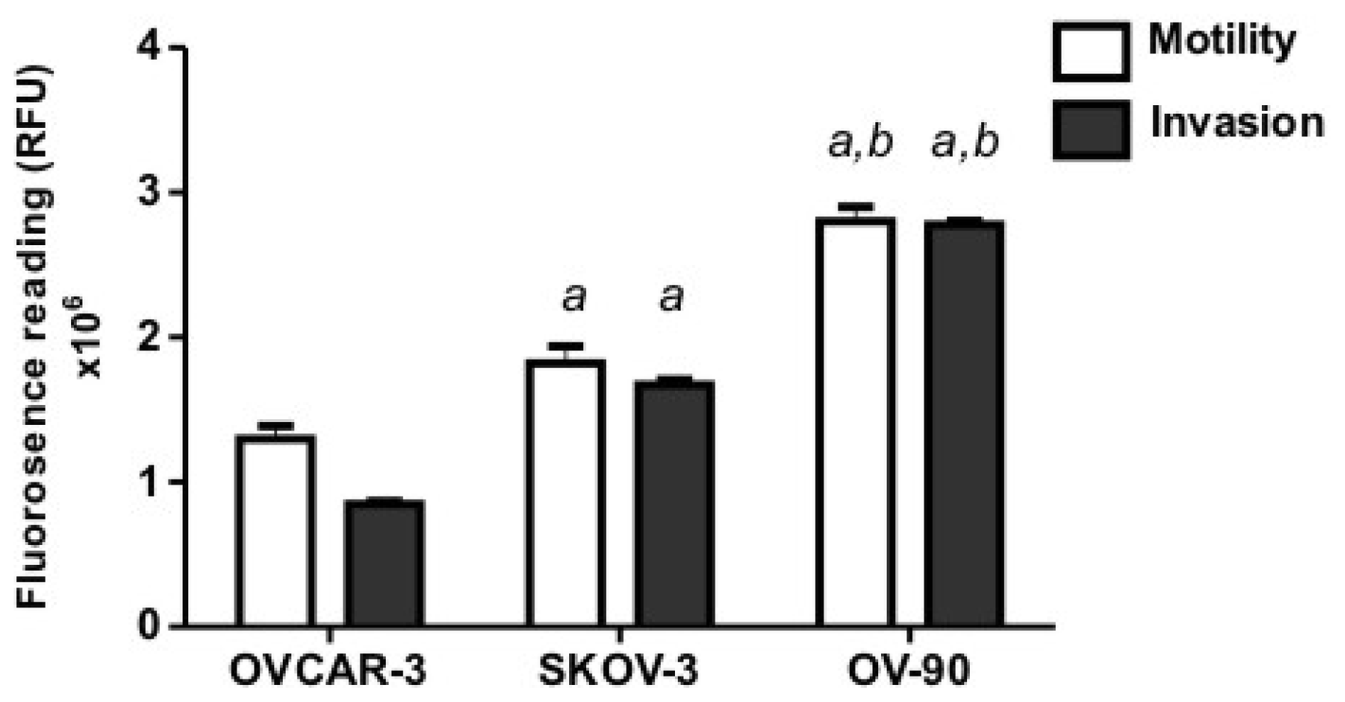
2.1. Human Ovarian Cancer Cell Motility and Invasion In Vitro</title>
<p>We compared the motility and invasion of three ovarian cancer cells (OVCAR-3, SKOV-3 and OV-90) using <italic>in vitro</italic> assays. We found OV-90 cells to be most invasive through an extracellular matrix and migrated faster through 12 μm pores towards a chemo attractant, compared to SKOV-3 and OVCAR-3 cells (<xref ref-type="fig" rid="f1-ijms-13-09959">Figure 1</xref>). OVCAR-3 cells were the least motile and invasive cell line in our study.</p></sec>
<sec>
<title>
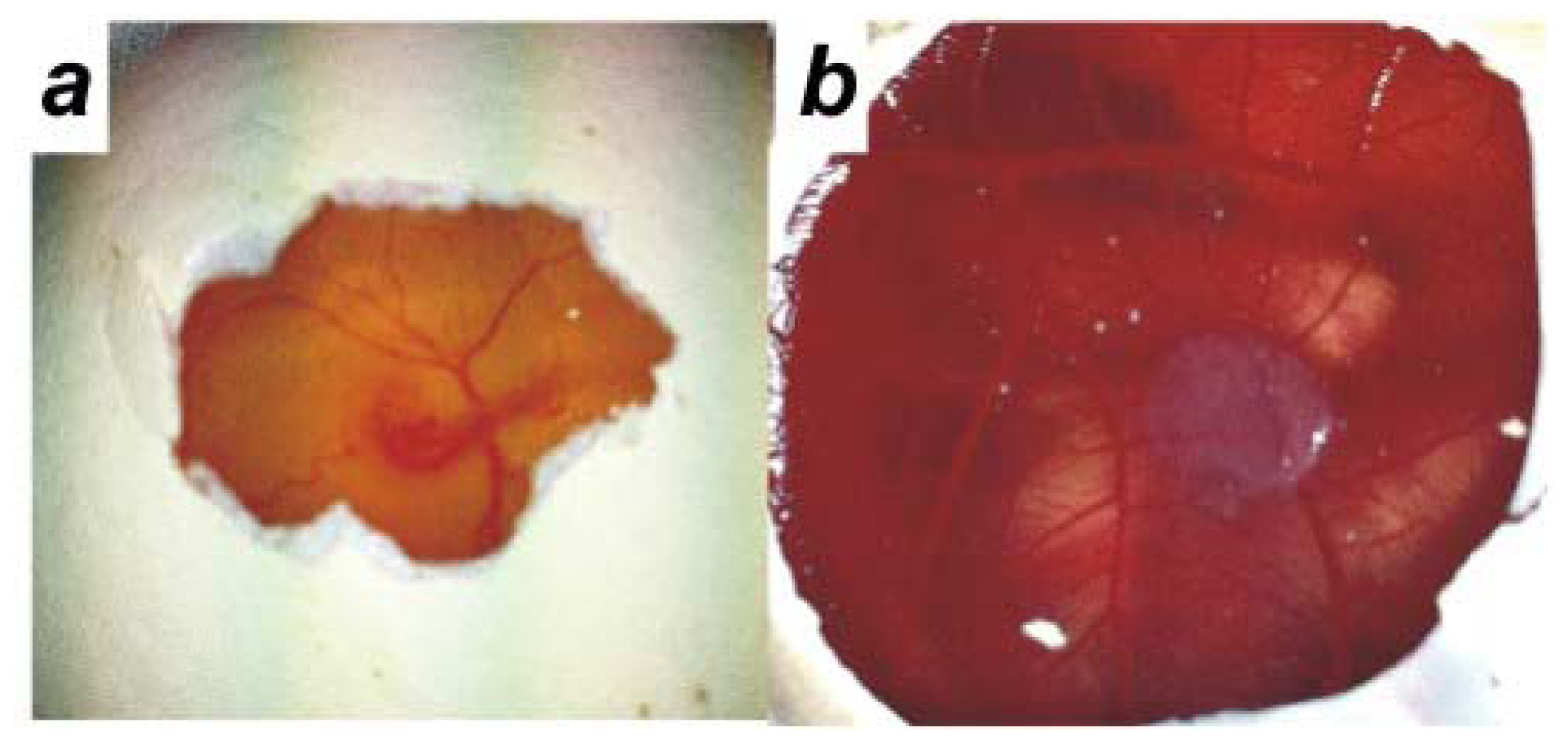
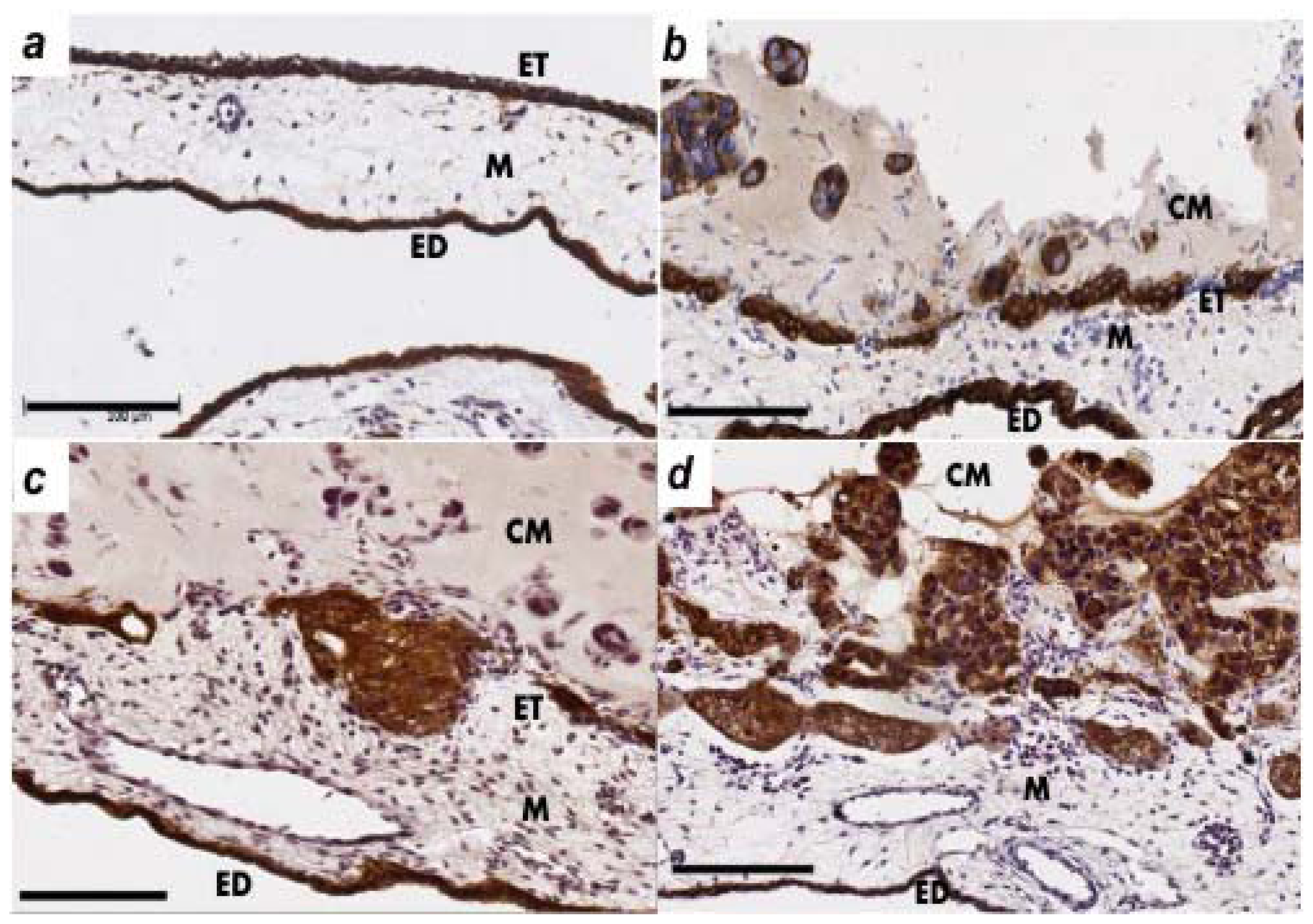
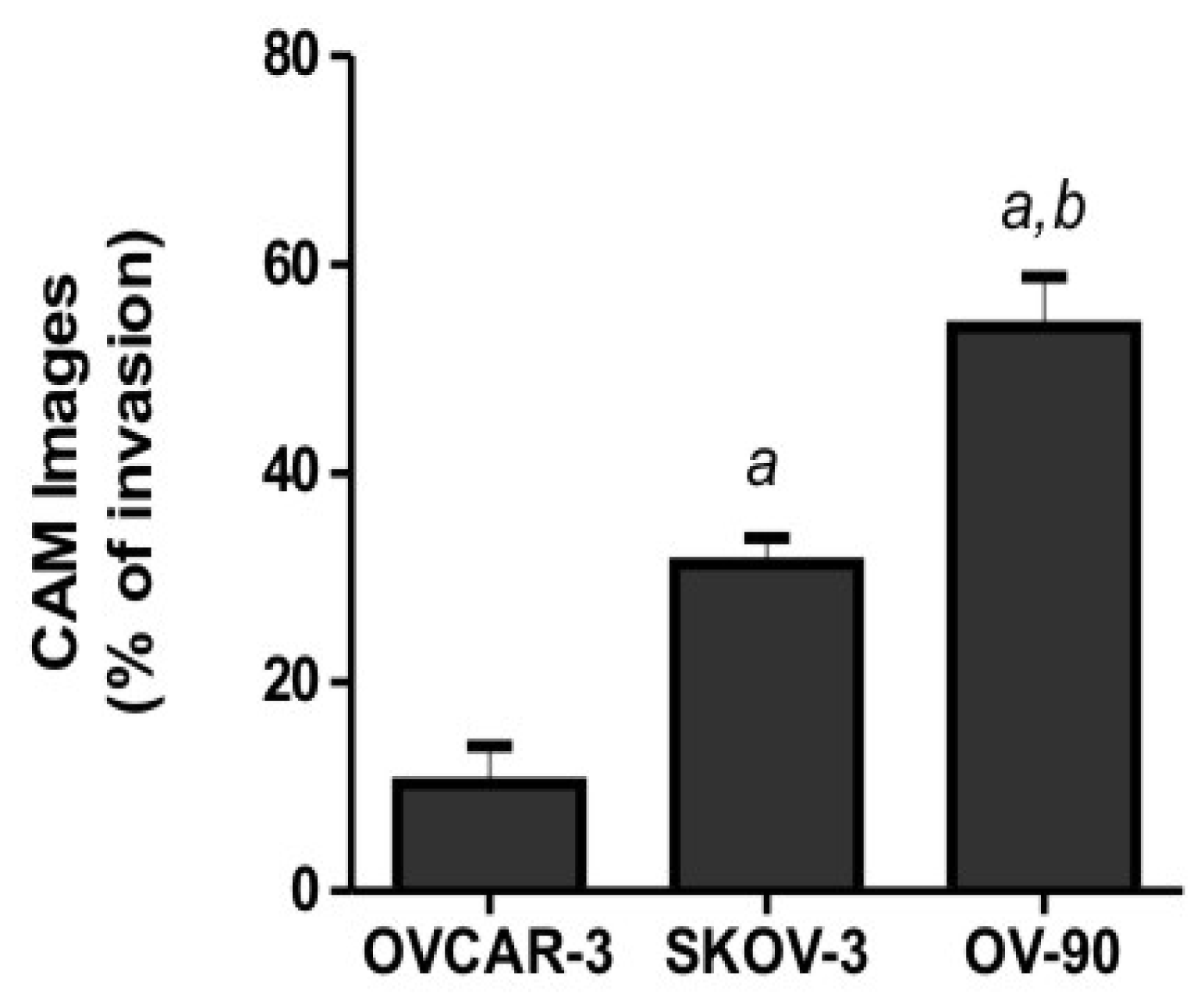
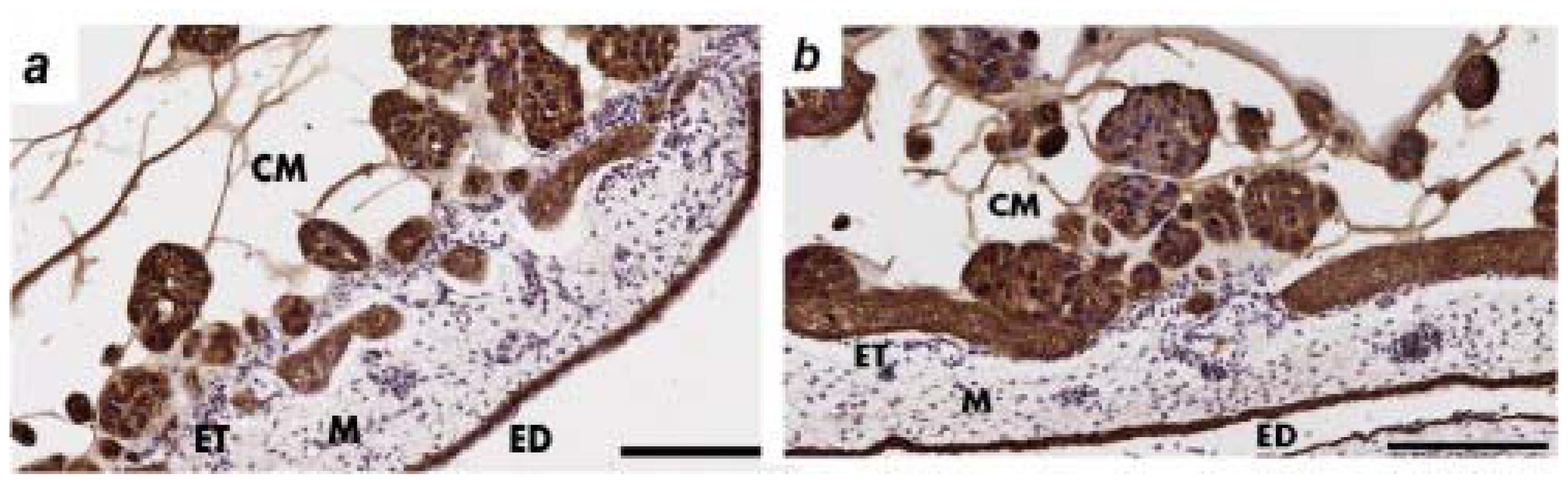
2.2. Human Ovarian Cancer Cell Invasion into the Chick Chorioallantoic Membrane (CAM)</title>
<p>We initially utilized an <italic>ex ovo</italic> method and incubated the chick embryos in plastic weigh boats as described previously [<xref ref-type="bibr" rid="b23-ijms-13-09959">23</xref>]. The <italic>ex ovo</italic> method has the advantage of allowing the application of a larger number of matrigel grafts as a wider area of the CAM is accessible. However, the survival rate for the <italic>ex ovo</italic> method was very low and only 10% of embryos survived to day 14. The <italic>in ovo</italic> method had a survival rate of 70% on day 14. In the <italic>in ovo</italic> method, a small window is made in the shell on day 3 of chick embryo development to detach the CAM layer from the egg shell (<xref ref-type="fig" rid="f2-ijms-13-09959">Figure 2a</xref>). Ovarian cancer cells (9 × 10<sup>5</sup> cells) were mixed with matrigel to form a gel and grafted on top of the CAM of day 11 chick embryos. The chick embryos were incubated with the matrigel grafts until day 14 of development. An example of a matrigel graft on day 14 is shown in <xref ref-type="fig" rid="f2-ijms-13-09959">Figure 2b</xref>.</p>
<p>The CAM layers; ectoderm (ET), mesoderm (M) and endoderm (ED) can be seen in <xref ref-type="fig" rid="f3-ijms-13-09959">Figure 3a</xref>. Cytokeratin immunohistochemistry was used to identify the CAM layer integrity and presence of ovarian cancer cells in the mesodermal layer.</p>
<p>The invasion of the ovarian cancer cells through the ectoderm into the mesoderm was assessed on day 14 of chick embryo development. OVCAR-3 cytokeratin immunohistochemistry showed some damage to the ectoderm layer but minimal invasion into the CAM mesoderm (<xref ref-type="fig" rid="f3-ijms-13-09959">Figure 3b</xref>). The SKOV-3 cells showed invasion into the mesoderm layer and minimal destruction of the ectoderm layer (<xref ref-type="fig" rid="f3-ijms-13-09959">Figure 3c</xref>). OV-90 cells were the most invasive cells in the CAM assay (<xref ref-type="fig" rid="f3-ijms-13-09959">Figure 3d</xref>), which agrees with the results of the <italic>in vitro</italic> assays (<xref ref-type="fig" rid="f1-ijms-13-09959">Figure 1</xref>). <xref ref-type="fig" rid="f3-ijms-13-09959">Figure 3d</xref> shows a large number of OV-90 cells invading into the mesoderm of the CAM, as well as the destruction of the CAM ectoderm layer.</p>
<p>Quantitative analysis of ovarian cancer cell invasion into the CAM was performed by determining the number of images (8 to 12 sections per chick embryo) with cancer cell invasion into the CAM mesoderm of day 14 chick embryos. Our results showed a significantly higher invasion of OV-90 and SKOV-3 cells into the CAM mesoderm, when compared with OVCAR-3 cells (<xref ref-type="fig" rid="f4-ijms-13-09959">Figure 4</xref>).</p></sec>
<sec>
<title>2.3. The Effects of Protein a Neutralizing Antibody on OV-90 Cancer Cell Invasion into the CAM</title>
<p>We used a neutralizing antibody against one of the novel proteins identified in our previous study investigating ovarian cancer-peritoneal cell interactions [<xref ref-type="bibr" rid="b21-ijms-13-09959">21</xref>,<xref ref-type="bibr" rid="b22-ijms-13-09959">22</xref>]. OV-90 cancer cells (9 × 10<sup>5</sup> cells) were mixed with matrigel and the control anti-mouse IgG or the neutralizing antibody against protein A before grafting onto day 11 chick embryos. Neutralizing antibody against protein A effectively inhibited OV-90 cancer cell invasion into the mesoderm of the CAM, compared with the control anti-mouse IgG, where OV-90 cancer cells invaded the mesoderm of the CAM and a destruction of the ectoderm layer was observed (<xref ref-type="fig" rid="f5-ijms-13-09959">Figure 5</xref>).</p></sec></sec>
<sec sec-type="discussion">
<title>3. Discussion</title>
<p>The CAM assay is a frequently applied model to study ovarian cancer angiogenesis [<xref ref-type="bibr" rid="b24-ijms-13-09959">24</xref>–<xref ref-type="bibr" rid="b27-ijms-13-09959">27</xref>]. However, there is only one study which has used CAM assays to assess ovarian cancer cell invasion and metastasis [<xref ref-type="bibr" rid="b20-ijms-13-09959">20</xref>]. Chang <italic>et al.</italic> described IGROV-1 ovarian cancer cell invasion and metastasis to the posterior CAM and lungs of chick embryos [<xref ref-type="bibr" rid="b20-ijms-13-09959">20</xref>]. We have developed a CAM assay protocol to monitor the metastatic properties of ovarian cancer cells (OVCAR-3, SKOV-3 and OV-90) and have successfully used it to study the effect of newly identified molecules <italic>in vivo</italic>. Our results show that the CAM assay is an effective model to study ovarian cancer metastasis. Importantly, our CAM model closely mimics the mode of ovarian cancer metastasis which involves ovarian cancer cell attachment and invasion into the peritoneum. The ectodermal layer of the CAM has many similarities with the peritoneum, which consists of a single layer of mesothelial cells covering the organs in the abdominal cavity.</p>
<p>We observed a higher survival rate with the <italic>in ovo</italic> method in comparison to the <italic>ex ovo</italic> method for monitoring of ovarian cancer cell growth in the chick embryos. Various methods have been used to graft cancer cells in the CAM model; such as collagen onplants [<xref ref-type="bibr" rid="b23-ijms-13-09959">23</xref>], plastic rings [<xref ref-type="bibr" rid="b19-ijms-13-09959">19</xref>], and matrigel grafts [<xref ref-type="bibr" rid="b28-ijms-13-09959">28</xref>]. Furthermore, cancer cells can also be inoculated by dropping the cell suspension on top of the CAM [<xref ref-type="bibr" rid="b29-ijms-13-09959">29</xref>], or administered intravenously to study metastasis of cancer cells in the chick embryos [<xref ref-type="bibr" rid="b18-ijms-13-09959">18</xref>]. Matrigel is one of the most suitable scaffolds used for implantation and grafting of cancer cells onto the CAM. In our model, ovarian cancer cells and matrigel were mixed with or without neutralizing antibodies before grafting onto the CAM of the chick embryos to assess ovarian cancer cell invasion. The grafting of the matrigel in the CAM model allows continuous visualization of the test site. Moreover, other studies have reported visible and solid tumors on the CAM of chick embryos a few days after cancer cell inoculation [<xref ref-type="bibr" rid="b11-ijms-13-09959">11</xref>,<xref ref-type="bibr" rid="b19-ijms-13-09959">19</xref>]. We used histological assessment by means of a pan-cytokeratin antibody, to allow the visualization of cancer cells invading into the mesoderm.</p>
<p>The CAM model has been previously employed to assess cancer metastasis [<xref ref-type="bibr" rid="b3-ijms-13-09959">3</xref>,<xref ref-type="bibr" rid="b29-ijms-13-09959">29</xref>]. In some studies quantitative <italic>alu</italic> PCR was used to assess the presence of metastatic human cancer cells in chick embryo organs [<xref ref-type="bibr" rid="b30-ijms-13-09959">30</xref>]. Several studies have compared both CAM assays and mouse models to assess tumor growth and metastasis. Colorectal cancer cells were reported to colonize the CAM similarly to the mouse model [<xref ref-type="bibr" rid="b10-ijms-13-09959">10</xref>]. Strojnik <italic>et al.</italic> conducted a study to compare the histological and immunohistochemical characteristics of glioma tumor protein expression in the CAM and an established rat model. They reported a similar profile of proteins expressed in both models [<xref ref-type="bibr" rid="b12-ijms-13-09959">12</xref>]. In addition, the CAM model has also been used concurrently with the nude mice model to assess tumor growth of fibrosarcoma (HT1080 cells) and human squamous carcinoma (Hep3 cells) cells [<xref ref-type="bibr" rid="b31-ijms-13-09959">31</xref>]. Lyu <italic>et al.</italic> also showed that over expression of urokinase-type plasminogen activator receptor (u-PAR) in Hep3 cells leads to an increase in cancer cell invasion in the CAM model as well as accelerated tumor growth in the SCID mice model [<xref ref-type="bibr" rid="b32-ijms-13-09959">32</xref>]. These studies demonstrate the validity of the CAM model for <italic>in vivo</italic> analysis of cancer cell invasion and metastasis.</p>
<p>The CAM model has many advantages. It is cost effective, allows large scale screening and is an easily reproducible <italic>in vivo</italic> model [<xref ref-type="bibr" rid="b4-ijms-13-09959">4</xref>,<xref ref-type="bibr" rid="b7-ijms-13-09959">7</xref>]. A comparison of the advantages and limitations of the CAM against the mouse model are summarized in <xref ref-type="table" rid="t1-ijms-13-09959">Table 1</xref>.</p>
<p>The CAM model has also been used in pre-clinical screening to assess the efficacy of drugs and inhibitors on tumor growth. Hagedorn <italic>et al.</italic> reported that treatment of human glioma cells with receptor tyrosine kinase inhibitors inhibited tumor growth in a CAM model [<xref ref-type="bibr" rid="b14-ijms-13-09959">14</xref>]. Bekes <italic>et al.</italic>, demonstrated that treatment of prostate cancer (PC3 cells) with u-PA activation blocking antibody mAb-112 significantly inhibited cancer cell invasion in the CAM model [<xref ref-type="bibr" rid="b35-ijms-13-09959">35</xref>]. Additionally, the CAM model has been used to test the efficacy of chemotherapy agents (such as doxorubicin) in human leukemia cell lines and has been shown to reduce cancer cell growth in the CAM [<xref ref-type="bibr" rid="b18-ijms-13-09959">18</xref>]. An important limitation of the CAM model is the inability to assess cancer–immune cell interactions. Examination of cancer–immune cell interactions requires the use of transgenic ovarian cancer models [<xref ref-type="bibr" rid="b36-ijms-13-09959">36</xref>], however, these models are not widely available, are not suitable for high throughput screening, and cannot be used with primary ovarian cancer cells derived from clinical samples. The CAM assay is therefore an attractive model to rapidly assess the effectiveness of novel candidate therapeutic drugs and the <italic>in vivo</italic> inhibition of specific tumor types and subtypes in one consistent model.</p>
<p>We have shown that OV-90 ovarian cancer cells invade into the mesoderm of the CAM within three days of implantation, therefore making the OV-90 CAM model ideal for studying ovarian cancer invasion and metastasis. In contrast, the OVCAR-3 cells showed limited invasion in the CAM over the three days of our assays, and would therefore be suitable for studying the role of molecules that promote ovarian cancer invasion. In conclusion, the CAM model provides a high throughput <italic>in vivo</italic> model for the assessment and evaluation of candidate pro-invasive molecules as well as potential therapeutic targets for ovarian cancer.</p></sec>
<sec sec-type="materials|methods">
<title>4. Materials and Methods</title>
<sec>
<title>4.1. Cell Culture</title>
<p>The human ovarian cancer cell lines OVCAR-3, SKOV-3 and OV-90 were obtained from American Type Culture Collection (ATCC, Manassas, VA, USA). All ovarian cancer cell lines were maintained in RPMI 1640 medium supplemented with 4 mM <sc>l</sc>-glutamine, antibiotics (100 U penicillin G, 100 μg/mL streptomycin sulfate and 0.25 μg/mL amphotericin B), (Sigma-Aldrich, St. Louis, MO, USA). OVCAR-3 and SKOV-3 cells were supplemented with 5% fetal bovine serum (FBS) (Sigma-Aldrich) and OV-90 cells were supplemented with 10% FBS. All cell lines were maintained at 37 °C in an environment of 5% CO<sub>2</sub>.</p></sec>
<sec>
<title>4.2. Cell Motility and Invasion Assays</title>
<p>Cell motility and invasion assays were performed as previously described [<xref ref-type="bibr" rid="b37-ijms-13-09959">37</xref>]. Briefly, the ovarian cancer cells (OVCAR-3, SKOV-3 and OV-90) were diluted to a cell concentration of 1 × 10<sup>6</sup> cells/mL and labeled with calcein-AM (1 μg/mL, Life Technologies, VIC, Australia). Ovarian cancer cells were mixed at room temperature for 1 hour in the dark on an oscillating platform. Ovarian cancer cells (50 μL, 40,000 cells) were loaded on top of uncoated 12 μm filter inserts (Disposable, 96-well plate, ChemoTx, Neuro Probe Inc, Gaitherburg, MD, USA) for motility assays or 12 μm filters coated with Geltrex (0.6 μL/well, 9 mg/mL, Life Technologies) for invasion assays. 10% FBS RPMI media was used as a chemo attractant. The cells were allowed to migrate and invade to the lower chamber for 6 hours. Non-migratory cells on the top of the filter were removed and the fluorescence was measured at 485–520 nm.</p></sec>
<sec>
<title>4.3. Chick Chorioallantoic Membrane Assay (CAM Assay)</title>
<p>Fertilized white leghorn chicken eggs were obtained from Hi-Chick, South Australia, Australia. Eggs were incubated in a MultiQuip Incubator (E2) at 37 °C with 60% humidity. Ethics approval was obtained by the University of Adelaide Animal Ethics Committee. A small window was made in the shell on day 3 of chick embryo development under aseptic conditions. The window was resealed with adhesive tape and eggs were returned to the incubator until day 11 of chick embryo development. On day 11, OVCAR-3, SKOV-3 and OV-90 cell suspensions (9 × 10<sup>5</sup>) were mixed with growth factor reduced matrigel (8.9 mg/mL, BD Biosciences, NSW, Australia) in a total volume of 30 μL. Control anti-mouse IgG (20 μg/mL) (Sigma Aldrich) and neutralizing antibody to protein A (20 μg/mL) (BD Biosciences) were mixed together with the OV-90 cancer cells and matrigel. Matrigel grafts were placed on top of the CAM and eggs were resealed and returned to the incubator for 72 hours until day 14 (n = 6 chicken embryos per cell line). Matrigel grafts with surrounding CAM were harvested from each embryo and fixed with 4% paraformaldehyde for 24 hours and embedded in paraffin. Serial sections (6 μm) were stained with hematoxylin and eosin. Slides were digitally scanned using the NanoZoomer (Hamamatsu Photonics K.K., Japan).</p></sec>
<sec>
<title>4.4. Immunohistochemistry</title>
<p>CAM paraffin sections (6 μm) were incubated on a heat plate at 60 °C for 2 hours. Tissue sections were dewaxed with xylene and ethanol, followed by PBS washes. Antigen retrieval was performed by using 1% protease (Sigma-Aldrich) for 10 minutes on a heat plate at 37 °C. The endogenous peroxidase activity of the sections was quenched with 0.3% H<sub>2</sub>O<sub>2</sub>. Each tissue sample was blocked with 5% goat serum for 30 minutes before incubation with monoclonal mouse anti-human cytokeratin clone AE1/AE3 (1:50, Dako, VIC, Australia) at 4 °C overnight. Subsequently, the tissues sections were incubated sequentially with biotinylated goat anti-mouse (1:400, Dako), followed by streptavidin-HRP conjugated (1:500, Dako) for 1 hour at room temperature. Immunoreactivity was detected using diaminobenzidine/H<sub>2</sub>O<sub>2</sub> substrate (Sigma-Aldrich). The sections were counterstained with 10% haematoxylin (Sigma-Aldrich), dehydrated and mounted in Pertex (Medite Medizintechnik, Germany). Slides were digitally scanned using the NanoZoomer (Hamamatsu Photonics K.K.). For quantitative analysis of ovarian cancer cell invasion into the mesoderm layer, 8 to 12 CAM images from each embryo were assessed by two independent researchers [<xref ref-type="bibr" rid="b16-ijms-13-09959">16</xref>].</p></sec>
<sec sec-type="methods">
<title>4.5. Statistical Analysis</title>
<p>All analyses were performed using SPSS 15.0 for Windows software (SPSS, Chicago, IL, USA). The student’s t-test and one-way ANOVA were used to determine statistical significance of ovarian cancer cell motility and invasion <italic>in vitro</italic>, and ovarian cancer cell invasion in the CAM model. Statistical significance was accepted at <italic>p</italic> &lt; 0.05.</p></sec></sec>
<sec sec-type="conclusions">
<title>5. Conclusions</title>
<p>The CAM assay is a robust technique that can be used to monitor invasion of ovarian cancer cell lines and to assess the role of novel molecules and potential therapeutic targets. It is a valuable alternative to murine <italic>in vivo</italic> models for the study of ovarian cancer invasion and metastasis.</p></sec></body>
<back>
<ack>
<title>Acknowledgments</title>
<p>The grant sponsors for this project were the Ovarian Cancer Research Foundation (OCRF), Australia, Cancer Council South Australia and South Australian Health and Medical Research Institute. We thank Simon Koblar from the School of Medicine, University of Adelaide, Australia for his help with setting up the CAM assay.</p></ack>
<ref-list>
<title>References</title>
<ref id="b1-ijms-13-09959"><label>1</label><citation citation-type="journal"><person-group person-group-type="author"><name><surname>Lengyel</surname><given-names>E.</given-names></name></person-group><article-title>Ovarian cancer development and metastasis</article-title><source>Am. J. Pathol</source><year>2010</year><volume>177</volume><fpage>1053</fpage><lpage>1064</lpage><pub-id pub-id-type="doi">10.2353/ajpath.2010.100105</pub-id><pub-id pub-id-type="pmid">20651229</pub-id></citation></ref>
<ref id="b2-ijms-13-09959"><label>2</label><citation citation-type="journal"><person-group person-group-type="author"><name><surname>Richardson</surname><given-names>M.</given-names></name><name><surname>Singh</surname><given-names>G.</given-names></name></person-group><article-title>Observations on the use of the avian chorioallantoic membrane (CAM) model in investigations into angiogenesis</article-title><source>Curr. Drug Targets-Cardiovasc. Hematol. Disord</source><year>2003</year><volume>3</volume><fpage>155</fpage><lpage>185</lpage><pub-id pub-id-type="doi">10.2174/1568006033481492</pub-id></citation></ref>
<ref id="b3-ijms-13-09959"><label>3</label><citation citation-type="journal"><person-group person-group-type="author"><name><surname>Deryugina</surname><given-names>E.I.</given-names></name><name><surname>Quigley</surname><given-names>J.P.</given-names></name></person-group><article-title>Chick embryo chorioallantoic membrane model systems to study and visualize human tumor cell metastasis</article-title><source>Histochem. Cell Biol</source><year>2008</year><volume>130</volume><fpage>1119</fpage><lpage>1130</lpage><pub-id pub-id-type="doi">10.1007/s00418-008-0536-2</pub-id><pub-id pub-id-type="pmid">19005674</pub-id></citation></ref>
<ref id="b4-ijms-13-09959"><label>4</label><citation citation-type="journal"><person-group person-group-type="author"><name><surname>Tufan</surname><given-names>A.C.</given-names></name><name><surname>Satiroglu-Tufan</surname><given-names>N.L.</given-names></name></person-group><article-title>The chick embryo chorioallantoic membrane as a model system for the study of tumor angiogenesis, invasion and development of anti-angiogenic agents</article-title><source>Curr. Cancer Drug Targets</source><year>2005</year><volume>5</volume><fpage>249</fpage><lpage>266</lpage><pub-id pub-id-type="doi">10.2174/1568009054064624</pub-id><pub-id pub-id-type="pmid">15975046</pub-id></citation></ref>
<ref id="b5-ijms-13-09959"><label>5</label><citation citation-type="journal"><person-group person-group-type="author"><name><surname>Ossowski</surname><given-names>L.</given-names></name></person-group><article-title><italic>In vivo</italic> invasion of modified chorioallantoic membrane by tumor cells: The role of cell surface-bound urokinase</article-title><source>J. Cell Biol</source><year>1988</year><volume>107</volume><fpage>2437</fpage><lpage>2445</lpage><pub-id pub-id-type="doi">10.1083/jcb.107.6.2437</pub-id><pub-id pub-id-type="pmid">2848851</pub-id></citation></ref>
<ref id="b6-ijms-13-09959"><label>6</label><citation citation-type="journal"><person-group person-group-type="author"><name><surname>Zhai</surname><given-names>Y.</given-names></name><name><surname>Kuick</surname><given-names>R.</given-names></name><name><surname>Nan</surname><given-names>B.</given-names></name><name><surname>Ota</surname><given-names>I.</given-names></name><name><surname>Weiss</surname><given-names>S.J.</given-names></name><name><surname>Trimble</surname><given-names>C.L.</given-names></name><name><surname>Fearon</surname><given-names>E.R.</given-names></name><name><surname>Cho</surname><given-names>K.R.</given-names></name></person-group><article-title>Gene expression analysis of preinvasive and invasive cervical squamous cell carcinomas identifies HOXC10 as a key mediator of invasion</article-title><source>Cancer Res</source><year>2007</year><volume>67</volume><fpage>10163</fpage><lpage>10172</lpage><pub-id pub-id-type="doi">10.1158/0008-5472.CAN-07-2056</pub-id><pub-id pub-id-type="pmid">17974957</pub-id></citation></ref>
<ref id="b7-ijms-13-09959"><label>7</label><citation citation-type="journal"><person-group person-group-type="author"><name><surname>Cimpean</surname><given-names>A.M.</given-names></name><name><surname>Ribatti</surname><given-names>D.</given-names></name><name><surname>Raica</surname><given-names>M.</given-names></name></person-group><article-title>The chick embryo chorioallantoic membrane as a model to study tumor metastasis</article-title><source>Angiogenesis</source><year>2008</year><volume>11</volume><fpage>311</fpage><lpage>319</lpage><pub-id pub-id-type="doi">10.1007/s10456-008-9117-1</pub-id><pub-id pub-id-type="pmid">18780151</pub-id></citation></ref>
<ref id="b8-ijms-13-09959"><label>8</label><citation citation-type="journal"><person-group person-group-type="author"><name><surname>Valdes</surname><given-names>T.I.</given-names></name><name><surname>Kreutzer</surname><given-names>D.</given-names></name><name><surname>Moussy</surname><given-names>F.</given-names></name></person-group><article-title>The chick chorioallantoic membrane as a novel <italic>in vivo</italic> model for the testing of biomaterials</article-title><source>J. Biomed. Mater. Res</source><year>2002</year><volume>62</volume><fpage>273</fpage><lpage>282</lpage><pub-id pub-id-type="doi">10.1002/jbm.10152</pub-id><pub-id pub-id-type="pmid">12209948</pub-id></citation></ref>
<ref id="b9-ijms-13-09959"><label>9</label><citation citation-type="journal"><person-group person-group-type="author"><name><surname>Giannopoulou</surname><given-names>E.</given-names></name><name><surname>Katsoris</surname><given-names>P.</given-names></name><name><surname>Hatziapostolou</surname><given-names>M.</given-names></name><name><surname>Kardamakis</surname><given-names>D.</given-names></name><name><surname>Kotsaki</surname><given-names>E.</given-names></name><name><surname>Polytarchou</surname><given-names>C.</given-names></name><name><surname>Parthymou</surname><given-names>A.</given-names></name><name><surname>Papaioannou</surname><given-names>S.</given-names></name><name><surname>Papadimitriou</surname><given-names>E.</given-names></name></person-group><article-title>X-Rays modulate extracellular matrix <italic>in vivo</italic></article-title><source>Int. J. Cancer</source><year>2001</year><volume>94</volume><fpage>690</fpage><lpage>698</lpage><pub-id pub-id-type="doi">10.1002/ijc.1535</pub-id><pub-id pub-id-type="pmid">11745464</pub-id></citation></ref>
<ref id="b10-ijms-13-09959"><label>10</label><citation citation-type="journal"><person-group person-group-type="author"><name><surname>Subauste</surname><given-names>M.C.</given-names></name><name><surname>Kupriyanova</surname><given-names>T.A.</given-names></name><name><surname>Conn</surname><given-names>E.M.</given-names></name><name><surname>Ardi</surname><given-names>V.C.</given-names></name><name><surname>Quigley</surname><given-names>J.P.</given-names></name><name><surname>Deryugina</surname><given-names>E.I.</given-names></name></person-group><article-title>Evaluation of metastatic and angiogenic potentials of human colon carcinoma cells in chick embryo model systems</article-title><source>Clin. Exp. Metastasis</source><year>2009</year><volume>26</volume><fpage>1033</fpage><lpage>1047</lpage><pub-id pub-id-type="doi">10.1007/s10585-009-9293-4</pub-id><pub-id pub-id-type="pmid">19842048</pub-id></citation></ref>
<ref id="b11-ijms-13-09959"><label>11</label><citation citation-type="journal"><person-group person-group-type="author"><name><surname>Demir</surname><given-names>R.</given-names></name><name><surname>Dimmler</surname><given-names>A.</given-names></name><name><surname>Naschberger</surname><given-names>E.</given-names></name><name><surname>Demir</surname><given-names>I.</given-names></name><name><surname>Papadopoulos</surname><given-names>T.</given-names></name><name><surname>Melling</surname><given-names>N.</given-names></name><name><surname>Sturzl</surname><given-names>M.</given-names></name><name><surname>Hohenberger</surname><given-names>W.</given-names></name></person-group><article-title>Malignant progression of invasive tumour cells seen in hypoxia present an accumulation of beta-catenin in the nucleus at the tumour front</article-title><source>Exp. Mol. Pathol</source><year>2009</year><volume>87</volume><fpage>109</fpage><lpage>116</lpage><pub-id pub-id-type="doi">10.1016/j.yexmp.2009.05.004</pub-id><pub-id pub-id-type="pmid">19481539</pub-id></citation></ref>
<ref id="b12-ijms-13-09959"><label>12</label><citation citation-type="journal"><person-group person-group-type="author"><name><surname>Strojnik</surname><given-names>T.</given-names></name><name><surname>Kavalar</surname><given-names>R.</given-names></name><name><surname>Barone</surname><given-names>T.A.</given-names></name><name><surname>Plunkett</surname><given-names>R.J.</given-names></name></person-group><article-title>Experimental model and immunohistochemical comparison of U87 human glioblastoma cell xenografts on the chicken chorioallantoic membrane and in rat brains</article-title><source>Anticancer Res</source><year>2010</year><volume>30</volume><fpage>4851</fpage><lpage>4860</lpage><pub-id pub-id-type="pmid">21187462</pub-id></citation></ref>
<ref id="b13-ijms-13-09959"><label>13</label><citation citation-type="journal"><person-group person-group-type="author"><name><surname>Balciuniene</surname><given-names>N.</given-names></name><name><surname>Tamasauskas</surname><given-names>A.</given-names></name><name><surname>Valanciute</surname><given-names>A.</given-names></name><name><surname>Deltuva</surname><given-names>V.</given-names></name><name><surname>Vaitiekaitis</surname><given-names>G.</given-names></name><name><surname>Gudinaviciene</surname><given-names>I.</given-names></name><name><surname>Weis</surname><given-names>J.</given-names></name><name><surname>von Keyserlingk</surname><given-names>D.G.</given-names></name></person-group><article-title>Histology of human glioblastoma transplanted on chicken chorioallantoic membrane</article-title><source>Medicina (Kaunas)</source><year>2009</year><volume>45</volume><fpage>123</fpage><lpage>131</lpage></citation></ref>
<ref id="b14-ijms-13-09959"><label>14</label><citation citation-type="journal"><person-group person-group-type="author"><name><surname>Hagedorn</surname><given-names>M.</given-names></name><name><surname>Javerzat</surname><given-names>S.</given-names></name><name><surname>Gilges</surname><given-names>D.</given-names></name><name><surname>Meyre</surname><given-names>A.</given-names></name><name><surname>de Lafarge</surname><given-names>B.</given-names></name><name><surname>Eichmann</surname><given-names>A.</given-names></name><name><surname>Bikfalvi</surname><given-names>A.</given-names></name></person-group><article-title>Accessing key steps of human tumor progression <italic>in vivo</italic> by using an avian embryo model</article-title><source>Proc. Natl. Acad. Sci. USA</source><year>2005</year><volume>102</volume><fpage>1643</fpage><lpage>1648</lpage><pub-id pub-id-type="doi">10.1073/pnas.0408622102</pub-id><pub-id pub-id-type="pmid">15665100</pub-id></citation></ref>
<ref id="b15-ijms-13-09959"><label>15</label><citation citation-type="journal"><person-group person-group-type="author"><name><surname>Kobayashi</surname><given-names>T.</given-names></name><name><surname>Koshida</surname><given-names>K.</given-names></name><name><surname>Endo</surname><given-names>Y.</given-names></name><name><surname>Imao</surname><given-names>T.</given-names></name><name><surname>Uchibayashi</surname><given-names>T.</given-names></name><name><surname>Sasaki</surname><given-names>T.</given-names></name><name><surname>Namiki</surname><given-names>M.</given-names></name></person-group><article-title>A chick embryo model for metastatic human prostate cancer</article-title><source>Eur. Urol</source><year>1998</year><volume>34</volume><fpage>154</fpage><lpage>160</lpage><pub-id pub-id-type="doi">10.1159/000019702</pub-id><pub-id pub-id-type="pmid">9693252</pub-id></citation></ref>
<ref id="b16-ijms-13-09959"><label>16</label><citation citation-type="journal"><person-group person-group-type="author"><name><surname>Wittig-Blaich</surname><given-names>S.M.</given-names></name><name><surname>Kacprzyk</surname><given-names>L.A.</given-names></name><name><surname>Eismann</surname><given-names>T.</given-names></name><name><surname>Bewerunge-Hudler</surname><given-names>M.</given-names></name><name><surname>Kruse</surname><given-names>P.</given-names></name><name><surname>Winkler</surname><given-names>E.</given-names></name><name><surname>Strauss</surname><given-names>W.S.</given-names></name><name><surname>Hibst</surname><given-names>R.</given-names></name><name><surname>Steiner</surname><given-names>R.</given-names></name><name><surname>Schrader</surname><given-names>M.</given-names></name><etal/></person-group><article-title>Matrix-dependent regulation of AKT in Hepsin-overexpressing PC3 prostate cancer cells</article-title><source>Neoplasia</source><year>2011</year><volume>13</volume><fpage>579</fpage><lpage>589</lpage><pub-id pub-id-type="pmid">21750652</pub-id></citation></ref>
<ref id="b17-ijms-13-09959"><label>17</label><citation citation-type="journal"><person-group person-group-type="author"><name><surname>Conn</surname><given-names>E.M.</given-names></name><name><surname>Botkjaer</surname><given-names>K.A.</given-names></name><name><surname>Kupriyanova</surname><given-names>T.A.</given-names></name><name><surname>Andreasen</surname><given-names>P.A.</given-names></name><name><surname>Deryugina</surname><given-names>E.I.</given-names></name><name><surname>Quigley</surname><given-names>J.P.</given-names></name></person-group><article-title>Comparative analysis of metastasis variants derived from human prostate carcinoma cells: Roles in intravasation of VEGF-mediated angiogenesis and uPA-mediated invasion</article-title><source>Am. J. Pathol</source><year>2009</year><volume>175</volume><fpage>1638</fpage><lpage>1652</lpage><pub-id pub-id-type="doi">10.2353/ajpath.2009.090384</pub-id><pub-id pub-id-type="pmid">19729488</pub-id></citation></ref>
<ref id="b18-ijms-13-09959"><label>18</label><citation citation-type="journal"><person-group person-group-type="author"><name><surname>Taizi</surname><given-names>M.</given-names></name><name><surname>Deutsch</surname><given-names>V.R.</given-names></name><name><surname>Leitner</surname><given-names>A.</given-names></name><name><surname>Ohana</surname><given-names>A.</given-names></name><name><surname>Goldstein</surname><given-names>R.S.</given-names></name></person-group><article-title>A novel and rapid <italic>in vivo</italic> system for testing therapeutics on human leukemias</article-title><source>Exp. Hematol</source><year>2006</year><volume>34</volume><fpage>1698</fpage><lpage>1708</lpage><pub-id pub-id-type="doi">10.1016/j.exphem.2006.07.005</pub-id><pub-id pub-id-type="pmid">17157167</pub-id></citation></ref>
<ref id="b19-ijms-13-09959"><label>19</label><citation citation-type="journal"><person-group person-group-type="author"><name><surname>Balke</surname><given-names>M.</given-names></name><name><surname>Neumann</surname><given-names>A.</given-names></name><name><surname>Kersting</surname><given-names>C.</given-names></name><name><surname>Agelopoulos</surname><given-names>K.</given-names></name><name><surname>Gebert</surname><given-names>C.</given-names></name><name><surname>Gosheger</surname><given-names>G.</given-names></name><name><surname>Buerger</surname><given-names>H.</given-names></name><name><surname>Hagedorn</surname><given-names>M</given-names></name></person-group><article-title>Morphologic characterization of osteosarcoma growth on the chick chorioallantoic membrane</article-title><source>BMC Res. Notes</source><year>2010</year><volume>3</volume><pub-id pub-id-type="doi">10.1186/1756-0500-3-58</pub-id></citation></ref>
<ref id="b20-ijms-13-09959"><label>20</label><citation citation-type="journal"><person-group person-group-type="author"><name><surname>Chang</surname><given-names>H.L.</given-names></name><name><surname>Pieretti-Vanmarcke</surname><given-names>R.</given-names></name><name><surname>Nicolaou</surname><given-names>F.</given-names></name><name><surname>Li</surname><given-names>X.</given-names></name><name><surname>Wei</surname><given-names>X.</given-names></name><name><surname>MacLaughlin</surname><given-names>D.T.</given-names></name><name><surname>Donahoe</surname><given-names>P.K.</given-names></name></person-group><article-title>Mullerian inhibiting substance inhibits invasion and migration of epithelial cancer cell lines</article-title><source>Gynecol. Oncol</source><year>2011</year><volume>120</volume><fpage>128</fpage><lpage>134</lpage><pub-id pub-id-type="doi">10.1016/j.ygyno.2010.09.017</pub-id><pub-id pub-id-type="pmid">21056908</pub-id></citation></ref>
<ref id="b21-ijms-13-09959"><label>21</label><citation citation-type="journal"><person-group person-group-type="author"><name><surname>Ween</surname><given-names>M.P.</given-names></name><name><surname>Lokman</surname><given-names>N.A.</given-names></name><name><surname>Hoffmann</surname><given-names>P.</given-names></name><name><surname>Rodgers</surname><given-names>R.J.</given-names></name><name><surname>Ricciardelli</surname><given-names>C.</given-names></name><name><surname>Oehler</surname><given-names>M.K.</given-names></name></person-group><article-title>Transforming growth factor-beta-induced protein secreted by peritoneal cells increases the metastatic potential of ovarian cancer cells</article-title><source>Int. J. Cancer</source><year>2011</year><volume>128</volume><fpage>1570</fpage><lpage>1584</lpage><pub-id pub-id-type="doi">10.1002/ijc.25494</pub-id><pub-id pub-id-type="pmid">20521251</pub-id></citation></ref>
<ref id="b22-ijms-13-09959"><label>22</label><citation citation-type="journal"><person-group person-group-type="author"><name><surname>Lokman</surname><given-names>N.A.</given-names></name><name><surname>Ween</surname><given-names>M.P.</given-names></name><name><surname>Oehler</surname><given-names>M.K.</given-names></name><name><surname>Ricciardelli</surname><given-names>C.</given-names></name></person-group><article-title>The role of annexin A2 in tumorigenesis and cancer progression</article-title><source>Cancer Microenviron</source><year>2011</year><volume>4</volume><fpage>199</fpage><lpage>208</lpage><pub-id pub-id-type="doi">10.1007/s12307-011-0064-9</pub-id><pub-id pub-id-type="pmid">21909879</pub-id></citation></ref>
<ref id="b23-ijms-13-09959"><label>23</label><citation citation-type="journal"><person-group person-group-type="author"><name><surname>Deryugina</surname><given-names>E.I.</given-names></name><name><surname>Quigley</surname><given-names>J.P.</given-names></name></person-group><article-title>Chapter 2. Chick embryo chorioallantoic membrane models to quantify angiogenesis induced by inflammatory and tumor cells or purified effector molecules</article-title><source>Methods Enzymol</source><year>2008</year><volume>444</volume><fpage>21</fpage><lpage>41</lpage><pub-id pub-id-type="doi">10.1016/S0076-6879(08)02802-4</pub-id><pub-id pub-id-type="pmid">19007659</pub-id></citation></ref>
<ref id="b24-ijms-13-09959"><label>24</label><citation citation-type="journal"><person-group person-group-type="author"><name><surname>Manenti</surname><given-names>L.</given-names></name><name><surname>Riccardi</surname><given-names>E.</given-names></name><name><surname>Marchini</surname><given-names>S.</given-names></name><name><surname>Naumova</surname><given-names>E.</given-names></name><name><surname>Floriani</surname><given-names>I.</given-names></name><name><surname>Garofalo</surname><given-names>A.</given-names></name><name><surname>Dossi</surname><given-names>R.</given-names></name><name><surname>Marrazzo</surname><given-names>E.</given-names></name><name><surname>Ribatti</surname><given-names>D.</given-names></name><name><surname>Scanziani</surname><given-names>E.</given-names></name><etal/></person-group><article-title>Circulating plasma vascular endothelial growth factor in mice bearing human ovarian carcinoma xenograft correlates with tumor progression and response to therapy</article-title><source>Mol. Cancer Ther</source><year>2005</year><volume>4</volume><fpage>715</fpage><lpage>725</lpage><pub-id pub-id-type="doi">10.1158/1535-7163.MCT-04-0305</pub-id><pub-id pub-id-type="pmid">15897235</pub-id></citation></ref>
<ref id="b25-ijms-13-09959"><label>25</label><citation citation-type="journal"><person-group person-group-type="author"><name><surname>Fang</surname><given-names>J.</given-names></name><name><surname>Zhou</surname><given-names>Q.</given-names></name><name><surname>Liu</surname><given-names>L.Z.</given-names></name><name><surname>Xia</surname><given-names>C.</given-names></name><name><surname>Hu</surname><given-names>X.</given-names></name><name><surname>Shi</surname><given-names>X.</given-names></name><name><surname>Jiang</surname><given-names>B.H.</given-names></name></person-group><article-title>Apigenin inhibits tumor angiogenesis through decreasing HIF-1α and VEGF expression</article-title><source>Carcinogenesis</source><year>2006</year><volume>28</volume><fpage>858</fpage><lpage>864</lpage><pub-id pub-id-type="doi">10.1093/carcin/bgl205</pub-id><pub-id pub-id-type="pmid">17071632</pub-id></citation></ref>
<ref id="b26-ijms-13-09959"><label>26</label><citation citation-type="journal"><person-group person-group-type="author"><name><surname>Luo</surname><given-names>H.</given-names></name><name><surname>Rankin</surname><given-names>G.O.</given-names></name><name><surname>Liu</surname><given-names>L.</given-names></name><name><surname>Daddysman</surname><given-names>M.K.</given-names></name><name><surname>Jiang</surname><given-names>B.H.</given-names></name><name><surname>Chen</surname><given-names>Y.C.</given-names></name></person-group><article-title>Kaempferol inhibits angiogenesis and VEGF expression through both HIF dependent and independent pathways in human ovarian cancer cells</article-title><source>Nutr. Cancer</source><year>2009</year><volume>61</volume><fpage>554</fpage><lpage>563</lpage><pub-id pub-id-type="doi">10.1080/01635580802666281</pub-id><pub-id pub-id-type="pmid">19838928</pub-id></citation></ref>
<ref id="b27-ijms-13-09959"><label>27</label><citation citation-type="journal"><person-group person-group-type="author"><name><surname>Liu</surname><given-names>L.Z.</given-names></name><name><surname>Jing</surname><given-names>Y.</given-names></name><name><surname>Jiang</surname><given-names>L.L.</given-names></name><name><surname>Jiang</surname><given-names>X.E.</given-names></name><name><surname>Jiang</surname><given-names>Y.</given-names></name><name><surname>Rojanasakul</surname><given-names>Y.</given-names></name><name><surname>Jiang</surname><given-names>B.H.</given-names></name></person-group><article-title>Acacetin inhibits VEGF expression, tumor angiogenesis and growth through AKT/HIF-1alpha pathway</article-title><source>Biochem. Biophys. Res. Commun</source><year>2011</year><volume>413</volume><fpage>299</fpage><lpage>305</lpage><pub-id pub-id-type="doi">10.1016/j.bbrc.2011.08.091</pub-id><pub-id pub-id-type="pmid">21893035</pub-id></citation></ref>
<ref id="b28-ijms-13-09959"><label>28</label><citation citation-type="journal"><person-group person-group-type="author"><name><surname>Kisker</surname><given-names>O.</given-names></name><name><surname>Onizuka</surname><given-names>S.</given-names></name><name><surname>Banyard</surname><given-names>J.</given-names></name><name><surname>Komiyama</surname><given-names>T.</given-names></name><name><surname>Becker</surname><given-names>C.M.</given-names></name><name><surname>Achilles</surname><given-names>E.G.</given-names></name><name><surname>Barnes</surname><given-names>C.M.</given-names></name><name><surname>O’Reilly</surname><given-names>M.S.</given-names></name><name><surname>Folkman</surname><given-names>J.</given-names></name><name><surname>Pirie-Shepherd</surname><given-names>S.R.</given-names></name></person-group><article-title>Generation of multiple angiogenesis inhibitors by human pancreatic cancer</article-title><source>Cancer Res</source><year>2001</year><volume>61</volume><fpage>7298</fpage><lpage>7304</lpage><pub-id pub-id-type="pmid">11585769</pub-id></citation></ref>
<ref id="b29-ijms-13-09959"><label>29</label><citation citation-type="journal"><person-group person-group-type="author"><name><surname>Petitclerc</surname><given-names>E.</given-names></name><name><surname>Brooks</surname><given-names>P.C.</given-names></name></person-group><article-title>The chick embryo metastasis model</article-title><source>Methods Mol. Med</source><year>2001</year><volume>58</volume><fpage>173</fpage><lpage>187</lpage><pub-id pub-id-type="pmid">21340857</pub-id></citation></ref>
<ref id="b30-ijms-13-09959"><label>30</label><citation citation-type="journal"><person-group person-group-type="author"><name><surname>Zijlstra</surname><given-names>A.</given-names></name><name><surname>Mellor</surname><given-names>R.</given-names></name><name><surname>Panzarella</surname><given-names>G.</given-names></name><name><surname>Aimes</surname><given-names>R.T.</given-names></name><name><surname>Hooper</surname><given-names>J.D.</given-names></name><name><surname>Marchenko</surname><given-names>N.D.</given-names></name><name><surname>Quigley</surname><given-names>J.P.</given-names></name></person-group><article-title>A quantitative analysis of rate-limiting steps in the metastatic cascade using human-specific real-time polymerase chain reaction</article-title><source>Cancer Res</source><year>2002</year><volume>62</volume><fpage>7083</fpage><lpage>7092</lpage><pub-id pub-id-type="pmid">12460930</pub-id></citation></ref>
<ref id="b31-ijms-13-09959"><label>31</label><citation citation-type="journal"><person-group person-group-type="author"><name><surname>Aguirre-Ghiso</surname><given-names>J.A.</given-names></name><name><surname>Ossowski</surname><given-names>L.</given-names></name><name><surname>Rosenbaum</surname><given-names>S.K.</given-names></name></person-group><article-title>Green fluorescent protein tagging of extracellular signal-regulated kinase and p38 pathways reveals novel dynamics of pathway activation during primary and metastatic growth</article-title><source>Cancer Res</source><year>2004</year><volume>64</volume><fpage>7336</fpage><lpage>7345</lpage><pub-id pub-id-type="doi">10.1158/0008-5472.CAN-04-0113</pub-id><pub-id pub-id-type="pmid">15492254</pub-id></citation></ref>
<ref id="b32-ijms-13-09959"><label>32</label><citation citation-type="journal"><person-group person-group-type="author"><name><surname>Lyu</surname><given-names>M.A.</given-names></name><name><surname>Choi</surname><given-names>Y.K.</given-names></name><name><surname>Park</surname><given-names>B.N.</given-names></name><name><surname>Kim</surname><given-names>B.J.</given-names></name><name><surname>Park</surname><given-names>I.K.</given-names></name><name><surname>Hyun</surname><given-names>B.H.</given-names></name><name><surname>Kook</surname><given-names>Y.H.</given-names></name></person-group><article-title>Over-expression of urokinase receptor in human epidermoid-carcinoma cell line (HEp3) increases tumorigenicity on chorio-allantoic membrane and in severe-combined-immunodeficient mice</article-title><source>Int. J. Cancer</source><year>1998</year><volume>77</volume><fpage>257</fpage><lpage>263</lpage><pub-id pub-id-type="doi">10.1002/(SICI)1097-0215(19980717)77:2&lt;257::AID-IJC15&gt;3.0.CO;2-8</pub-id><pub-id pub-id-type="pmid">9650562</pub-id></citation></ref>
<ref id="b33-ijms-13-09959"><label>33</label><citation citation-type="journal"><person-group person-group-type="author"><name><surname>Ray</surname><given-names>P.</given-names></name><name><surname>Lewin</surname><given-names>S.A.</given-names></name><name><surname>Mihalko</surname><given-names>L.A.</given-names></name><name><surname>Schmidt</surname><given-names>B.T.</given-names></name><name><surname>Luker</surname><given-names>K.E.</given-names></name><name><surname>Luker</surname><given-names>G.D.</given-names></name></person-group><article-title>Noninvasive imaging reveals inhibition of ovarian cancer by targeting CXCL12-CXCR4</article-title><source>Neoplasia (New York, NY, USA)</source><year>2011</year><volume>13</volume><fpage>1152</fpage><lpage>1161</lpage></citation></ref>
<ref id="b34-ijms-13-09959"><label>34</label><citation citation-type="journal"><person-group person-group-type="author"><name><surname>Sale</surname><given-names>S.</given-names></name><name><surname>Orsulic</surname><given-names>S.</given-names></name></person-group><article-title>Models of ovarian cancer metastasis: Murine models</article-title><source>Drug Discov. Today Dis. Models</source><year>2006</year><volume>3</volume><fpage>149</fpage><lpage>154</lpage><pub-id pub-id-type="doi">10.1016/j.ddmod.2006.05.006</pub-id><pub-id pub-id-type="pmid">19337569</pub-id></citation></ref>
<ref id="b35-ijms-13-09959"><label>35</label><citation citation-type="journal"><person-group person-group-type="author"><name><surname>Bekes</surname><given-names>E.M.</given-names></name><name><surname>Deryugina</surname><given-names>E.I.</given-names></name><name><surname>Kupriyanova</surname><given-names>T.A.</given-names></name><name><surname>Zajac</surname><given-names>E.</given-names></name><name><surname>Botkjaer</surname><given-names>K.A.</given-names></name><name><surname>Andreasen</surname><given-names>P.A.</given-names></name><name><surname>Quigley</surname><given-names>J.P.</given-names></name></person-group><article-title>Activation of pro-uPA is critical for initial escape from the primary tumor and hematogenous dissemination of human carcinoma cells</article-title><source>Neoplasia</source><year>2011</year><volume>13</volume><fpage>806</fpage><lpage>821</lpage><pub-id pub-id-type="pmid">21969814</pub-id></citation></ref>
<ref id="b36-ijms-13-09959"><label>36</label><citation citation-type="journal"><person-group person-group-type="author"><name><surname>Mullany</surname><given-names>L.K.</given-names></name><name><surname>Richards</surname><given-names>J.A.S.</given-names></name></person-group><article-title>Minireview: Animal models and mechanisms of ovarian cancer development</article-title><source>Endocrinology</source><year>2012</year><volume>153</volume><fpage>1585</fpage><lpage>1592</lpage><pub-id pub-id-type="doi">10.1210/en.2011-2121</pub-id><pub-id pub-id-type="pmid">22396450</pub-id></citation></ref>
<ref id="b37-ijms-13-09959"><label>37</label><citation citation-type="journal"><person-group person-group-type="author"><name><surname>Ween</surname><given-names>M.P.</given-names></name><name><surname>Hummitzsch</surname><given-names>K.</given-names></name><name><surname>Rodgers</surname><given-names>R.J.</given-names></name><name><surname>Oehler</surname><given-names>M.K.</given-names></name><name><surname>Ricciardelli</surname><given-names>C.</given-names></name></person-group><article-title>Versican induces a pro-metastatic ovarian cancer cell behavior which can be inhibited by small hyaluronan oligosaccharides</article-title><source>Clin. Exp. Metastasis</source><year>2011</year><volume>28</volume><fpage>113</fpage><lpage>125</lpage><pub-id pub-id-type="doi">10.1007/s10585-010-9363-7</pub-id><pub-id pub-id-type="pmid">21153687</pub-id></citation></ref></ref-list>
<sec sec-type="display-objects">
<title>Figures and Table</title>
<fig id="f1-ijms-13-09959" position="float">
<label>Figure 1</label>
<caption>
<p>Motility and invasion of human ovarian cancer cell lines (OVCAR-3, SKOV-3 and OV-90) <italic>in vitro</italic>. The fluorescence reading represents the cancer cells that have migrated through the pores or invaded through the extracellular matrix (Geltrex). Data represents the mean ± SEM from two independent experiments performed in quadruplicate. (<italic>a</italic>) Indicates significant difference from OVCAR-3 cells; and (<italic>b</italic>) indicates significant difference from SKOV-3 cells, <italic>p</italic> &lt; 0.05.</p></caption>
<graphic xlink:href="ijms-13-09959f1.gif"/></fig>
<fig id="f2-ijms-13-09959" position="float">
<label>Figure 2</label>
<caption>
<p>(<bold>a</bold>) Day 3 chick embryo; (<bold>b</bold>) Ovarian cancer cells and matrigel graft on the chick chorioallantoic membrane (CAM) on day 14.</p></caption>
<graphic xlink:href="ijms-13-09959f2.gif"/></fig>
<fig id="f3-ijms-13-09959" position="float">
<label>Figure 3</label>
<caption>
<p>Invasion of ovarian cancer cells in the chick chorioallantoic membrane (CAM). (<bold>a</bold>) Control showing the normal structure of the CAM layers; ectoderm (ET), mesoderm (M) and endoderm (ED); (<bold>b</bold>) OVCAR-3; (<bold>c</bold>) SKOV-3; and (<bold>d</bold>) OV-90 cancer cell matrigel grafts (CM) were placed on top of the ectoderm layer and cancer cell invasion into the CAM mesoderm was assessed in day 14 chick embryos. CAM paraffin sections (6 μm) were immunostained with cytokeratin antibody. Original magnification ×200, scale bar 100 μm.</p></caption>
<graphic xlink:href="ijms-13-09959f3.gif"/></fig>
<fig id="f4-ijms-13-09959" position="float">
<label>Figure 4</label>
<caption>
<p>Chick chorioallantoic membrane (CAM) invasion by ovarian cancer cells in day 14 chick embryos. Data generated from 48–60 images from 6 chicken embryos per cell line. Data represents the percentage of images with invasion into the mesoderm, mean ± SEM from two independent experiments. (<italic>a</italic>) Indicates significant difference from OVCAR-3 cells; and (<italic>b</italic>) indicates significant difference from SKOV-3 cells, <italic>p</italic> &lt; 0.05.</p></caption>
<graphic xlink:href="ijms-13-09959f4.gif"/></fig>
<fig id="f5-ijms-13-09959" position="float">
<label>Figure 5</label>
<caption>
<p>Effects of protein A neutralizing antibody on OV-90 cancer cell invasion into the chick chorioallantoic membrane (CAM). OV-90 cancer cells were mixed with matrigel and (<bold>a</bold>) control anti-mouse IgG (20 μg/mL); or (<bold>b</bold>) neutralizing antibody against protein A (20 μg/mL). CAM paraffin sections (6 μm) were immunostained with a pan-cytokeratin antibody. ET = ectoderm. M = mesoderm. ED = endoderm. CM = cancer cell matrigel grafts. Original magnification ×200, scale bar 100 μm.</p></caption>
<graphic xlink:href="ijms-13-09959f5.gif"/></fig>
<table-wrap id="t1-ijms-13-09959" position="float">
<label>Table 1</label>
<caption>
<p>Comparison of the advantages and limitations of the chick chorioallantoic membrane (CAM) and mouse model.</p></caption>
<table frame="box" rules="all">
<thead>
<tr>
<th align="center" valign="top"><italic>In vivo</italic> model</th>
<th align="center" valign="top">Advantages</th>
<th align="center" valign="top">Reference</th>
<th align="center" valign="top">Limitations</th>
<th align="center" valign="top">Reference</th></tr></thead>
<tbody>
<tr>
<td align="center" valign="middle" rowspan="12"><bold>CAM</bold></td>
<td align="center" valign="middle">Short experimental assay (days)</td>
<td align="center" valign="middle"/>
<td align="center" valign="middle">Short observation period (days)</td>
<td align="center" valign="middle"/></tr>
<tr>
<td align="center" valign="middle">Inexpensive</td>
<td align="center" valign="middle"/>
<td align="center" valign="middle">Cannot examine cancer-immune cell interactions</td>
<td align="center" valign="middle"/></tr>
<tr>
<td align="center" valign="middle">Easily reproducible and high throughput</td>
<td align="center" valign="middle"/>
<td align="center" valign="middle">Rapid morphological changes</td>
<td align="center" valign="middle">[<xref ref-type="bibr" rid="b4-ijms-13-09959">4</xref>]</td></tr>
<tr>
<td align="center" valign="middle">Closed system—allows assessment of small quantities of therapeutic agents</td>
<td align="center" valign="middle"/>
<td align="center" valign="middle">Limited antibodies to chicken tissues for characterization</td>
<td align="center" valign="middle">[<xref ref-type="bibr" rid="b7-ijms-13-09959">7</xref>]</td></tr>
<tr>
<td align="center" valign="middle">Naturally immunodeficient</td>
<td align="center" valign="middle">[<xref ref-type="bibr" rid="b7-ijms-13-09959">7</xref>]</td>
<td colspan="2" align="center" valign="middle" rowspan="8"/></tr>
<tr>
<td align="center" valign="middle">Multiple tests per individual CAM</td>
<td align="center" valign="middle" rowspan="3">[<xref ref-type="bibr" rid="b4-ijms-13-09959">4</xref>]</td></tr>
<tr>
<td align="center" valign="middle">Allows large scale screening</td></tr>
<tr>
<td align="center" valign="middle">Biology and physiology well known</td></tr>
<tr>
<td align="center" valign="middle">Availability of <italic>in vivo</italic> imaging</td>
<td align="center" valign="middle">[<xref ref-type="bibr" rid="b3-ijms-13-09959">3</xref>]</td></tr>
<tr>
<td align="center" valign="middle">Allows direct visualization</td>
<td align="center" valign="middle" rowspan="2">[<xref ref-type="bibr" rid="b8-ijms-13-09959">8</xref>]</td></tr>
<tr>
<td align="center" valign="middle">Animals do not have to be restrained</td></tr>
<tr>
<td align="center" valign="middle">Can be used with primary human cell lines</td>
<td align="center" valign="middle"/></tr>
<tr>
<td align="center" valign="middle" rowspan="6"><bold>Mouse</bold></td>
<td align="center" valign="middle">Longer observation period (weeks to months)</td>
<td align="center" valign="middle"/>
<td align="center" valign="middle">Long experimental length (months to years)</td>
<td align="center" valign="middle" rowspan="5">[<xref ref-type="bibr" rid="b4-ijms-13-09959">4</xref>]</td></tr>
<tr>
<td align="center" valign="middle">Biology and physiology well known, but also complex</td>
<td align="center" valign="middle">[<xref ref-type="bibr" rid="b4-ijms-13-09959">4</xref>]</td>
<td align="center" valign="middle">Costly</td></tr>
<tr>
<td align="center" valign="middle">Availability of <italic>in vivo</italic> imaging</td>
<td align="center" valign="middle">[<xref ref-type="bibr" rid="b33-ijms-13-09959">33</xref>]</td>
<td align="center" valign="middle">Mature immune system</td></tr>
<tr>
<td align="center" valign="middle">Defined genetic background</td>
<td align="center" valign="middle">[<xref ref-type="bibr" rid="b34-ijms-13-09959">34</xref>]</td>
<td align="center" valign="middle">Reproducibility is expensive</td></tr>
<tr>
<td colspan="2" align="center" valign="middle" rowspan="2"/>
<td align="center" valign="middle">Large number of animals required</td></tr>
<tr>
<td align="center" valign="middle">Animals have to be restrained</td>
<td align="center" valign="middle">[<xref ref-type="bibr" rid="b8-ijms-13-09959">8</xref>]</td></tr></tbody></table></table-wrap></sec></back></article>
