Article

Homology Modeling and Analysis of Structure Predictions of the Bovine Rhinitis B Virus RNA Dependent RNA Polymerase (RdRp)

Foreign Animal Disease Research Unit, United States Department of Agriculture, Agricultural Research Service, Plum Island Animal Disease Center, Greenport, NY 11944, USA
*
Author to whom correspondence should be addressed.
Int. J. Mol. Sci. 2012, 13(7), 8998-9013; https://doi.org/10.3390/ijms13078998
Submission received: 3 May 2012 / Revised: 3 July 2012 / Accepted: 11 July 2012 / Published: 19 July 2012
(This article belongs to the Special Issue Advances in Biomolecular Simulation)

Abstract

:
Bovine Rhinitis B Virus (BRBV) is a picornavirus responsible for mild respiratory infection of cattle. It is probably the least characterized among the aphthoviruses. BRBV is the closest relative known to Foot and Mouth Disease virus (FMDV) with a ~43% identical polyprotein sequence and as much as 67% identical sequence for the RNA dependent RNA polymerase (RdRp), which is also known as 3D polymerase (3Dpol). In the present study we carried out phylogenetic analysis, structure based sequence alignment and prediction of three-dimensional structure of BRBV 3Dpol using a combination of different computational tools. Model structures of BRBV 3Dpol were verified for their stereochemical quality and accuracy. The BRBV 3Dpol structure predicted by SWISS-MODEL exhibited highest scores in terms of stereochemical quality and accuracy, which were in the range of 2Å resolution crystal structures. The active site, nucleic acid binding site and overall structure were observed to be in agreement with the crystal structure of unliganded as well as template/primer (T/P), nucleotide tri-phosphate (NTP) and pyrophosphate (PPi) bound FMDV 3Dpol (PDB, 1U09 and 2E9Z). The closest proximity of BRBV and FMDV 3Dpol as compared to human rhinovirus type 16 (HRV-16) and rabbit hemorrhagic disease virus (RHDV) 3Dpols is also substantiated by phylogeny analysis and root-mean square deviation (RMSD) between C-α traces of the polymerase structures. The absence of positively charged α-helix at C terminal, significant differences in non-covalent interactions especially salt bridges and CH-pi interactions around T/P channel of BRBV 3Dpol compared to FMDV 3Dpol, indicate that despite a very high homology to FMDV 3Dpol, BRBV 3Dpol may adopt a different mechanism for handling its substrates and adapting to physiological requirements. Our findings will be valuable in the design of structure-function interventions and identification of molecular targets for drug design applicable to Aphthovirus RdRps.

1. Introduction

Viruses belonging to family Picornaviridae, cause a wide range of human and animal diseases. The family Picornaviridae consists of 12 distinct genera: Enterovirus, Cardiovirus, Aphthovirus, Hepatovirus, Parechovirus, Erbovirus, Kobuvirus, Teschovirus, Sapelovirus, Senecavirus, Tremovirus and Avihepatovirus. Picornaviruses are genetically diverse, small non-enveloped viruses with a single strand positive sense RNA genome of 7–8.9 Kb [1]. Their classification is based on physiochemical characteristics, genome and mode of replication.
Rhinoviruses were originally classified as separate genus of the family Picornaviridae. Based on available sequence and phylogenetic analysis, the International Committee on Taxonomy of Viruses (ICTV) has recently deleted the genus Rhinovirus and placed human rhinoviruses in the genus Enterovirus and bovine rhinoviruses in the genus Aphthovirus [2]. Bovine rhinitis viruses (BRV) are represented by two species, bovine rhinitis A virus (BRAV-1 and BRAV-2) and bovine rhinitis B virus (BRBV). The latter has only one serotype, bovine rhinitis B virus type-1 (BRBV-1). Initially a BRBV isolate EC-11, also known as BRV-2, was isolated from the lung of a specific pathogen-free calf that unexpectedly developed a respiratory disease [3]. In a recent study the virus was reported to be genetically closer to FMDV than HRV on the basis of phylogenetic characterization and full genome analysis [2].
The BRBV genomic RNA encodes a large polyprotein that is processed into structural (capsid) and non-structural polypeptide products by viral proteases during the infection. Picornavirus replication is catalyzed by the RdRp enzyme. RdRp is encoded by the 3D region of the genome at the C-terminus of the polyprotein and hence termed as 3Dpol. The available crystal structures of a number of picornaviral 3Dpols show right hand structure with thumb, palm and finger domains common to all oligonucleotide polymerases [49]. 3Dpols have an additional fingertip region bridging finger and thumb domains [7].
In contrast to DNA polymerases that undergo conformational change to accommodate the substrate viral RdRps are not thought to involve conformational motions during the process of RNA replication. However, recent studies do show some evidence of conformational rearrangements in RdRps [10,11]. The crystal structure of FMDV RdRp in the unliganded form and in complex with nucleic acid and NTP revealed extensive similarities in terms of overall structural attributes with other RdRps [12]. The putative structural motifs involved in nucleotide recognition and binding (A and B), phosphoryl transfer (A and C), structural integrity of the palm domain (D) and priming nucleotide binding (E) [12,13] are conserved among polymerases [8,1416]. Motif F in finger domain, a unique feature of RdRps, is involved in interactions with the incoming nucleotide [7].
The availability of the sequence information of the bovine rhinitis virus genome and the high resolution crystal structure of related RdRps (picornaviruses such as FMDV, poliovirus, coxsackievirus, HRV and caliciviruses such as RHDV) allow us to model the structure of the BRBV 3Dpol protein, which can potentially reveal new information about the general feature as well as specific properties of viral RdRps. Therefore, in this study we examined the functional domains, sequence features and the three dimensional structure of the modeled BRBV 3Dpol, which could broaden our understanding of the structure-function relationship in RdRps among aphthoviruses.

2. Results and Discussion

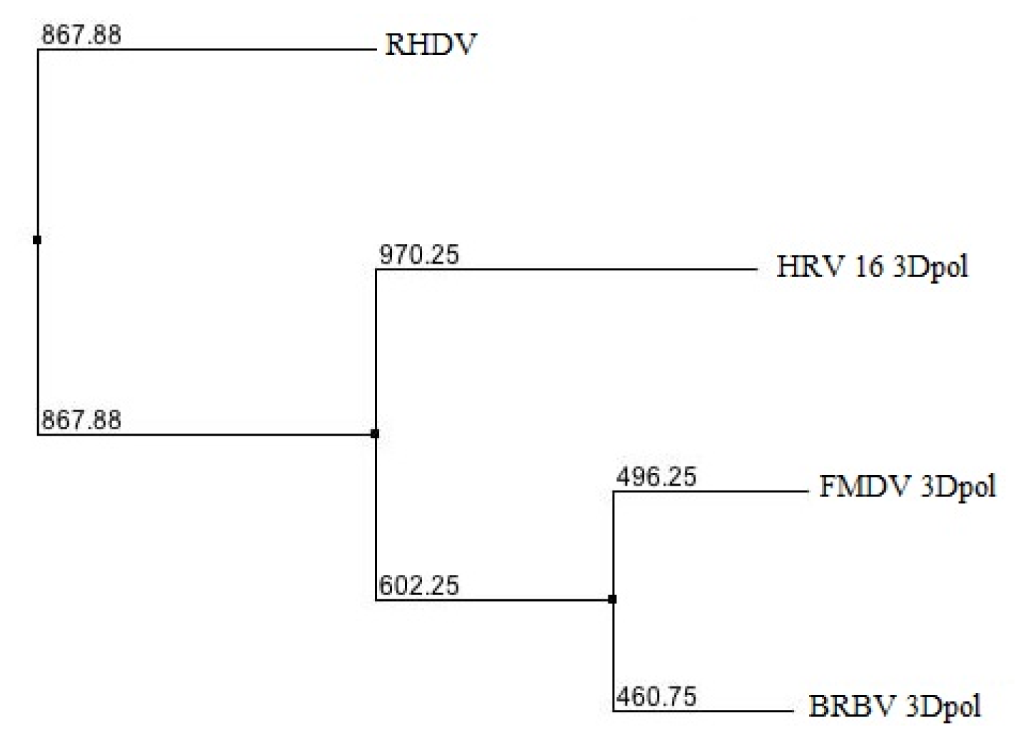
2.1. Phylogenetic Analysis of BRBV RdRp

As shown in Figure 1, the distance map generated by BLOSUM62 scoring scoring matrix imbedded in Jalview platform [17,18] shows that BRBV 3Dpol is genetically closest to FMDV followed by HRV and RHDV 3Dpol, respectively. The result supports the earlier study reporting close proximity of BRBV (previously known as BRV-2) to FMDV [3].

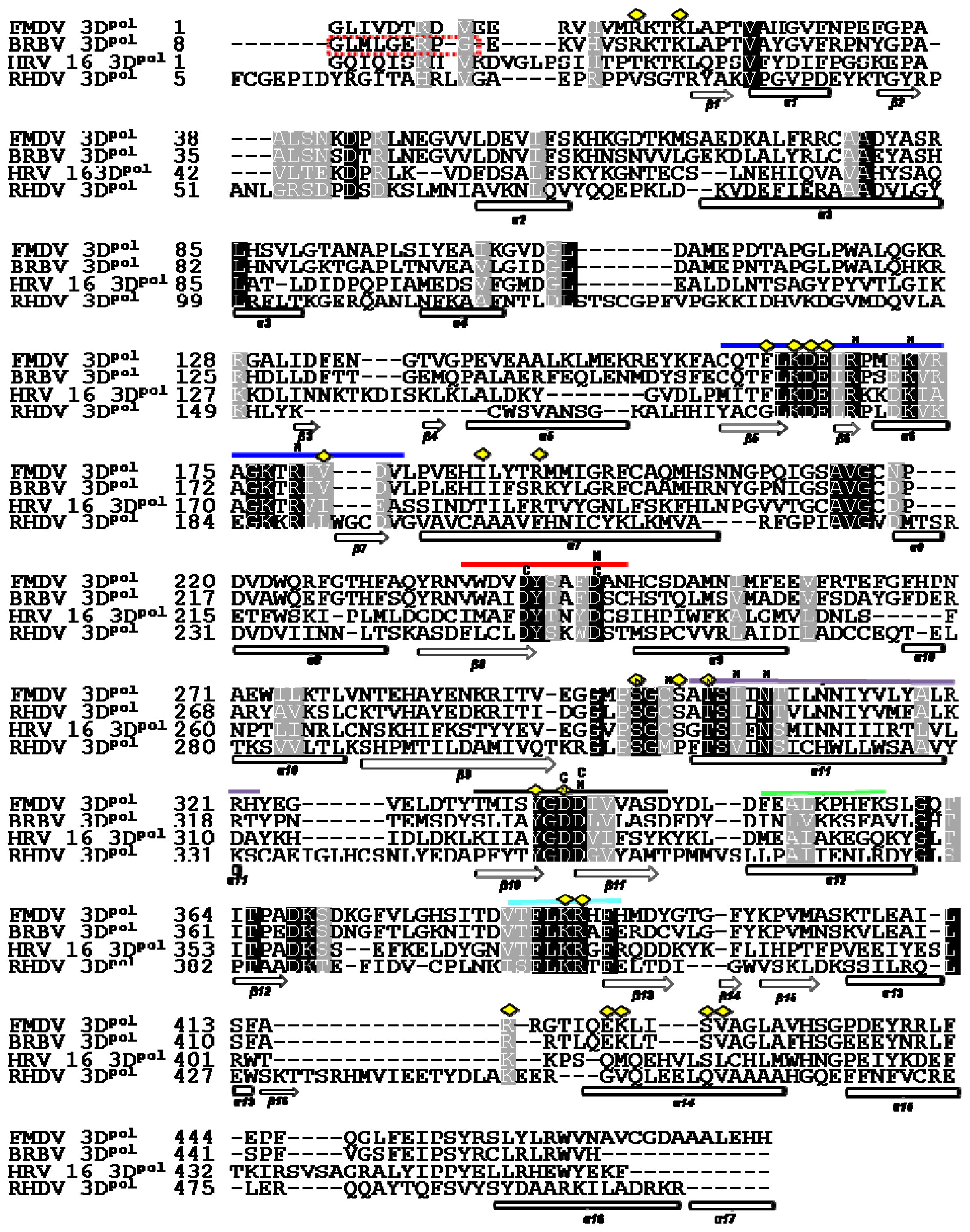
2.2. Structure Based Sequence Alignment

The structure based alignment of the sequences of 3Dpol proteins of BRBV, FMDV, HRV-16 and RHDV was carried out using magic fit algorithm followed by iterative magic fit refinement on Deep View platform [19]. The alignment was verified and curated manually by looking at the superposed structures in order to define the secondary structure elements (SSEs) and functional motifs of all RdRps accurately. The results indicated that the BRBV 3Dpol shares 64.03% sequence identity with the FMDV protein counterpart, followed by HRV-16 3Dpol (29.39%) and RHDV 3Dpol (20.21%) respectively (Figure 2). BRBV 3Dpol shows 76.9, 94.12, 85.7, 45, 100 and 95.8% conservation for motifs A, B, C, D, E and F, respectively. These motifs are conserved in HRV-16 3Dpol as well, though to a lesser extent. BRBV, HRV-16 and FMDV 3Dpol seem to utilize the same T/P binding, NTP binding and active site. RHDV 3Dpol has a noticeably different T/P binding interface with fewer positive residues (Figure 2). The catalytic site is absolutely conserved among all the polymerases analyzed. The structure based sequence alignment suggests a close proximity between BRBV and FMDV 3Dpol followed by HRV-16 and RHDV 3Dpol.

2.3. Search for Structural Homologues to BRBV 3Dpol via deconSTRUCT Web Server

The modeled structures of BRBV 3Dpol prepared by Geno3D and SWISS-MODEL [2026] were submitted to the deconSTRUCT web server. dconSTRUCT finds homologous proteins via a totally different approach as discussed in the materials and methods section [27]. The details of the modeling protocol and analysis of structures are discussed in sections 2.4–2.8. The results from the structure based homologue search generated by deconSTRUCT suggested that the BRBV 3Dpol has the highest homology with the FMDV 3Dpol followed by Hepatitis C virus and RHDV polymerases (Table 1). However, as shown in Figure 2, when we superposed the structures of FMDV, BRBV, HRV-16 and RHDV RdRps and analyzed key structural motifs, BRBV 3Dpol was found to be closest to FMDV followed by HRV-16 and RHDV 3Dpol. This discrepancy of prediction could be attributed to the methodology adopted by deconSTRUCT. Since it compares parts of proteins with fewer SSEs, this could result in a stronger signal for a distantly related protein. The superposition of structures on the other hand provides better control over comparison of overall as well as in part structure of proteins.

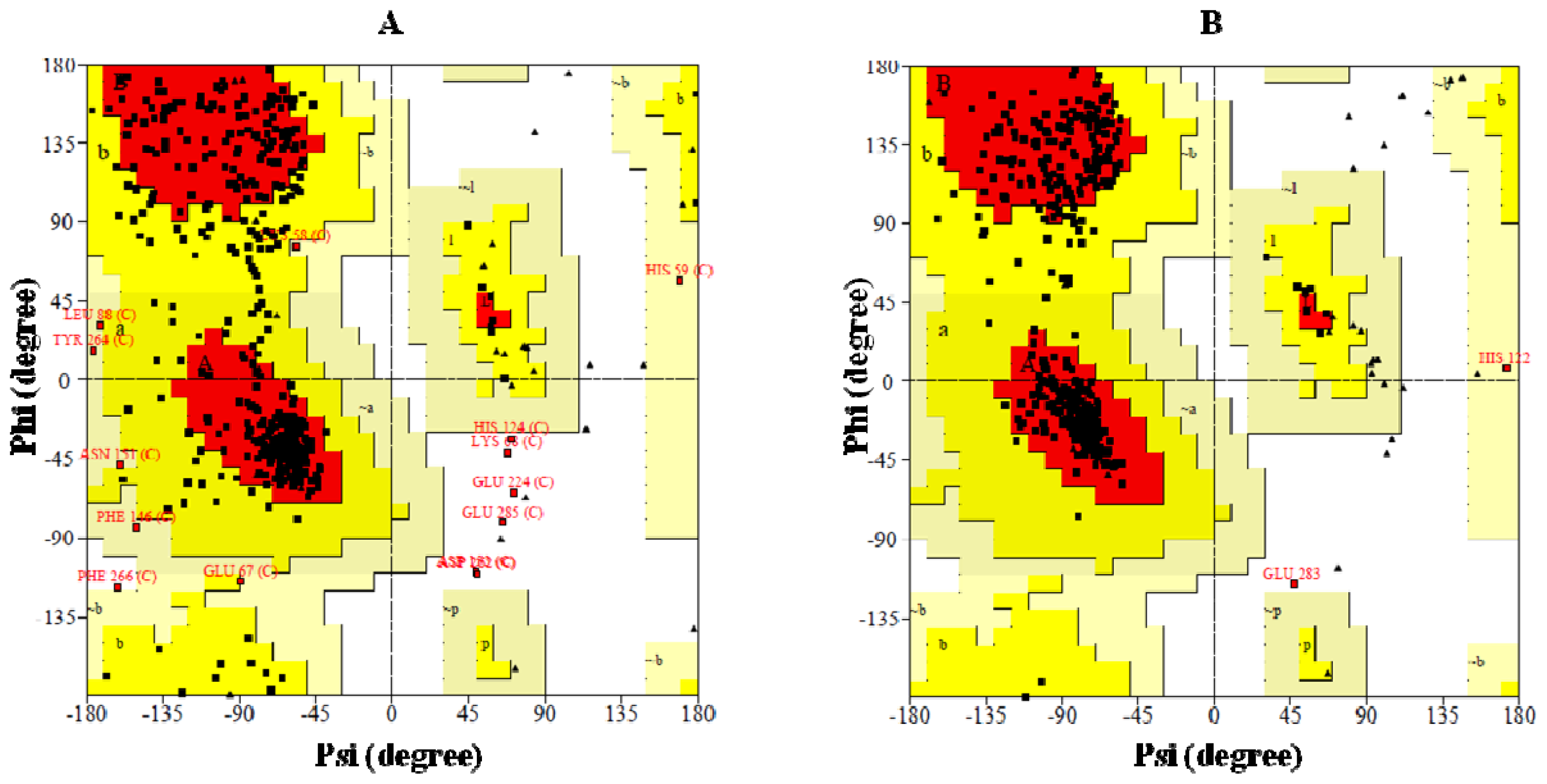
2.4. Preparation of Homology Model of BRBV 3Dpol and the Assessment of Model Accuracy

BRBV 3Dpol was modeled on FMDV and FMDV+HRV-16 3Dpol via SWISS-MODEL and Geno3D web servers, respectively. The stereochemical quality and accuracy of the models were tested using PROCHECK [28]. Results from PROCHECK are reported as Ramachandran plots (Figure 3). The main and side chain parameters are summarized in (Tables 2A and 3A) and (Tables 2B and 3B), respectively and discussed in the context of accuracy of the structure prediction. Ramachandran plot is one of the oldest yet most reliable methods of determination of the quality of protein structure. A structure with ≥90% of its residues in the most favored regions A, B and L of Ramachandran plot is considered to be as accurate as a 2Å-resolution crystal structure [28]. Only 73.1% residues in the Geno3D generated model of BRBV 3Dpol are in the most favorable regions of Ramachandran plot (Figure 3A, Table 1A). In contrast SWISS-MODEL generated structure has 90.15% of its residues in the most favorable region of Ramachandran plot (Figure 3B, Table 1B). Other main chain parameters such as omega angle standard deviation, bad contacts/100 residues, zeta angle standard deviation, hydrogen bond energy standard deviation and overall G factor, which underscores to structural accuracy of a structure as determined by PROCHECK, also show that the SWISS-MODEL generated structure has better structural attributes (Tables 1A and 2A).
Side chain parameters were shown as chi1-chi2 side chain torsion angle combinations for all residue types whose side chains are long enough to have both these angles. The chi1-chi2 plots which were presented as side chain parameters in Tables 2A and 2B indicate SWISS-MODEL generated structure exhibited higher score in terms of overall structural accuracy. In conclusion, the results from PROCHECK analysis clearly suggested that the modeled structure of BRBV 3Dpol generated by SWISS-MODEL program was superior in quality. Therefore, further characterization of the modeled structure of BRBV was therefore, carried out on SWISS-MODEL generated structure.

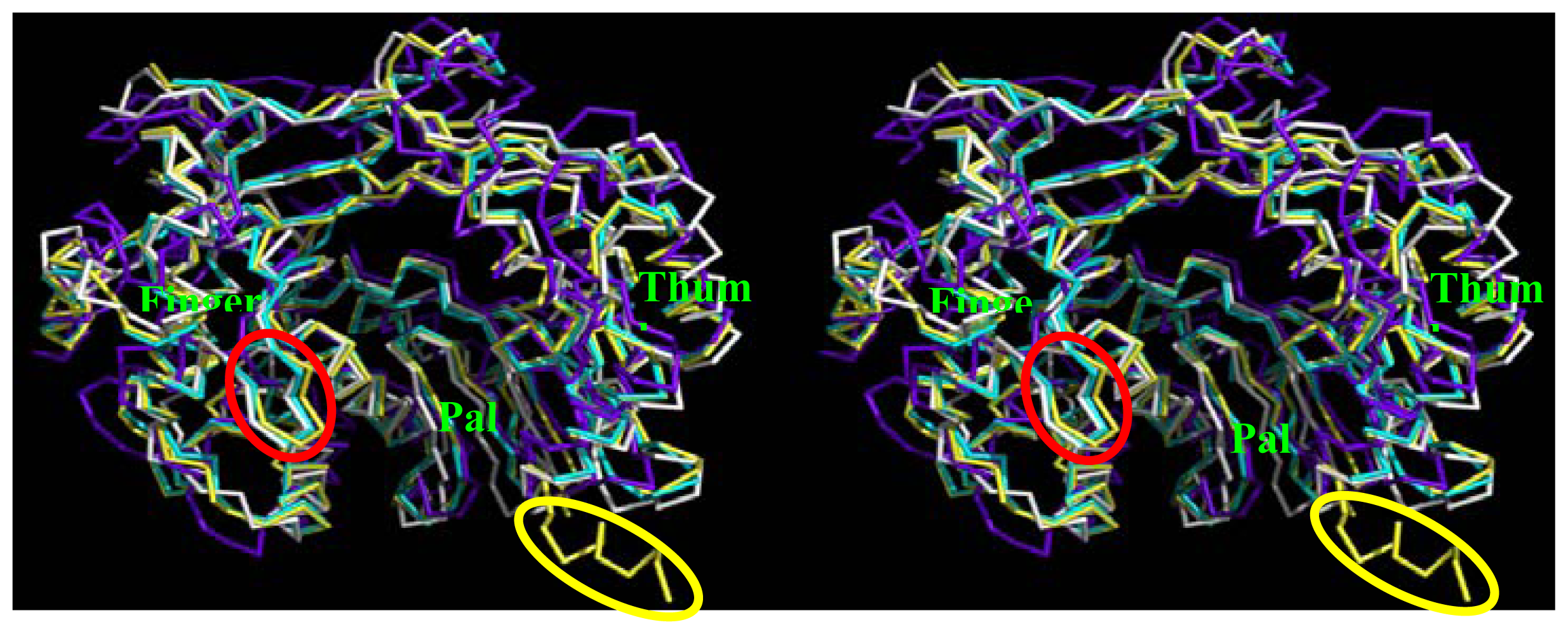
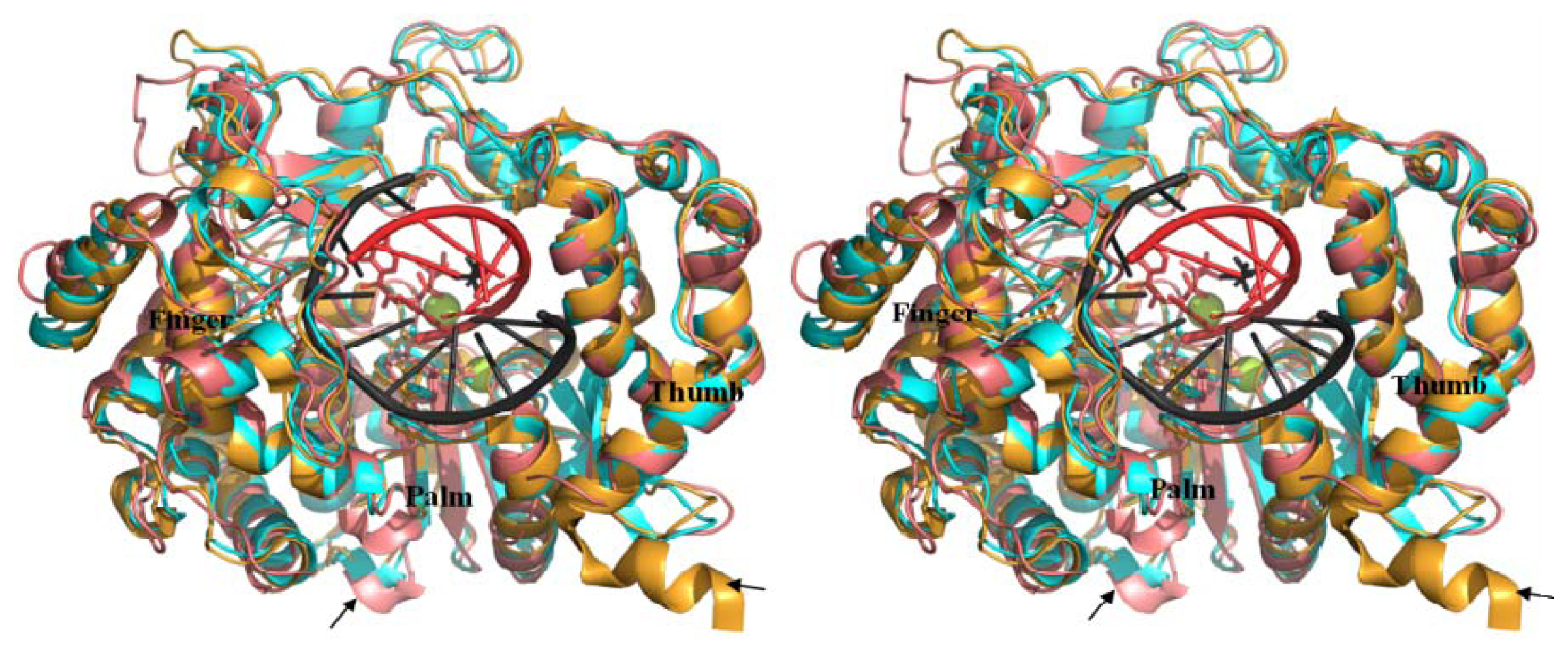
2.5. Comparison of BRBV 3Dpol and Other 3Dpol Crystal Structures

PDB coordinate file can be found as supplementary information. As shown in Figure 4, the modeled structure of BRBV (cyan) and crystal structures of HRV-16 (white) as well as RHDV 3Dpol (blue) were superimposed on FMDV 3Dpol (yellow) (PDB, 1U09). BRBV Model showed high degree of homology to FMDV 3Dpol followed by HRV-16 and RHDV counterparts. The thumb domain of RHDV 3Dpol was observed to be shifted inwards as compared to the FMDV and other RdRps. RHDV polymerase lacks critical positively charged loop (shown in red oval, Figure 4), which aligns the template-binding surface observed in FMDV. BRBV 3Dpol appears to lack C-terminal α-helix as compared to FMDV counterpart (marked with yellow oval). FMDV 3Dpol C-α trace RMSD were 0.758, 1.518, and 4.607 for BRBV 3Dpol model, HRV-16 and RHDV 3Dpols, respectively. It is important to note that the RMSD score of ≤1.0 Å indicates a very good agreement between the template and homology model. The comparison of three dimensional structures of the RdRps also supports the close relatedness of BRBV 3Dpol to its FMDV counterpart. As indicated with arrows in Figure 5 the modeled structure generated by Geno3D (pink color) has an important α-helix disordered in the finger domain. In addition, the loop aligning the template channel is pushed upwards in this model. In contrast the SWISS-MODEL generated structure (cyan color) has higher degree of conservation of these structural features in the context of FMDV 3Dpol.

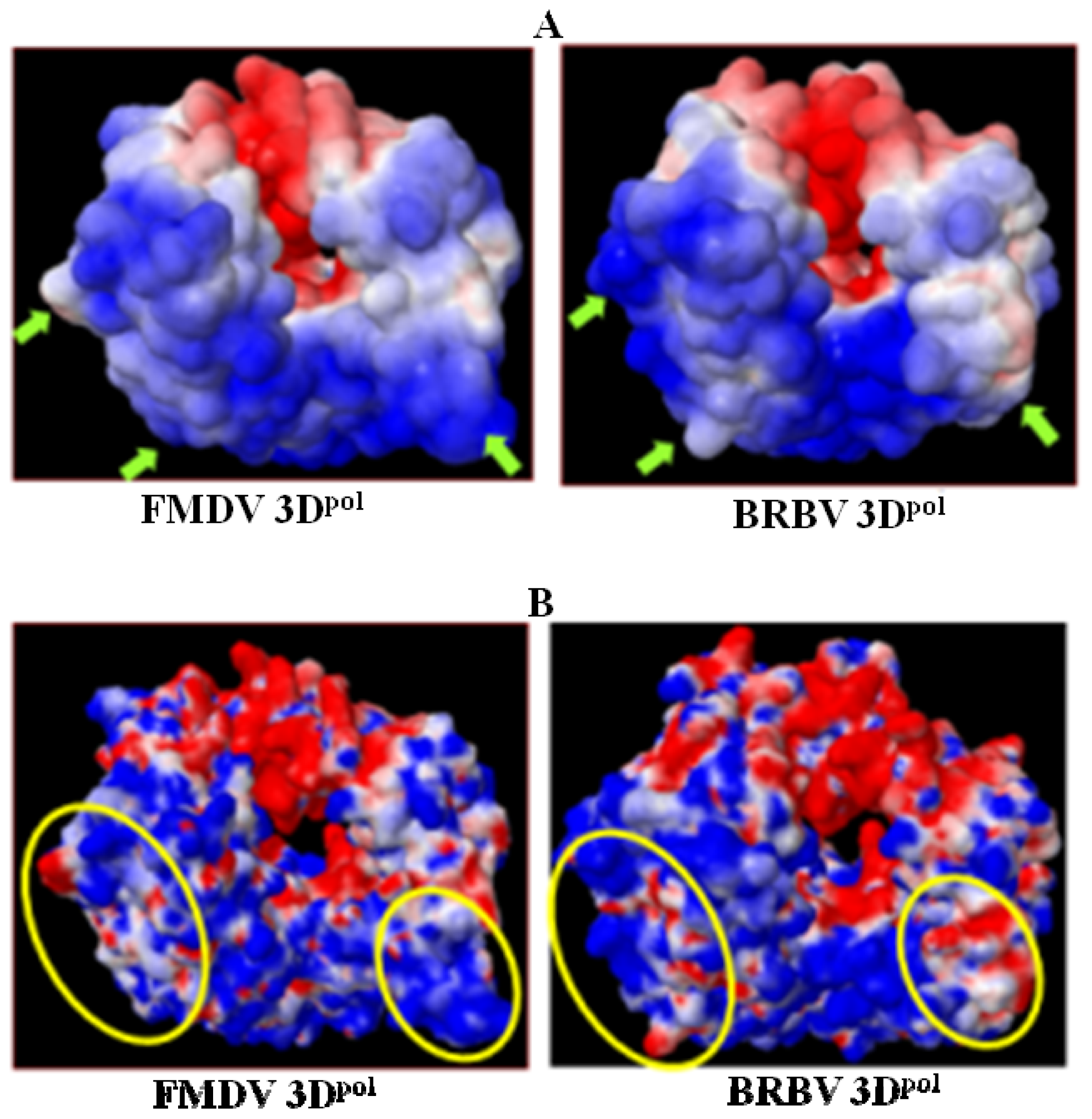
2.6. Analysis of Key Structural Features of FMDV 3Dpol and BRBV Homology Model

The electrostatic surface potentials of modeled BRBV 3Dpol and FMDV 3Dpol apo enzyme were calculated using the Adaptive Poison Boltzman Solver (APBS) via PDB2PQR web server [29,30]. Solvent exposed and solvent excluded electrostatic surface structures were prepared for both FMDV and BRBV 3Dpol (Figure 6A,B). Total electrostatic energies were recorded to be in total 4.15 × 10⁵ and 4.29 × 10⁵ kilo-Joule per mole (kJ/mol), respectively for FMDV and BRBV 3Dpol. The similar distribution of the electrostatic potential energy also substantiates similar structural attributes of the two polymerases. However, as reflected in Figure 6A,B the BRBV 3Dpol lacks the C-terminal α-helix with positively charged surface observed in FMDV counterpart. Other key differences in the distribution of positively charged, neutral and negatively charged residues have been indicated by green arrows and yellow ovals in Figure 6A,B, respectively. It is important to note that BRBV 3Dpol has more positively charged residues at the base of finger sub domain as opposed to FMDV 3Dpol, which has more neutral amino acids in this region (white). In contrast the base of thumb sub domain is positively charged in FMDV 3Dpol, C terminus of BRBV 3Dpol has an uncharged surface. These differences could individually or collectively contribute to physiochemical behavior as well as the function of the two enzymes.

2.7. Analysis on Non-Covalent Interactions in FMDV and BRBV 3Dpol

Non-covalent interactions such as hydrogen bonds, salt bridges, van der waals interactions, hydrophobic interactions, cation-pi and CH-pi bonds are individually weak but collectively they determine the structure and behavior of proteins. Hydrogen bonds and salt bridges were calculated using VMD [31]. BRBV 3Dpol was observed to forms an extensive network of hydrogen bonds (46 hydrogen bonds) exactly similar to FMDV 3Dpol. The distribution of these bonds was also very similar between FMDV and BRV 3Dpol. Since hydrogen bonds are major contributors of protein folding and function, it is conceivable that given other similar features BRBV 3Dpol is homologous to its FMDV counterpart. However, the number of salt bridges was strikingly different between BRBV FMDV 3Dpol, which form 1 and 17 salt bridges, respectively. Since salt bridges are critical for the thermal behavior of proteins it would be interesting to explore the temperature sensitivity of the protein and compare it to FMDV and other closely related proteins. The cation-pi interactions, formed between positively charged amino acids and aromatic amino acids, when they are in close proximity, contribute significantly to the protein structure [32]. Arginine (R) is more likely than lysine (K) to participate in a cation-pi interaction. The order of participation of aromatic side chains in cation-pi interactions is Tryptophan (W) > Tyrosine (Y)> Phenylalanine (F). We recorded 4 energetically significant cation-pi interactions in FMDV 3Dpol, which exist between R17-F162, R153-W273, R388-W223, and K400-W462. In contrast BRBV 3Dpol possesses only 1 energetically favorable cation-pi interaction, which is formed between R385 and W220.

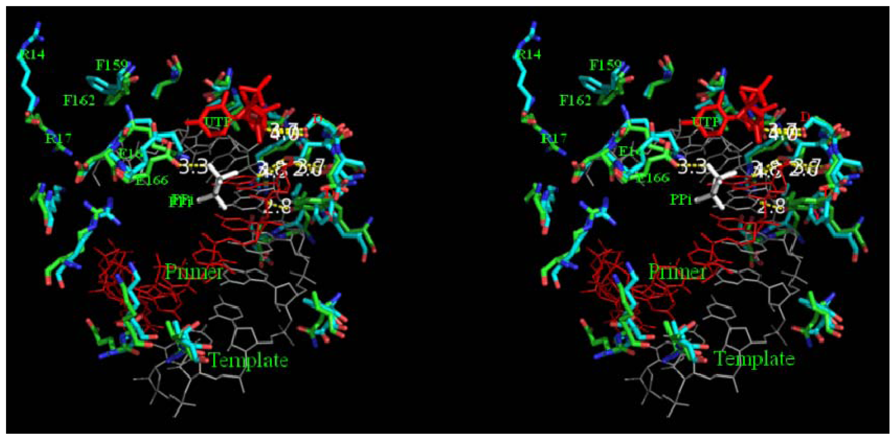
2.8. Analysis of Template/Primer (T/P) Binding Interface and Active Site of BRBV 3Dpol

T/P binding interface of BRBV 3Dpol was mapped on to FMDV 3Dpol bound to decameric RNA, uridine tri-phosphate and pyrophosphate (PDB, 2E9Z). As shown in Figure 7, the template binding interface of both BRBV 3Dpol and FMDV 3Dpol are aligned with positively charged amino acids, which is an adaptation of the RdRp to accommodate negatively charged nucleic acid substrate. It is interesting to note that the side-chains of R14 and F 159 of BRBV have been flipped out of the plane in comparison to FMDV 3Dpol. Their counterparts in FMDV 3Dpol form a strong cation-pi interaction. This might be a key stabilizing interaction, which holds the β-loop and keeps the T/P binding interface of the polymerase intact. The cation-pi interaction between W220 and R385 in BRBV 3Dpol could be an important determinant of its function. This bond is formed between β-13 strand and α-8 helix in the palm region that align the catalytic motif of the polymerase. Glutamate 163 is also moved out of the template channel losing an important hydrogen bond interaction with pyrophosphate in BRBV 3Dpol (Figure 7). Some of the differences in the bonding pattern could also be attributed to the use of apo structure of FMDV 3Dpol as a template for homology modeling. Since, it is an unliganded structure, there is more rotational freedom for the amino acids as compared to RNA and NTP bound polymerase. Such rotations may yield the altered conformations of amino acid side chains and hence alter the bonding pattern as well to some extent. Alternatively, it is also possible that in a replicating polymerase it is the alternate conformation of these amino acids that is predominant. Such structural rearrangements are gaining attention as a mechanism that RdRps adopt to facilitate the replication activity of their polymerases [10,11]. It is important to note that the available literature suggests that growth of BRBV in tissue culture is difficult to achieve [33] and virus infected cells incubated at 31 or 33 °C display more pronounced CPE than when incubated at 37 °C [2]. These studies also support a different physiological and thermal behavior of BRBV 3Dpol suggested herein on the basis of the structure and bonding pattern.

3. Experimental Section

3.1. Sequence

BRBV polypeptide sequence (NCBI Reference Sequence: YP_003355055.1) was utilized to retrieve the putative 3Dpol region, which was later utilized for sequence comparisons and homology modeling.

3.2. Phylogenetic Analysis

Phylogeny tree was prepared in order to determine the relatedness of BRBV 3Dpol to other related polymerases. The tree was calculated from distance matrices determined from percent identity or aggregate BLOSUM62 score using average distance as implemented in JalView [17,18]. In this method, pairwise distances used to cluster the sequences are represented as the percentage of mismatches between two sequences. The branch lengths are the percentage mismatch between two nodes; the leaves show the sequence IDs.

3.3. Structure based Alignment of the Polymerase Sequences

The structure based alignment of BRBV 3Dpol with other RdRps was performed using iterative magic fit function incorporated in the Deep view-[19]. The aligned sequences were manually curated to characterize the key structural motifs in each protein accurately. The key structural features such as functional motifs as well as T/P and nucleotide triphosphate interacting residues were assigned different color and symbols manually.

3.4. Preparation of Homology Model of BRBV 3Dpol

The homology models of the protein were prepared via:
  • Geno3D web server
    Crystal structures of FMDV (PDB, 1U09) and HRV-16 3Dpol (PDB, 1XR7) were utilized as templates for model generation. After the completion of run 10 homology models were prepared and their quality was assessed through PROCHECK [20,28]. Geno3D ( http://geno3d-pbil.ibcp.fr) is an automated web server for protein molecular modeling [20]. Starting with a query protein sequence, the server performs the homology modeling in six successive steps: first, it identifies homologous proteins with known structures by using PSI-BLAST [21]. This provides the user all potential templates for target selection. After the user defines the templates and submits the job, the server performs the alignment of both query and subject sequences and extracts geometrical restraints (dihedral angles and distances) for corresponding atoms between the query and the template. Lastly, it performs the 3D construction of the protein by using a distance geometry approach.
  • SWISS-MODEL
    The crystal structure of FMDV 3Dpol (PDB, 1U09) was identified as the closest match to the BRV counterpart and later utilized as template by SWISS-MODEL workspace, which is an integrated web-based modeling environment. For a given target protein it searches a library of experimental protein structures to identify suitable templates. On the basis of a sequence alignment between the target protein and the template structure, a three-dimensional model for the target protein is generated. The alignment produced by SWISS-MODEL was verified using another alignment algorithm (T-coffee). In homology modeling the most crucial steps is the evaluation and refinement of the raw model and SWISS-MODEL utilizes a set of unique analysis tools in addition to commonly tools such as PROCHECK and WHAT CHECK [2226,28,34] to achieve this. These tools include atomic empirical mean force potential (ANOLEA) which performs energy calculations on a protein chain, evaluating the “Non- Local Environment” (NLE) of each heavy atom in the molecule and is used to assess packing quality of the models; QMEAN which is a composite scoring function for both the estimation of the global quality of the entire model as well as for the local per-residue analysis of different regions within a model; DFIRE, which is an all-atom statistical potential based on a distance-scaled finite ideal-gas reference state and reflects the quality of the model to indicate that a that a model is lower energy closer to the native conformation [3537]. Finally, it minimizes the structure using GROMOS9639 performing 200 cycles by the steepest descent method and 300 cycles by the conjugate gradient method to minimize the steric clashes [38]. With stringent quality controls SWISS-MODEL workspace usually generates models with a reasonably reliable quality.

3.5. DeconSTRUCT Analysis of the Homology Models of BRBV 3Dpol

In order to find the degree of homology of BRBV 3Dpol to other viral RdRps, the best scoring homology models of BRBV 3Dpol prepared by both SWISS-MODEL and Geno3D were submitted to the deconSTRUCT web server, which offers an interface to a protein database search engine that detects similar protein sub-structures. Firstly, it deconstructs the query structure into its SSEs and finds the match to the target by requiring a (tunable) degree of similarity in the direction and sequential order of SSEs. The search engine of deconSTRUCT utilizes the hierarchical organization and judicious use of the information about protein structures to achieve the sensitivity and specificity of the established search engines at orders of magnitude increased speed, without tying up irretrievably the substructure information in the form of a hash.

3.6. Calculation of Electrostatic Surface Potential

Electrostatic surface potential energies for BRBV and FMDV 3Dpol were calculated using APBS via PDB2PQR web portal ( http://kryptonite.nbcr.net/pdb2pqr/) [29,30]. The input files were prepared from the PDB files of the two polymerases (BRBV and FMDV). Atomic charges and ionic radii were assigned according to Assisted Model Building with Energy Refinement (AMBER) force field. PROPKA was used to assign the protonation state to the proteins at pH 7.0. The proteins were assigned a low dielectric constant of 2.0 with a solvent dielectric constant of 78.54. Solvent accessible and solvent excluded surfaces of the two proteins were prepared. −5 and +5 kT/e (k is Boltzman Constant, T is temperature and e is the charge of electron) settings were used to color most positively charged and most negatively charged surface.

3.7. Calculation of Non-Covalent Interactions

The total number of hydrogen bonds was calculated in modeled as well as FMDV 3Dpol crystal structure, which was used as template to generate the model. The salt bridges were calculated using VMD [31]. Cation-pi interactions are formed when a cationic side-chain (Lys or Arg) is near an aromatic side-chain (Phe, Tyr, or Trp). Under such conditions, the geometry of aromatic ring is biased toward one that would experience a favorable cation-π interaction. Energetically favorable cation-pi interactions were alculated in both the modeled BRBV 3Dpol as well as FMDV 3Dpol structures via CaPTURE ( http://capture.caltech.edu/) program developed by Justin Gallivan [32].

3.8. Preparation of Structures

Different representations of the structures of all the proteins were created Pymol version 1.3, wherever not mentioned otherwise [39].

4. Conclusions

In this study we built a high quality homology model of the 3D polymerase of bovine rhinitis B virus, which shares the highest homology to the FMDV counterpart. The modeled structures generated by different methods were characterized extensively for accuracy and relevance in terms of other closely related RdRps. We found that BRBV 3Dpol lacks C-terminal α-helix, which appears as an extension of thumb sub-domain in FMDV 3Dpol. The active site and palm sub-domain show high conservation similar to most polymerases. The significant differences in the number and pattern of non-covalent interactions as compared to FMDV 3Dpol could be an important determinant of the thermal enzyme behavior of BRBV 3Dpol. Although, the template binding (97.5%) and active sites (100%) are highly conserved between FMDV and BRBV 3Dpol, the differences in adjacent residues, as well as bonding, could be important in determining the overall functionality of the protein. The finding of the present study would help broaden understanding of Aphthovirus RdRp, in particular, understanding the conformational changes in aphthoviral polymerases, which is as yet not an established phenomenon.

Acknowledgements

This research was supported in part by the Plum Island Animal Disease Research Participation Program administered by the Oak Ridge Institute for Science and Education through an interagency agreement between the U.S. Department of Energy and the U.S. Department of Agriculture (appointment of Devendra Rai); by CRIS project no. 1940-32000-057-00D, Agricultural Research Service (ARS), U.S. Department of Agriculture (Elizabeth Rieder). We thank Sabena Uddowla for sources of BRBV proteins and for fruitful discussions; Paul Lawrence for critical reading of the manuscript and useful suggestions.
  • Conflict of InterestThe authors declare that they have no conflict of interest.

References

  1. Knowles, N.J.; Hovi, T; Hyypiä, T.; King, A.M.Q.; Lindberg, M.; Pallansch, M.A.; Palmenberg, A.C.; Simmonds, P.; Skern, T.; Stanway, G.; et al. Picornaviridae. In Virus Taxonomy: Classification and Nomenclature of Viruses: Ninth Report of the International Committee on Taxonomy of Viruses; King, A.M.Q., Adams, M.J., Carstens, E.B., Lefkowitz, E.J., Eds.; Elsevier: San Diego, CA, USA, 2011; pp. 855–880. [Google Scholar]
  2. Hollister, J.R.; Vagnozzi, A.; Knowles, N.J.; Rieder, E. Molecular and phylogenetic analyses of bovine rhinovirus type 2 shows it is closely related to foot-and-mouth disease virus. Virology 2008, 373, 411–425. [Google Scholar]
  3. Betts, A.O.; Edington, N.; Jennings, A.R.; Reed, S.E. Studies on a rhinovirus (EC11) derived from a calf. II. Disease in calves. J. Comp. Pathol 1971, 81, 41–48. [Google Scholar]
  4. Ng, K.K.; Cherney, M.M.; Vazquez, A.L.; Machin, A.; Alonso, J.M.; Parra, F.; James, M.N. Crystal structures of active and inactive conformations of a caliciviral RNA-dependent RNA polymerase. J. Biol. Chem 2002, 277, 1381–1387. [Google Scholar]
  5. Love, R.A.; Maegley, K.A.; Yu, X.; Ferre, R.A.; Lingardo, L.K.; Diehl, W.; Parge, H.E.; Dragovich, P.S.; Fuhrman, S.A. The crystal structure of the RNA-dependent RNA polymerase from human rhinovirus: A dual function target for common cold antiviral therapy. Structure 2004, 12, 1533–1544. [Google Scholar]
  6. Thompson, A.A.; Peersen, O.B. Structural basis for proteolysis-dependent activation of the poliovirus RNA-dependent RNA polymerase. EMBO J 2004, 23, 3462–3471. [Google Scholar]
  7. Ferrer-Orta, C.; Arias, A.; Escarmís, C.; Verdaguer, N. A comparison of viral RNA-dependent RNA polymerases. Curr. Opin. Struct. Biol 2006, 16, 27–34. [Google Scholar]
  8. Ferrer-Orta, C.; Arias, A.; Perez-Luque, R.; Escarmis, C.; Domingo, E.; Verdaguer, N. Structure of foot-and-mouth disease virus RNA-dependent RNA polymerase and its complex with a template-primer RNA. J. Biol. Chem 2005, 279, 47212–47221. [Google Scholar]
  9. Gruez, A.; Selisko, B.; Roberts, M.; Bricogne, G.; Bussetta, C.; Jabafi, I.; Coutard, B.; de Palma, A.M.; Neyts, J.; Canard, B. The crystal structure of coxsackievirus B3 RNA-dependent RNA polymerase in complex with its protein primer VPg confirms the existence of a second VPg binding site on Picornaviridae polymerases. J. Virol 2008, 82, 9577–9790. [Google Scholar]
  10. Steitz, T.A. DNA and RNA polymerases: Structural diversity and common mechanisms. Harvey Lect. 1997–1998, 93, 75–93. [Google Scholar]
  11. Gong, P.; Peersen, O.B. Structural basis for active site closure by the poliovirus RNA-dependent RNA polymerase. Proc. Natl. Acad. Sci. USA 2010, 107, 22505–22510. [Google Scholar]
  12. Bruenn, J.A. A structural and primary sequence comparison of the viral RNA-dependent RNA polymerases. Nucleic Acids Res 2003, 31, 1821–1829. [Google Scholar]
  13. Cameron, C.E.; Gohara, D.W.; Arnold, J.J. Poliovirus RNA-Dependent RNA Polymerase (3Dpol): Structure, Function and Mechanisms. In Molecular Biology of Picornaviruses; Semler, B.L., Wimmer, E., Eds.; ASM Press: Washington DC, USA, 2002; pp. 255–267. [Google Scholar]
  14. Van Dijk, A.A.; Makeyev, E.V.; Bamford, D.H. Structural basis for proteolysis-dependent activation of the poliovirus RNA-dependent RNA polymerase. EMBO J 2004, 23, 3462–3471. [Google Scholar]
  15. Thompson, A.A.; Albertini, R.A.; Peersen, O.B. Stabilization of poliovirus polymerase by NTP binding and fingers-thumb interactions. J. Mol. Biol 2007, 366, 1459–1474. [Google Scholar]
  16. Kok, C.; McMinn, P. Picornavirus RNA-dependent RNA polymerase. Int. J. Biochem. Cell Biol 2009, 41, 498–502. [Google Scholar]
  17. Waterhouse, A.M.; Procter, J.B.; Martin, D.M.A.; Clamp, M.; Barton, G.J. Jalview version 2: A multiple sequence alignment and analysis workbench. Bioinformatics 2009, 25, 1189–1191. [Google Scholar]
  18. Styczynski, M.P.; Jensen, K.L.; Rigoutsos, I.; Stephanopoulos, G. BLOSUM62 miscalculations improve search performance. Nat. Biotechnol 2008, 26, 274–275. [Google Scholar]
  19. Guex, N.; Peitsch, M.C. SWISS-MODEL and the Swiss-PDBViewer: An environment for comparative protein modeling. Electrophoresis 1997, 18, 2714–2723. [Google Scholar]
  20. Combet, C.; Jambon, M.; Deléage, G.; Geourjon, C. Geno3D: Automatic comparative molecular modelling of protein. Bioinformatics 2002, 18, 213–214. [Google Scholar]
  21. Müller, A.; MacCallum, R.M.; Sternberg, M.J. Benchmarking PSI-BLAST in genome annotation. J. Mol. Biol 1999, 293, 1257–1271. [Google Scholar]
  22. Arnold, K.; Bordoli, L.; Kopp, J.; Schwede, T. The SWISS-MODEL workspace: A web-based environment for protein structure homology modelling. Bioinformatics 2006, 22, 195–201. [Google Scholar]
  23. Kiefer, F.; Arnold, K.; Künzli, M.; Bordoli, L.; Schwede, T. The SWISS-MODEL repository and associated resources. Nucleic Acids Res 2009, 37, D387–D392. [Google Scholar]
  24. Kopp, J.; Schwede, T. The SWISS-MODEL Repository: New features and functionalities. Nucleic Acids Res 2006, 34, D315–D318. [Google Scholar]
  25. Schwede, T.; Kopp, J.; Guex, N.; Peitsch, M.C. SWISS-MODEL: An automated protein homology-modeling server. Nucleic Acids Res 2003, 31, 3381–3385. [Google Scholar]
  26. Arnold, K.; Bordoli, L.; Kopp, J.; Schwede, T. The SWISS-MODEL workspace: A web-based environment for protein structure homology modelling. Bioinformatics 2006, 22, 195–201. [Google Scholar]
  27. Zhang, Z.H.; Bharatham, K.; Sherman, W.A.; Mihalek, I. deconSTRUCT: General purpose protein database search on the substructure level. Nucleic Acids Res 2010, 38, W590–W594. [Google Scholar]
  28. Laskowski, R.A.; MacArthur, M.W.; Moss, D.S.; Thornton, J.M. PROCHECK-a program to check the stereochemical quality of protein structures. In J. Appl. Cryst; 1996; Volume 26, pp. 283–291. [Google Scholar]
  29. Dolinsky, T.J.; Nielsen, J.E.; McCammon, J.A.; Baker, N.A. PDB2PQR: An automated pipeline for the setup, execution, and analysis of Poisson-Boltzmann electrostatics calculations. Nucleic Acids Res 2010, 32, W665–W667. [Google Scholar]
  30. Baker, N.A.; Sept, D.; Joseph, S.; Holst, M.J.; McCammon, J.A. Electrostatics of nanosystems: Application to microtubules and the ribosome. Proc. Natl. Acad. Sci. USA 2001, 98, 10037–10041. [Google Scholar]
  31. Humphrey, W.; Dalke, A.; Schulten, K. VMD: Visual molecular dynamics. J. Mol. Graph 1996, 14, 33–38. [Google Scholar]
  32. Gallivan, J.P.; Dougherty, D.A. Cation-pi interactions in structructural biology. Proc. Natl. Acad. Sci. USA 1999, 96, 9459–9464. [Google Scholar]
  33. Reed, S.E.; Tyrrell, D.A.; Betts, A.O.; Watt, R.G. Studies on a rhinovirus (EC11) derived from a calf. I. Isolation in calf tracheal organ cultures and characterization of the virus. J. Comp. Pathol 1971, 81, 33–40. [Google Scholar]
  34. Hooft, R.W.; Vriend, G.; Sander, C.; Abola, E. Errors in protein structures. Nature 1996, 381. [Google Scholar] [CrossRef]
  35. Melo, F.; Feytmans, E. Assessing protein structures with a non-local atomic interaction energy. J. Mol. Biol 1998, 277, 1141–1152. [Google Scholar]
  36. Benkert, P.; Tosatto, S.C.E.; Schomburg, D. QMEAN: A comprehensive scoring function for model quality assessment. Proteins Struct. Funct. Bioinforma 1998, 71, 261–277. [Google Scholar]
  37. Zhou, H.; Zhou, Y. Distance-scaled, finite ideal-gas reference state improves structure-derived potentials of mean force for structure selection and stability prediction. Protein Sci 2002, 11, 2714–2726. [Google Scholar]
  38. Van Gunsteren, W.F.; Billeter, S.R.; Eising, A.A.; Hünenberger, P.H.; Krüger, P.; Mark, A.E.; Scott, W.R.P.; Tironi, I.G. Biomolecular Simulation: The GROMOS96 Manual and User Guide; Verlag der Fachvereine Hochschulverlag AG an der ETH Zurich: Zurich, Switzerland, 1996. [Google Scholar]
  39. The PyMOL Molecular Graphics System, version 1.3. Available online: http://www.pymol.org/ accessed on 19 July 2012.
Figure 1. Phylogenetic analysis of Bovine Rhinitis B Virus (BRBV) 3Dpol: An un-rooted tree showing the genetic relatedness of BRBV 3Dpol to other RdRps. The distance represents the difference from BRBV 3Dpol sequence conservation.
Ijms 13 08998f1
Figure 2. Structure based alignment of BRBV 3Dpol with other related RdRPs: The conserved regions are shaded black and gray to mark identical and similar residues, respectively. Each polymerase sequence is labeled starting from the left margin and the numbers next to polymerase in each row represent the sequence position. The red broken rectangle shows the sequence of BRBV 3Dpol excluded while preparing the homology model. Functionally important motifs A, B, C, D, E and F are marked with red, purple, black, green, light blue and blue bars, respectively. α-helices and β-sheets are numbered starting from the N-terminus of protein and marked with open cylinders and open arrows. Functional motifs A, B, C, D, E and F are marked with red, purple, black, green, light blue and blue bars, respectively. Their putative function has been discussed in the introduction. The nucleic acid binding residues are marked with the yellow diamonds. The nucleotide binding and catalytic sites are marked with the letters N and C. The motifs and other key structural features are based on the structure of FMDV 3Dpol described earlier [7].
Ijms 13 08998f2
Figure 3. Ramachandran plot analysis of BRBV3Dpol structures: The Ramachandran plots of modeled BRBV structure generated by Geno3D (A) and SWISS-MODEL (B). The most favored regions are marked as {A, B, L}. The additional allowed regions are marked as {a, b, l, p}. All non-glycine and proline residues are shown as filled black squares, whereas, glycines (non-end) are shown as filled black triangles. Disallowed residues are colored red.
Ijms 13 08998f3
Figure 4. Stereoview of superimposed RdRp structures: A stereoview of ribbon representation of BRBV, HRV-16 and rabbit hemorrhagic disease virus (RHDV) RdRp 3Dpols superimposed on to foot and Mouth Disease virus (FMDV) 3Dpol (PDB, 1U09). Finger, palm and thumb domains are labeled. FMDV, BRBV, HRV-16 and RHDV 3Dpols are colored yellow, cyan, white and blue, respectively. An important loop, which aligns the template channel, is not present in RHDV 3Dpol (red oval). BRBV 3Dpol lacks the C-terminal α-helix in comparison to FMDV 3Dpol (yellow oval). The parts covering palm domain and the loop aligning T/P channel, have been removed to facilitate visualization of the regions with significant differences.
Ijms 13 08998f4
Figure 5. Stereoview of modeled BRBV 3Dpol superimposed on FMDV3Dpol: Polymerases and RNA template/primer are presented as cartoon structures. Uridine 5′-triphosphate (UTP) and pyrophosphate (PPi) are presented as sticks. Metals (Mg++) are presented as green spheres. The modeled structures generated by Geno3D [A] and SWISS-MODEL [B] are colored pink and cyan, respectively. FMDV 3Dpol protein is colored as bright orange. The template and primer RNA are colored as gray and red, respectively. UTP and PPi are colored red, and gray, respectively. Finger, palm and thumb domains of the polymerase are marked. Major structural differences are highlighted with black arrows and discussed in the text.
Ijms 13 08998f5
Figure 6. Electrostatic surface of BRBV 3Dpol: Electrostatic surface of BRBV 3Dpol and FMDV 3Dpol are colored red to blue ranging from most negative (−5 kT/e) to most positively (+5 kT/e) charged regions. Neutral surface is shown in white color. Major differences in the distribution of solvent exposed and solvent excluded surface residues are indicated with green arrows (A) and yellow ovals (B).
Ijms 13 08998f6
Figure 7. Stereoview of functionally important residues of BRBV 3Dpol and FMDV 3Dpol: Template/primer binding residues and active site of BRBV 3Dpol (cyan) and FMDV 3Dpol (green) are shown is stick representation. The nitrogen and oxygen atoms of the amino acids are colored blue and red, respectively. Template and primer are colored gray and red respectively and shown as lines. UTP and PPi are shown as sticks colored red and light gray, respectively. Amino acid residues are labeled as single letter amino acid codes with the identifiers. Catalytic tyrosine and glutamate residues are red colored one letter residue symbols. Arginine 14 and phenyl alanine 159 are flipped out of the nucleic acid binding interface in BRBV 3Dpol. Hydrogen bonds are shown with yellow dotted lines and marked with bond length.
Ijms 13 08998f7
Table 1. Alignment score from deconSTRUCT search.
PDB idAln. ScoreAln. LengthRMSD (Å)Avg. dLGeom. ZTarget Molecule
1U09A330.503701.421.88−7.20FMDV RdRp
2B43A273.363372.652.11−4.04Hepatitis C virus (HCV) RdRp
1KHWA259.873162.432.75−3.44RHDV RdRp
1S48A182.592312.872.500.51Bovine viral diarrhea virus (BVDV Rna-dependent rna polymerase
2JL9A116.571482.941.671.58Phi6 RNA polymerase
Aln. Score is the alignment value obtained from the web server after the search for the closely resembling protein structures available in PDB; Aln. Length is the length of the residues aligned to the query sequence.
Table 2A. Summary of main chain parameters for the structure generated by Geno3D.
Stereochemical qualityNo. of data pointsParameter valueComparison typical valueValue band widthNo. of band widths from meanInterpretation
% residues in A,B,L41373.683.810−1.0WORSE
Omega angle St. Dev.4630.66.03.0−1.8BETTER
Bad Contact/100 Residue004.210−0.4INSIDE
Zeta angle St. Dev.4331.13.11.6−1.3BETTER
H-bond Energy St.Dev.2830.70.80.2−0.5INSIDE
Overall G-Factor4650.2−0.40.31.9BETTER
St. Dev. denotes the standard deviation of the score observed. The accuracy of structure is depicted in the order BETTER > INSIDE > WORSE each parameter.
Table 2B. Summary of main chain parameters for the structure generated by SWISS-MODEL.
Stereochemical qualityNo of data pointsParameter valueComparison typical valueValue band widthNo. of band widths from meanInterpretation
% residues in A,B,L40590.176.6101.4WORSE
Omega angle St. dev.4525.26.03.0−0.3BETTER
Bad Contact/100 Residue0010.510−1.1INSIDE
Zeta angle St. Dev.4251.03.11.6−1.3BETTER
H-bond Energy St. Dev.3200.70.90.2−1.1INSIDE
Overall G-Factor4540.1−0.60.32.2BETTER
St. Dev. denotes the standard deviation of the score observed. The accuracy of structure is depicted in the order BETTER > INSIDE > WORSE each parameter.
Table 3A. Summary of side chain parameters for the structure generated by Geno3D.
Stereochemical qualityNo. of data pointsParameter valueComparison typical valueValue band widthNo. of band widths from meanInterpretation
Chi-1 gauche minus St. Dev.5210.022.76.5−1.9BETTER
Chi-1 trans St. Dev11810.122.75.3−2.4BETTER
Chi-1 gauche plus St. Dev.20010.121.34.9−2.3BETTER
Chi-1 pooled St. Dev.37010.222.04.8−2.4BETTER
Chi-2 transst dev1088.623.15.0−2.9BETTER
St Dev. denotes the standard deviation of the score observed. The accuracy of structure is depicted in the order BETTER > INSIDE > WORSE each parameter.
Table 3B. Summary of side chain parameters for the structure generated by SWISS-MODEL.
Stereochemical qualityNo. of data pointsParameter valueComparison typical valueValue band widthNo. of band widths from meanInterpretation
Chi-1 gauche minus St. Dev.6016.618.16.5−0.2INSIDE
Chi-1 trans St. Dev.13211.9195.3−1.3BETTER
Chi-1 gauche plus St. Dev.18513.817.54.9−0.8INSIDE
Chi-1 pooled St. Dev.37714.218.24.8−0.8INSIDE
Chi-2 trans St. Dev.8614.720.45.0−1.1INSIDE
St. Dev. denotes the standard deviation of the score observed. The accuracy of structure is depicted in the order BETTER > INSIDE > WORSE each parameter.

