<?xml version="1.0" encoding="UTF-8"?>
<!DOCTYPE article PUBLIC "-//NLM//DTD Journal Publishing DTD v2.3 20070202//EN" "journalpublishing.dtd">
<article xmlns:xlink="http://www.w3.org/1999/xlink" xml:lang="en" article-type="review-article">
<front>
<journal-meta>
<journal-id journal-id-type="publisher-id">ijms</journal-id>
<journal-title>International Journal of Molecular Sciences</journal-title>
<abbrev-journal-title>Int. J. Mol. Sci.</abbrev-journal-title>
<issn pub-type="epub">1422-0067</issn>
<publisher>
<publisher-name>Molecular Diversity Preservation International (MDPI)</publisher-name></publisher></journal-meta>
<article-meta>
<article-id pub-id-type="doi">10.3390/ijms131217160</article-id>
<article-id pub-id-type="publisher-id">ijms-13-17160</article-id>
<article-categories>
<subj-group>
<subject>Review</subject></subj-group></article-categories>
<title-group>
<article-title>Nitric Oxide in Skeletal Muscle: Role on Mitochondrial Biogenesis and Function</article-title></title-group>
<contrib-group>
<contrib contrib-type="author">
<name><surname>Tengan</surname><given-names>Celia Harumi</given-names></name><xref ref-type="aff" rid="af1-ijms-13-17160">1</xref><xref ref-type="corresp" rid="c1-ijms-13-17160">*</xref></contrib>
<contrib contrib-type="author">
<name><surname>Rodrigues</surname><given-names>Gabriela Silva</given-names></name><xref ref-type="aff" rid="af1-ijms-13-17160">1</xref></contrib>
<contrib contrib-type="author">
<name><surname>Godinho</surname><given-names>Rosely Oliveira</given-names></name><xref ref-type="aff" rid="af2-ijms-13-17160">2</xref></contrib></contrib-group>
<aff id="af1-ijms-13-17160">
<label>1</label>Department of Neurology and Neurosurgery, Paulista School of Medicine, Federal University of São Paulo, Sao Paulo 04039-032, Brazil; E-Mail: <email>1000gabi@gmail.com</email></aff>
<aff id="af2-ijms-13-17160">
<label>2</label>Department of Pharmacology, Paulista School of Medicine, Federal University of São Paulo, Sao Paulo 04044-020, Brazil; E-Mail: <email>godinho@unifesp.br</email></aff>
<author-notes>
<corresp id="c1-ijms-13-17160">
<label>*</label>Author to whom correspondence should be addressed; E-Mail: <email>chtengan@unifesp.br</email>; Tel.: +55-11-5576-4465; Fax: +55-11-5085-5000.</corresp></author-notes>
<pub-date pub-type="collection">
<year>2012</year></pub-date>
<pub-date pub-type="epub">
<day>14</day>
<month>12</month>
<year>2012</year></pub-date>
<volume>13</volume>
<issue>12</issue>
<fpage>17160</fpage>
<lpage>17184</lpage>
<history>
<date date-type="received">
<day>01</day>
<month>11</month>
<year>2012</year></date>
<date date-type="rev-recd">
<day>04</day>
<month>12</month>
<year>2012</year></date>
<date date-type="accepted">
<day>05</day>
<month>12</month>
<year>2012</year></date></history>
<permissions>
<copyright-statement>© 2012 by the authors; licensee Molecular Diversity Preservation International, Basel, Switzerland.</copyright-statement>
<copyright-year>2012</copyright-year>
<license license-type="open-access" xlink:href="http://creativecommons.org/licenses/by/3.0">
<p>This article is an open-access article distributed under the terms and conditions of the Creative Commons Attribution license (http://creativecommons.org/licenses/by/3.0/).</p></license></permissions>
<abstract>
<p>Nitric oxide (NO) has been implicated in several cellular processes as a signaling molecule and also as a source of reactive nitrogen species (RNS). NO is produced by three isoenzymes called nitric oxide synthases (NOS), all present in skeletal muscle. While neuronal NOS (nNOS) and endothelial NOS (eNOS) are isoforms constitutively expressed, inducible NOS (iNOS) is mainly expressed during inflammatory responses. Recent studies have demonstrated that NO is also involved in the mitochondrial biogenesis pathway, having PGC-1α as the main signaling molecule. Increased NO synthesis has been demonstrated in the sarcolemma of skeletal muscle fiber and NO can also reversibly inhibit cytochrome <italic>c</italic> oxidase (Complex IV of the respiratory chain). Investigation on cultured skeletal myotubes treated with NO donors, NO precursors or NOS inhibitors have also showed a bimodal effect of NO that depends on the concentration used. The present review will discuss the new insights on NO roles on mitochondrial biogenesis and function in skeletal muscle. We will also focus on potential therapeutic strategies based on NO precursors or analogs to treat patients with myopathies and mitochondrial deficiency.</p></abstract>
<kwd-group>
<kwd>nitric oxide</kwd>
<kwd>nitric oxide synthase</kwd>
<kwd>mitochondrial biogenesis</kwd>
<kwd>mitochondria</kwd>
<kwd>mitochondrial disease</kwd></kwd-group></article-meta></front>
<body>
<sec sec-type="intro">
<title>1. Introduction</title>
<p>Nitric oxide (NO·) has been studied in different areas of biomedical sciences due to its multiple roles, acting as signaling molecule in several cellular processes and as a free radical, involved in oxidative damage. A simple search in PubMed (12/01/2012) using the term “nitric oxide” and “mitochondria” resulted in 2733 articles and 1940 articles were published in the last decade. These data show the great importance of this molecule in biomedical sciences with studies in different areas, aiming the elucidation of physiological pathways, pathogenesis and treatment strategies based on NO function. In fact, the use of NO in clinical practice is not new. Sodium nitroprusside, an NO donor, is a good example of clinical application that is still in use as a potent vasodilator, resulting in a rapid control of dangerous levels of arterial hypertension [<xref ref-type="bibr" rid="b1-ijms-13-17160">1</xref>]. With the recent discoveries of other functions of NO, novel treatments may be developed in different areas of medicine, including treatment of mitochondrial abnormalities.</p>
<p>NO is involved in different signaling pathways in mitochondria, including control of mitochondrial respiration, apoptosis, free radical generation and more recently, mitochondrial biogenesis [<xref ref-type="bibr" rid="b2-ijms-13-17160">2</xref>–<xref ref-type="bibr" rid="b5-ijms-13-17160">5</xref>]. The present review will discuss the new insights of the roles of NO on mitochondrial biogenesis and function in skeletal muscle. We will also focus on potential therapeutic strategies based on NO precursors or analogs to treat patients with myopathies and other diseases associated with mitochondrial deficiency.</p></sec>
<sec>
<title>
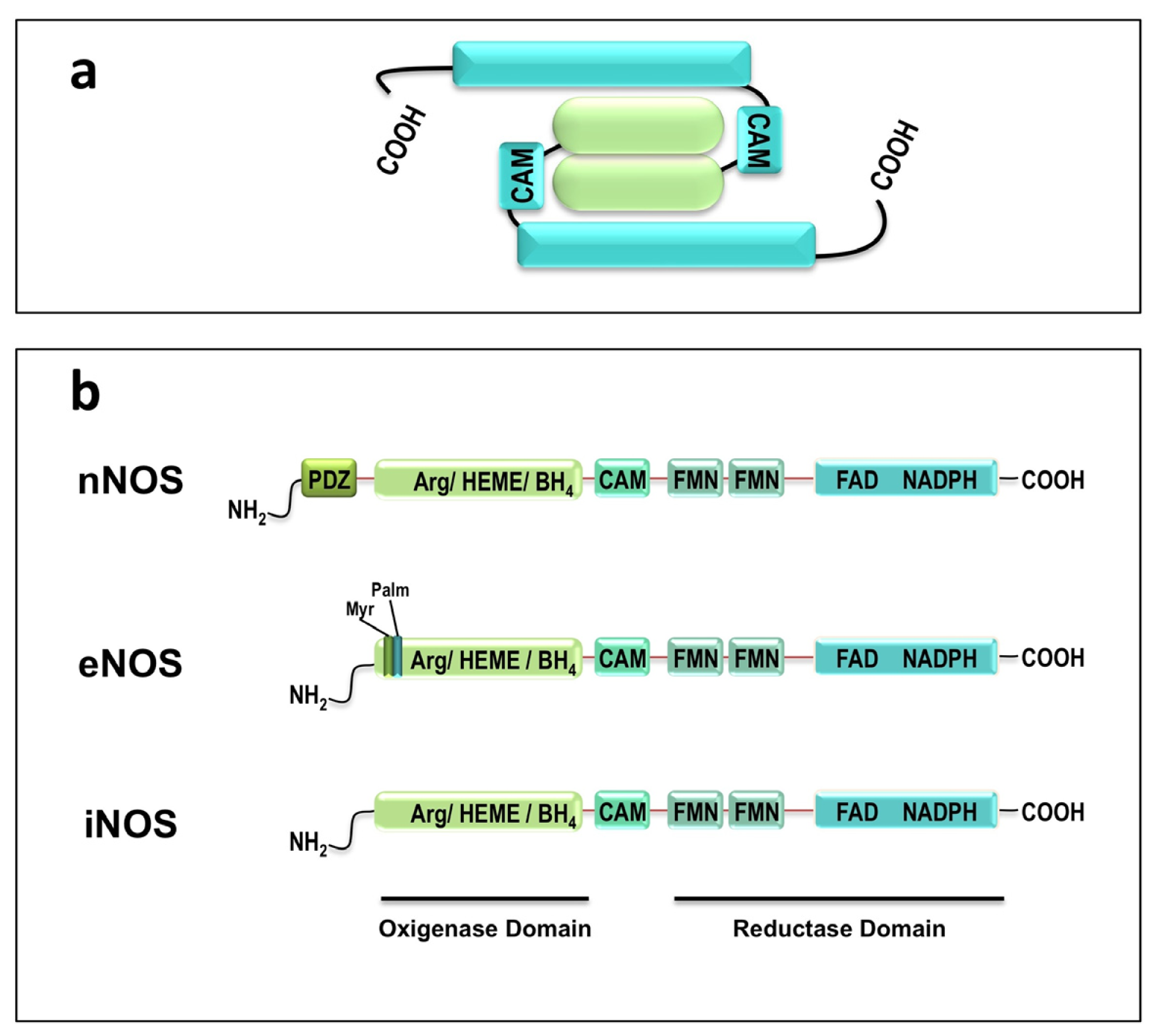
2. NO and Nitric Oxide Synthase</title>
<p>NO is a gaseous, inorganic, uncharged, diatomic molecule and a free radical with one unpaired electron in its external orbital [<xref ref-type="bibr" rid="b3-ijms-13-17160">3</xref>,<xref ref-type="bibr" rid="b6-ijms-13-17160">6</xref>]. It is highly lipophilic and diffusible, so NO can pass through multiple cell membranes to reach its final target at some distance from the sites of NO synthesis [<xref ref-type="bibr" rid="b7-ijms-13-17160">7</xref>]. Considering its very short half-life, less than 5 s, NO cannot be stored in free form and is generally synthesized on demand with specific biological effects [<xref ref-type="bibr" rid="b7-ijms-13-17160">7</xref>]. This molecule is produced by three isoforms of NO synthases (NOS): neuronal NOS (nNOS, EC 1.14.13.39), endothelial NOS (eNOS, EC 1.14.13.39) and inducible NOS (iNOS, EC 1.14.13.39). These isoforms were originally named because of the tissues from where they were first purified (neuronal and endothelial) and the mode of activation in the case of iNOS; but because they were also identified in several other cells, these denominations became inappropriate. Despite the proposal of other nomenclatures (numerical and descriptive) [<xref ref-type="bibr" rid="b8-ijms-13-17160">8</xref>] the original denominations are still used. In the numerical nomenclature nNOS is type I, iNOS is type II and eNOS is type III enzyme. In the descriptive nomenclature the dependence of Ca<sup>2+</sup> is indicated by a letter “c”, such as ncNOS and ecNOS, for neuronal and endothelial isoforms, respectively. These differences in nomenclature cause great confusion in the literature but in this review, we will only consider the original denominations: nNOS, eNOS and iNOS. All three types of NOS are expressed in skeletal muscle [<xref ref-type="bibr" rid="b9-ijms-13-17160">9</xref>].</p>
<p>NOS uses molecular oxygen to oxidize the guanidinium nitrogen atom of <sc>l</sc>-arginine to produce NO and <sc>l</sc>-citrulline [<xref ref-type="bibr" rid="b6-ijms-13-17160">6</xref>]. Two isoforms, nNOS and eNOS, are constitutive and regulated by the interaction of Ca<sup>2+</sup> with calmodulin [<xref ref-type="bibr" rid="b6-ijms-13-17160">6</xref>]. On the other hand, iNOS is usually only expressed during defense mechanisms against infections or inflammation [<xref ref-type="bibr" rid="b8-ijms-13-17160">8</xref>] and it is not regulated by Ca<sup>2+</sup> because it forms a complex with calmodulin at very low concentrations of Ca<sup>2+</sup>[<xref ref-type="bibr" rid="b2-ijms-13-17160">2</xref>]. Four co-factors are required for NO synthesis: tetrahydrobiopterin (BH4), flavin adenine dinucleotide (FAD), flavin monucleotide (FMN) and nicotinamide adenine dinucleotide phosphate (NADPH) [<xref ref-type="bibr" rid="b3-ijms-13-17160">3</xref>].</p>
<p>Although all three NOS isoforms are products of different genes, they have similar structure with 50%–60% sequence identity [<xref ref-type="bibr" rid="b7-ijms-13-17160">7</xref>] and are only active as dimer [<xref ref-type="bibr" rid="b10-ijms-13-17160">10</xref>] (<xref ref-type="fig" rid="f1-ijms-13-17160">Figure 1</xref>). Each subunit of this dimeric complex is composed by two domains, oxygenase and reductase domains. The oxygenase domain contains the binding sites for <sc>l</sc>-arginine, BH4 and heme (Fe) and the reductase domain binds to FMN, FAD, NADPH and calmodulin [<xref ref-type="bibr" rid="b7-ijms-13-17160">7</xref>,<xref ref-type="bibr" rid="b10-ijms-13-17160">10</xref>,<xref ref-type="bibr" rid="b11-ijms-13-17160">11</xref>]. Both subunits are attached at the oxygenase domain, forming the dimeric conformation. In skeletal muscle, the nNOS isoform has an additional 250 amino acid <italic>N</italic>-terminal sequence, the PDZ motif, that target the enzyme to the sarcolemma, in skeletal muscle [<xref ref-type="bibr" rid="b7-ijms-13-17160">7</xref>]. Myristoylation and palmitoylation sites are typically present in eNOS <italic>N</italic>-terminal. Acylation by myristate and palmitate is required for the subcellular localization of eNOS in the caveolae of endothelial cells [<xref ref-type="bibr" rid="b11-ijms-13-17160">11</xref>].</p>
<p>Since the first isolation and characterization of NOS [<xref ref-type="bibr" rid="b12-ijms-13-17160">12</xref>], several splice variants have been discovered [<xref ref-type="bibr" rid="b13-ijms-13-17160">13</xref>,<xref ref-type="bibr" rid="b14-ijms-13-17160">14</xref>]. nNOS is encoded by the gene <italic>NOS1</italic> localized in chromosome 12q24.22, contains 29 exons and has at least ten transcript variants [<xref ref-type="bibr" rid="b15-ijms-13-17160">15</xref>]. The classical isoform (nNOSα) is predominantly found in neuronal cells, contains both exon 2, which is responsible for the catalytic activity of the enzyme and the PDZ domain, which interacts with membrane proteins [<xref ref-type="bibr" rid="b16-ijms-13-17160">16</xref>]. nNOSβ and nNOSδ lack the PDZ domain, maintain the catalytic domains and have different exon 1 sequence [<xref ref-type="bibr" rid="b15-ijms-13-17160">15</xref>]. Another variant, nNOSμ, contains an additional in-frame exon between exon 16 and 17. Primarily found in mouse skeletal and cardiac muscles, it was later found in other human organs, such as aorta, bladder, colon, corpus cavernosum and placenta [<xref ref-type="bibr" rid="b17-ijms-13-17160">17</xref>]. A human testis specific variant, TnNOS, which is analogue to the mouse nNOSδ, was also described and characterized with a shorter <italic>N</italic>-terminus [<xref ref-type="bibr" rid="b18-ijms-13-17160">18</xref>].</p>
<p>The isoform eNOS is encoded by the gene <italic>NOS3</italic>, located on chromosome 7q36; and highly expressed in intestine, parathyroid, placenta, ovary, kidney, platelet and myocyte [<xref ref-type="bibr" rid="b19-ijms-13-17160">19</xref>,<xref ref-type="bibr" rid="b20-ijms-13-17160">20</xref>]. There are three transcript variants, with shorter distinct-<italic>C</italic>-terminus due to alternate 3′ exon and polyadenylation site, known as eNOS13A, eNOS13B and eNOS13C [<xref ref-type="bibr" rid="b21-ijms-13-17160">21</xref>]. iNOS is encoded by the gene <italic>NOS2</italic>, located on chromosome 17q11.2 and is expressed in several cell types and tissue in response to inflammatory agents and cytokines [<xref ref-type="bibr" rid="b22-ijms-13-17160">22</xref>]. In addition, NO can also be generated through non enzymatic sources, such as the reduction of nitrite to NO in disease states, such as ischemia, and under the acidic and highly reduced conditions [<xref ref-type="bibr" rid="b23-ijms-13-17160">23</xref>].</p>
<sec>
<title>2.1. Mitochondrial NOS</title>
<p>The finding of co-localization of eNOS expression with mitochondrial markers in skeletal muscle, suggested a close relationship between eNOS and mitochondria [<xref ref-type="bibr" rid="b24-ijms-13-17160">24</xref>]. In fact, using gold labeling and electron microscopy, immunoreactivity to eNOS was found in rat liver and brain mitochondria [<xref ref-type="bibr" rid="b25-ijms-13-17160">25</xref>]. It was, therefore, hypothesized that a mitochondrial NOS (mtNOS) could be eNOS or another NOS with significant homology to this form [<xref ref-type="bibr" rid="b25-ijms-13-17160">25</xref>]. The co-localization of eNOS and mitochondria was demonstrated in other studies, where immunoreactivity to eNOS was found associated to skeletal muscle mitochondria in skeletal muscle [<xref ref-type="bibr" rid="b26-ijms-13-17160">26</xref>,<xref ref-type="bibr" rid="b27-ijms-13-17160">27</xref>]. Actually, NO production by isolated mitochondria strongly suggested the existence of an mtNOS [<xref ref-type="bibr" rid="b28-ijms-13-17160">28</xref>,<xref ref-type="bibr" rid="b29-ijms-13-17160">29</xref>]. Lacza <italic>et al.</italic> confirmed the presence of NOS activity in liver, using the assay of conversion of <sc>l</sc>-arginine to citrulline and also found immunoreactivity to eNOS by Western blot and immuno-gold labeling with electron microscopy [<xref ref-type="bibr" rid="b30-ijms-13-17160">30</xref>]. However, the same group was not able to confirm these results in a later study, when NOS activity and NO production were not detected in liver mitochondria [<xref ref-type="bibr" rid="b31-ijms-13-17160">31</xref>]. They also identified specific immunoreactivity to nNOS, iNOS and eNOS in liver mitochondria obtained from nNOS, iNOS and eNOS knockout mice, excluding these isoforms as the mtNOS. Studies on this subject have been controversial because they are based on the presence of NOS activity, NO production or nitrite/peroxynitrite in isolated mitochondria from rat liver [<xref ref-type="bibr" rid="b28-ijms-13-17160">28</xref>,<xref ref-type="bibr" rid="b32-ijms-13-17160">32</xref>,<xref ref-type="bibr" rid="b33-ijms-13-17160">33</xref>], PC12 (rat neuronal cells) and COS-1 (monkey fibroblasts) cells [<xref ref-type="bibr" rid="b34-ijms-13-17160">34</xref>], porcine heart [<xref ref-type="bibr" rid="b35-ijms-13-17160">35</xref>], rat kidney [<xref ref-type="bibr" rid="b36-ijms-13-17160">36</xref>], rat heart [<xref ref-type="bibr" rid="b37-ijms-13-17160">37</xref>]; while others do not confirm these findings in murine liver [<xref ref-type="bibr" rid="b31-ijms-13-17160">31</xref>,<xref ref-type="bibr" rid="b38-ijms-13-17160">38</xref>,<xref ref-type="bibr" rid="b39-ijms-13-17160">39</xref>]. Other studies using different approaches have suggested that mtNOS might be eNOS [<xref ref-type="bibr" rid="b25-ijms-13-17160">25</xref>,<xref ref-type="bibr" rid="b26-ijms-13-17160">26</xref>,<xref ref-type="bibr" rid="b40-ijms-13-17160">40</xref>], iNOS [<xref ref-type="bibr" rid="b32-ijms-13-17160">32</xref>,<xref ref-type="bibr" rid="b33-ijms-13-17160">33</xref>,<xref ref-type="bibr" rid="b35-ijms-13-17160">35</xref>,<xref ref-type="bibr" rid="b37-ijms-13-17160">37</xref>] or even nNOS [<xref ref-type="bibr" rid="b36-ijms-13-17160">36</xref>,<xref ref-type="bibr" rid="b41-ijms-13-17160">41</xref>–<xref ref-type="bibr" rid="b43-ijms-13-17160">43</xref>]. These discrepancies can be explained by differences in methods of purification of mitochondrial fraction, specificity of the method of NOS detection and different tissue/cell sources. Studies with knockout mice have also given discrepant results [<xref ref-type="bibr" rid="b31-ijms-13-17160">31</xref>,<xref ref-type="bibr" rid="b44-ijms-13-17160">44</xref>]. Kanai <italic>et al.</italic> (2001) studied heart from knockout (nNOS −/−, iNOS −/− and eNOS −/−) mice and observed absence of mitochondrial NO production only in nNOS −/− knockout mice, indicating that nNOS should be the mtNOS, specifically the nNOSα variant, which was the gene knocked out in these mice [<xref ref-type="bibr" rid="b44-ijms-13-17160">44</xref>]. Lacza <italic>et al</italic>., however, have not confirmed NO production in nNOS knockout mice [<xref ref-type="bibr" rid="b45-ijms-13-17160">45</xref>].</p>
<p>Only a few studies focused on the existence and identity of mtNOS in skeletal muscle [<xref ref-type="bibr" rid="b40-ijms-13-17160">40</xref>,<xref ref-type="bibr" rid="b46-ijms-13-17160">46</xref>]. In rat muscle, eNOS was localized in mitochondria by electron microscopy [<xref ref-type="bibr" rid="b40-ijms-13-17160">40</xref>] which is supported by the findings of co-localization of eNOS with mitochondrial markers in rat [<xref ref-type="bibr" rid="b24-ijms-13-17160">24</xref>] and human muscle fibers with mitochondrial proliferation [<xref ref-type="bibr" rid="b26-ijms-13-17160">26</xref>,<xref ref-type="bibr" rid="b27-ijms-13-17160">27</xref>]. Studying the insulin induced effects on NO synthesis in rat skeletal muscle, Finocchietto <italic>et al.</italic> showed an increased mitochondrial NO synthesis via Akt phosphorylation that was dimished in nNOS silenced rat muscle. Based on these results they concluded that the mtNOS would be a variant of nNOS that could be subjected to pos-translational modifications [<xref ref-type="bibr" rid="b47-ijms-13-17160">47</xref>]. It was also suggested that nNOS could be translocated to mitochondria but with unclear mechanisms [<xref ref-type="bibr" rid="b48-ijms-13-17160">48</xref>]. Recently, Aguirre <italic>et al.</italic>[<xref ref-type="bibr" rid="b46-ijms-13-17160">46</xref>] tried to identify the nature of mtNOS performing a study on skeletal muscle mitochondria of mice injected with lipopolysaccharide (LPS), considering that mtNOS expression was reported to increase in response to endotoxin [<xref ref-type="bibr" rid="b49-ijms-13-17160">49</xref>]. The authors observed that in the presence of a NOS substrate, mitochondria from LPS treated mice had lower respiration rate, which was prevented by a NOS inhibitor. Because they could not find the expression of the known iNOS, they suggested that the mtNOS could be an endotoxin-inducible NOS but distinct from iNOS and the other cytosolic isoforms. In skeletal muscle there are some evidences suggesting that, at least in this tissue, eNOS or a similar variant may be the mtNOS, but more clarification is still needed.</p></sec>
<sec>
<title>2.2. NOS Isoforms in Skeletal Muscle</title>
<p>In skeletal muscle, nNOS and eNOS isoforms are expressed constitutively while iNOS is only expressed during inflammatory responses [<xref ref-type="bibr" rid="b9-ijms-13-17160">9</xref>,<xref ref-type="bibr" rid="b50-ijms-13-17160">50</xref>]. Each NOS isoform has a specific localization in different compartments of muscle fibers. The nNOS isoform is considered the main source of NO in skeletal muscle, however it is not the same isoform expressed in brain. A splice variant is expressed in skeletal muscle, nNOSμ, characterized by an insertion between exon 16 and 17, encoding a 34-aminoacid segment [<xref ref-type="bibr" rid="b51-ijms-13-17160">51</xref>]. This isoform is located on the sarcolemma (muscle membrane) [<xref ref-type="bibr" rid="b26-ijms-13-17160">26</xref>,<xref ref-type="bibr" rid="b27-ijms-13-17160">27</xref>], with higher activity in type II (fast twitch) fibers and linked to the dystrophin complex [<xref ref-type="bibr" rid="b26-ijms-13-17160">26</xref>,<xref ref-type="bibr" rid="b52-ijms-13-17160">52</xref>,<xref ref-type="bibr" rid="b53-ijms-13-17160">53</xref>]. nNOS expression in the sarcoplasm was also reported in type I and II fibers, but more intense in type I fibers [<xref ref-type="bibr" rid="b54-ijms-13-17160">54</xref>]. It was demonstrated that nNOSμ is also present in soluble fractions of gastrocnemius muscle homogenates in mice, showing the existence of a soluble cytoplasmic nNOSμ [<xref ref-type="bibr" rid="b55-ijms-13-17160">55</xref>]. Studies with mice with reduced sarcolemmal nNOS but preserved soluble nNOS show that NO produced by sarcolemmal nNOS acts in the regulation of α-adrenergic vasoconstriction and blood supply in contracting skeletal muscle [<xref ref-type="bibr" rid="b55-ijms-13-17160">55</xref>,<xref ref-type="bibr" rid="b56-ijms-13-17160">56</xref>]. In the absence of sarcolemmal nNOS there is a considerable decrease in the blood supply during electrically evoked muscle contraction in mice [<xref ref-type="bibr" rid="b55-ijms-13-17160">55</xref>], which demonstrates the important role of this isoform in exercise capacity. However, when sarcolemmal nNOS is absent, cytoplasmic nNOSμ is not able to compensate the NO deficiency [<xref ref-type="bibr" rid="b55-ijms-13-17160">55</xref>]. Another nNOS splice variant, nNOSβ, was recently identified and localized in the Golgi complex [<xref ref-type="bibr" rid="b56-ijms-13-17160">56</xref>]. The loss of nNOSμ and β in genetically modified mice showed disruption of the microtubule cytoskeleton and abnormal mitochondrial morphology, suggesting that both splice variants have an important role in maintaining normal mitochondrial health [<xref ref-type="bibr" rid="b56-ijms-13-17160">56</xref>]. The function of the cytoplasmic nNOSμ remains unclear.</p>
<p>Expression of eNOS is primarily found in endothelial cells of vessels and microvessels [<xref ref-type="bibr" rid="b54-ijms-13-17160">54</xref>] but sarcoplasmic expression has also been reported [<xref ref-type="bibr" rid="b26-ijms-13-17160">26</xref>,<xref ref-type="bibr" rid="b27-ijms-13-17160">27</xref>]. This pattern is confirmed by NADPH diaphorase histochemistry, which is considered an indirect method to evaluate NOS activity [<xref ref-type="bibr" rid="b26-ijms-13-17160">26</xref>,<xref ref-type="bibr" rid="b27-ijms-13-17160">27</xref>,<xref ref-type="bibr" rid="b53-ijms-13-17160">53</xref>]. Abnormalities in specific isoforms such as nNOS and eNOS have been reported in muscle diseases with mitochondrial deficiencies [<xref ref-type="bibr" rid="b26-ijms-13-17160">26</xref>,<xref ref-type="bibr" rid="b27-ijms-13-17160">27</xref>], indicating that specific NOS activities and expression may be involved in the pathogenesis of these diseases. Increased nNOS activity and expression were observed in muscle fibers with mitochondrial proliferation, suggesting that it is related to mitochondrial biogenesis [<xref ref-type="bibr" rid="b27-ijms-13-17160">27</xref>]. However, the exact mechanisms involved in these abnormalities are not clear.</p></sec></sec>
<sec>
<title>3. Regulation of NOS</title>
<p>Due to the various physiological roles, specific tissue expression and different isoforms, it is reasonable to believe that the regulatory mechanisms controlling NOS activity, expression and localization are very complex and multifactorial. All types of NOS are transcriptionally modulated and exhibit inducible and constitutive patterns of expression in different tissue environments [<xref ref-type="bibr" rid="b7-ijms-13-17160">7</xref>]. The mechanisms of regulation can be summarized in alternative mRNA splicing, protein-protein interactions (e.g., to Ca<sup>2+</sup>/calmodulin complex, PDZ domains, PIN, caveolin-1 and 3, Hsp90, ENAP-1, kalirin), covalent modifications (phosphorylation, myristoylation, palmitoylation) and redox signaling [<xref ref-type="bibr" rid="b7-ijms-13-17160">7</xref>,<xref ref-type="bibr" rid="b11-ijms-13-17160">11</xref>,<xref ref-type="bibr" rid="b57-ijms-13-17160">57</xref>]. These regulatory mechanisms control the expression, localization and activity of NOS.</p>
<p>The expression of nNOS and eNOS in specific tissues is controlled in some cases by alternate mRNA splicing, as in the case of TnNOS, expressed in testis [<xref ref-type="bibr" rid="b18-ijms-13-17160">18</xref>,<xref ref-type="bibr" rid="b58-ijms-13-17160">58</xref>], while the presence of a 220 aminoacid sequence in the <italic>N</italic>-terminal of nNOS, containing the PDZ domain, targets nNOS to the brain and skeletal muscle [<xref ref-type="bibr" rid="b16-ijms-13-17160">16</xref>]. In skeletal muscle, PDZ domain interactions mediate binding to syntrophin, a dystrophin associated protein, localizing the muscle nNOS to the sarcolemmal membrane. When this complex is absent, such as in Duchenne muscular dystrophy, nNOS is not attached to the membrane and localizes in the cytosol [<xref ref-type="bibr" rid="b59-ijms-13-17160">59</xref>].</p>
<p>The processes of myristoylation and palmitoylation (acylation with the fatty acids myristate and palmitate) are required for the proper localization of enzyme in the caveolae of endothelial cells [<xref ref-type="bibr" rid="b60-ijms-13-17160">60</xref>,<xref ref-type="bibr" rid="b61-ijms-13-17160">61</xref>]. In cardiac myocytes, eNOS is localized in plasmalemmal caveolae due to the process of palmitoylation [<xref ref-type="bibr" rid="b62-ijms-13-17160">62</xref>]. Caveolae are microdomains of the plasmalemmal membrane and it is coated by proteins called caveolin. Caveolin 1 and 3 are negative regulator of eNOS and nNOS [<xref ref-type="bibr" rid="b63-ijms-13-17160">63</xref>].</p>
<p>The interaction between calmodulin and Ca<sup>2+</sup> is required for nNOS and eNOS activity. However several proteins can interact and regulate the enzyme activity. The FMN-binding sub-domain can act as an enzyme inhibitor, by destabilizing calmodulin binding at low Ca<sup>2+</sup> level. PIN, an 89-amino-acid protein that specifically interacts with the <italic>N</italic>-terminal of nNOS, has been associated with inhibition of the isoform activity [<xref ref-type="bibr" rid="b64-ijms-13-17160">64</xref>,<xref ref-type="bibr" rid="b65-ijms-13-17160">65</xref>], but it has also been argued that this inhibition may not be specific to nNOS [<xref ref-type="bibr" rid="b66-ijms-13-17160">66</xref>]. The molecular chaperone Hsp90 increases eNOS activity [<xref ref-type="bibr" rid="b67-ijms-13-17160">67</xref>] whereas kalirin, a cytosolic protein, inhibits iNOS by preventing dimer formation [<xref ref-type="bibr" rid="b68-ijms-13-17160">68</xref>]. Structures such as BH4 and Zinc binding are not required for dimer formation but are important for stabilization of this structure, acting in the regulation of NOS activity [<xref ref-type="bibr" rid="b69-ijms-13-17160">69</xref>]. Phosphorylation can have an inhibitory effect on nNOS activity while on eNOS the activity is increased [<xref ref-type="bibr" rid="b70-ijms-13-17160">70</xref>–<xref ref-type="bibr" rid="b73-ijms-13-17160">73</xref>].</p>
<p>The influence of redox signaling on NOS activity has been demonstrated for the endothelial isoform. ROS can affect eNOS through pos-translational modifications such as <italic>S</italic>-glutathionylation, <italic>S</italic>-nitrosylation; H<sub>2</sub>O<sub>2</sub>-dependent activation or interactions affecting signaling pathways of eNOS activation [<xref ref-type="bibr" rid="b7-ijms-13-17160">7</xref>,<xref ref-type="bibr" rid="b57-ijms-13-17160">57</xref>,<xref ref-type="bibr" rid="b74-ijms-13-17160">74</xref>]. The importance of <italic>S</italic>-glutathionylation in the regulation of endothelial function and vascular tone has been demonstrated by the finding of increased <italic>S</italic>-glutathionylation in hypertensive vessels, that was restored by thiol-specific reducing agents, which reverse this <italic>S</italic>-glutathionylation [<xref ref-type="bibr" rid="b75-ijms-13-17160">75</xref>].</p>
<p><italic>S</italic>-glutathionylation induces a reversible uncoupling of eNOS, which during oxidative stress, could function to prevent irreversible oxidative damage of the thiols critical for eNOS function [<xref ref-type="bibr" rid="b76-ijms-13-17160">76</xref>]. <italic>S</italic>-nitrosylation of eNOS is an important mechanism of activity control. It was demonstrated that eNOS is constitutively <italic>S</italic>-nitrosylated in resting endothelial cells but with the addition of an eNOS agonist, eNOS is denitrosylated and then progressively renitrosylated, resuming its original level. An inverse relationship between eNOS activity and <italic>S</italic>-nitrosylation was also found in endothelial cells. These results show that eNOS <italic>S</italic>-nitrosylation leads to a decrease in enzyme activity [<xref ref-type="bibr" rid="b77-ijms-13-17160">77</xref>]. <italic>S</italic>-nitrosylation is also a dynamic mechanism of control of NOS activity. Targeting of eNOS to cellular membranes, such as caveolae, was also demonstrated as a requirement of eNOS <italic>S</italic>-nitrosylation while when the enzyme location in the cytosol promotes denitrosylation [<xref ref-type="bibr" rid="b77-ijms-13-17160">77</xref>,<xref ref-type="bibr" rid="b78-ijms-13-17160">78</xref>]. Mass spectrometry analysis showed that the location of <italic>S</italic>-nitrosylation is the zinc-tetrathiolate of eNOS[<xref ref-type="bibr" rid="b78-ijms-13-17160">78</xref>]. However the roles of these mechanisms on regulation of NOS isoforms in skeletal muscle are yet to be clarified.</p>
<p>H<sub>2</sub>O<sub>2</sub> is a key modulator of eNOS activation in vascular endothelial cells [<xref ref-type="bibr" rid="b79-ijms-13-17160">79</xref>] and cardiac myocytes [<xref ref-type="bibr" rid="b80-ijms-13-17160">80</xref>]. Using a highly sensitive fluorescent probe it was possible to demonstrate that low concentrations of H<sub>2</sub>O<sub>2</sub> promote NO synthesis, through activation of eNOS by phosphorylation on multiple residues [<xref ref-type="bibr" rid="b80-ijms-13-17160">80</xref>]. Comparing the effects of H<sub>2</sub>O<sub>2</sub> treatment on cardiac myocytes from eNOS<sup>null</sup> and nNOS<sup>null</sup>, Sartoretto <italic>et al.</italic> found that H<sub>2</sub>O<sub>2</sub> treatment effects were abolished in eNOS<sup>null</sup> cells while nNOS<sup>null</sup> myocytes H<sub>2</sub>O<sub>2</sub> effects were maintained, suggesting that eNOS is the main source of H<sub>2</sub>O<sub>2</sub> induced NO synthesis [<xref ref-type="bibr" rid="b80-ijms-13-17160">80</xref>]. Interestingly, Waypa <italic>et al.</italic>[<xref ref-type="bibr" rid="b81-ijms-13-17160">81</xref>] demonstrated that H<sub>2</sub>O<sub>2</sub> production in pulmonary smooth muscle cells under hypoxic conditions is increased in the mitochondrial intermembrane space but not in the matrix, which favours the idea of compartmentalized redox regulation.</p>
<p>Signaling pathways of NOS activation can also be affected by redox modifications in several intermediaries (for review see [<xref ref-type="bibr" rid="b74-ijms-13-17160">74</xref>]).</p></sec>
<sec>
<title>
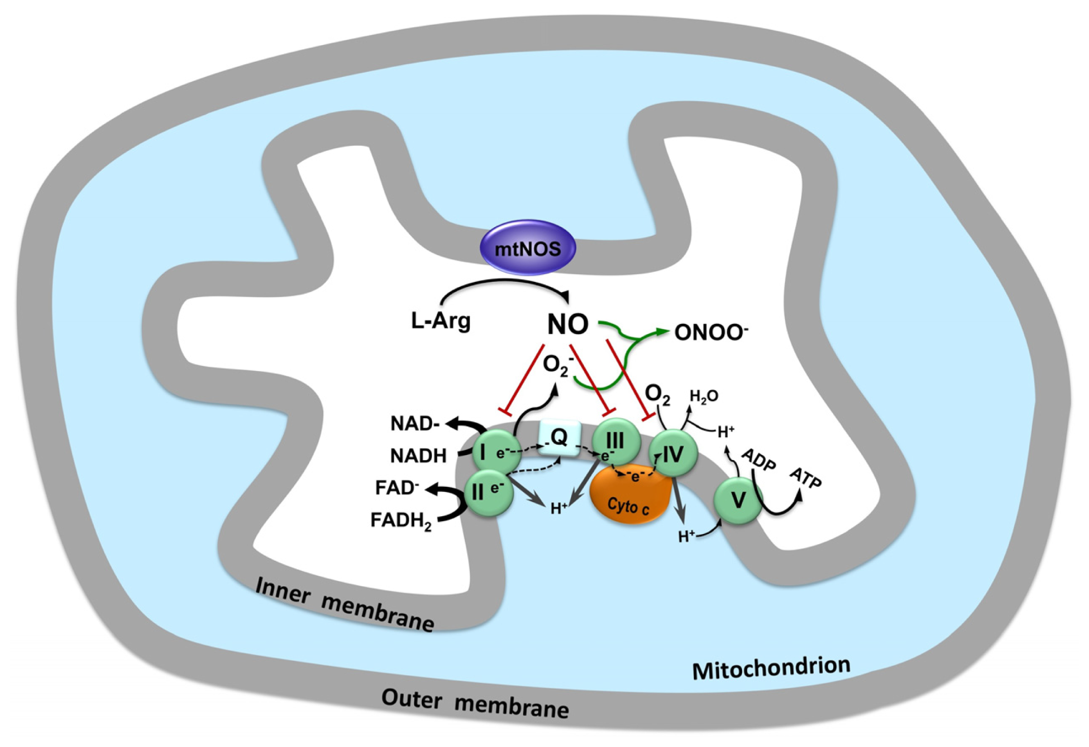
4. NO and Mitochondrial Function</title>
<p>NO diffuses from mitochondria to cytosol, as well as from cytosol to mitochondria, what is called mitochondria-cytosol NO cross-talk [<xref ref-type="bibr" rid="b82-ijms-13-17160">82</xref>]. Through the interaction with other molecules or proteins, NO exerts its function on signaling physiological events or promoting cell damage (<xref ref-type="fig" rid="f2-ijms-13-17160">Figure 2</xref>).</p>
<p>Nitrosative and oxidative modifications generally constitute redox-related signaling events in several physiological pathways or nitrosative/oxidative stress promoting mitochondrial damage [<xref ref-type="bibr" rid="b9-ijms-13-17160">9</xref>]. NO can react to three types of targets: (a) molecular oxygen and superoxide anions; (b) transition metals such as heme iron and iron-sulfur centers; and (c) reduced thiols [<xref ref-type="bibr" rid="b83-ijms-13-17160">83</xref>].</p>
<p>The reaction of NO with molecular oxygen or superoxide anions generates low molecular weight NO derivatives that participate in electron transfer reactions, as retain redox activity [<xref ref-type="bibr" rid="b84-ijms-13-17160">84</xref>]. The formation of peroxynitrite from the combination of NO and superoxide, however, mediates oxidative damage [<xref ref-type="bibr" rid="b9-ijms-13-17160">9</xref>].</p>
<p>The reaction with transition metals represents a mechanism of NO buffering or regulation. Protein activity can be modulated through reversible reaction with transition metals and thiols [<xref ref-type="bibr" rid="b84-ijms-13-17160">84</xref>]. The interaction of NO with heme iron of soluble glanylyl cyclase induces a conformational change activating this enzyme and increasing cyclic guanosine monophosphate (cGMP) levels [<xref ref-type="bibr" rid="b84-ijms-13-17160">84</xref>,<xref ref-type="bibr" rid="b85-ijms-13-17160">85</xref>]. This increase activates cGMP-dependent protein kinases or cGMP-gated ion channels, characterizing the NO mediate physiological actions [<xref ref-type="bibr" rid="b82-ijms-13-17160">82</xref>].</p>
<p>NO can also interact with other heme-containing proteins, such as, cytochrome <italic>c</italic> oxidase, or with iron-sulfur clusters present in complex I of the respiratory chain, both promoting enzyme inhibition [<xref ref-type="bibr" rid="b9-ijms-13-17160">9</xref>].</p>
<p>NO reacts with protein thiol (RSH, RS-) via <italic>S</italic>-nitrosylation to form RS–NO groups, this reaction is reversed by NO transfer to other sulphur centers. <italic>S</italic>-nitrosylation of glutathione and other non-regulatory thiols can act as NO buffering and regulate protein function by altering conformation, by accelerating disulphide formation, or by influencing the reactivity of nearby metal centers [<xref ref-type="bibr" rid="b86-ijms-13-17160">86</xref>].</p>
<p>Finocchietto <italic>et al.</italic>[<xref ref-type="bibr" rid="b47-ijms-13-17160">47</xref>] demonstrated the role of NO on glucose metabolism. These studies demonstrated that insulin increased muscle oxidative rate via mitochondrial NO, resulting in decline of mitochondrial O<sub>2</sub> uptake. In the absence of NO, mitochondrial O<sub>2</sub> uptake is completely released leading to a preferential oxidation of glucose to CO<sub>2</sub> and H<sub>2</sub>O, while in the presence of NO, glucose utilization is delivered to glycogen synthesis.</p>
<sec>
<title>4.1. Control of Mitochondrial Respiration</title>
<p>NO affects mitochondrial function in many ways. Because of its vascular smooth muscle relaxing effect, NO regulates blood flow to skeletal muscle fiber, facilitating the supply of respiratory substrates to mitochondria [<xref ref-type="bibr" rid="b9-ijms-13-17160">9</xref>]. NO also regulates binding and release of O<sub>2</sub> from hemoglobin in red blood cells [<xref ref-type="bibr" rid="b87-ijms-13-17160">87</xref>], thereby regulating oxygen delivery to tissues and consequently the O<sub>2</sub> supply to the mitochondria [<xref ref-type="bibr" rid="b88-ijms-13-17160">88</xref>]. The classical concept of control of mitochondrial respiration is based on the mitochondrial metabolic states, which is controlled by ADP availability [<xref ref-type="bibr" rid="b89-ijms-13-17160">89</xref>]. In state 4, there is availability of respiratory substrate but not of ADP, while in state 3, both respiratory substrate and ADP are available. State 3 corresponds to the maximal physiological rate of ATP production and O<sub>2</sub> consumption. Under non-stimulated physiological conditions only 35% of the ATP-producing capacity is used [<xref ref-type="bibr" rid="b90-ijms-13-17160">90</xref>]. Boveris <italic>et al.</italic> proposed a new concept of regulation of cellular respiration that depends on energy demands, which is controlled by the availability of ADP to F1-ATPase and of O<sub>2</sub> and NO to cytochrome <italic>c</italic> oxidase [<xref ref-type="bibr" rid="b3-ijms-13-17160">3</xref>].</p>
<p>The influence of NO on mitochondrial respiratory chain can be summarized as follow: (a) NO inhibits cytochrome <italic>c</italic> oxidase activity by competing with oxygen; (b) NO inhibits electron transfer between cytochrome <italic>b</italic> and <italic>c</italic> and increases mitochondrial production of O<sub>2</sub><sup>−</sup>; and (c) NO inhibits electron transfer and NADH-dehydrogenase function in Complex I [<xref ref-type="bibr" rid="b3-ijms-13-17160">3</xref>].</p></sec>
<sec>
<title>4.2. NO Inhibits Cytochrome <italic>c</italic> Oxidase Activity</title>
<p>NO was first recognized as an inhibitor of mitochondrial electron transfer in 1994 [<xref ref-type="bibr" rid="b91-ijms-13-17160">91</xref>,<xref ref-type="bibr" rid="b92-ijms-13-17160">92</xref>], with the observations that low NO concentrations was able to reversibly inhibit brain and muscle cytochrome <italic>c</italic> oxidase through competitive binding at O<sub>2</sub> site. This NO binding occurs at physiological concentrations of NO and inhibits the electron flow in the respiratory chain and therefore decreases O<sub>2</sub> consumption and ATP formation [<xref ref-type="bibr" rid="b2-ijms-13-17160">2</xref>,<xref ref-type="bibr" rid="b93-ijms-13-17160">93</xref>]. Differently from O<sub>2</sub>, NO reversibly binds to both reduced and oxidized cytochrome <italic>c</italic> oxidase. At low O<sub>2</sub> concentration, NO binds to the heme <italic>a3</italic> domains of reduced form of cytochrome <italic>c</italic> oxidase, as a competitive inhibitor of O<sub>2</sub>. However, at high O<sub>2</sub> level, NO binds to the oxidized cytochrome <italic>c</italic> oxidase, via the copper moiety of the binuclear center instead of the iron moiety. This reaction results in nitrite (NO<sup>2 −</sup>) formation and consequently in O<sub>2</sub> consumption [<xref ref-type="bibr" rid="b94-ijms-13-17160">94</xref>]. A persistent inhibition of cytochrome <italic>c</italic> oxidase can promote, at an early stage, the release of small amount of hydrogen peroxide, acting as cellular signaling defense molecule. But at a later stage, high concentrations of hydrogen peroxide may lead to peroxynitrite generation and induce apoptosis or necrosis [<xref ref-type="bibr" rid="b82-ijms-13-17160">82</xref>].</p></sec>
<sec>
<title>4.3. NO Inhibits Electron Transfer between Cytochrome <italic>b</italic> and <italic>c</italic></title>
<p>Another NO sensitive site in the respiratory chain is the electron transfer at complex III, ubiquinol-cytochrome <italic>c</italic> reductase [<xref ref-type="bibr" rid="b3-ijms-13-17160">3</xref>,<xref ref-type="bibr" rid="b90-ijms-13-17160">90</xref>]. NO is able to inhibit succinate-cytochrome <italic>c</italic> reductase and NADH-cytochrome <italic>c</italic> reductase, with similar effect on both reductases but in a lower extent than cytochrome <italic>c</italic> oxidase [<xref ref-type="bibr" rid="b95-ijms-13-17160">95</xref>]. Inhibition at this site increases the production of O<sub>2</sub><sup>−</sup> and H<sub>2</sub>O<sub>2</sub>, which are not only involved with oxidative damage, but together with NO, are considered as part of an integrated system of mitochondrial signaling for cellular regulation [<xref ref-type="bibr" rid="b82-ijms-13-17160">82</xref>].NO is also able to reduce cytochrome <italic>b</italic>, probably by its interaction with an iron-sulfur center, which is reversible but is not affected by the O<sub>2</sub>/NO ratio [<xref ref-type="bibr" rid="b3-ijms-13-17160">3</xref>].</p></sec>
<sec>
<title>4.4. NO Inhibits Electron Transfer and NADH-Dehydrogenase Function in Complex I</title>
<p>It has been shown that prolonged exposure of NO also leads to inhibition of complex I activity in murine macrophage cultured cells, with a concomitant decrease in the content of intracellular reduced glutathione [<xref ref-type="bibr" rid="b96-ijms-13-17160">96</xref>]. At first this inhibition is reversible but it becomes persistent with time. The mechanisms of complex I inhibition are not completely clear. There are a few hypothetical mechanisms but three are the most likely: <italic>S</italic>-nitrosation, tyrosine nitration and damage to Fe–S centers [<xref ref-type="bibr" rid="b97-ijms-13-17160">97</xref>]. <italic>S</italic>-nitrosation was suggested as an important mechanism of complex I inhibition because the inhibition was reverted by either light or reduced thiols, which are treatments that typically revert <italic>S</italic>-nitrosation [<xref ref-type="bibr" rid="b96-ijms-13-17160">96</xref>,<xref ref-type="bibr" rid="b98-ijms-13-17160">98</xref>]. Rat mitochondria from different tissues such as heart, liver and brain, exhibited inhibition of complex I after prolonged exposure to NO. The same effect was obtained after addition of peroxynitrite with tyrosine nitration, suggesting that tyrosine nitration induced by peroxynitrite would be one of the mechanisms involved in complex I inhibition [<xref ref-type="bibr" rid="b99-ijms-13-17160">99</xref>]. This idea is supported by the finding of tyrosine nitration in Complex I subunits of bovine and human mitochondria after exposure to peroxynitrite [<xref ref-type="bibr" rid="b100-ijms-13-17160">100</xref>].</p>
<p>It was hypothesized that inhibition of complex I, II and aconitase induced by NO could be caused by damage to iron-sulfur clusters and release of iron from these centers. NO would react directly with the iron, displacing elemental sulfur or cysteine residues [<xref ref-type="bibr" rid="b97-ijms-13-17160">97</xref>]. However, Pearce <italic>et al.</italic> (2005) did not find any effect of NO and peroxynitrite on the cofactors of complex I and III (hemes, iron-sulfur cluster or flavin) [<xref ref-type="bibr" rid="b101-ijms-13-17160">101</xref>]. The same result was found with Complex II, with the exception of the cofactor in aconitase, thus it was suggested that mammalian mitochondrial iron-sulfur cluster are resistant to degradation from oxidative/nitrosative stress [<xref ref-type="bibr" rid="b97-ijms-13-17160">97</xref>,<xref ref-type="bibr" rid="b101-ijms-13-17160">101</xref>,<xref ref-type="bibr" rid="b102-ijms-13-17160">102</xref>].</p></sec></sec>
<sec>
<title>
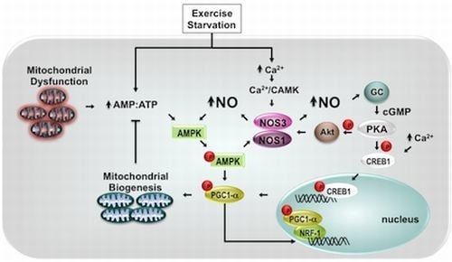
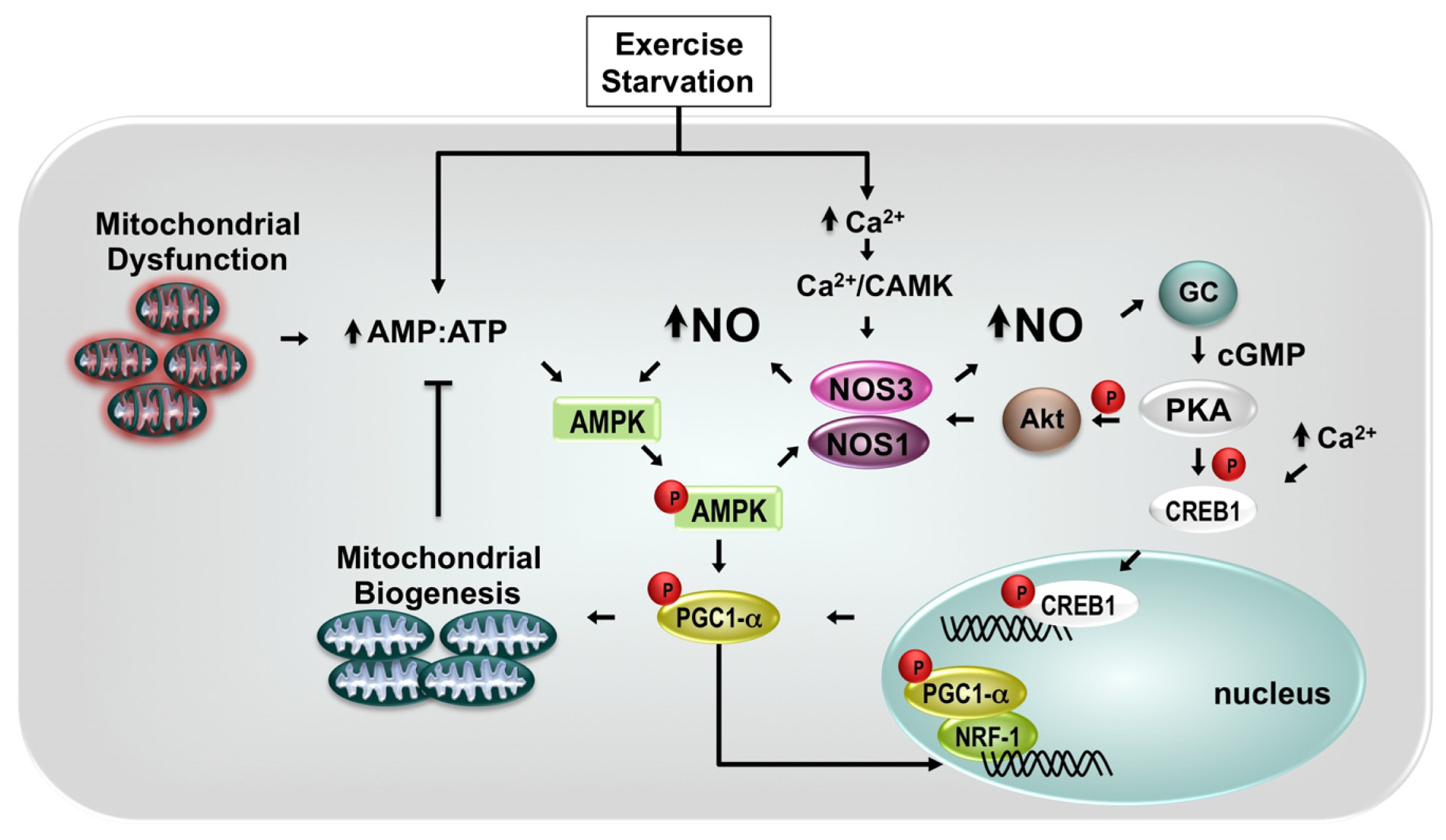
5. NO and Mitochondrial Biogenesis</title>
<p>NO plays an important role in mitochondrial biogenesis of skeletal muscle [<xref ref-type="bibr" rid="b88-ijms-13-17160">88</xref>]. Several studies have shown that treatment of cells with NO donor increase mitochondrial markers, demonstrating induction of biosynthesis of functional mitochondria able to generate ATP via oxidative phosphorylation [<xref ref-type="bibr" rid="b5-ijms-13-17160">5</xref>,<xref ref-type="bibr" rid="b103-ijms-13-17160">103</xref>,<xref ref-type="bibr" rid="b104-ijms-13-17160">104</xref>]. Treatment of rat primary skeletal muscle cultures with the NO donor <italic>S</italic>-nitroso-<italic>N</italic>-acetylpenicillamine (SNAP) led to a significant increase in mitochondrial content [<xref ref-type="bibr" rid="b27-ijms-13-17160">27</xref>], a phenomenon also observed in other tissues. For example, studies performed in the primary cultures of brown adipocytes, which express a larger number of mitochondria than other cell types, have shown that incubation with SNAP increases the mitochondrial size and mitochondrial DNA (mtDNA) content in a concentration dependent manner [<xref ref-type="bibr" rid="b4-ijms-13-17160">4</xref>]. The inductive effect of SNAP was completely abolished by supplementation of the medium with the NO scavenger oxyhemoglobin, indicating that the stimulatory effect depends on generation of NO.</p>
<p>The exact molecular pathways involved in mitochondrial proliferation in response to NO signaling are not entirely understood, but in many tissues and cells, they have been associated to peroxisome proliferator-activated receptor gamma coactivator-1 (PGC-1α) [<xref ref-type="bibr" rid="b5-ijms-13-17160">5</xref>,<xref ref-type="bibr" rid="b103-ijms-13-17160">103</xref>,<xref ref-type="bibr" rid="b104-ijms-13-17160">104</xref>] (<xref ref-type="fig" rid="f3-ijms-13-17160">Figure 3</xref>). In brown adipocyte tissue, NO-induced mitochondrial biogenesis is mimicked by incubation of cells with 8-Br-cGMP, indicating that it depends on activation of guanylil cyclase and generation of guanosine 3′,5′-monophosphate (cGMP). The downstream mechanism requires induction of PGC-1α expression since PGC-1α antisense oligomers reduced the SNAP-dependent increase in mtDNA content [<xref ref-type="bibr" rid="b4-ijms-13-17160">4</xref>]. In addition, ectopic expression of PGC-1α into skeletal muscle cell lineage C<sub>2</sub>C<sub>12</sub> induces a 2-fold increase in mtDNA content which was associated to a parallel increase (~60%) in mitochondrial density (mitochondrial number/cytoplasmic area) [<xref ref-type="bibr" rid="b105-ijms-13-17160">105</xref>]. The PGC-1α-dependent increment in mitochondrial biogenesis occurs in parallel with increase in the basal oxygen consumption and in the expression (transcripts/proteins) of respiratory chain components, including the nuclear-encoded cytochrome <italic>c</italic> oxidase subunit IV and the mitochondrial-encoded cytochrome <italic>c</italic> oxidase subunit II and cytochrome <italic>c</italic>[<xref ref-type="bibr" rid="b105-ijms-13-17160">105</xref>].</p>
<p>Stimulation of mitochondrial biogenesis by NO also requires the expression of transcriptional factors, that include CREB1 (cAMP response element-binding protein 1) and the nuclear respiratory factors 1 and 2 proteins (NRF-1 and NRF-2), which mediate expression of multiple nuclear genes encoding for mitochondrial proteins, such as the mitochondrial rate-limiting enzyme for heme biosynthesis, 5-aminolevulinate synthase [<xref ref-type="bibr" rid="b106-ijms-13-17160">106</xref>] and, the subunits of the respiratory chain complexes [<xref ref-type="bibr" rid="b107-ijms-13-17160">107</xref>].</p>
<p>This process involves an orchestrated cascade of phosphorylation events performed by distinct serine/threonine kinases. For example, while both serine/threonone kinases Akt [<xref ref-type="bibr" rid="b108-ijms-13-17160">108</xref>] and PKA (cyclic AMP-dependent protein kinase) are able to activate eNOS via phosphorylation of Ser1177, leading to increased production of NO, phosphorylation of CREB transcription factor by PKA results in increased expression of PGC-1α, which in turn increases mitochondrial biogenesis. Actually, the most potent activator of PGC-1α transcription is the coactivator of CREB, named TORC1 (transducer of regulated CREB binding protein 1). According to Wu <italic>el al.</italic> (2006) [<xref ref-type="bibr" rid="b109-ijms-13-17160">109</xref>], TORC 1, 2 and 3 markedly increases PGC-1α promoter activity, in a CREB-dependent fashion in primary mouse skeletal muscle cultures. The authors have shown that transduction of mouse primary myotubes with an adenovirus expressing hTORC1, hTORC2, hTORC3 resulted in increased mRNA levels of PGC-1α and mitochondrial markers <italic>Cyt c</italic>, <italic>Cox II</italic>, and <italic>IDH3</italic>α (isocitrate dehydrogenase 3α). In addition, Akt may form a physical complex with PKA serving as a substrate of PKA [<xref ref-type="bibr" rid="b110-ijms-13-17160">110</xref>]. Once phosphorylated, Akt activates eNOS, leading to increased NO production. Conversely, Akt may induce mitochondrial biogenesis through the phosphorylation of NRF-1 and CREB1, enabling their nuclear translocation and activation of target genes, such as mitochondrial transcription factor A (Tfam), which is required for mtDNA transcription and replication [<xref ref-type="bibr" rid="b111-ijms-13-17160">111</xref>] (for review, see [<xref ref-type="bibr" rid="b112-ijms-13-17160">112</xref>]).</p>
<p>The molecular mechanisms of NO-dependent regulation of PGC-1α/ mitochondrial biogenesis also involve 5′-AMP-activated protein kinase (AMPK), a heterotrimeric kinase allosterically activated by AMP and inhibited by ATP, which serves as an energy sensor, under conditions of low energy charge (decrease in ATP and increase in AMP). Thus, conditions that cause significant cellular energy stress, such as exercise, starvation or mitochondrial dysfunction are able to increase AMPK activity (for review, see [<xref ref-type="bibr" rid="b113-ijms-13-17160">113</xref>]) and promote mitochondrial biogenesis [<xref ref-type="bibr" rid="b114-ijms-13-17160">114</xref>–<xref ref-type="bibr" rid="b116-ijms-13-17160">116</xref>]. For example, exercise training performed in rat or humans increases mitochondrial content and capacity [<xref ref-type="bibr" rid="b117-ijms-13-17160">117</xref>–<xref ref-type="bibr" rid="b119-ijms-13-17160">119</xref>]. In fact contraction induced by electrical stimulation of isolated rat skeletal muscle also increases NO release from muscle [<xref ref-type="bibr" rid="b120-ijms-13-17160">120</xref>] with a significant contribution of both calcium-calmodulin-dependent isoforms of NOS, nNOS and eNOS [<xref ref-type="bibr" rid="b121-ijms-13-17160">121</xref>]. In working tissues, such as skeletal and cardiac muscle, increased levels of intracellular calcium during contraction have been associated to physiological induction of mitochondrial biogenesis. Intermittent exposure of L6 myotubes to the Ca<sup>2+</sup> ionophore ionomycin (5 h/day) or to caffeine or W7, which release Ca<sup>2+</sup> from the sarcoplasmic reticulum induced an increase in mitochondrial enzymes [<xref ref-type="bibr" rid="b122-ijms-13-17160">122</xref>]. Interestingly, skeletal muscles from transgenic mice that selectively express a constitutively active form of calcium/calmodulin-dependent protein kinase IV showed augmented mtDNA replication and mitochondrial biogenesis. These effects are associated to up-regulation of mitochondrial enzymes involved in fatty acid metabolism and electron transport, and reduced susceptibility to fatigue during repetitive contractions. In mice C2C12 myotubes, the effect of CaMK was associated to activated the PGC-1α gene promoter and increased expression of PGC-1α [<xref ref-type="bibr" rid="b123-ijms-13-17160">123</xref>]. Thus, calcium-regulated signaling pathways are able to induce mitochondrial biogenesis via activation of NOS or stimulation of Ca<sup>2+</sup>-dependent transcription factors. Interestingly, during exercise in humans, AMPK phosphorylates skeletal muscle nNOSμ at Ser1451 [<xref ref-type="bibr" rid="b124-ijms-13-17160">124</xref>], which may account for the increase in glucose uptake during exercise. Indeed, taking into account that AMPK is able to phosphorylate and activate eNOS/nNOS during contracting human skeletal muscle [<xref ref-type="bibr" rid="b125-ijms-13-17160">125</xref>], these data suggest that phosphorylation of NOS by AMPK may be, at least in part, involved in mitochondrial biogenesis in muscle cells.</p>
<p>This correlation of AMPK, PGC-1α, NO and mitochondrial biogenesis in L6 myogenic cell lineage was recently confirmed by McConell <italic>et al.</italic>[<xref ref-type="bibr" rid="b103-ijms-13-17160">103</xref>]. Incubation of L6 myotubes with AICAR, which activates AMP-activated protein kinase (AMPK), increased myotube mitochondrial biogenesis. The AMPK activator also increased the expression of PGC-1α, subunits I and IV of cytochrome <italic>c</italic> oxidase. Actually, while SNAP increased phospho-AMPK and markers of mitochondrial biogenesis (PGC-1α, NRF-1, COX-1 and COX-4), inhibition of NOS with <sc>l</sc>-NAME attenuated the AICAR-induced increases in COX-1 and COX-4 protein, indicating that activation of AMPK in L6 myotubes increases mitochondrial biogenesis, at least in part, via interactions with NOS.</p></sec>
<sec>
<title>6. NO and Myogenic Differentiation</title>
<p>Differentiation of embryonic muscle cells involves fusion of mononucleated myoblasts derived from pluripotent mesodermic cells to form multinucleated myotubes. Myotubes differentiate into mature skeletal muscle fibers with functional contractile apparatus and compartmentalized expression of synaptic proteins [<xref ref-type="bibr" rid="b126-ijms-13-17160">126</xref>]. Beyond this stage, mitosis and DNA synthesis arrest almost completely and generation of new fibers depends on quiescent precursor cells named “satellite cells”, located under the basal lamina of individual postnatal and adult myofibers.</p>
<p>Both embryonic and adult myogenic cells are influenced by changes in NO levels. While embryonic myoblasts have NOS activity, as measured by their ability to convert arginine to citrulline [<xref ref-type="bibr" rid="b127-ijms-13-17160">127</xref>], adult satellite cells are activated by nitric oxide (NO) produced in muscle fibers, which may contribute to post-injury regeneration of skeletal muscle. According to Lee <italic>et al.</italic>[<xref ref-type="bibr" rid="b127-ijms-13-17160">127</xref>] NOS activity dramatically increases in chick embryonic myoblasts that are competent for fusion, and coincides with increment in intracellular cGMP levels. The authors also showed that while NO donor SNP (sodium nitroprusside) accelerated myoblast fusion, the NOS inhibitor <sc>l</sc>-NMMA (NG-methyl-<sc>l</sc>-arginine) delayed the fusion that is reversed by treatment with 8-Br-cGMP, an analog of cGMP.</p>
<p>Differentiation of myogenic cell also requires mitochondrial elongation that depends on the cellular generation of NO. Using myogenic precursor cells isolated from the muscles of newborn mice, De Palma <italic>et al</italic>. [<xref ref-type="bibr" rid="b128-ijms-13-17160">128</xref>] showed that inhibition of NO synthesis lead to inhibition of both mitochondrial elongation and myogenic differentiation. This phenomenon was associated with NO-dependent inhibition of dynamin-related protein-1 (Drp1) [<xref ref-type="bibr" rid="b128-ijms-13-17160">128</xref>], a GTPase protein that induces mitochondrial fission and, as consequence, interferes with maintenance of mitochondrial integrity (for review see [<xref ref-type="bibr" rid="b129-ijms-13-17160">129</xref>–<xref ref-type="bibr" rid="b131-ijms-13-17160">131</xref>]. By inhibiting Drp1, NO increases the formation of mitochondrial network, due to the ongoing mitochondrial fusion, and stimulates myogenic differentiation.</p>
<p>A better understanding of NO interference on myogenic differentiation may be of crucial importance to development of new therapeutic strategies for enhancing skeletal muscle mass and performance, opening promising perspectives for treatment of conditions associated with reduced proliferation of myogenic cells, such as aging or degenerative diseases.</p></sec>
<sec>
<title>7. Potential Therapeutic Strategies</title>
<p>Several drugs with NO-donating properties are currently in use in clinical practice, mostly for cardiovascular diseases, such as nitrovasodilator, drugs affecting NOS mRNA and/or protein levels with special modulation of eNOS [<xref ref-type="bibr" rid="b7-ijms-13-17160">7</xref>]. A specific treatment of muscle diseases based on NO physiology is still not available, however several strategies have been studied in recent years. Focusing on skeletal muscle and mitochondrial dysfunction, NO-directed approaches can be summarized in: diet, exercise and pharmacological approaches to increase NO in subcellular compartments [<xref ref-type="bibr" rid="b132-ijms-13-17160">132</xref>]. The main objective when focusing on mitochondrial deficiencies is to increase the energy supply by improving the oxidative phosphorylation efficiency or increasing mitochondrial content.</p>
<p>It has been shown that calorie restriction lowers whole-body energy expenditure and induces mitochondrial biogenesis in overweight non-obese humans [<xref ref-type="bibr" rid="b133-ijms-13-17160">133</xref>]. This study also demonstrated a decrease in DNA damage and proposed that caloric restriction would induce biogenesis of “efficient” mitochondria in human skeletal muscle as an adaptive mechanism, reducing oxidative stress. Calorie restriction was also able to increase mitochondrial biogenesis, eNOS expression and Akt phosphorylation in mice skeletal muscle, indicating that the NO pathway is activated [<xref ref-type="bibr" rid="b134-ijms-13-17160">134</xref>].</p>
<p>Several studies using rodent models have shown that exercise training induces an increase in skeletal muscle mitochondrial content [<xref ref-type="bibr" rid="b135-ijms-13-17160">135</xref>], which was demonstrated by increased expression of mitochondrial markers. As discussed in section 5, the mechanism of exercise induction of mitochondrial biogenesis involves the PGC-1α pathway [<xref ref-type="bibr" rid="b136-ijms-13-17160">136</xref>]. It is suggested that the source of NO in exercise induced mitochondrial biogenesis is eNOS, as Nisoli <italic>et al</italic>. (2004) [<xref ref-type="bibr" rid="b104-ijms-13-17160">104</xref>] reported that eNOS −/− mice had reduced mitochondrial content in skeletal muscle. However, Wadley <italic>et al.</italic> (2007) could not confirm the involvement of either eNOS or nNOS in the exercise induced mitochondrial biogenesis, studying eNOS −/− and nNOS −/− mice [<xref ref-type="bibr" rid="b135-ijms-13-17160">135</xref>]. Later, Lee-Young <italic>et al.</italic> (2010) showed that eNOS is critical to muscle cell signaling during exercise <italic>in vivo</italic>, by showing that ablation of eNOS in eNOS −/− mice resulted in impaired exercise capacity [<xref ref-type="bibr" rid="b137-ijms-13-17160">137</xref>]. Studies evaluating the exercise training in patients with mitochondrial myopathy, a genetic disorder with oxidative phosphorylation deficiency, aim the stimulation of mitochondrial biogenesis and a gene shifting, with increase of mitochondrial content with normal mtDNA [<xref ref-type="bibr" rid="b138-ijms-13-17160">138</xref>–<xref ref-type="bibr" rid="b140-ijms-13-17160">140</xref>]. In fact, improvement of muscle strength and oxidative capacity have been demonstrated after exercise training [<xref ref-type="bibr" rid="b140-ijms-13-17160">140</xref>] but further investigations are still needed to determine the safety, effective benefits and the most appropriate exercise training protocol.</p>
<p>Supplementation with arginine or citrulline has also been proposed for treatment of mitochondrial disorders with NO deficiency [<xref ref-type="bibr" rid="b141-ijms-13-17160">141</xref>]. <sc>l</sc>-arginine was first used in the treatment of a mitochondrial encephalopathy with stroke like syndrome (MELAS) as an NO precursor with the objective to promote vasodilation in cerebral vessels [<xref ref-type="bibr" rid="b142-ijms-13-17160">142</xref>,<xref ref-type="bibr" rid="b143-ijms-13-17160">143</xref>]. The rationale of this approach is that patients with MELAS probably have a deficiency in smooth muscle vascular relaxation, which could be responsible for the symptoms of cerebral ischemia. This therapeutic approach is supported by the finding of low levels of serum arginine [<xref ref-type="bibr" rid="b142-ijms-13-17160">142</xref>], low levels of citrulline [<xref ref-type="bibr" rid="b144-ijms-13-17160">144</xref>] and endothelium dysfunction [<xref ref-type="bibr" rid="b145-ijms-13-17160">145</xref>] in patients with MELAS. This treatment showed promising results with significant decrease in stroke frequency, improvement of neurological deficits [<xref ref-type="bibr" rid="b142-ijms-13-17160">142</xref>,<xref ref-type="bibr" rid="b143-ijms-13-17160">143</xref>,<xref ref-type="bibr" rid="b145-ijms-13-17160">145</xref>,<xref ref-type="bibr" rid="b146-ijms-13-17160">146</xref>] and increased NO production [<xref ref-type="bibr" rid="b147-ijms-13-17160">147</xref>]. Based on these findings and the observation of NO deficiency in muscle fibers with cytochrome <italic>c</italic> oxidase deficiency of patients with mitochondrial myopathy [<xref ref-type="bibr" rid="b27-ijms-13-17160">27</xref>], it was proposed that arginine and citrulline supplementation could have a broader application in mitochondrial diseases, especially in those with muscular symptoms [<xref ref-type="bibr" rid="b141-ijms-13-17160">141</xref>].</p>
<p>Mitochondrial biogenesis can also be activated with the use of drugs, such as resveratrol (3,5,4′-trihydroxystilbene), a diet-derived polyphenol. Treatment of rodents with resveratrol induced increased mitochondrial content and improved function in skeletal muscle [<xref ref-type="bibr" rid="b148-ijms-13-17160">148</xref>,<xref ref-type="bibr" rid="b149-ijms-13-17160">149</xref>]. The mechanism of action involves the decrease in PGC-1α acetylation with resulting increase in PGC-1α activity [<xref ref-type="bibr" rid="b148-ijms-13-17160">148</xref>] and AMPK activation [<xref ref-type="bibr" rid="b150-ijms-13-17160">150</xref>]. Studies using endothelial cells demonstrated that resveratrol induces an up-regulation of eNOS [<xref ref-type="bibr" rid="b151-ijms-13-17160">151</xref>]. Other drugs acting specifically on eNOS have been largely studied in cardiovascular diseases [<xref ref-type="bibr" rid="b132-ijms-13-17160">132</xref>]. One example is statin that is classically known by the effects on dyslipidemia, however other beneficial pleiotropic effects have been reported [<xref ref-type="bibr" rid="b132-ijms-13-17160">132</xref>]. Statin pleiotropic effects are mainly reported in studies with endothelial cells showing that eNOS activity may be increased [<xref ref-type="bibr" rid="b152-ijms-13-17160">152</xref>]. However, it is well known that statins, such as sinvastatin and atorvastatin, can induce myopathy with rhabdomyolysis. A recent study in mice has demonstrated that the treatment with a novel class of statin, an NO-donating atorvastatin was able to prevent the atorvastatin induced skeletal muscle dysfunction [<xref ref-type="bibr" rid="b153-ijms-13-17160">153</xref>]. The authors showed that the NO-donating atorvastatin maintained the lipid lowering effect with absence of the other side effects observed with atorvastatin, such as sarcolemmal damage and mitochondrial dysfunction.</p>
<p>NO-donating drugs have also been tested in Duchenne muscular dystrophy in order to improve muscle regeneration and restore NO intracellular level, which is diminished due to the loss of nNOS. Brunelli <italic>et al.</italic>[<xref ref-type="bibr" rid="b154-ijms-13-17160">154</xref>] studied the treatment with HCT 1026, a derivative of flurbiprofen, a nonsteroidal anti-inflammatory drug, that releases NO and with no severe side effects. They showed that HCT 1026 slowed down disease progression and ameliorated the morphological, biochemical and functional phenotype. In the same study arterially delivered donor stem cells in association with HCT 1026 presented an enhanced therapeutic efficacy. This treatment demonstrated better results than the combination of <sc>l</sc>-arginine and deflazacort [<xref ref-type="bibr" rid="b155-ijms-13-17160">155</xref>]. Recently, Buono <italic>et al.</italic>[<xref ref-type="bibr" rid="b156-ijms-13-17160">156</xref>] demonstrated a therapeutic effect of molsidomine, another NO releasing drug, in dystrophic mice. This treatment increased regenerating muscle fibers, as well as led to functional amelioration of the muscle, due to increased proliferation of satellite cells. These studies open up a new possibilities for treatment of muscle diseases including those with mitochondrial dysfunction, especially because they are drugs already tested in humans and could be used in long term treatments.</p></sec>
<sec sec-type="conclusions">
<title>8. Conclusions</title>
<p>NO has important roles on the regulation of mitochondrial function and induction of mitochondrial biogenesis. The pathways involved in NO signaling are complex but an increasing number of articles have addressed this subject in recent years. Several therapeutic approaches have been proposed with the aim of increasing mitochondrial content to compensate mitochondrial deficiency and to increase energy supply. However a lot of work has to be done to elucidate the exact mechanisms involved to make this approaches viable for clinical practice.</p></sec></body>
<back>
<ack>
<title>Acknowledgments</title>
<p>The authors were funded by research grants from Fundação de Amparo a Pesquisa de São Paulo (CHT, GSR, ROG) and Conselho Nacional de Desenvolvimento Científico e Tecnológico (ROG).</p></ack>
<fn-group><fn id="fn1-ijms-13-17160">
<p><bold>Conflict of Interest</bold></p>
<p>The authors declare no conflict of interest.</p></fn></fn-group>
<ref-list>
<title>References</title>
<ref id="b1-ijms-13-17160"><label>1</label><citation citation-type="journal"><person-group person-group-type="author"><name><surname>Cohn</surname><given-names>J.N.</given-names></name><name><surname>McInnes</surname><given-names>G.T.</given-names></name><name><surname>Shepherd</surname><given-names>A.M.</given-names></name></person-group><article-title>Direct-acting vasodilators</article-title><source>J. Clin. Hypertens</source><year>2011</year><volume>13</volume><fpage>690</fpage><lpage>692</lpage><pub-id pub-id-type="doi">10.1111/j.1751-7176.2011.00507.x</pub-id></citation></ref>
<ref id="b2-ijms-13-17160"><label>2</label><citation citation-type="journal"><person-group person-group-type="author"><name><surname>Ghafourifar</surname><given-names>P.</given-names></name><name><surname>Cadenas</surname><given-names>E.</given-names></name></person-group><article-title>Mitochondrial nitric oxide synthase</article-title><source>Trends Pharmacol. Sci</source><year>2005</year><volume>26</volume><fpage>190</fpage><lpage>195</lpage><pub-id pub-id-type="doi">10.1016/j.tips.2005.02.005</pub-id><pub-id pub-id-type="pmid">15808343</pub-id></citation></ref>
<ref id="b3-ijms-13-17160"><label>3</label><citation citation-type="journal"><person-group person-group-type="author"><name><surname>Boveris</surname><given-names>A.</given-names></name><name><surname>Costa</surname><given-names>L.E.</given-names></name><name><surname>Poderoso</surname><given-names>J.J.</given-names></name><name><surname>Carreras</surname><given-names>M.C.</given-names></name><name><surname>Cadenas</surname><given-names>E.</given-names></name></person-group><article-title>Regulation of mitochondrial respiration by oxygen and nitric oxide</article-title><source>Ann. N. Y. Acad. Sci</source><year>2000</year><volume>899</volume><fpage>121</fpage><lpage>135</lpage><pub-id pub-id-type="pmid">10863534</pub-id></citation></ref>
<ref id="b4-ijms-13-17160"><label>4</label><citation citation-type="journal"><person-group person-group-type="author"><name><surname>Nisoli</surname><given-names>E.</given-names></name><name><surname>Clementi</surname><given-names>E.</given-names></name><name><surname>Paolucci</surname><given-names>C.</given-names></name><name><surname>Cozzi</surname><given-names>V.</given-names></name><name><surname>Tonello</surname><given-names>C.</given-names></name><name><surname>Sciorati</surname><given-names>C.</given-names></name><name><surname>Bracale</surname><given-names>R.</given-names></name><name><surname>Valerio</surname><given-names>A.</given-names></name><name><surname>Francolini</surname><given-names>M.</given-names></name><name><surname>Moncada</surname><given-names>S.</given-names></name><etal/></person-group><article-title>Mitochondrial biogenesis in mammals: The role of endogenous nitric oxide</article-title><source>Science</source><year>2003</year><volume>299</volume><fpage>896</fpage><lpage>899</lpage><pub-id pub-id-type="doi">10.1126/science.1079368</pub-id><pub-id pub-id-type="pmid">12574632</pub-id></citation></ref>
<ref id="b5-ijms-13-17160"><label>5</label><citation citation-type="journal"><person-group person-group-type="author"><name><surname>Lira</surname><given-names>V.A.</given-names></name><name><surname>Brown</surname><given-names>D.L.</given-names></name><name><surname>Lira</surname><given-names>A.K.</given-names></name><name><surname>Kavazis</surname><given-names>A.N.</given-names></name><name><surname>Soltow</surname><given-names>Q.A.</given-names></name><name><surname>Zeanah</surname><given-names>E.H.</given-names></name><name><surname>Criswell</surname><given-names>D.S.</given-names></name></person-group><article-title>Nitric oxide and ampk cooperatively regulate pgc-1 in skeletal muscle cells</article-title><source>J. Physiol</source><year>2010</year><volume>588</volume><fpage>3551</fpage><lpage>3566</lpage><pub-id pub-id-type="doi">10.1113/jphysiol.2010.194035</pub-id><pub-id pub-id-type="pmid">20643772</pub-id></citation></ref>
<ref id="b6-ijms-13-17160"><label>6</label><citation citation-type="journal"><person-group person-group-type="author"><name><surname>Toledo</surname><given-names>J.C.</given-names><suffix>Jr</suffix></name><name><surname>Augusto</surname><given-names>O.</given-names></name></person-group><article-title>Connecting the chemical and biological properties of nitric oxide</article-title><source>Chem. Res. Toxicol.</source><year>2012</year><volume>25</volume><fpage>975</fpage><lpage>989</lpage><pub-id pub-id-type="doi">10.1021/tx300042g</pub-id><pub-id pub-id-type="pmid">22449080</pub-id></citation></ref>
<ref id="b7-ijms-13-17160"><label>7</label><citation citation-type="journal"><person-group person-group-type="author"><name><surname>Dudzinski</surname><given-names>D.M.</given-names></name><name><surname>Igarashi</surname><given-names>J.</given-names></name><name><surname>Greif</surname><given-names>D.</given-names></name><name><surname>Michel</surname><given-names>T.</given-names></name></person-group><article-title>The regulation and pharmacology of endothelial nitric oxide synthase</article-title><source>Annu. Rev. Pharmacol. Toxicol</source><year>2006</year><volume>46</volume><fpage>235</fpage><lpage>276</lpage><pub-id pub-id-type="doi">10.1146/annurev.pharmtox.44.101802.121844</pub-id><pub-id pub-id-type="pmid">16402905</pub-id></citation></ref>
<ref id="b8-ijms-13-17160"><label>8</label><citation citation-type="journal"><person-group person-group-type="author"><name><surname>Nathan</surname><given-names>C.</given-names></name><name><surname>Xie</surname><given-names>Q.W.</given-names></name></person-group><article-title>Nitric oxide synthases: Roles, tolls, and controls</article-title><source>Cell</source><year>1994</year><volume>78</volume><fpage>915</fpage><lpage>918</lpage><pub-id pub-id-type="doi">10.1016/0092-8674(94)90266-6</pub-id><pub-id pub-id-type="pmid">7522969</pub-id></citation></ref>
<ref id="b9-ijms-13-17160"><label>9</label><citation citation-type="journal"><person-group person-group-type="author"><name><surname>Stamler</surname><given-names>J.S.</given-names></name><name><surname>Meissner</surname><given-names>G.</given-names></name></person-group><article-title>Physiology of nitric oxide in skeletal muscle</article-title><source>Physiol. Rev</source><year>2001</year><volume>81</volume><fpage>209</fpage><lpage>237</lpage><pub-id pub-id-type="pmid">11152758</pub-id></citation></ref>
<ref id="b10-ijms-13-17160"><label>10</label><citation citation-type="journal"><person-group person-group-type="author"><name><surname>Stuehr</surname><given-names>D.J.</given-names></name></person-group><article-title>Structure-function aspects in the nitric oxide synthases</article-title><source>Annu. Rev. Pharmacol. Toxicol</source><year>1997</year><volume>37</volume><fpage>339</fpage><lpage>359</lpage><pub-id pub-id-type="doi">10.1146/annurev.pharmtox.37.1.339</pub-id><pub-id pub-id-type="pmid">9131257</pub-id></citation></ref>
<ref id="b11-ijms-13-17160"><label>11</label><citation citation-type="journal"><person-group person-group-type="author"><name><surname>Alderton</surname><given-names>W.K.</given-names></name><name><surname>Cooper</surname><given-names>C.E.</given-names></name><name><surname>Knowles</surname><given-names>R.G.</given-names></name></person-group><article-title>Nitric oxide synthases: Structure, function and inhibition</article-title><source>Biochem. J</source><year>2001</year><volume>357</volume><fpage>593</fpage><lpage>615</lpage><pub-id pub-id-type="doi">10.1042/0264-6021:3570593</pub-id><pub-id pub-id-type="pmid">11463332</pub-id></citation></ref>
<ref id="b12-ijms-13-17160"><label>12</label><citation citation-type="journal"><person-group person-group-type="author"><name><surname>Bredt</surname><given-names>D.S.</given-names></name><name><surname>Snyder</surname><given-names>S.H.</given-names></name></person-group><article-title>Isolation of nitric oxide synthetase, a calmodulin-requiring enzyme</article-title><source>Proc. Natl. Acad. Sci. USA</source><year>1990</year><volume>87</volume><fpage>682</fpage><lpage>685</lpage><pub-id pub-id-type="doi">10.1073/pnas.87.2.682</pub-id><pub-id pub-id-type="pmid">1689048</pub-id></citation></ref>
<ref id="b13-ijms-13-17160"><label>13</label><citation citation-type="journal"><person-group person-group-type="author"><name><surname>Eissa</surname><given-names>N.T.</given-names></name><name><surname>Yuan</surname><given-names>J.W.</given-names></name><name><surname>Haggerty</surname><given-names>C.M.</given-names></name><name><surname>Choo</surname><given-names>E.K.</given-names></name><name><surname>Palmer</surname><given-names>C.D.</given-names></name><name><surname>Moss</surname><given-names>J.</given-names></name></person-group><article-title>Cloning and characterization of human inducible nitric oxide synthase splice variants: A domain, encoded by exons 8 and 9, is critical for dimerization</article-title><source>Proc. Natl. Acad. Sci. USA</source><year>1998</year><volume>95</volume><fpage>7625</fpage><lpage>7630</lpage><pub-id pub-id-type="doi">10.1073/pnas.95.13.7625</pub-id><pub-id pub-id-type="pmid">9636200</pub-id></citation></ref>
<ref id="b14-ijms-13-17160"><label>14</label><citation citation-type="journal"><person-group person-group-type="author"><name><surname>Forstermann</surname><given-names>U.</given-names></name><name><surname>Boissel</surname><given-names>J.P.</given-names></name><name><surname>Kleinert</surname><given-names>H.</given-names></name></person-group><article-title>Expressional control of the “constitutive” isoforms of nitric oxide synthase (nos i and nos iii)</article-title><source>FASEB J</source><year>1998</year><volume>12</volume><fpage>773</fpage><lpage>790</lpage><pub-id pub-id-type="pmid">9657518</pub-id></citation></ref>
<ref id="b15-ijms-13-17160"><label>15</label><citation citation-type="journal"><person-group person-group-type="author"><name><surname>Eliasson</surname><given-names>M.J.</given-names></name><name><surname>Blackshaw</surname><given-names>S.</given-names></name><name><surname>Schell</surname><given-names>M.J.</given-names></name><name><surname>Snyder</surname><given-names>S.H.</given-names></name></person-group><article-title>Neuronal nitric oxide synthase alternatively spliced forms: Prominent functional localizations in the brain</article-title><source>Proc. Natl. Acad. Sci. USA</source><year>1997</year><volume>94</volume><fpage>3396</fpage><lpage>3401</lpage><pub-id pub-id-type="doi">10.1073/pnas.94.7.3396</pub-id><pub-id pub-id-type="pmid">9096405</pub-id></citation></ref>
<ref id="b16-ijms-13-17160"><label>16</label><citation citation-type="journal"><person-group person-group-type="author"><name><surname>Brenman</surname><given-names>J.E.</given-names></name><name><surname>Chao</surname><given-names>D.S.</given-names></name><name><surname>Gee</surname><given-names>S.H.</given-names></name><name><surname>McGee</surname><given-names>A.W.</given-names></name><name><surname>Craven</surname><given-names>S.E.</given-names></name><name><surname>Santillano</surname><given-names>D.R.</given-names></name><name><surname>Wu</surname><given-names>Z.</given-names></name><name><surname>Huang</surname><given-names>F.</given-names></name><name><surname>Xia</surname><given-names>H.</given-names></name><name><surname>Peters</surname><given-names>M.F.</given-names></name><etal/></person-group><article-title>Interaction of nitric oxide synthase with the postsynaptic density protein psd-95 and alpha1-syntrophin mediated by pdz domains</article-title><source>Cell</source><year>1996</year><volume>84</volume><fpage>757</fpage><lpage>767</lpage><pub-id pub-id-type="doi">10.1016/S0092-8674(00)81053-3</pub-id><pub-id pub-id-type="pmid">8625413</pub-id></citation></ref>
<ref id="b17-ijms-13-17160"><label>17</label><citation citation-type="journal"><person-group person-group-type="author"><name><surname>Larsson</surname><given-names>B.</given-names></name><name><surname>Phillips</surname><given-names>S.C.</given-names></name></person-group><article-title>Isolation and characterization of a novel, human neuronal nitric oxide synthase cdna</article-title><source>Biochem. Biophys. Res. Commun</source><year>1998</year><volume>251</volume><fpage>898</fpage><lpage>902</lpage><pub-id pub-id-type="doi">10.1006/bbrc.1998.9578</pub-id><pub-id pub-id-type="pmid">9791007</pub-id></citation></ref>
<ref id="b18-ijms-13-17160"><label>18</label><citation citation-type="journal"><person-group person-group-type="author"><name><surname>Wang</surname><given-names>Y.</given-names></name><name><surname>Goligorsky</surname><given-names>M.S.</given-names></name><name><surname>Lin</surname><given-names>M.</given-names></name><name><surname>Wilcox</surname><given-names>J.N.</given-names></name><name><surname>Marsden</surname><given-names>P.A.</given-names></name></person-group><article-title>A novel, testis-specific mrna transcript encoding an nh2-terminal truncated nitric-oxide synthase</article-title><source>J. Biol. Chem</source><year>1997</year><volume>272</volume><fpage>11392</fpage><lpage>11401</lpage><pub-id pub-id-type="doi">10.1074/jbc.272.17.11392</pub-id><pub-id pub-id-type="pmid">9111048</pub-id></citation></ref>
<ref id="b19-ijms-13-17160"><label>19</label><citation citation-type="journal"><person-group person-group-type="author"><name><surname>Marsden</surname><given-names>P.A.</given-names></name><name><surname>Schappert</surname><given-names>K.T.</given-names></name><name><surname>Chen</surname><given-names>H.S.</given-names></name><name><surname>Flowers</surname><given-names>M.</given-names></name><name><surname>Sundell</surname><given-names>C.L.</given-names></name><name><surname>Wilcox</surname><given-names>J.N.</given-names></name><name><surname>Lamas</surname><given-names>S.</given-names></name><name><surname>Michel</surname><given-names>T.</given-names></name></person-group><article-title>Molecular cloning and characterization of human endothelial nitric oxide synthase</article-title><source>FEBS Lett</source><year>1992</year><volume>307</volume><fpage>287</fpage><lpage>293</lpage><pub-id pub-id-type="doi">10.1016/0014-5793(92)80697-F</pub-id><pub-id pub-id-type="pmid">1379542</pub-id></citation></ref>
<ref id="b20-ijms-13-17160"><label>20</label><citation citation-type="journal"><person-group person-group-type="author"><name><surname>Miyahara</surname><given-names>K.</given-names></name><name><surname>Kawamoto</surname><given-names>T.</given-names></name><name><surname>Sase</surname><given-names>K.</given-names></name><name><surname>Yui</surname><given-names>Y.</given-names></name><name><surname>Toda</surname><given-names>K.</given-names></name><name><surname>Yang</surname><given-names>L.X.</given-names></name><name><surname>Hattori</surname><given-names>R.</given-names></name><name><surname>Aoyama</surname><given-names>T.</given-names></name><name><surname>Yamamoto</surname><given-names>Y.</given-names></name><name><surname>Doi</surname><given-names>Y.</given-names></name><etal/></person-group><article-title>Cloning and structural characterization of the human endothelial nitric-oxide-synthase gene</article-title><source>Eur. J. Biochem</source><year>1994</year><volume>223</volume><fpage>719</fpage><lpage>726</lpage><pub-id pub-id-type="doi">10.1111/j.1432-1033.1994.tb19045.x</pub-id><pub-id pub-id-type="pmid">7519987</pub-id></citation></ref>
<ref id="b21-ijms-13-17160"><label>21</label><citation citation-type="journal"><person-group person-group-type="author"><name><surname>Lorenz</surname><given-names>M.</given-names></name><name><surname>Hewing</surname><given-names>B.</given-names></name><name><surname>Hui</surname><given-names>J.</given-names></name><name><surname>Zepp</surname><given-names>A.</given-names></name><name><surname>Baumann</surname><given-names>G.</given-names></name><name><surname>Bindereif</surname><given-names>A.</given-names></name><name><surname>Stangl</surname><given-names>V.</given-names></name><name><surname>Stangl</surname><given-names>K.</given-names></name></person-group><article-title>Alternative splicing in intron 13 of the human enos gene: A potential mechanism for regulating enos activity</article-title><source>FASEB J</source><year>2007</year><volume>21</volume><fpage>1556</fpage><lpage>1564</lpage><pub-id pub-id-type="doi">10.1096/fj.06-7434com</pub-id><pub-id pub-id-type="pmid">17264164</pub-id></citation></ref>
<ref id="b22-ijms-13-17160"><label>22</label><citation citation-type="journal"><person-group person-group-type="author"><name><surname>Chartrain</surname><given-names>N.A.</given-names></name><name><surname>Geller</surname><given-names>D.A.</given-names></name><name><surname>Koty</surname><given-names>P.P.</given-names></name><name><surname>Sitrin</surname><given-names>N.F.</given-names></name><name><surname>Nussler</surname><given-names>A.K.</given-names></name><name><surname>Hoffman</surname><given-names>E.P.</given-names></name><name><surname>Billiar</surname><given-names>T.R.</given-names></name><name><surname>Hutchinson</surname><given-names>N.I.</given-names></name><name><surname>Mudgett</surname><given-names>J.S.</given-names></name></person-group><article-title>Molecular cloning, structure, and chromosomal localization of the human inducible nitric oxide synthase gene</article-title><source>J. Biol. Chem</source><year>1994</year><volume>269</volume><fpage>6765</fpage><lpage>6772</lpage><pub-id pub-id-type="pmid">7509810</pub-id></citation></ref>
<ref id="b23-ijms-13-17160"><label>23</label><citation citation-type="journal"><person-group person-group-type="author"><name><surname>Zweier</surname><given-names>J.L.</given-names></name><name><surname>Samouilov</surname><given-names>A.</given-names></name><name><surname>Kuppusamy</surname><given-names>P.</given-names></name></person-group><article-title>Non-enzymatic nitric oxide synthesis in biological systems</article-title><source>Biochim. Biophys. Acta</source><year>1999</year><volume>1411</volume><fpage>250</fpage><lpage>262</lpage><pub-id pub-id-type="doi">10.1016/S0005-2728(99)00018-3</pub-id><pub-id pub-id-type="pmid">10320661</pub-id></citation></ref>
<ref id="b24-ijms-13-17160"><label>24</label><citation citation-type="journal"><person-group person-group-type="author"><name><surname>Kobzik</surname><given-names>L.</given-names></name><name><surname>Stringer</surname><given-names>B.</given-names></name><name><surname>Balligand</surname><given-names>J.L.</given-names></name><name><surname>Reid</surname><given-names>M.B.</given-names></name><name><surname>Stamler</surname><given-names>J.S.</given-names></name></person-group><article-title>Endothelial type nitric oxide synthase in skeletal muscle fibers: Mitochondrial relationships</article-title><source>Biochem. Biophys. Res. Commun</source><year>1995</year><volume>211</volume><fpage>375</fpage><lpage>381</lpage><pub-id pub-id-type="doi">10.1006/bbrc.1995.1824</pub-id><pub-id pub-id-type="pmid">7540837</pub-id></citation></ref>
<ref id="b25-ijms-13-17160"><label>25</label><citation citation-type="journal"><person-group person-group-type="author"><name><surname>Bates</surname><given-names>T.E.</given-names></name><name><surname>Loesch</surname><given-names>A.</given-names></name><name><surname>Burnstock</surname><given-names>G.</given-names></name><name><surname>Clark</surname><given-names>J.B.</given-names></name></person-group><article-title>Immunocytochemical evidence for a mitochondrially located nitric oxide synthase in brain and liver</article-title><source>Biochem. Biophys. Res. Commun</source><year>1995</year><volume>213</volume><fpage>896</fpage><lpage>900</lpage><pub-id pub-id-type="doi">10.1006/bbrc.1995.2213</pub-id><pub-id pub-id-type="pmid">7544582</pub-id></citation></ref>
<ref id="b26-ijms-13-17160"><label>26</label><citation citation-type="journal"><person-group person-group-type="author"><name><surname>Ohkoshi</surname><given-names>N.</given-names></name><name><surname>Mizusawa</surname><given-names>H.</given-names></name><name><surname>Fujita</surname><given-names>T.</given-names></name><name><surname>Shoji</surname><given-names>S.</given-names></name></person-group><article-title>Histological determination of nitric oxide synthase (nos) and nadph-diaphorase in ragged-red fibers from patients with mitochondrial encephalomyopathies</article-title><source>J. Neurol. Sci</source><year>1997</year><volume>149</volume><fpage>151</fpage><lpage>156</lpage><pub-id pub-id-type="doi">10.1016/S0022-510X(97)05385-9</pub-id><pub-id pub-id-type="pmid">9171323</pub-id></citation></ref>
<ref id="b27-ijms-13-17160"><label>27</label><citation citation-type="journal"><person-group person-group-type="author"><name><surname>Tengan</surname><given-names>C.H.</given-names></name><name><surname>Kiyomoto</surname><given-names>B.H.</given-names></name><name><surname>Godinho</surname><given-names>R.O.</given-names></name><name><surname>Gamba</surname><given-names>J.</given-names></name><name><surname>Neves</surname><given-names>A.C.</given-names></name><name><surname>Schmidt</surname><given-names>B.</given-names></name><name><surname>Oliveira</surname><given-names>A.S.</given-names></name><name><surname>Gabbai</surname><given-names>A.A.</given-names></name></person-group><article-title>The role of nitric oxide in muscle fibers with oxidative phosphorylation defects</article-title><source>Biochem. Biophys. Res. Commun</source><year>2007</year><volume>359</volume><fpage>771</fpage><lpage>777</lpage><pub-id pub-id-type="doi">10.1016/j.bbrc.2007.05.184</pub-id><pub-id pub-id-type="pmid">17560547</pub-id></citation></ref>
<ref id="b28-ijms-13-17160"><label>28</label><citation citation-type="journal"><person-group person-group-type="author"><name><surname>Giulivi</surname><given-names>C.</given-names></name><name><surname>Poderoso</surname><given-names>J.J.</given-names></name><name><surname>Boveris</surname><given-names>A.</given-names></name></person-group><article-title>Production of nitric oxide by mitochondria</article-title><source>J. Biol. Chem</source><year>1998</year><volume>273</volume><fpage>11038</fpage><lpage>11043</lpage><pub-id pub-id-type="doi">10.1074/jbc.273.18.11038</pub-id><pub-id pub-id-type="pmid">9556586</pub-id></citation></ref>
<ref id="b29-ijms-13-17160"><label>29</label><citation citation-type="journal"><person-group person-group-type="author"><name><surname>Ghafourifar</surname><given-names>P.</given-names></name><name><surname>Richter</surname><given-names>C.</given-names></name></person-group><article-title>Nitric oxide synthase activity in mitochondria</article-title><source>FEBS Lett</source><year>1997</year><volume>418</volume><fpage>291</fpage><lpage>296</lpage><pub-id pub-id-type="doi">10.1016/S0014-5793(97)01397-5</pub-id><pub-id pub-id-type="pmid">9428730</pub-id></citation></ref>
<ref id="b30-ijms-13-17160"><label>30</label><citation citation-type="journal"><person-group person-group-type="author"><name><surname>Lacza</surname><given-names>Z.</given-names></name><name><surname>Puskar</surname><given-names>M.</given-names></name><name><surname>Figueroa</surname><given-names>J.P.</given-names></name><name><surname>Zhang</surname><given-names>J.</given-names></name><name><surname>Rajapakse</surname><given-names>N.</given-names></name><name><surname>Busija</surname><given-names>D.W.</given-names></name></person-group><article-title>Mitochondrial nitric oxide synthase is constitutively active and is functionally upregulated in hypoxia</article-title><source>Free Radic. Biol. Med</source><year>2001</year><volume>31</volume><fpage>1609</fpage><lpage>1615</lpage><pub-id pub-id-type="doi">10.1016/S0891-5849(01)00754-7</pub-id><pub-id pub-id-type="pmid">11744335</pub-id></citation></ref>
<ref id="b31-ijms-13-17160"><label>31</label><citation citation-type="journal"><person-group person-group-type="author"><name><surname>Lacza</surname><given-names>Z.</given-names></name><name><surname>Snipes</surname><given-names>J.A.</given-names></name><name><surname>Zhang</surname><given-names>J.</given-names></name><name><surname>Horvath</surname><given-names>E.M.</given-names></name><name><surname>Figueroa</surname><given-names>J.P.</given-names></name><name><surname>Szabo</surname><given-names>C.</given-names></name><name><surname>Busija</surname><given-names>D.W.</given-names></name></person-group><article-title>Mitochondrial nitric oxide synthase is not enos, nnos or inos</article-title><source>Free Radic. Biol. Med</source><year>2003</year><volume>35</volume><fpage>1217</fpage><lpage>1228</lpage><pub-id pub-id-type="doi">10.1016/S0891-5849(03)00510-0</pub-id><pub-id pub-id-type="pmid">14607521</pub-id></citation></ref>
<ref id="b32-ijms-13-17160"><label>32</label><citation citation-type="journal"><person-group person-group-type="author"><name><surname>Tatoyan</surname><given-names>A.</given-names></name><name><surname>Giulivi</surname><given-names>C.</given-names></name></person-group><article-title>Purification and characterization of a nitric-oxide synthase from rat liver mitochondria</article-title><source>J. Biol. Chem</source><year>1998</year><volume>273</volume><fpage>11044</fpage><lpage>11048</lpage><pub-id pub-id-type="doi">10.1074/jbc.273.18.11044</pub-id><pub-id pub-id-type="pmid">9556587</pub-id></citation></ref>
<ref id="b33-ijms-13-17160"><label>33</label><citation citation-type="journal"><person-group person-group-type="author"><name><surname>Carreras</surname><given-names>M.C.</given-names></name><name><surname>Peralta</surname><given-names>J.G.</given-names></name><name><surname>Converso</surname><given-names>D.P.</given-names></name><name><surname>Finocchietto</surname><given-names>P.V.</given-names></name><name><surname>Rebagliati</surname><given-names>I.</given-names></name><name><surname>Zaninovich</surname><given-names>A.A.</given-names></name><name><surname>Poderoso</surname><given-names>J.J.</given-names></name></person-group><article-title>Modulation of liver mitochondrial nos is implicated in thyroid-dependent regulation of o(2) uptake</article-title><source>Am. J. Physiol. Heart Circ. Physiol</source><year>2001</year><volume>281</volume><fpage>H2282</fpage><lpage>H2288</lpage><pub-id pub-id-type="pmid">11709393</pub-id></citation></ref>
<ref id="b34-ijms-13-17160"><label>34</label><citation citation-type="journal"><person-group person-group-type="author"><name><surname>Lopez-Figueroa</surname><given-names>M.O.</given-names></name><name><surname>Caamano</surname><given-names>C.</given-names></name><name><surname>Morano</surname><given-names>M.I.</given-names></name><name><surname>Ronn</surname><given-names>L.C.</given-names></name><name><surname>Akil</surname><given-names>H.</given-names></name><name><surname>Watson</surname><given-names>S.J.</given-names></name></person-group><article-title>Direct evidence of nitric oxide presence within mitochondria</article-title><source>Biochem. Biophys. Res. Commun</source><year>2000</year><volume>272</volume><fpage>129</fpage><lpage>133</lpage><pub-id pub-id-type="doi">10.1006/bbrc.2000.2748</pub-id><pub-id pub-id-type="pmid">10872815</pub-id></citation></ref>
<ref id="b35-ijms-13-17160"><label>35</label><citation citation-type="journal"><person-group person-group-type="author"><name><surname>French</surname><given-names>S.</given-names></name><name><surname>Giulivi</surname><given-names>C.</given-names></name><name><surname>Balaban</surname><given-names>R.S.</given-names></name></person-group><article-title>Nitric oxide synthase in porcine heart mitochondria: Evidence for low physiological activity</article-title><source>Am. J. Physiol. Heart Circ. Physiol</source><year>2001</year><volume>280</volume><fpage>H2863</fpage><lpage>H2867</lpage><pub-id pub-id-type="pmid">11356646</pub-id></citation></ref>
<ref id="b36-ijms-13-17160"><label>36</label><citation citation-type="journal"><person-group person-group-type="author"><name><surname>Vinas</surname><given-names>J.L.</given-names></name><name><surname>Sola</surname><given-names>A.</given-names></name><name><surname>Hotter</surname><given-names>G.</given-names></name></person-group><article-title>Mitochondrial nos upregulation during renal i/r causes apoptosis in a peroxynitrite-dependent manner</article-title><source>Kidney Int</source><year>2006</year><volume>69</volume><fpage>1403</fpage><lpage>1409</lpage><pub-id pub-id-type="pmid">16557219</pub-id></citation></ref>
<ref id="b37-ijms-13-17160"><label>37</label><citation citation-type="journal"><person-group person-group-type="author"><name><surname>Xu</surname><given-names>C.</given-names></name><name><surname>Yi</surname><given-names>C.</given-names></name><name><surname>Wang</surname><given-names>H.</given-names></name><name><surname>Bruce</surname><given-names>I.C.</given-names></name><name><surname>Xia</surname><given-names>Q.</given-names></name></person-group><article-title>Mitochondrial nitric oxide synthase participates in septic shock myocardial depression by nitric oxide overproduction and mitochondrial permeability transition pore opening</article-title><source>Shock</source><year>2012</year><volume>37</volume><fpage>110</fpage><lpage>115</lpage><pub-id pub-id-type="doi">10.1097/SHK.0b013e3182391831</pub-id><pub-id pub-id-type="pmid">21993446</pub-id></citation></ref>
<ref id="b38-ijms-13-17160"><label>38</label><citation citation-type="journal"><person-group person-group-type="author"><name><surname>Venkatakrishnan</surname><given-names>P.</given-names></name><name><surname>Nakayasu</surname><given-names>E.S.</given-names></name><name><surname>Almeida</surname><given-names>I.C.</given-names></name><name><surname>Miller</surname><given-names>R.T.</given-names></name></person-group><article-title>Absence of nitric-oxide synthase in sequentially purified rat liver mitochondria</article-title><source>J. Biol. Chem</source><year>2009</year><volume>284</volume><fpage>19843</fpage><lpage>19855</lpage><pub-id pub-id-type="doi">10.1074/jbc.M109.003301</pub-id><pub-id pub-id-type="pmid">19372221</pub-id></citation></ref>
<ref id="b39-ijms-13-17160"><label>39</label><citation citation-type="journal"><person-group person-group-type="author"><name><surname>Tay</surname><given-names>Y.M.</given-names></name><name><surname>Lim</surname><given-names>K.S.</given-names></name><name><surname>Sheu</surname><given-names>F.S.</given-names></name><name><surname>Jenner</surname><given-names>A.</given-names></name><name><surname>Whiteman</surname><given-names>M.</given-names></name><name><surname>Wong</surname><given-names>K.P.</given-names></name><name><surname>Halliwell</surname><given-names>B.</given-names></name></person-group><article-title>Do mitochondria make nitric oxide? No?</article-title><source>Free Radic. Res</source><year>2004</year><volume>38</volume><fpage>591</fpage><lpage>599</lpage><pub-id pub-id-type="doi">10.1080/10715760410001694008</pub-id><pub-id pub-id-type="pmid">15346650</pub-id></citation></ref>
<ref id="b40-ijms-13-17160"><label>40</label><citation citation-type="journal"><person-group person-group-type="author"><name><surname>Bates</surname><given-names>T.E.</given-names></name><name><surname>Loesch</surname><given-names>A.</given-names></name><name><surname>Burnstock</surname><given-names>G.</given-names></name><name><surname>Clark</surname><given-names>J.B.</given-names></name></person-group><article-title>Mitochondrial nitric oxide synthase: A ubiquitous regulator of oxidative phosphorylation?</article-title><source>Biochem. Biophys. Res. Commun</source><year>1996</year><volume>218</volume><fpage>40</fpage><lpage>44</lpage><pub-id pub-id-type="doi">10.1006/bbrc.1996.0008</pub-id><pub-id pub-id-type="pmid">8573169</pub-id></citation></ref>
<ref id="b41-ijms-13-17160"><label>41</label><citation citation-type="journal"><person-group person-group-type="author"><name><surname>Marks</surname><given-names>J.D.</given-names></name><name><surname>Boriboun</surname><given-names>C.</given-names></name><name><surname>Wang</surname><given-names>J.</given-names></name></person-group><article-title>Mitochondrial nitric oxide mediates decreased vulnerability of hippocampal neurons from immature animals to nmda</article-title><source>J. Neurosci</source><year>2005</year><volume>25</volume><fpage>6561</fpage><lpage>6575</lpage><pub-id pub-id-type="doi">10.1523/JNEUROSCI.1450-05.2005</pub-id><pub-id pub-id-type="pmid">16014717</pub-id></citation></ref>
<ref id="b42-ijms-13-17160"><label>42</label><citation citation-type="journal"><person-group person-group-type="author"><name><surname>Bustamante</surname><given-names>J.</given-names></name><name><surname>Czerniczyniec</surname><given-names>A.</given-names></name><name><surname>Cymeryng</surname><given-names>C.</given-names></name><name><surname>Lores-Arnaiz</surname><given-names>S.</given-names></name></person-group><article-title>Age related changes from youth to adulthood in rat brain cortex: Nitric oxide synthase and mitochondrial respiratory function</article-title><source>Neurochem. Res</source><year>2008</year><volume>33</volume><fpage>1216</fpage><lpage>1223</lpage><pub-id pub-id-type="doi">10.1007/s11064-007-9570-z</pub-id><pub-id pub-id-type="pmid">18259860</pub-id></citation></ref>
<ref id="b43-ijms-13-17160"><label>43</label><citation citation-type="journal"><person-group person-group-type="author"><name><surname>Haynes</surname><given-names>V.</given-names></name><name><surname>Elfering</surname><given-names>S.</given-names></name><name><surname>Traaseth</surname><given-names>N.</given-names></name><name><surname>Giulivi</surname><given-names>C.</given-names></name></person-group><article-title>Mitochondrial nitric-oxide synthase: Enzyme expression, characterization, and regulation</article-title><source>J. Bioenerget. Biomembr</source><year>2004</year><volume>36</volume><fpage>341</fpage><lpage>346</lpage><pub-id pub-id-type="doi">10.1023/B:JOBB.0000041765.27145.08</pub-id></citation></ref>
<ref id="b44-ijms-13-17160"><label>44</label><citation citation-type="journal"><person-group person-group-type="author"><name><surname>Kanai</surname><given-names>A.J.</given-names></name><name><surname>Pearce</surname><given-names>L.L.</given-names></name><name><surname>Clemens</surname><given-names>P.R.</given-names></name><name><surname>Birder</surname><given-names>L.A.</given-names></name><name><surname>VanBibber</surname><given-names>M.M.</given-names></name><name><surname>Choi</surname><given-names>S.Y.</given-names></name><name><surname>de Groat</surname><given-names>W.C.</given-names></name><name><surname>Peterson</surname><given-names>J.</given-names></name></person-group><article-title>Identification of a neuronal nitric oxide synthase in isolated cardiac mitochondria using electrochemical detection</article-title><source>Proc. Natl. Acad. Sci. USA</source><year>2001</year><volume>98</volume><fpage>14126</fpage><lpage>14131</lpage><pub-id pub-id-type="doi">10.1073/pnas.241380298</pub-id><pub-id pub-id-type="pmid">11717466</pub-id></citation></ref>
<ref id="b45-ijms-13-17160"><label>45</label><citation citation-type="journal"><person-group person-group-type="author"><name><surname>Lacza</surname><given-names>Z.</given-names></name><name><surname>Horn</surname><given-names>T.F.</given-names></name><name><surname>Snipes</surname><given-names>J.A.</given-names></name><name><surname>Zhang</surname><given-names>J.</given-names></name><name><surname>Roychowdhury</surname><given-names>S.</given-names></name><name><surname>Horvath</surname><given-names>E.M.</given-names></name><name><surname>Figueroa</surname><given-names>J.P.</given-names></name><name><surname>Kollai</surname><given-names>M.</given-names></name><name><surname>Szabo</surname><given-names>C.</given-names></name><name><surname>Busija</surname><given-names>D.W.</given-names></name></person-group><article-title>Lack of mitochondrial nitric oxide production in the mouse brain</article-title><source>J. Neurochem</source><year>2004</year><volume>90</volume><fpage>942</fpage><lpage>951</lpage><pub-id pub-id-type="doi">10.1111/j.1471-4159.2004.02553.x</pub-id><pub-id pub-id-type="pmid">15287900</pub-id></citation></ref>
<ref id="b46-ijms-13-17160"><label>46</label><citation citation-type="journal"><person-group person-group-type="author"><name><surname>Aguirre</surname><given-names>E.</given-names></name><name><surname>Lopez-Bernardo</surname><given-names>E.</given-names></name><name><surname>Cadenas</surname><given-names>S.</given-names></name></person-group><article-title>Functional evidence for nitric oxide production by skeletal-muscle mitochondria from lipopolysaccharide-treated mice</article-title><source>Mitochondrion</source><year>2012</year><volume>12</volume><fpage>126</fpage><lpage>131</lpage><pub-id pub-id-type="doi">10.1016/j.mito.2011.05.010</pub-id><pub-id pub-id-type="pmid">21664300</pub-id></citation></ref>
<ref id="b47-ijms-13-17160"><label>47</label><citation citation-type="journal"><person-group person-group-type="author"><name><surname>Finocchietto</surname><given-names>P.</given-names></name><name><surname>Barreyro</surname><given-names>F.</given-names></name><name><surname>Holod</surname><given-names>S.</given-names></name><name><surname>Peralta</surname><given-names>J.</given-names></name><name><surname>Franco</surname><given-names>M.C.</given-names></name><name><surname>Mendez</surname><given-names>C.</given-names></name><name><surname>Converso</surname><given-names>D.P.</given-names></name><name><surname>Estevez</surname><given-names>A.</given-names></name><name><surname>Carreras</surname><given-names>M.C.</given-names></name><name><surname>Poderoso</surname><given-names>J.J.</given-names></name></person-group><article-title>Control of muscle mitochondria by insulin entails activation of akt2-mtnos pathway: Implications for the metabolic syndrome</article-title><source>PLoS One</source><year>2008</year><volume>3</volume><fpage>e1749</fpage><pub-id pub-id-type="doi">10.1371/journal.pone.0001749</pub-id><pub-id pub-id-type="pmid">18335029</pub-id></citation></ref>
<ref id="b48-ijms-13-17160"><label>48</label><citation citation-type="journal"><person-group person-group-type="author"><name><surname>Finocchietto</surname><given-names>P.V.</given-names></name><name><surname>Franco</surname><given-names>M.C.</given-names></name><name><surname>Holod</surname><given-names>S.</given-names></name><name><surname>Gonzalez</surname><given-names>A.S.</given-names></name><name><surname>Converso</surname><given-names>D.P.</given-names></name><name><surname>antico Arciuch</surname><given-names>V.G.</given-names></name><name><surname>Serra</surname><given-names>M.P.</given-names></name><name><surname>Poderoso</surname><given-names>J.J.</given-names></name><name><surname>Carreras</surname><given-names>M.C.</given-names></name></person-group><article-title>Mitochondrial nitric oxide synthase: A masterpiece of metabolic adaptation, cell growth, transformation, and death</article-title><source>Exp. Biol. Med</source><year>2009</year><volume>234</volume><fpage>1020</fpage><lpage>1028</lpage><pub-id pub-id-type="doi">10.3181/0902-MR-81</pub-id></citation></ref>
<ref id="b49-ijms-13-17160"><label>49</label><citation citation-type="journal"><person-group person-group-type="author"><name><surname>Alvarez</surname><given-names>S.</given-names></name><name><surname>Boveris</surname><given-names>A.</given-names></name></person-group><article-title>Mitochondrial nitric oxide metabolism in rat muscle during endotoxemia</article-title><source>Free Radic. Biol. Med</source><year>2004</year><volume>37</volume><fpage>1472</fpage><lpage>1478</lpage><pub-id pub-id-type="doi">10.1016/j.freeradbiomed.2004.06.034</pub-id><pub-id pub-id-type="pmid">15454287</pub-id></citation></ref>
<ref id="b50-ijms-13-17160"><label>50</label><citation citation-type="journal"><person-group person-group-type="author"><name><surname>Hussain</surname><given-names>S.N.</given-names></name><name><surname>El-Dwairi</surname><given-names>Q.</given-names></name><name><surname>Abdul-Hussain</surname><given-names>M.N.</given-names></name><name><surname>Sakkal</surname><given-names>D.</given-names></name></person-group><article-title>Expression of nitric oxide synthase isoforms in normal ventilatory and limb muscles</article-title><source>J. Appl. Physiol</source><year>1997</year><volume>83</volume><fpage>348</fpage><lpage>353</lpage><pub-id pub-id-type="pmid">9262425</pub-id></citation></ref>
<ref id="b51-ijms-13-17160"><label>51</label><citation citation-type="journal"><person-group person-group-type="author"><name><surname>Silvagno</surname><given-names>F.</given-names></name><name><surname>Xia</surname><given-names>H.</given-names></name><name><surname>Bredt</surname><given-names>D.S.</given-names></name></person-group><article-title>Neuronal nitric-oxide synthase-mu, an alternatively spliced isoform expressed in differentiated skeletal muscle</article-title><source>J. Biol. Chem</source><year>1996</year><volume>271</volume><fpage>11204</fpage><lpage>11208</lpage><pub-id pub-id-type="doi">10.1074/jbc.271.19.11204</pub-id><pub-id pub-id-type="pmid">8626668</pub-id></citation></ref>
<ref id="b52-ijms-13-17160"><label>52</label><citation citation-type="journal"><person-group person-group-type="author"><name><surname>Grozdanovic</surname><given-names>Z.</given-names></name></person-group><article-title>No message from muscle</article-title><source>Microsc. Res. Tech</source><year>2001</year><volume>55</volume><fpage>148</fpage><lpage>153</lpage><pub-id pub-id-type="doi">10.1002/jemt.1165</pub-id><pub-id pub-id-type="pmid">11747089</pub-id></citation></ref>
<ref id="b53-ijms-13-17160"><label>53</label><citation citation-type="journal"><person-group person-group-type="author"><name><surname>Planitzer</surname><given-names>G.</given-names></name><name><surname>Baum</surname><given-names>O.</given-names></name><name><surname>Gossrau</surname><given-names>R.</given-names></name></person-group><article-title>Skeletal muscle fibres show nadph diaphorase activity associated with mitochondria, the sarcoplasmic reticulum and the nos-1-containing sarcolemma</article-title><source>Histochem. J</source><year>2000</year><volume>32</volume><fpage>303</fpage><lpage>312</lpage><pub-id pub-id-type="doi">10.1023/A:1004041129915</pub-id><pub-id pub-id-type="pmid">10939518</pub-id></citation></ref>
<ref id="b54-ijms-13-17160"><label>54</label><citation citation-type="journal"><person-group person-group-type="author"><name><surname>Frandsen</surname><given-names>U.</given-names></name><name><surname>Lopez-Figueroa</surname><given-names>M.</given-names></name><name><surname>Hellsten</surname><given-names>Y.</given-names></name></person-group><article-title>Localization of nitric oxide synthase in human skeletal muscle</article-title><source>Biochem. Biophys. Res. Commun</source><year>1996</year><volume>227</volume><fpage>88</fpage><lpage>93</lpage><pub-id pub-id-type="doi">10.1006/bbrc.1996.1472</pub-id><pub-id pub-id-type="pmid">8858108</pub-id></citation></ref>
<ref id="b55-ijms-13-17160"><label>55</label><citation citation-type="journal"><person-group person-group-type="author"><name><surname>Thomas</surname><given-names>G.D.</given-names></name><name><surname>Shaul</surname><given-names>P.W.</given-names></name><name><surname>Yuhanna</surname><given-names>I.S.</given-names></name><name><surname>Froehner</surname><given-names>S.C.</given-names></name><name><surname>Adams</surname><given-names>M.E.</given-names></name></person-group><article-title>Vasomodulation by skeletal muscle-derived nitric oxide requires alpha-syntrophin-mediated sarcolemmal localization of neuronal nitric oxide synthase</article-title><source>Circ. Res</source><year>2003</year><volume>92</volume><fpage>554</fpage><lpage>560</lpage><pub-id pub-id-type="doi">10.1161/01.RES.0000061570.83105.52</pub-id><pub-id pub-id-type="pmid">12600881</pub-id></citation></ref>
<ref id="b56-ijms-13-17160"><label>56</label><citation citation-type="journal"><person-group person-group-type="author"><name><surname>Percival</surname><given-names>J.M.</given-names></name><name><surname>Anderson</surname><given-names>K.N.</given-names></name><name><surname>Huang</surname><given-names>P.</given-names></name><name><surname>Adams</surname><given-names>M.E.</given-names></name><name><surname>Froehner</surname><given-names>S.C.</given-names></name></person-group><article-title>Golgi and sarcolemmal neuronal nos differentially regulate contraction-induced fatigue and vasoconstriction in exercising mouse skeletal muscle</article-title><source>J. Clin. Invest</source><year>2010</year><volume>120</volume><fpage>816</fpage><lpage>826</lpage><pub-id pub-id-type="doi">10.1172/JCI40736</pub-id><pub-id pub-id-type="pmid">20124730</pub-id></citation></ref>
<ref id="b57-ijms-13-17160"><label>57</label><citation citation-type="journal"><person-group person-group-type="author"><name><surname>Ullrich</surname><given-names>V.</given-names></name><name><surname>Kissner</surname><given-names>R.</given-names></name></person-group><article-title>Redox signaling: Bioinorganic chemistry at its best</article-title><source>J. Inorg. Biochem</source><year>2006</year><volume>100</volume><fpage>2079</fpage><lpage>2086</lpage><pub-id pub-id-type="doi">10.1016/j.jinorgbio.2006.09.019</pub-id><pub-id pub-id-type="pmid">17095095</pub-id></citation></ref>
<ref id="b58-ijms-13-17160"><label>58</label><citation citation-type="journal"><person-group person-group-type="author"><name><surname>Wang</surname><given-names>Y.</given-names></name><name><surname>Newton</surname><given-names>D.C.</given-names></name><name><surname>Miller</surname><given-names>T.L.</given-names></name><name><surname>Teichert</surname><given-names>A.M.</given-names></name><name><surname>Phillips</surname><given-names>M.J.</given-names></name><name><surname>Davidoff</surname><given-names>M.S.</given-names></name><name><surname>Marsden</surname><given-names>P.A.</given-names></name></person-group><article-title>An alternative promoter of the human neuronal nitric oxide synthase gene is expressed specifically in leydig cells</article-title><source>Am. J. Pathol</source><year>2002</year><volume>160</volume><fpage>369</fpage><lpage>380</lpage><pub-id pub-id-type="doi">10.1016/S0002-9440(10)64380-5</pub-id><pub-id pub-id-type="pmid">11786430</pub-id></citation></ref>
<ref id="b59-ijms-13-17160"><label>59</label><citation citation-type="journal"><person-group person-group-type="author"><name><surname>Brenman</surname><given-names>J.E.</given-names></name><name><surname>Chao</surname><given-names>D.S.</given-names></name><name><surname>Xia</surname><given-names>H.</given-names></name><name><surname>Aldape</surname><given-names>K.</given-names></name><name><surname>Bredt</surname><given-names>D.S.</given-names></name></person-group><article-title>Nitric oxide synthase complexed with dystrophin and absent from skeletal muscle sarcolemma in duchenne muscular dystrophy</article-title><source>Cell</source><year>1995</year><volume>82</volume><fpage>743</fpage><lpage>752</lpage><pub-id pub-id-type="doi">10.1016/0092-8674(95)90471-9</pub-id><pub-id pub-id-type="pmid">7545544</pub-id></citation></ref>
<ref id="b60-ijms-13-17160"><label>60</label><citation citation-type="journal"><person-group person-group-type="author"><name><surname>Shaul</surname><given-names>P.W.</given-names></name><name><surname>Smart</surname><given-names>E.J.</given-names></name><name><surname>Robinson</surname><given-names>L.J.</given-names></name><name><surname>German</surname><given-names>Z.</given-names></name><name><surname>Yuhanna</surname><given-names>I.S.</given-names></name><name><surname>Ying</surname><given-names>Y.</given-names></name><name><surname>Anderson</surname><given-names>R.G.</given-names></name><name><surname>Michel</surname><given-names>T.</given-names></name></person-group><article-title>Acylation targets emdothelial nitric-oxide synthase to plasmalemmal caveolae</article-title><source>J. Biol. Chem</source><year>1996</year><volume>271</volume><fpage>6518</fpage><lpage>6522</lpage><pub-id pub-id-type="doi">10.1074/jbc.271.11.6518</pub-id><pub-id pub-id-type="pmid">8626455</pub-id></citation></ref>
<ref id="b61-ijms-13-17160"><label>61</label><citation citation-type="journal"><person-group person-group-type="author"><name><surname>Liu</surname><given-names>J.</given-names></name><name><surname>Garcia-Cardena</surname><given-names>G.</given-names></name><name><surname>Sessa</surname><given-names>W.C.</given-names></name></person-group><article-title>Biosynthesis and palmitoylation of endothelial nitric oxide synthase: Mutagenesis of palmitoylation sites, cysteines-15 and/or -26, argues against depalmitoylation-induced translocation of the enzyme</article-title><source>Biochemistry</source><year>1995</year><volume>34</volume><fpage>12333</fpage><lpage>12340</lpage><pub-id pub-id-type="doi">10.1021/bi00038a029</pub-id><pub-id pub-id-type="pmid">7547976</pub-id></citation></ref>
<ref id="b62-ijms-13-17160"><label>62</label><citation citation-type="journal"><person-group person-group-type="author"><name><surname>Belhassen</surname><given-names>L.</given-names></name><name><surname>Feron</surname><given-names>O.</given-names></name><name><surname>Kaye</surname><given-names>D.M.</given-names></name><name><surname>Michel</surname><given-names>T.</given-names></name><name><surname>Kelly</surname><given-names>R.A.</given-names></name></person-group><article-title>Regulation by camp of post-translational processing and subcellular targeting of endothelial nitric-oxide synthase (type 3) in cardiac myocytes</article-title><source>J. Biol. Chem</source><year>1997</year><volume>272</volume><fpage>11198</fpage><lpage>11204</lpage><pub-id pub-id-type="doi">10.1074/jbc.272.17.11198</pub-id><pub-id pub-id-type="pmid">9111020</pub-id></citation></ref>
<ref id="b63-ijms-13-17160"><label>63</label><citation citation-type="journal"><person-group person-group-type="author"><name><surname>Garcia-Cardena</surname><given-names>G.</given-names></name><name><surname>Martasek</surname><given-names>P.</given-names></name><name><surname>Masters</surname><given-names>B.S.</given-names></name><name><surname>Skidd</surname><given-names>P.M.</given-names></name><name><surname>Couet</surname><given-names>J.</given-names></name><name><surname>Li</surname><given-names>S.</given-names></name><name><surname>Lisanti</surname><given-names>M.P.</given-names></name><name><surname>Sessa</surname><given-names>W.C.</given-names></name></person-group><article-title>Dissecting the interaction between nitric oxide synthase (nos) and caveolin. Functional significance of the nos caveolin binding domain <italic>in vivo</italic></article-title><source>J. Biol. Chem</source><year>1997</year><volume>272</volume><fpage>25437</fpage><lpage>25440</lpage><pub-id pub-id-type="doi">10.1074/jbc.272.41.25437</pub-id><pub-id pub-id-type="pmid">9325253</pub-id></citation></ref>
<ref id="b64-ijms-13-17160"><label>64</label><citation citation-type="journal"><person-group person-group-type="author"><name><surname>Jaffrey</surname><given-names>S.R.</given-names></name><name><surname>Snyder</surname><given-names>S.H.</given-names></name></person-group><article-title>Pin: An associated protein inhibitor of neuronal nitric oxide synthase</article-title><source>Science</source><year>1996</year><volume>274</volume><fpage>774</fpage><lpage>777</lpage><pub-id pub-id-type="doi">10.1126/science.274.5288.774</pub-id><pub-id pub-id-type="pmid">8864115</pub-id></citation></ref>
<ref id="b65-ijms-13-17160"><label>65</label><citation citation-type="journal"><person-group person-group-type="author"><name><surname>Fan</surname><given-names>J.S.</given-names></name><name><surname>Zhang</surname><given-names>Q.</given-names></name><name><surname>Li</surname><given-names>M.</given-names></name><name><surname>Tochio</surname><given-names>H.</given-names></name><name><surname>Yamazaki</surname><given-names>T.</given-names></name><name><surname>Shimizu</surname><given-names>M.</given-names></name><name><surname>Zhang</surname><given-names>M.</given-names></name></person-group><article-title>Protein inhibitor of neuronal nitric-oxide synthase, pin, binds to a 17-amino acid residue fragment of the enzyme</article-title><source>J. Biol. Chem</source><year>1998</year><volume>273</volume><fpage>33472</fpage><lpage>33481</lpage><pub-id pub-id-type="doi">10.1074/jbc.273.50.33472</pub-id><pub-id pub-id-type="pmid">9837926</pub-id></citation></ref>
<ref id="b66-ijms-13-17160"><label>66</label><citation citation-type="journal"><person-group person-group-type="author"><name><surname>Hemmens</surname><given-names>B.</given-names></name><name><surname>Woschitz</surname><given-names>S.</given-names></name><name><surname>Pitters</surname><given-names>E.</given-names></name><name><surname>Klosch</surname><given-names>B.</given-names></name><name><surname>Volker</surname><given-names>C.</given-names></name><name><surname>Schmidt</surname><given-names>K.</given-names></name><name><surname>Mayer</surname><given-names>B.</given-names></name></person-group><article-title>The protein inhibitor of neuronal nitric oxide synthase (pin): Characterization of its action on pure nitric oxide synthases</article-title><source>FEBS Lett</source><year>1998</year><volume>430</volume><fpage>397</fpage><lpage>400</lpage><pub-id pub-id-type="doi">10.1016/S0014-5793(98)00704-2</pub-id><pub-id pub-id-type="pmid">9688579</pub-id></citation></ref>
<ref id="b67-ijms-13-17160"><label>67</label><citation citation-type="journal"><person-group person-group-type="author"><name><surname>Garcia-Cardena</surname><given-names>G.</given-names></name><name><surname>Fan</surname><given-names>R.</given-names></name><name><surname>Shah</surname><given-names>V.</given-names></name><name><surname>Sorrentino</surname><given-names>R.</given-names></name><name><surname>Cirino</surname><given-names>G.</given-names></name><name><surname>Papapetropoulos</surname><given-names>A.</given-names></name><name><surname>Sessa</surname><given-names>W.C.</given-names></name></person-group><article-title>Dynamic activation of endothelial nitric oxide synthase by hsp90</article-title><source>Nature</source><year>1998</year><volume>392</volume><fpage>821</fpage><lpage>824</lpage><pub-id pub-id-type="doi">10.1038/33934</pub-id><pub-id pub-id-type="pmid">9580552</pub-id></citation></ref>
<ref id="b68-ijms-13-17160"><label>68</label><citation citation-type="journal"><person-group person-group-type="author"><name><surname>Ratovitski</surname><given-names>E.A.</given-names></name><name><surname>Alam</surname><given-names>M.R.</given-names></name><name><surname>Quick</surname><given-names>R.A.</given-names></name><name><surname>McMillan</surname><given-names>A.</given-names></name><name><surname>Bao</surname><given-names>C.</given-names></name><name><surname>Kozlovsky</surname><given-names>C.</given-names></name><name><surname>Hand</surname><given-names>T.A.</given-names></name><name><surname>Johnson</surname><given-names>R.C.</given-names></name><name><surname>Mains</surname><given-names>R.E.</given-names></name><name><surname>Eipper</surname><given-names>B.A.</given-names></name><etal/></person-group><article-title>Kalirin inhibition of inducible nitric-oxide synthase</article-title><source>J. Biol. Chem</source><year>1999</year><volume>274</volume><fpage>993</fpage><lpage>999</lpage><pub-id pub-id-type="doi">10.1074/jbc.274.2.993</pub-id><pub-id pub-id-type="pmid">9873042</pub-id></citation></ref>
<ref id="b69-ijms-13-17160"><label>69</label><citation citation-type="journal"><person-group person-group-type="author"><name><surname>Li</surname><given-names>H.</given-names></name><name><surname>Poulos</surname><given-names>T.L.</given-names></name></person-group><article-title>Structure-function studies on nitric oxide synthases</article-title><source>J. Inorg. Biochem</source><year>2005</year><volume>99</volume><fpage>293</fpage><lpage>305</lpage><pub-id pub-id-type="doi">10.1016/j.jinorgbio.2004.10.016</pub-id><pub-id pub-id-type="pmid">15598508</pub-id></citation></ref>
<ref id="b70-ijms-13-17160"><label>70</label><citation citation-type="journal"><person-group person-group-type="author"><name><surname>Boo</surname><given-names>Y.C.</given-names></name><name><surname>Kim</surname><given-names>H.J.</given-names></name><name><surname>Song</surname><given-names>H.</given-names></name><name><surname>Fulton</surname><given-names>D.</given-names></name><name><surname>Sessa</surname><given-names>W.</given-names></name><name><surname>Jo</surname><given-names>H.</given-names></name></person-group><article-title>Coordinated regulation of endothelial nitric oxide synthase activity by phosphorylation and subcellular localization</article-title><source>Free Radic. Biol. Med</source><year>2006</year><volume>41</volume><fpage>144</fpage><lpage>153</lpage><pub-id pub-id-type="doi">10.1016/j.freeradbiomed.2006.03.024</pub-id><pub-id pub-id-type="pmid">16781462</pub-id></citation></ref>
<ref id="b71-ijms-13-17160"><label>71</label><citation citation-type="journal"><person-group person-group-type="author"><name><surname>Song</surname><given-names>T.</given-names></name><name><surname>Hatano</surname><given-names>N.</given-names></name><name><surname>Kume</surname><given-names>K.</given-names></name><name><surname>Sugimoto</surname><given-names>K.</given-names></name><name><surname>Yamaguchi</surname><given-names>F.</given-names></name><name><surname>Tokuda</surname><given-names>M.</given-names></name><name><surname>Watanabe</surname><given-names>Y.</given-names></name></person-group><article-title>Inhibition of neuronal nitric-oxide synthase by phosphorylation at threonine1296 in ng108–15 neuronal cells</article-title><source>FEBS Lett</source><year>2005</year><volume>579</volume><fpage>5658</fpage><lpage>5662</lpage><pub-id pub-id-type="doi">10.1016/j.febslet.2005.09.037</pub-id><pub-id pub-id-type="pmid">16214135</pub-id></citation></ref>
<ref id="b72-ijms-13-17160"><label>72</label><citation citation-type="journal"><person-group person-group-type="author"><name><surname>Komeima</surname><given-names>K.</given-names></name><name><surname>Hayashi</surname><given-names>Y.</given-names></name><name><surname>Naito</surname><given-names>Y.</given-names></name><name><surname>Watanabe</surname><given-names>Y.</given-names></name></person-group><article-title>Inhibition of neuronal nitric-oxide synthase by calcium/calmodulin-dependent protein kinase iialpha through ser847 phosphorylation in ng108-15 neuronal cells</article-title><source>J. Biol. Chem</source><year>2000</year><volume>275</volume><fpage>28139</fpage><lpage>28143</lpage><pub-id pub-id-type="pmid">10874031</pub-id></citation></ref>
<ref id="b73-ijms-13-17160"><label>73</label><citation citation-type="journal"><person-group person-group-type="author"><name><surname>Corson</surname><given-names>M.A.</given-names></name><name><surname>James</surname><given-names>N.L.</given-names></name><name><surname>Latta</surname><given-names>S.E.</given-names></name><name><surname>Nerem</surname><given-names>R.M.</given-names></name><name><surname>Berk</surname><given-names>B.C.</given-names></name><name><surname>Harrison</surname><given-names>D.G.</given-names></name></person-group><article-title>Phosphorylation of endothelial nitric oxide synthase in response to fluid shear stress</article-title><source>Circ. Res</source><year>1996</year><volume>79</volume><fpage>984</fpage><lpage>991</lpage><pub-id pub-id-type="doi">10.1161/01.RES.79.5.984</pub-id><pub-id pub-id-type="pmid">8888690</pub-id></citation></ref>
<ref id="b74-ijms-13-17160"><label>74</label><citation citation-type="journal"><person-group person-group-type="author"><name><surname>Maron</surname><given-names>B.A.</given-names></name><name><surname>Michel</surname><given-names>T.</given-names></name></person-group><article-title>Subcellular localization of oxidants and redox modulation of endothelial nitric oxide synthase</article-title><source>Circ. J</source><year>2012</year><volume>76</volume><fpage>2497</fpage><lpage>2512</lpage><pub-id pub-id-type="doi">10.1253/circj.CJ-12-1207</pub-id><pub-id pub-id-type="pmid">23075817</pub-id></citation></ref>
<ref id="b75-ijms-13-17160"><label>75</label><citation citation-type="journal"><person-group person-group-type="author"><name><surname>Chen</surname><given-names>C.A.</given-names></name><name><surname>Wang</surname><given-names>T.Y.</given-names></name><name><surname>Varadharaj</surname><given-names>S.</given-names></name><name><surname>Reyes</surname><given-names>L.A.</given-names></name><name><surname>Hemann</surname><given-names>C.</given-names></name><name><surname>Talukder</surname><given-names>M.A.</given-names></name><name><surname>Chen</surname><given-names>Y.R.</given-names></name><name><surname>Druhan</surname><given-names>L.J.</given-names></name><name><surname>Zweier</surname><given-names>J.L.</given-names></name></person-group><article-title><italic>S</italic>-glutathionylation uncouples enos and regulates its cellular and vascular function</article-title><source>Nature</source><year>2010</year><volume>468</volume><fpage>1115</fpage><lpage>1118</lpage><pub-id pub-id-type="doi">10.1038/nature09599</pub-id><pub-id pub-id-type="pmid">21179168</pub-id></citation></ref>
<ref id="b76-ijms-13-17160"><label>76</label><citation citation-type="journal"><person-group person-group-type="author"><name><surname>Zweier</surname><given-names>J.L.</given-names></name><name><surname>Chen</surname><given-names>C.A.</given-names></name><name><surname>Druhan</surname><given-names>L.J.</given-names></name></person-group><article-title><italic>S</italic>-glutathionylation reshapes our understanding of endothelial nitric oxide synthase uncoupling and nitric oxide/reactive oxygen species-mediated signaling</article-title><source>Antioxid. Redox Signal</source><year>2011</year><volume>14</volume><fpage>1769</fpage><lpage>1775</lpage><pub-id pub-id-type="doi">10.1089/ars.2011.3904</pub-id><pub-id pub-id-type="pmid">21261471</pub-id></citation></ref>
<ref id="b77-ijms-13-17160"><label>77</label><citation citation-type="journal"><person-group person-group-type="author"><name><surname>Erwin</surname><given-names>P.A.</given-names></name><name><surname>Lin</surname><given-names>A.J.</given-names></name><name><surname>Golan</surname><given-names>D.E.</given-names></name><name><surname>Michel</surname><given-names>T.</given-names></name></person-group><article-title>Receptor-regulated dynamic <italic>S</italic>-nitrosylation of endothelial nitric-oxide synthase in vascular endothelial cells</article-title><source>J. Biol. Chem</source><year>2005</year><volume>280</volume><fpage>19888</fpage><lpage>19894</lpage><pub-id pub-id-type="doi">10.1074/jbc.M413058200</pub-id><pub-id pub-id-type="pmid">15774480</pub-id></citation></ref>
<ref id="b78-ijms-13-17160"><label>78</label><citation citation-type="journal"><person-group person-group-type="author"><name><surname>Erwin</surname><given-names>P.A.</given-names></name><name><surname>Mitchell</surname><given-names>D.A.</given-names></name><name><surname>Sartoretto</surname><given-names>J.</given-names></name><name><surname>Marletta</surname><given-names>M.A.</given-names></name><name><surname>Michel</surname><given-names>T.</given-names></name></person-group><article-title>Subcellular targeting and differential <italic>S</italic>-nitrosylation of endothelial nitric-oxide synthase</article-title><source>J. Biol. Chem</source><year>2006</year><volume>281</volume><fpage>151</fpage><lpage>157</lpage><pub-id pub-id-type="pmid">16286475</pub-id></citation></ref>
<ref id="b79-ijms-13-17160"><label>79</label><citation citation-type="journal"><person-group person-group-type="author"><name><surname>Jin</surname><given-names>B.Y.</given-names></name><name><surname>Sartoretto</surname><given-names>J.L.</given-names></name><name><surname>Gladyshev</surname><given-names>V.N.</given-names></name><name><surname>Michel</surname><given-names>T.</given-names></name></person-group><article-title>Endothelial nitric oxide synthase negatively regulates hydrogen peroxide-stimulated amp-activated protein kinase in endothelial cells</article-title><source>Proc. Natl. Acad. Sci. USA</source><year>2009</year><volume>106</volume><fpage>17343</fpage><lpage>17348</lpage><pub-id pub-id-type="doi">10.1073/pnas.0907409106</pub-id><pub-id pub-id-type="pmid">19805165</pub-id></citation></ref>
<ref id="b80-ijms-13-17160"><label>80</label><citation citation-type="journal"><person-group person-group-type="author"><name><surname>Sartoretto</surname><given-names>J.L.</given-names></name><name><surname>Kalwa</surname><given-names>H.</given-names></name><name><surname>Pluth</surname><given-names>M.D.</given-names></name><name><surname>Lippard</surname><given-names>S.J.</given-names></name><name><surname>Michel</surname><given-names>T.</given-names></name></person-group><article-title>Hydrogen peroxide differentially modulates cardiac myocyte nitric oxide synthesis</article-title><source>Proc. Natl. Acad. Sci. USA</source><year>2011</year><volume>108</volume><fpage>15792</fpage><lpage>15797</lpage><pub-id pub-id-type="doi">10.1073/pnas.1111331108</pub-id><pub-id pub-id-type="pmid">21896719</pub-id></citation></ref>
<ref id="b81-ijms-13-17160"><label>81</label><citation citation-type="journal"><person-group person-group-type="author"><name><surname>Waypa</surname><given-names>G.B.</given-names></name><name><surname>Marks</surname><given-names>J.D.</given-names></name><name><surname>Guzy</surname><given-names>R.</given-names></name><name><surname>Mungai</surname><given-names>P.T.</given-names></name><name><surname>Schriewer</surname><given-names>J.</given-names></name><name><surname>Dokic</surname><given-names>D.</given-names></name><name><surname>Schumacker</surname><given-names>P.T.</given-names></name></person-group><article-title>Hypoxia triggers subcellular compartmental redox signaling in vascular smooth muscle cells</article-title><source>Circ. Res</source><year>2010</year><volume>106</volume><fpage>526</fpage><lpage>535</lpage><pub-id pub-id-type="doi">10.1161/CIRCRESAHA.109.206334</pub-id><pub-id pub-id-type="pmid">20019331</pub-id></citation></ref>
<ref id="b82-ijms-13-17160"><label>82</label><citation citation-type="journal"><person-group person-group-type="author"><name><surname>Moncada</surname><given-names>S.</given-names></name><name><surname>Erusalimsky</surname><given-names>J.D.</given-names></name></person-group><article-title>Does nitric oxide modulate mitochondrial energy generation and apoptosis?</article-title><source>Nat. Rev. Mol. Cell Biol</source><year>2002</year><volume>3</volume><fpage>214</fpage><lpage>220</lpage><pub-id pub-id-type="doi">10.1038/nrm762</pub-id><pub-id pub-id-type="pmid">11994742</pub-id></citation></ref>
<ref id="b83-ijms-13-17160"><label>83</label><citation citation-type="journal"><person-group person-group-type="author"><name><surname>Reid</surname><given-names>M.B.</given-names></name></person-group><article-title>Role of nitric oxide in skeletal muscle: Synthesis, distribution and functional importance</article-title><source>Acta Physiol. Scand</source><year>1998</year><volume>162</volume><fpage>401</fpage><lpage>409</lpage><pub-id pub-id-type="doi">10.1046/j.1365-201X.1998.0303f.x</pub-id><pub-id pub-id-type="pmid">9578386</pub-id></citation></ref>
<ref id="b84-ijms-13-17160"><label>84</label><citation citation-type="journal"><person-group person-group-type="author"><name><surname>Stamler</surname><given-names>J.S.</given-names></name><name><surname>Singel</surname><given-names>D.J.</given-names></name><name><surname>Loscalzo</surname><given-names>J.</given-names></name></person-group><article-title>Biochemistry of nitric oxide and its redox-activated forms</article-title><source>Science</source><year>1992</year><volume>258</volume><fpage>1898</fpage><lpage>1902</lpage><pub-id pub-id-type="doi">10.1126/science.1281928</pub-id><pub-id pub-id-type="pmid">1281928</pub-id></citation></ref>
<ref id="b85-ijms-13-17160"><label>85</label><citation citation-type="journal"><person-group person-group-type="author"><name><surname>Kobzik</surname><given-names>L.</given-names></name><name><surname>Reid</surname><given-names>M.B.</given-names></name><name><surname>Bredt</surname><given-names>D.S.</given-names></name><name><surname>Stamler</surname><given-names>J.S.</given-names></name></person-group><article-title>Nitric oxide in skeletal muscle</article-title><source>Nature</source><year>1994</year><volume>372</volume><fpage>546</fpage><lpage>548</lpage><pub-id pub-id-type="doi">10.1038/372546a0</pub-id><pub-id pub-id-type="pmid">7527495</pub-id></citation></ref>
<ref id="b86-ijms-13-17160"><label>86</label><citation citation-type="journal"><person-group person-group-type="author"><name><surname>Stamler</surname><given-names>J.S.</given-names></name></person-group><article-title>Redox signaling: Nitrosylation and related target interactions of nitric oxide</article-title><source>Cell</source><year>1994</year><volume>78</volume><fpage>931</fpage><lpage>936</lpage><pub-id pub-id-type="doi">10.1016/0092-8674(94)90269-0</pub-id><pub-id pub-id-type="pmid">7923362</pub-id></citation></ref>
<ref id="b87-ijms-13-17160"><label>87</label><citation citation-type="journal"><person-group person-group-type="author"><name><surname>Wolzt</surname><given-names>M.</given-names></name><name><surname>MacAllister</surname><given-names>R.J.</given-names></name><name><surname>Davis</surname><given-names>D.</given-names></name><name><surname>Feelisch</surname><given-names>M.</given-names></name><name><surname>Moncada</surname><given-names>S.</given-names></name><name><surname>Vallance</surname><given-names>P.</given-names></name><name><surname>Hobbs</surname><given-names>A.J.</given-names></name></person-group><article-title>Biochemical characterization of <italic>S</italic>-nitrosohemoglobin. Mechanisms underlying synthesis, no release, and biological activity</article-title><source>J. Biol. Chem</source><year>1999</year><volume>274</volume><fpage>28983</fpage><lpage>28990</lpage><pub-id pub-id-type="doi">10.1074/jbc.274.41.28983</pub-id><pub-id pub-id-type="pmid">10506146</pub-id></citation></ref>
<ref id="b88-ijms-13-17160"><label>88</label><citation citation-type="journal"><person-group person-group-type="author"><name><surname>Nisoli</surname><given-names>E.</given-names></name><name><surname>Carruba</surname><given-names>M.O.</given-names></name></person-group><article-title>Nitric oxide and mitochondrial biogenesis</article-title><source>J. Cell Sci</source><year>2006</year><volume>119</volume><fpage>2855</fpage><lpage>2862</lpage><pub-id pub-id-type="doi">10.1242/jcs.03062</pub-id><pub-id pub-id-type="pmid">16825426</pub-id></citation></ref>
<ref id="b89-ijms-13-17160"><label>89</label><citation citation-type="journal"><person-group person-group-type="author"><name><surname>Chance</surname><given-names>B.</given-names></name><name><surname>Williams</surname><given-names>G.R.</given-names></name></person-group><article-title>Respiratory enzymes in oxidative phosphorylation</article-title><source>J. Biol. Chem</source><year>1955</year><volume>217</volume><fpage>383</fpage><lpage>427</lpage><pub-id pub-id-type="pmid">13271402</pub-id></citation></ref>
<ref id="b90-ijms-13-17160"><label>90</label><citation citation-type="journal"><person-group person-group-type="author"><name><surname>Cadenas</surname><given-names>E.</given-names></name></person-group><article-title>Mitochondrial free radical production and cell signaling</article-title><source>Mol. Asp. Med</source><year>2004</year><volume>25</volume><fpage>17</fpage><lpage>26</lpage><pub-id pub-id-type="doi">10.1016/j.mam.2004.02.005</pub-id></citation></ref>
<ref id="b91-ijms-13-17160"><label>91</label><citation citation-type="journal"><person-group person-group-type="author"><name><surname>Cleeter</surname><given-names>M.W.</given-names></name><name><surname>Cooper</surname><given-names>J.M.</given-names></name><name><surname>Darley-Usmar</surname><given-names>V.M.</given-names></name><name><surname>Moncada</surname><given-names>S.</given-names></name><name><surname>Schapira</surname><given-names>A.H.</given-names></name></person-group><article-title>Reversible inhibition of cytochrome <italic>c</italic> oxidase, the terminal enzyme of the mitochondrial respiratory chain, by nitric oxide. Implications for neurodegenerative diseases</article-title><source>FEBS Lett</source><year>1994</year><volume>345</volume><fpage>50</fpage><lpage>54</lpage><pub-id pub-id-type="doi">10.1016/0014-5793(94)00424-2</pub-id><pub-id pub-id-type="pmid">8194600</pub-id></citation></ref>
<ref id="b92-ijms-13-17160"><label>92</label><citation citation-type="journal"><person-group person-group-type="author"><name><surname>Brown</surname><given-names>G.C.</given-names></name><name><surname>Cooper</surname><given-names>C.E.</given-names></name></person-group><article-title>Nanomolar concentrations of nitric oxide reversibly inhibit synaptosomal respiration by competing with oxygen at cytochrome oxidase</article-title><source>FEBS Lett</source><year>1994</year><volume>356</volume><fpage>295</fpage><lpage>298</lpage><pub-id pub-id-type="doi">10.1016/0014-5793(94)01290-3</pub-id><pub-id pub-id-type="pmid">7805858</pub-id></citation></ref>
<ref id="b93-ijms-13-17160"><label>93</label><citation citation-type="journal"><person-group person-group-type="author"><name><surname>Giulivi</surname><given-names>C.</given-names></name></person-group><article-title>Characterization and function of mitochondrial nitric-oxide synthase</article-title><source>Free Radic. Biol. Med</source><year>2003</year><volume>34</volume><fpage>397</fpage><lpage>408</lpage><pub-id pub-id-type="doi">10.1016/S0891-5849(02)01298-4</pub-id><pub-id pub-id-type="pmid">12566065</pub-id></citation></ref>
<ref id="b94-ijms-13-17160"><label>94</label><citation citation-type="journal"><person-group person-group-type="author"><name><surname>Taylor</surname><given-names>C.T.</given-names></name><name><surname>Moncada</surname><given-names>S.</given-names></name></person-group><article-title>Nitric oxide, cytochrome <italic>c</italic> oxidase, and the cellular response to hypoxia</article-title><source>Arterioscler. Thromb. Vasc. Biol</source><year>2010</year><volume>30</volume><fpage>643</fpage><lpage>647</lpage><pub-id pub-id-type="doi">10.1161/ATVBAHA.108.181628</pub-id><pub-id pub-id-type="pmid">19713530</pub-id></citation></ref>
<ref id="b95-ijms-13-17160"><label>95</label><citation citation-type="journal"><person-group person-group-type="author"><name><surname>Poderoso</surname><given-names>J.J.</given-names></name><name><surname>Carreras</surname><given-names>M.C.</given-names></name><name><surname>Lisdero</surname><given-names>C.</given-names></name><name><surname>Riobo</surname><given-names>N.</given-names></name><name><surname>Schopfer</surname><given-names>F.</given-names></name><name><surname>Boveris</surname><given-names>A.</given-names></name></person-group><article-title>Nitric oxide inhibits electron transfer and increases superoxide radical production in rat heart mitochondria and submitochondrial particles</article-title><source>Arch. Biochem. Biophys</source><year>1996</year><volume>328</volume><fpage>85</fpage><lpage>92</lpage><pub-id pub-id-type="doi">10.1006/abbi.1996.0146</pub-id><pub-id pub-id-type="pmid">8638942</pub-id></citation></ref>
<ref id="b96-ijms-13-17160"><label>96</label><citation citation-type="journal"><person-group person-group-type="author"><name><surname>Clementi</surname><given-names>E.</given-names></name><name><surname>Brown</surname><given-names>G.C.</given-names></name><name><surname>Feelisch</surname><given-names>M.</given-names></name><name><surname>Moncada</surname><given-names>S.</given-names></name></person-group><article-title>Persistent inhibition of cell respiration by nitric oxide: Crucial role of <italic>S</italic>-nitrosylation of mitochondrial complex i and protective action of glutathione</article-title><source>Proc. Natl Acad. Sci. USA</source><year>1998</year><volume>95</volume><fpage>7631</fpage><lpage>7636</lpage><pub-id pub-id-type="doi">10.1073/pnas.95.13.7631</pub-id><pub-id pub-id-type="pmid">9636201</pub-id></citation></ref>
<ref id="b97-ijms-13-17160"><label>97</label><citation citation-type="journal"><person-group person-group-type="author"><name><surname>Brown</surname><given-names>G.C.</given-names></name><name><surname>Borutaite</surname><given-names>V.</given-names></name></person-group><article-title>Inhibition of mitochondrial respiratory complex i by nitric oxide, peroxynitrite and <italic>S</italic>-nitrosothiols</article-title><source>Biochim. Biophys. Acta</source><year>2004</year><volume>1658</volume><fpage>44</fpage><lpage>49</lpage><pub-id pub-id-type="doi">10.1016/j.bbabio.2004.03.016</pub-id><pub-id pub-id-type="pmid">15282173</pub-id></citation></ref>
<ref id="b98-ijms-13-17160"><label>98</label><citation citation-type="journal"><person-group person-group-type="author"><name><surname>Beltran</surname><given-names>B.</given-names></name><name><surname>Orsi</surname><given-names>A.</given-names></name><name><surname>Clementi</surname><given-names>E.</given-names></name><name><surname>Moncada</surname><given-names>S.</given-names></name></person-group><article-title>Oxidative stress and <italic>S</italic>-nitrosylation of proteins in cells</article-title><source>Br. J. Pharmacol</source><year>2000</year><volume>129</volume><fpage>953</fpage><lpage>960</lpage><pub-id pub-id-type="doi">10.1038/sj.bjp.0703147</pub-id><pub-id pub-id-type="pmid">10696095</pub-id></citation></ref>
<ref id="b99-ijms-13-17160"><label>99</label><citation citation-type="journal"><person-group person-group-type="author"><name><surname>Riobo</surname><given-names>N.A.</given-names></name><name><surname>Clementi</surname><given-names>E.</given-names></name><name><surname>Melani</surname><given-names>M.</given-names></name><name><surname>Boveris</surname><given-names>A.</given-names></name><name><surname>Cadenas</surname><given-names>E.</given-names></name><name><surname>Moncada</surname><given-names>S.</given-names></name><name><surname>Poderoso</surname><given-names>J.J.</given-names></name></person-group><article-title>Nitric oxide inhibits mitochondrial nadh:Ubiquinone reductase activity through peroxynitrite formation</article-title><source>Biochem. J</source><year>2001</year><volume>359</volume><fpage>139</fpage><lpage>145</lpage><pub-id pub-id-type="doi">10.1042/0264-6021:3590139</pub-id><pub-id pub-id-type="pmid">11563977</pub-id></citation></ref>
<ref id="b100-ijms-13-17160"><label>100</label><citation citation-type="journal"><person-group person-group-type="author"><name><surname>Murray</surname><given-names>J.</given-names></name><name><surname>Taylor</surname><given-names>S.W.</given-names></name><name><surname>Zhang</surname><given-names>B.</given-names></name><name><surname>Ghosh</surname><given-names>S.S.</given-names></name><name><surname>Capaldi</surname><given-names>R.A.</given-names></name></person-group><article-title>Oxidative damage to mitochondrial complex i due to peroxynitrite: Identification of reactive tyrosines by mass spectrometry</article-title><source>J. Biol. Chem</source><year>2003</year><volume>278</volume><fpage>37223</fpage><lpage>37230</lpage><pub-id pub-id-type="doi">10.1074/jbc.M305694200</pub-id><pub-id pub-id-type="pmid">12857734</pub-id></citation></ref>
<ref id="b101-ijms-13-17160"><label>101</label><citation citation-type="journal"><person-group person-group-type="author"><name><surname>Pearce</surname><given-names>L.L.</given-names></name><name><surname>Kanai</surname><given-names>A.J.</given-names></name><name><surname>Epperly</surname><given-names>M.W.</given-names></name><name><surname>Peterson</surname><given-names>J.</given-names></name></person-group><article-title>Nitrosative stress results in irreversible inhibition of purified mitochondrial complexes i and iii without modification of cofactors</article-title><source>Nitric Oxide</source><year>2005</year><volume>13</volume><fpage>254</fpage><lpage>263</lpage><pub-id pub-id-type="doi">10.1016/j.niox.2005.07.010</pub-id><pub-id pub-id-type="pmid">16185902</pub-id></citation></ref>
<ref id="b102-ijms-13-17160"><label>102</label><citation citation-type="journal"><person-group person-group-type="author"><name><surname>Pearce</surname><given-names>L.L.</given-names></name><name><surname>Martinez-Bosch</surname><given-names>S.</given-names></name><name><surname>Manzano</surname><given-names>E.L.</given-names></name><name><surname>Winnica</surname><given-names>D.E.</given-names></name><name><surname>Epperly</surname><given-names>M.W.</given-names></name><name><surname>Peterson</surname><given-names>J.</given-names></name></person-group><article-title>The resistance of electron-transport chain fe-s clusters to oxidative damage during the reaction of peroxynitrite with mitochondrial complex ii and rat-heart pericardium</article-title><source>Nitric Oxide</source><year>2009</year><volume>20</volume><fpage>135</fpage><lpage>142</lpage><pub-id pub-id-type="doi">10.1016/j.niox.2008.12.001</pub-id><pub-id pub-id-type="pmid">19118636</pub-id></citation></ref>
<ref id="b103-ijms-13-17160"><label>103</label><citation citation-type="journal"><person-group person-group-type="author"><name><surname>McConell</surname><given-names>G.K.</given-names></name><name><surname>Phillips</surname><given-names>M.</given-names></name><name><surname>Ruan</surname><given-names>Z.</given-names></name><name><surname>Macaulay</surname><given-names>S.L.</given-names></name><name><surname>Wadley</surname><given-names>G.D.</given-names></name></person-group><article-title>Central role of nitric oxide synthase in aicar and caffeine-induced mitochondrial biogenesis in l6 myocytes</article-title><source>J. Appl. Physiol</source><year>2010</year><volume>108</volume><fpage>589</fpage><lpage>595</lpage><pub-id pub-id-type="pmid">20044477</pub-id></citation></ref>
<ref id="b104-ijms-13-17160"><label>104</label><citation citation-type="journal"><person-group person-group-type="author"><name><surname>Nisoli</surname><given-names>E.</given-names></name><name><surname>Falcone</surname><given-names>S.</given-names></name><name><surname>Tonello</surname><given-names>C.</given-names></name><name><surname>Cozzi</surname><given-names>V.</given-names></name><name><surname>Palomba</surname><given-names>L.</given-names></name><name><surname>Fiorani</surname><given-names>M.</given-names></name><name><surname>Pisconti</surname><given-names>A.</given-names></name><name><surname>Brunelli</surname><given-names>S.</given-names></name><name><surname>Cardile</surname><given-names>A.</given-names></name><name><surname>Francolini</surname><given-names>M.</given-names></name><etal/></person-group><article-title>Mitochondrial biogenesis by no yields functionally active mitochondria in mammals</article-title><source>Proc. Natl. Acad. Sci. USA</source><year>2004</year><volume>101</volume><fpage>16507</fpage><lpage>16512</lpage><pub-id pub-id-type="doi">10.1073/pnas.0405432101</pub-id><pub-id pub-id-type="pmid">15545607</pub-id></citation></ref>
<ref id="b105-ijms-13-17160"><label>105</label><citation citation-type="journal"><person-group person-group-type="author"><name><surname>Wu</surname><given-names>Z.</given-names></name><name><surname>Puigserver</surname><given-names>P.</given-names></name><name><surname>Andersson</surname><given-names>U.</given-names></name><name><surname>Zhang</surname><given-names>C.</given-names></name><name><surname>Adelmant</surname><given-names>G.</given-names></name><name><surname>Mootha</surname><given-names>V.</given-names></name><name><surname>Troy</surname><given-names>A.</given-names></name><name><surname>Cinti</surname><given-names>S.</given-names></name><name><surname>Lowell</surname><given-names>B.</given-names></name><name><surname>Scarpulla</surname><given-names>R.C.</given-names></name><etal/></person-group><article-title>Mechanisms controlling mitochondrial biogenesis and respiration through the thermogenic coactivator pgc-1</article-title><source>Cell</source><year>1999</year><volume>98</volume><fpage>115</fpage><lpage>124</lpage><pub-id pub-id-type="doi">10.1016/S0092-8674(00)80611-X</pub-id><pub-id pub-id-type="pmid">10412986</pub-id></citation></ref>
<ref id="b106-ijms-13-17160"><label>106</label><citation citation-type="journal"><person-group person-group-type="author"><name><surname>Braidotti</surname><given-names>G.</given-names></name><name><surname>Borthwick</surname><given-names>I.A.</given-names></name><name><surname>May</surname><given-names>B.K.</given-names></name></person-group><article-title>Identification of regulatory sequences in the gene for 5-aminolevulinate synthase from rat</article-title><source>J. Biol. Chem</source><year>1993</year><volume>268</volume><fpage>1109</fpage><lpage>1117</lpage><pub-id pub-id-type="pmid">8093450</pub-id></citation></ref>
<ref id="b107-ijms-13-17160"><label>107</label><citation citation-type="journal"><person-group person-group-type="author"><name><surname>Scarpulla</surname><given-names>R.C.</given-names></name></person-group><article-title>Nuclear control of respiratory chain expression in mammalian cells</article-title><source>J. Bioenerg. Biomembr</source><year>1997</year><volume>29</volume><fpage>109</fpage><lpage>119</lpage><pub-id pub-id-type="doi">10.1023/A:1022681828846</pub-id><pub-id pub-id-type="pmid">9239537</pub-id></citation></ref>
<ref id="b108-ijms-13-17160"><label>108</label><citation citation-type="journal"><person-group person-group-type="author"><name><surname>Dimmeler</surname><given-names>S.</given-names></name><name><surname>Fleming</surname><given-names>I.</given-names></name><name><surname>Fisslthaler</surname><given-names>B.</given-names></name><name><surname>Hermann</surname><given-names>C.</given-names></name><name><surname>Busse</surname><given-names>R.</given-names></name><name><surname>Zeiher</surname><given-names>A.M.</given-names></name></person-group><article-title>Activation of nitric oxide synthase in endothelial cells by akt-dependent phosphorylation</article-title><source>Nature</source><year>1999</year><volume>399</volume><fpage>601</fpage><lpage>605</lpage><pub-id pub-id-type="doi">10.1038/21224</pub-id><pub-id pub-id-type="pmid">10376603</pub-id></citation></ref>
<ref id="b109-ijms-13-17160"><label>109</label><citation citation-type="journal"><person-group person-group-type="author"><name><surname>Wu</surname><given-names>Z.</given-names></name><name><surname>Huang</surname><given-names>X.</given-names></name><name><surname>Feng</surname><given-names>Y.</given-names></name><name><surname>Handschin</surname><given-names>C.</given-names></name><name><surname>Feng</surname><given-names>Y.</given-names></name><name><surname>Gullicksen</surname><given-names>P.S.</given-names></name><name><surname>Bare</surname><given-names>O.</given-names></name><name><surname>Labow</surname><given-names>M.</given-names></name><name><surname>Spiegelman</surname><given-names>B.</given-names></name><name><surname>Stevenson</surname><given-names>S.C.</given-names></name></person-group><article-title>Transducer of regulated creb-binding proteins (torcs) induce pgc-1alpha transcription and mitochondrial biogenesis in muscle cells</article-title><source>Proc. Natl. Acad. Sci. USA</source><year>2006</year><volume>103</volume><fpage>14379</fpage><lpage>14384</lpage><pub-id pub-id-type="doi">10.1073/pnas.0606714103</pub-id><pub-id pub-id-type="pmid">16980408</pub-id></citation></ref>
<ref id="b110-ijms-13-17160"><label>110</label><citation citation-type="journal"><person-group person-group-type="author"><name><surname>Bellis</surname><given-names>A.</given-names></name><name><surname>Castaldo</surname><given-names>D.</given-names></name><name><surname>Trimarco</surname><given-names>V.</given-names></name><name><surname>Monti</surname><given-names>M.G.</given-names></name><name><surname>Chivasso</surname><given-names>P.</given-names></name><name><surname>Sadoshima</surname><given-names>J.</given-names></name><name><surname>Trimarco</surname><given-names>B.</given-names></name><name><surname>Morisco</surname><given-names>C.</given-names></name></person-group><article-title>Cross-talk between pka and akt protects endothelial cells from apoptosis in the late ischemic preconditioning</article-title><source>Arterioscler. Thrombosis Vasc. Biol</source><year>2009</year><volume>29</volume><fpage>1207</fpage><lpage>1212</lpage><pub-id pub-id-type="doi">10.1161/ATVBAHA.109.184135</pub-id></citation></ref>
<ref id="b111-ijms-13-17160"><label>111</label><citation citation-type="journal"><person-group person-group-type="author"><name><surname>Piantadosi</surname><given-names>C.A.</given-names></name><name><surname>Suliman</surname><given-names>H.B.</given-names></name></person-group><article-title>Mitochondrial transcription factor a induction by redox activation of nuclear respiratory factor 1</article-title><source>J. Biol. Chem</source><year>2006</year><volume>281</volume><fpage>324</fpage><lpage>333</lpage><pub-id pub-id-type="pmid">16230352</pub-id></citation></ref>
<ref id="b112-ijms-13-17160"><label>112</label><citation citation-type="journal"><person-group person-group-type="author"><name><surname>Piantadosi</surname><given-names>C.A.</given-names></name><name><surname>Suliman</surname><given-names>H.B.</given-names></name></person-group><article-title>Redox regulation of mitochondrial biogenesis</article-title><source>Free Radic. Biol. Med</source><year>2012</year><volume>53</volume><fpage>2043</fpage><lpage>2053</lpage><pub-id pub-id-type="doi">10.1016/j.freeradbiomed.2012.09.014</pub-id><pub-id pub-id-type="pmid">23000245</pub-id></citation></ref>
<ref id="b113-ijms-13-17160"><label>113</label><citation citation-type="journal"><person-group person-group-type="author"><name><surname>Steinberg</surname><given-names>G.R.</given-names></name><name><surname>Kemp</surname><given-names>B.E.</given-names></name></person-group><article-title>Ampk in health and disease</article-title><source>Physiol. Rev</source><year>2009</year><volume>89</volume><fpage>1025</fpage><lpage>1078</lpage><pub-id pub-id-type="doi">10.1152/physrev.00011.2008</pub-id><pub-id pub-id-type="pmid">19584320</pub-id></citation></ref>
<ref id="b114-ijms-13-17160"><label>114</label><citation citation-type="journal"><person-group person-group-type="author"><name><surname>Bergeron</surname><given-names>R.</given-names></name><name><surname>Ren</surname><given-names>J.M.</given-names></name><name><surname>Cadman</surname><given-names>K.S.</given-names></name><name><surname>Moore</surname><given-names>I.K.</given-names></name><name><surname>Perret</surname><given-names>P.</given-names></name><name><surname>Pypaert</surname><given-names>M.</given-names></name><name><surname>Young</surname><given-names>L.H.</given-names></name><name><surname>Semenkovich</surname><given-names>C.F.</given-names></name><name><surname>Shulman</surname><given-names>G.I.</given-names></name></person-group><article-title>Chronic activation of amp kinase results in nrf-1 activation and mitochondrial biogenesis</article-title><source>Am. J. Phys. Endocrinol. Metab</source><year>2001</year><volume>281</volume><fpage>E1340</fpage><lpage>E1346</lpage></citation></ref>
<ref id="b115-ijms-13-17160"><label>115</label><citation citation-type="journal"><person-group person-group-type="author"><name><surname>Zong</surname><given-names>H.</given-names></name><name><surname>Ren</surname><given-names>J.M.</given-names></name><name><surname>Young</surname><given-names>L.H.</given-names></name><name><surname>Pypaert</surname><given-names>M.</given-names></name><name><surname>Mu</surname><given-names>J.</given-names></name><name><surname>Birnbaum</surname><given-names>M.J.</given-names></name><name><surname>Shulman</surname><given-names>G.I.</given-names></name></person-group><article-title>Amp kinase is required for mitochondrial biogenesis in skeletal muscle in response to chronic energy deprivation</article-title><source>Proc. Natl. Acad. Sci. USA</source><year>2002</year><volume>99</volume><fpage>15983</fpage><lpage>15987</lpage><pub-id pub-id-type="doi">10.1073/pnas.252625599</pub-id><pub-id pub-id-type="pmid">12444247</pub-id></citation></ref>
<ref id="b116-ijms-13-17160"><label>116</label><citation citation-type="journal"><person-group person-group-type="author"><name><surname>Reznick</surname><given-names>R.M.</given-names></name><name><surname>Shulman</surname><given-names>G.I.</given-names></name></person-group><article-title>The role of amp-activated protein kinase in mitochondrial biogenesis</article-title><source>J. Physiol</source><year>2006</year><volume>574</volume><fpage>33</fpage><lpage>39</lpage><pub-id pub-id-type="doi">10.1113/jphysiol.2006.109512</pub-id><pub-id pub-id-type="pmid">16709637</pub-id></citation></ref>
<ref id="b117-ijms-13-17160"><label>117</label><citation citation-type="journal"><person-group person-group-type="author"><name><surname>Gollnick</surname><given-names>P.D.</given-names></name><name><surname>King</surname><given-names>D.W.</given-names></name></person-group><article-title>Effect of exercise and training on mitochondria of rat skeletal muscle</article-title><source>Am. J. Phys</source><year>1969</year><volume>216</volume><fpage>1502</fpage><lpage>1509</lpage></citation></ref>
<ref id="b118-ijms-13-17160"><label>118</label><citation citation-type="journal"><person-group person-group-type="author"><name><surname>Hoppeler</surname><given-names>H.</given-names></name><name><surname>Luthi</surname><given-names>P.</given-names></name><name><surname>Claassen</surname><given-names>H.</given-names></name><name><surname>Weibel</surname><given-names>E.R.</given-names></name><name><surname>Howald</surname><given-names>H.</given-names></name></person-group><article-title>The ultrastructure of the normal human skeletal muscle. A morphometric analysis on untrained men, women and well-trained orienteers</article-title><source>Pflugers Arch</source><year>1973</year><volume>344</volume><fpage>217</fpage><lpage>232</lpage><pub-id pub-id-type="doi">10.1007/BF00588462</pub-id><pub-id pub-id-type="pmid">4797912</pub-id></citation></ref>
<ref id="b119-ijms-13-17160"><label>119</label><citation citation-type="journal"><person-group person-group-type="author"><name><surname>Holloszy</surname><given-names>J.O.</given-names></name><name><surname>Booth</surname><given-names>F.W.</given-names></name></person-group><article-title>Biochemical adaptations to endurance exercise in muscle</article-title><source>Ann. Rev. Physiol</source><year>1976</year><volume>38</volume><fpage>273</fpage><lpage>291</lpage><pub-id pub-id-type="doi">10.1146/annurev.ph.38.030176.001421</pub-id></citation></ref>
<ref id="b120-ijms-13-17160"><label>120</label><citation citation-type="journal"><person-group person-group-type="author"><name><surname>Balon</surname><given-names>T.W.</given-names></name><name><surname>Nadler</surname><given-names>J.L.</given-names></name></person-group><article-title>Nitric oxide release is present from incubated skeletal muscle preparations</article-title><source>J. Appl. Physiol</source><year>1994</year><volume>77</volume><fpage>2519</fpage><lpage>2521</lpage><pub-id pub-id-type="pmid">7896585</pub-id></citation></ref>
<ref id="b121-ijms-13-17160"><label>121</label><citation citation-type="journal"><person-group person-group-type="author"><name><surname>Roberts</surname><given-names>C.K.</given-names></name><name><surname>Barnard</surname><given-names>R.J.</given-names></name><name><surname>Jasman</surname><given-names>A.</given-names></name><name><surname>Balon</surname><given-names>T.W.</given-names></name></person-group><article-title>Acute exercise increases nitric oxide synthase activity in skeletal muscle</article-title><source>Am. J. Physiol</source><year>1999</year><volume>277</volume><fpage>E390</fpage><lpage>E394</lpage><pub-id pub-id-type="pmid">10444436</pub-id></citation></ref>
<ref id="b122-ijms-13-17160"><label>122</label><citation citation-type="journal"><person-group person-group-type="author"><name><surname>Ojuka</surname><given-names>E.O.</given-names></name><name><surname>Jones</surname><given-names>T.E.</given-names></name><name><surname>Han</surname><given-names>D.H.</given-names></name><name><surname>Chen</surname><given-names>M.</given-names></name><name><surname>Wamhoff</surname><given-names>B.R.</given-names></name><name><surname>Sturek</surname><given-names>M.</given-names></name><name><surname>Holloszy</surname><given-names>J.O.</given-names></name></person-group><article-title>Intermittent increases in cytosolic Ca<sup>2+</sup> stimulate mitochondrial biogenesis in muscle cells</article-title><source>Am. J. Physiol. Endocrinol. Metab</source><year>2002</year><volume>283</volume><fpage>E1040</fpage><lpage>E1045</lpage><pub-id pub-id-type="pmid">12376333</pub-id></citation></ref>
<ref id="b123-ijms-13-17160"><label>123</label><citation citation-type="journal"><person-group person-group-type="author"><name><surname>Wu</surname><given-names>H.</given-names></name><name><surname>Kanatous</surname><given-names>S.B.</given-names></name><name><surname>Thurmond</surname><given-names>F.A.</given-names></name><name><surname>Gallardo</surname><given-names>T.</given-names></name><name><surname>Isotani</surname><given-names>E.</given-names></name><name><surname>Bassel-Duby</surname><given-names>R.</given-names></name><name><surname>Williams</surname><given-names>R.S.</given-names></name></person-group><article-title>Regulation of mitochondrial biogenesis in skeletal muscle by camk</article-title><source>Science</source><year>2002</year><volume>296</volume><fpage>349</fpage><lpage>352</lpage><pub-id pub-id-type="doi">10.1126/science.1071163</pub-id><pub-id pub-id-type="pmid">11951046</pub-id></citation></ref>
<ref id="b124-ijms-13-17160"><label>124</label><citation citation-type="journal"><person-group person-group-type="author"><name><surname>Chen</surname><given-names>Z.P.</given-names></name><name><surname>Stephens</surname><given-names>T.J.</given-names></name><name><surname>Murthy</surname><given-names>S.</given-names></name><name><surname>Canny</surname><given-names>B.J.</given-names></name><name><surname>Hargreaves</surname><given-names>M.</given-names></name><name><surname>Witters</surname><given-names>L.A.</given-names></name><name><surname>Kemp</surname><given-names>B.E.</given-names></name><name><surname>McConell</surname><given-names>G.K.</given-names></name></person-group><article-title>Effect of exercise intensity on skeletal muscle ampk signaling in humans</article-title><source>Diabetes</source><year>2003</year><volume>52</volume><fpage>2205</fpage><lpage>2212</lpage><pub-id pub-id-type="doi">10.2337/diabetes.52.9.2205</pub-id><pub-id pub-id-type="pmid">12941758</pub-id></citation></ref>
<ref id="b125-ijms-13-17160"><label>125</label><citation citation-type="journal"><person-group person-group-type="author"><name><surname>Chen</surname><given-names>Z.P.</given-names></name><name><surname>McConell</surname><given-names>G.K.</given-names></name><name><surname>Michell</surname><given-names>B.J.</given-names></name><name><surname>Snow</surname><given-names>R.J.</given-names></name><name><surname>Canny</surname><given-names>B.J.</given-names></name><name><surname>Kemp</surname><given-names>B.E.</given-names></name></person-group><article-title>Ampk signaling in contracting human skeletal muscle: Acetyl-coa carboxylase and no synthase phosphorylation</article-title><source>Am. J. Physiol. Endocrinol. Metab</source><year>2000</year><volume>279</volume><fpage>E1202</fpage><lpage>E1206</lpage><pub-id pub-id-type="pmid">11052978</pub-id></citation></ref>
<ref id="b126-ijms-13-17160"><label>126</label><citation citation-type="journal"><person-group person-group-type="author"><name><surname>Godinho</surname><given-names>R.O.</given-names></name></person-group><article-title><italic>In vitro</italic> development of skeletal muscle fiber</article-title><source>Braz. J. Morphol. Sci</source><year>2006</year><volume>23</volume><fpage>173</fpage><lpage>186</lpage></citation></ref>
<ref id="b127-ijms-13-17160"><label>127</label><citation citation-type="journal"><person-group person-group-type="author"><name><surname>Lee</surname><given-names>K.H.</given-names></name><name><surname>Baek</surname><given-names>M.Y.</given-names></name><name><surname>Moon</surname><given-names>K.Y.</given-names></name><name><surname>Song</surname><given-names>W.K.</given-names></name><name><surname>Chung</surname><given-names>C.H.</given-names></name><name><surname>Ha</surname><given-names>D.B.</given-names></name><name><surname>Kang</surname><given-names>M.S.</given-names></name></person-group><article-title>Nitric oxide as a messenger molecule for myoblast fusion</article-title><source>J. Biol. Chem</source><year>1994</year><volume>269</volume><fpage>14371</fpage><lpage>14374</lpage><pub-id pub-id-type="pmid">7514168</pub-id></citation></ref>
<ref id="b128-ijms-13-17160"><label>128</label><citation citation-type="journal"><person-group person-group-type="author"><name><surname>De Palma</surname><given-names>C.</given-names></name><name><surname>Falcone</surname><given-names>S.</given-names></name><name><surname>Pisoni</surname><given-names>S.</given-names></name><name><surname>Cipolat</surname><given-names>S.</given-names></name><name><surname>Panzeri</surname><given-names>C.</given-names></name><name><surname>Pambianco</surname><given-names>S.</given-names></name><name><surname>Pisconti</surname><given-names>A.</given-names></name><name><surname>Allevi</surname><given-names>R.</given-names></name><name><surname>Bassi</surname><given-names>M.T.</given-names></name><name><surname>Cossu</surname><given-names>G.</given-names></name><etal/></person-group><article-title>Nitric oxide inhibition of drp1-mediated mitochondrial fission is critical for myogenic differentiation</article-title><source>Cell Death Differ</source><year>2010</year><volume>17</volume><fpage>1684</fpage><lpage>1696</lpage><pub-id pub-id-type="doi">10.1038/cdd.2010.48</pub-id><pub-id pub-id-type="pmid">20467441</pub-id></citation></ref>
<ref id="b129-ijms-13-17160"><label>129</label><citation citation-type="journal"><person-group person-group-type="author"><name><surname>Okamoto</surname><given-names>K.</given-names></name><name><surname>Shaw</surname><given-names>J.M.</given-names></name></person-group><article-title>Mitochondrial morphology and dynamics in yeast and multicellular eukaryotes</article-title><source>Ann. Rev. Genet</source><year>2005</year><volume>39</volume><fpage>503</fpage><lpage>536</lpage><pub-id pub-id-type="doi">10.1146/annurev.genet.38.072902.093019</pub-id><pub-id pub-id-type="pmid">16285870</pub-id></citation></ref>
<ref id="b130-ijms-13-17160"><label>130</label><citation citation-type="journal"><person-group person-group-type="author"><name><surname>Suen</surname><given-names>D.F.</given-names></name><name><surname>Norris</surname><given-names>K.L.</given-names></name><name><surname>Youle</surname><given-names>R.J.</given-names></name></person-group><article-title>Mitochondrial dynamics and apoptosis</article-title><source>Genes Dev</source><year>2008</year><volume>22</volume><fpage>1577</fpage><lpage>1590</lpage><pub-id pub-id-type="doi">10.1101/gad.1658508</pub-id><pub-id pub-id-type="pmid">18559474</pub-id></citation></ref>
<ref id="b131-ijms-13-17160"><label>131</label><citation citation-type="journal"><person-group person-group-type="author"><name><surname>Hoppins</surname><given-names>S.</given-names></name><name><surname>Lackner</surname><given-names>L.</given-names></name><name><surname>Nunnari</surname><given-names>J.</given-names></name></person-group><article-title>The machines that divide and fuse mitochondria</article-title><source>Ann. Rev. Biochem</source><year>2007</year><volume>76</volume><fpage>751</fpage><lpage>780</lpage><pub-id pub-id-type="doi">10.1146/annurev.biochem.76.071905.090048</pub-id><pub-id pub-id-type="pmid">17362197</pub-id></citation></ref>
<ref id="b132-ijms-13-17160"><label>132</label><citation citation-type="journal"><person-group person-group-type="author"><name><surname>Levine</surname><given-names>A.B.</given-names></name><name><surname>Punihaole</surname><given-names>D.</given-names></name><name><surname>Levine</surname><given-names>T.B.</given-names></name></person-group><article-title>Characterization of the role of nitric oxide and its clinical applications</article-title><source>Cardiology</source><year>2012</year><volume>122</volume><fpage>55</fpage><lpage>68</lpage><pub-id pub-id-type="doi">10.1159/000338150</pub-id><pub-id pub-id-type="pmid">22722323</pub-id></citation></ref>
<ref id="b133-ijms-13-17160"><label>133</label><citation citation-type="journal"><person-group person-group-type="author"><name><surname>Civitarese</surname><given-names>A.E.</given-names></name><name><surname>Carling</surname><given-names>S.</given-names></name><name><surname>Heilbronn</surname><given-names>L.K.</given-names></name><name><surname>Hulver</surname><given-names>M.H.</given-names></name><name><surname>Ukropcova</surname><given-names>B.</given-names></name><name><surname>Deutsch</surname><given-names>W.A.</given-names></name><name><surname>Smith</surname><given-names>S.R.</given-names></name><name><surname>Ravussin</surname><given-names>E.</given-names></name></person-group><article-title>Calorie restriction increases muscle mitochondrial biogenesis in healthy humans</article-title><source>PLoS Med</source><year>2007</year><volume>4</volume><fpage>e76</fpage><pub-id pub-id-type="doi">10.1371/journal.pmed.0040076</pub-id><pub-id pub-id-type="pmid">17341128</pub-id></citation></ref>
<ref id="b134-ijms-13-17160"><label>134</label><citation citation-type="journal"><person-group person-group-type="author"><name><surname>Cerqueira</surname><given-names>F.M.</given-names></name><name><surname>Laurindo</surname><given-names>F.R.</given-names></name><name><surname>Kowaltowski</surname><given-names>A.J.</given-names></name></person-group><article-title>Mild mitochondrial uncoupling and calorie restriction increase fasting enos, akt and mitochondrial biogenesis</article-title><source>PLoS One</source><year>2011</year><volume>6</volume><fpage>e18433</fpage><pub-id pub-id-type="doi">10.1371/journal.pone.0018433</pub-id><pub-id pub-id-type="pmid">21483800</pub-id></citation></ref>
<ref id="b135-ijms-13-17160"><label>135</label><citation citation-type="journal"><person-group person-group-type="author"><name><surname>Wadley</surname><given-names>G.D.</given-names></name><name><surname>Choate</surname><given-names>J.</given-names></name><name><surname>McConell</surname><given-names>G.K.</given-names></name></person-group><article-title>Nos isoform-specific regulation of basal but not exercise-induced mitochondrial biogenesis in mouse skeletal muscle</article-title><source>J. Physiol</source><year>2007</year><volume>585</volume><fpage>253</fpage><lpage>262</lpage><pub-id pub-id-type="doi">10.1113/jphysiol.2007.141309</pub-id><pub-id pub-id-type="pmid">17916611</pub-id></citation></ref>
<ref id="b136-ijms-13-17160"><label>136</label><citation citation-type="journal"><person-group person-group-type="author"><name><surname>Lira</surname><given-names>V.A.</given-names></name><name><surname>Benton</surname><given-names>C.R.</given-names></name><name><surname>Yan</surname><given-names>Z.</given-names></name><name><surname>Bonen</surname><given-names>A.</given-names></name></person-group><article-title>Pgc-1alpha regulation by exercise training and its influences on muscle function and insulin sensitivity</article-title><source>Am. J. Physiol. Endocrinol. Metab</source><year>2010</year><volume>299</volume><fpage>E145</fpage><lpage>E161</lpage><pub-id pub-id-type="pmid">20371735</pub-id></citation></ref>
<ref id="b137-ijms-13-17160"><label>137</label><citation citation-type="journal"><person-group person-group-type="author"><name><surname>Lee-Young</surname><given-names>R.S.</given-names></name><name><surname>Ayala</surname><given-names>J.E.</given-names></name><name><surname>Hunley</surname><given-names>C.F.</given-names></name><name><surname>James</surname><given-names>F.D.</given-names></name><name><surname>Bracy</surname><given-names>D.P.</given-names></name><name><surname>Kang</surname><given-names>L.</given-names></name><name><surname>Wasserman</surname><given-names>D.H.</given-names></name></person-group><article-title>Endothelial nitric oxide synthase is central to skeletal muscle metabolic regulation and enzymatic signaling during exercise <italic>in vivo</italic></article-title><source>Am. J. Physiol. Regul. Integr. Comp. Physiol</source><year>2010</year><volume>298</volume><fpage>R1399</fpage><lpage>R1408</lpage><pub-id pub-id-type="doi">10.1152/ajpregu.00004.2010</pub-id><pub-id pub-id-type="pmid">20200137</pub-id></citation></ref>
<ref id="b138-ijms-13-17160"><label>138</label><citation citation-type="journal"><person-group person-group-type="author"><name><surname>Adhihetty</surname><given-names>P.J.</given-names></name><name><surname>Taivassalo</surname><given-names>T.</given-names></name><name><surname>Haller</surname><given-names>R.G.</given-names></name><name><surname>Walkinshaw</surname><given-names>D.R.</given-names></name><name><surname>Hood</surname><given-names>D.A.</given-names></name></person-group><article-title>The effect of training on the expression of mitochondrial biogenesis- and apoptosis-related proteins in skeletal muscle of patients with mtdna defects</article-title><source>Am. J. Physiol. Endocrinol. Metab</source><year>2007</year><volume>293</volume><fpage>E672</fpage><lpage>E680</lpage><pub-id pub-id-type="doi">10.1152/ajpendo.00043.2007</pub-id><pub-id pub-id-type="pmid">17551003</pub-id></citation></ref>
<ref id="b139-ijms-13-17160"><label>139</label><citation citation-type="journal"><person-group person-group-type="author"><name><surname>Taivassalo</surname><given-names>T.</given-names></name><name><surname>Haller</surname><given-names>R.G.</given-names></name></person-group><article-title>Exercise and training in mitochondrial myopathies</article-title><source>Med. Sci. Sports Exercise</source><year>2005</year><volume>37</volume><fpage>2094</fpage><lpage>2101</lpage><pub-id pub-id-type="doi">10.1249/01.mss.0000177446.97671.2a</pub-id></citation></ref>
<ref id="b140-ijms-13-17160"><label>140</label><citation citation-type="journal"><person-group person-group-type="author"><name><surname>Murphy</surname><given-names>J.L.</given-names></name><name><surname>Blakely</surname><given-names>E.L.</given-names></name><name><surname>Schaefer</surname><given-names>A.M.</given-names></name><name><surname>He</surname><given-names>L.</given-names></name><name><surname>Wyrick</surname><given-names>P.</given-names></name><name><surname>Haller</surname><given-names>R.G.</given-names></name><name><surname>Taylor</surname><given-names>R.W.</given-names></name><name><surname>Turnbull</surname><given-names>D.M.</given-names></name><name><surname>Taivassalo</surname><given-names>T.</given-names></name></person-group><article-title>Resistance training in patients with single, large-scale deletions of mitochondrial DNA</article-title><source>Brain</source><year>2008</year><volume>131</volume><fpage>2832</fpage><lpage>2840</lpage><pub-id pub-id-type="doi">10.1093/brain/awn252</pub-id><pub-id pub-id-type="pmid">18984605</pub-id></citation></ref>
<ref id="b141-ijms-13-17160"><label>141</label><citation citation-type="journal"><person-group person-group-type="author"><name><surname>El-Hattab</surname><given-names>A.W.</given-names></name><name><surname>Emrick</surname><given-names>L.T.</given-names></name><name><surname>Craigen</surname><given-names>W.J.</given-names></name><name><surname>Scaglia</surname><given-names>F.</given-names></name></person-group><article-title>Citrulline and arginine utility in treating nitric oxide deficiency in mitochondrial disorders</article-title><source>Mol. Genet. Metab</source><year>2012</year><volume>107</volume><fpage>247</fpage><lpage>252</lpage><pub-id pub-id-type="doi">10.1016/j.ymgme.2012.06.018</pub-id><pub-id pub-id-type="pmid">22819233</pub-id></citation></ref>
<ref id="b142-ijms-13-17160"><label>142</label><citation citation-type="journal"><person-group person-group-type="author"><name><surname>Koga</surname><given-names>Y.</given-names></name><name><surname>Akita</surname><given-names>Y.</given-names></name><name><surname>Nishioka</surname><given-names>J.</given-names></name><name><surname>Yatsuga</surname><given-names>S.</given-names></name><name><surname>Povalko</surname><given-names>N.</given-names></name><name><surname>Tanabe</surname><given-names>Y.</given-names></name><name><surname>Fujimoto</surname><given-names>S.</given-names></name><name><surname>Matsuishi</surname><given-names>T.</given-names></name></person-group><article-title>l-arginine improves the symptoms of strokelike episodes in melas</article-title><source>Neurology</source><year>2005</year><volume>64</volume><fpage>710</fpage><lpage>712</lpage><pub-id pub-id-type="doi">10.1212/01.WNL.0000151976.60624.01</pub-id><pub-id pub-id-type="pmid">15728297</pub-id></citation></ref>
<ref id="b143-ijms-13-17160"><label>143</label><citation citation-type="journal"><person-group person-group-type="author"><name><surname>Koga</surname><given-names>Y.</given-names></name><name><surname>Ishibashi</surname><given-names>M.</given-names></name><name><surname>Ueki</surname><given-names>I.</given-names></name><name><surname>Yatsuga</surname><given-names>S.</given-names></name><name><surname>Fukiyama</surname><given-names>R.</given-names></name><name><surname>Akita</surname><given-names>Y.</given-names></name><name><surname>Matsuishi</surname><given-names>T.</given-names></name></person-group><article-title>Effects of l-arginine on the acute phase of strokes in three patients with melas</article-title><source>Neurology</source><year>2002</year><volume>58</volume><fpage>827</fpage><lpage>828</lpage><pub-id pub-id-type="doi">10.1212/WNL.58.5.827</pub-id><pub-id pub-id-type="pmid">11889254</pub-id></citation></ref>
<ref id="b144-ijms-13-17160"><label>144</label><citation citation-type="journal"><person-group person-group-type="author"><name><surname>Naini</surname><given-names>A.</given-names></name><name><surname>Kaufmann</surname><given-names>P.</given-names></name><name><surname>Shanske</surname><given-names>S.</given-names></name><name><surname>Engelstad</surname><given-names>K.</given-names></name><name><surname>de Vivo</surname><given-names>D.C.</given-names></name><name><surname>Schon</surname><given-names>E.A.</given-names></name></person-group><article-title>Hypocitrullinemia in patients with melas: An insight into the “melas paradox”</article-title><source>J. Neurol. Sci.</source><year>2005</year><volume>229–230</volume><fpage>187</fpage><lpage>193</lpage><pub-id pub-id-type="pmid">15760638</pub-id></citation></ref>
<ref id="b145-ijms-13-17160"><label>145</label><citation citation-type="journal"><person-group person-group-type="author"><name><surname>Koga</surname><given-names>Y.</given-names></name><name><surname>Akita</surname><given-names>Y.</given-names></name><name><surname>Junko</surname><given-names>N.</given-names></name><name><surname>Yatsuga</surname><given-names>S.</given-names></name><name><surname>Povalko</surname><given-names>N.</given-names></name><name><surname>Fukiyama</surname><given-names>R.</given-names></name><name><surname>Ishii</surname><given-names>M.</given-names></name><name><surname>Matsuishi</surname><given-names>T.</given-names></name></person-group><article-title>Endothelial dysfunction in melas improved by l-arginine supplementation</article-title><source>Neurology</source><year>2006</year><volume>66</volume><fpage>1766</fpage><lpage>1769</lpage><pub-id pub-id-type="doi">10.1212/01.wnl.0000220197.36849.1e</pub-id><pub-id pub-id-type="pmid">16769961</pub-id></citation></ref>
<ref id="b146-ijms-13-17160"><label>146</label><citation citation-type="journal"><person-group person-group-type="author"><name><surname>Koga</surname><given-names>Y.</given-names></name><name><surname>Akita</surname><given-names>Y.</given-names></name><name><surname>Nishioka</surname><given-names>J.</given-names></name><name><surname>Yatsuga</surname><given-names>S.</given-names></name><name><surname>Povalko</surname><given-names>N.</given-names></name><name><surname>Katayama</surname><given-names>K.</given-names></name><name><surname>Matsuishi</surname><given-names>T.</given-names></name></person-group><article-title>Melas and l-arginine therapy</article-title><source>Mitochondrion</source><year>2007</year><volume>7</volume><fpage>133</fpage><lpage>139</lpage><pub-id pub-id-type="doi">10.1016/j.mito.2006.11.006</pub-id><pub-id pub-id-type="pmid">17276739</pub-id></citation></ref>
<ref id="b147-ijms-13-17160"><label>147</label><citation citation-type="journal"><person-group person-group-type="author"><name><surname>El-Hattab</surname><given-names>A.W.</given-names></name><name><surname>Hsu</surname><given-names>J.W.</given-names></name><name><surname>Emrick</surname><given-names>L.T.</given-names></name><name><surname>Wong</surname><given-names>L.J.</given-names></name><name><surname>Craigen</surname><given-names>W.J.</given-names></name><name><surname>Jahoor</surname><given-names>F.</given-names></name><name><surname>Scaglia</surname><given-names>F.</given-names></name></person-group><article-title>Restoration of impaired nitric oxide production in melas syndrome with citrulline and arginine supplementation</article-title><source>Mol. Genet. Metab</source><year>2012</year><volume>105</volume><fpage>607</fpage><lpage>614</lpage><pub-id pub-id-type="doi">10.1016/j.ymgme.2012.01.016</pub-id><pub-id pub-id-type="pmid">22325939</pub-id></citation></ref>
<ref id="b148-ijms-13-17160"><label>148</label><citation citation-type="journal"><person-group person-group-type="author"><name><surname>Lagouge</surname><given-names>M.</given-names></name><name><surname>Argmann</surname><given-names>C.</given-names></name><name><surname>Gerhart-Hines</surname><given-names>Z.</given-names></name><name><surname>Meziane</surname><given-names>H.</given-names></name><name><surname>Lerin</surname><given-names>C.</given-names></name><name><surname>Daussin</surname><given-names>F.</given-names></name><name><surname>Messadeq</surname><given-names>N.</given-names></name><name><surname>Milne</surname><given-names>J.</given-names></name><name><surname>Lambert</surname><given-names>P.</given-names></name><name><surname>Elliott</surname><given-names>P.</given-names></name><etal/></person-group><article-title>Resveratrol improves mitochondrial function and protects against metabolic disease by activating sirt1 and pgc-1alpha</article-title><source>Cell</source><year>2006</year><volume>127</volume><fpage>1109</fpage><lpage>1122</lpage><pub-id pub-id-type="doi">10.1016/j.cell.2006.11.013</pub-id><pub-id pub-id-type="pmid">17112576</pub-id></citation></ref>
<ref id="b149-ijms-13-17160"><label>149</label><citation citation-type="journal"><person-group person-group-type="author"><name><surname>Zheng</surname><given-names>J.</given-names></name><name><surname>Chen</surname><given-names>L.L.</given-names></name><name><surname>Zhang</surname><given-names>H.H.</given-names></name><name><surname>Hu</surname><given-names>X.</given-names></name><name><surname>Kong</surname><given-names>W.</given-names></name><name><surname>Hu</surname><given-names>D.</given-names></name></person-group><article-title>Resveratrol improves insulin resistance of catch-up growth by increasing mitochondrial complexes and antioxidant function in skeletal muscle</article-title><source>Metabolism</source><year>2012</year><volume>61</volume><fpage>954</fpage><lpage>965</lpage><pub-id pub-id-type="doi">10.1016/j.metabol.2011.11.005</pub-id><pub-id pub-id-type="pmid">22209670</pub-id></citation></ref>
<ref id="b150-ijms-13-17160"><label>150</label><citation citation-type="journal"><person-group person-group-type="author"><name><surname>Price</surname><given-names>N.L.</given-names></name><name><surname>Gomes</surname><given-names>A.P.</given-names></name><name><surname>Ling</surname><given-names>A.J.</given-names></name><name><surname>Duarte</surname><given-names>F.V.</given-names></name><name><surname>Martin-Montalvo</surname><given-names>A.</given-names></name><name><surname>North</surname><given-names>B.J.</given-names></name><name><surname>Agarwal</surname><given-names>B.</given-names></name><name><surname>Ye</surname><given-names>L.</given-names></name><name><surname>Ramadori</surname><given-names>G.</given-names></name><name><surname>Teodoro</surname><given-names>J.S.</given-names></name><etal/></person-group><article-title>Sirt1 is required for ampk activation and the beneficial effects of resveratrol on mitochondrial function</article-title><source>Cell Metab</source><year>2012</year><volume>15</volume><fpage>675</fpage><lpage>690</lpage><pub-id pub-id-type="doi">10.1016/j.cmet.2012.04.003</pub-id><pub-id pub-id-type="pmid">22560220</pub-id></citation></ref>
<ref id="b151-ijms-13-17160"><label>151</label><citation citation-type="journal"><person-group person-group-type="author"><name><surname>Csiszar</surname><given-names>A.</given-names></name><name><surname>Labinskyy</surname><given-names>N.</given-names></name><name><surname>Pinto</surname><given-names>J.T.</given-names></name><name><surname>Ballabh</surname><given-names>P.</given-names></name><name><surname>Zhang</surname><given-names>H.</given-names></name><name><surname>Losonczy</surname><given-names>G.</given-names></name><name><surname>Pearson</surname><given-names>K.</given-names></name><name><surname>de Cabo</surname><given-names>R.</given-names></name><name><surname>Pacher</surname><given-names>P.</given-names></name><name><surname>Zhang</surname><given-names>C.</given-names></name><etal/></person-group><article-title>Resveratrol induces mitochondrial biogenesis in endothelial cells</article-title><source>Am. J. Physiol. Heart Circ. Physiol</source><year>2009</year><volume>297</volume><fpage>H13</fpage><lpage>H20</lpage><pub-id pub-id-type="doi">10.1152/ajpheart.00368.2009</pub-id><pub-id pub-id-type="pmid">19429820</pub-id></citation></ref>
<ref id="b152-ijms-13-17160"><label>152</label><citation citation-type="journal"><person-group person-group-type="author"><name><surname>Zhou</surname><given-names>Q.</given-names></name><name><surname>Liao</surname><given-names>J.K.</given-names></name></person-group><article-title>Pleiotropic effects of statins—Basic research and clinical perspectives</article-title><source>Circ. J</source><year>2010</year><volume>74</volume><fpage>818</fpage><lpage>826</lpage><pub-id pub-id-type="doi">10.1253/circj.CJ-10-0110</pub-id><pub-id pub-id-type="pmid">20424337</pub-id></citation></ref>
<ref id="b153-ijms-13-17160"><label>153</label><citation citation-type="journal"><person-group person-group-type="author"><name><surname>D’Antona</surname><given-names>G.</given-names></name><name><surname>Mascaro</surname><given-names>A.</given-names></name><name><surname>Monopoli</surname><given-names>A.</given-names></name><name><surname>Miglietta</surname><given-names>D.</given-names></name><name><surname>Ongini</surname><given-names>E.</given-names></name><name><surname>Bottinelli</surname><given-names>R</given-names></name></person-group><article-title>Nitric oxide prevents atorvastatin-induced skeletal muscle dysfunction and alterations in mice</article-title><source>Muscle Nerve</source><year>2012</year><pub-id pub-id-type="doi">10.1002/mus.23465</pub-id></citation></ref>
<ref id="b154-ijms-13-17160"><label>154</label><citation citation-type="journal"><person-group person-group-type="author"><name><surname>Brunelli</surname><given-names>S.</given-names></name><name><surname>Sciorati</surname><given-names>C.</given-names></name><name><surname>D’Antona</surname><given-names>G.</given-names></name><name><surname>Innocenzi</surname><given-names>A.</given-names></name><name><surname>Covarello</surname><given-names>D.</given-names></name><name><surname>Galvez</surname><given-names>B.G.</given-names></name><name><surname>Perrotta</surname><given-names>C.</given-names></name><name><surname>Monopoli</surname><given-names>A.</given-names></name><name><surname>Sanvito</surname><given-names>F.</given-names></name><name><surname>Bottinelli</surname><given-names>R.</given-names></name><etal/></person-group><article-title>Nitric oxide release combined with nonsteroidal antiinflammatory activity prevents muscular dystrophy pathology and enhances stem cell therapy</article-title><source>Proc. Natl. Acad. Sci. USA</source><year>2007</year><volume>104</volume><fpage>264</fpage><lpage>269</lpage><pub-id pub-id-type="doi">10.1073/pnas.0608277104</pub-id><pub-id pub-id-type="pmid">17182743</pub-id></citation></ref>
<ref id="b155-ijms-13-17160"><label>155</label><citation citation-type="journal"><person-group person-group-type="author"><name><surname>Archer</surname><given-names>J.D.</given-names></name><name><surname>Vargas</surname><given-names>C.C.</given-names></name><name><surname>Anderson</surname><given-names>J.E.</given-names></name></person-group><article-title>Persistent and improved functional gain in mdx dystrophic mice after treatment with l-arginine and deflazacort</article-title><source>FASEB J</source><year>2006</year><volume>20</volume><fpage>738</fpage><lpage>740</lpage><pub-id pub-id-type="pmid">16464957</pub-id></citation></ref>
<ref id="b156-ijms-13-17160"><label>156</label><citation citation-type="journal"><person-group person-group-type="author"><name><surname>Buono</surname><given-names>R.</given-names></name><name><surname>Vantaggiato</surname><given-names>C.</given-names></name><name><surname>Pisa</surname><given-names>V.</given-names></name><name><surname>Azzoni</surname><given-names>E.</given-names></name><name><surname>Bassi</surname><given-names>M.T.</given-names></name><name><surname>Brunelli</surname><given-names>S.</given-names></name><name><surname>Sciorati</surname><given-names>C.</given-names></name><name><surname>Clementi</surname><given-names>E.</given-names></name></person-group><article-title>Nitric oxide sustains long-term skeletal muscle regeneration by regulating fate of satellite cells via signaling pathways requiring vangl2 and cyclic gmp</article-title><source>Stem Cells</source><year>2012</year><volume>30</volume><fpage>197</fpage><lpage>209</lpage><pub-id pub-id-type="doi">10.1002/stem.783</pub-id><pub-id pub-id-type="pmid">22084027</pub-id></citation></ref></ref-list>
<sec sec-type="display-objects">
<title>Figures</title>
<fig id="f1-ijms-13-17160" position="float">
<label>Figure 1</label>
<caption>
<p>Schematic diagram illustrating the organization of human nitric oxide synthases. (<bold>a</bold>) Schematic figure showing the dimeric conformation of nitric oxide synthases (NOS) with both subunits attached at the oxygenase domains (green); (<bold>b</bold>) Main structure differences between the three types of NOS isoforms. PDZ domain is typically present in neuronal NOS (nNOS), the presence of myristoylation (Myr) and palmitoylation (Palm) sites are specific to endothelial NOS (eNOS). All isoforms the oxygenase domain contains binding sites for <sc>l</sc>-arginine (Arg), Heme and tetrahydrobiopterin (BH4) while the reductase domain binds to calmodulin (CAM), FMN, FAD and NADPH. iNOS = induced NOS.</p></caption>
<graphic xlink:href="ijms-13-17160f1.gif"/></fig>
<fig id="f2-ijms-13-17160" position="float">
<label>Figure 2</label>
<caption>
<p>The influence of NO on mitochondrial respiratory chain. The main site of inhibition of respiratory chain by NO is at complex IV (cytochrome <italic>c</italic> oxidase) by competition with oxygen. NO can also inhibits the electron transport chain at complex I (NADH dehydrogenase) and III (ubiquinol cytochrome <italic>c</italic> oxido reductase). The disturbance in the electron transport chain favors the formation of superoxide anions (O<sub>2</sub><sup>−</sup>). The reaction between superoxide anions and NO, results in formation of peroxynitrite (ONOO<sup>−</sup>) inducing macromolecular damage and cell death.</p></caption>
<graphic xlink:href="ijms-13-17160f2.gif"/></fig>
<fig id="f3-ijms-13-17160" position="float">
<label>Figure 3</label>
<caption>
<p>Schematic diagram illustrating the major NO pathways in the activation of mitochondrial biogenesis. Intracellular calcium release activates calcium/calmodulin kinase II (CaMK) triggering sequential activation of NOS and guanylate cyclase (GC) to generate cyclic GMP, which in turn activates protein kinase A (PKA). PKA phosphorylates CREB1 allowing its nuclear translocation and activation of the <italic>PGC-1</italic> gene (peroxisome proliferator-activated receptor gamma co-activator 1), a co-activator for NRF-1 (nuclear respiratory factor-1), a transcription factors for mitochondrial biogenesis. The NOS-dependent induction of mitochondrial biogenesis also involves activation of AMP-activated kinase (AMPK), allowing phosphorylation of PGC-1.</p></caption>
<graphic xlink:href="ijms-13-17160f3.gif"/></fig></sec></back></article>
