<?xml version="1.0" encoding="UTF-8"?>
<!DOCTYPE article PUBLIC "-//NLM//DTD Journal Publishing DTD v2.3 20070202//EN" "journalpublishing.dtd">
<article xmlns:xlink="http://www.w3.org/1999/xlink" xml:lang="en" article-type="research-article">
<front>
<journal-meta>
<journal-id journal-id-type="publisher-id">ijms</journal-id>
<journal-title>International Journal of Molecular Sciences</journal-title>
<abbrev-journal-title>Int. J. Mol. Sci.</abbrev-journal-title>
<issn pub-type="epub">1422-0067</issn>
<publisher>
<publisher-name>Molecular Diversity Preservation International (MDPI)</publisher-name></publisher></journal-meta>
<article-meta>
<article-id pub-id-type="doi">10.3390/ijms131216255</article-id>
<article-id pub-id-type="publisher-id">ijms-13-16255</article-id>
<article-categories>
<subj-group>
<subject>Communication</subject></subj-group></article-categories>
<title-group>
<article-title>Isolation and Characterization of Saponin-Producing Fungal Endophytes from <italic>Aralia elata</italic> in Northeast China</article-title></title-group>
<contrib-group>
<contrib contrib-type="author">
<name><surname>Wu</surname><given-names>Hao</given-names></name></contrib>
<contrib contrib-type="author">
<name><surname>Yang</surname><given-names>Hongyan</given-names></name></contrib>
<contrib contrib-type="author">
<name><surname>You</surname><given-names>Xiangling</given-names></name></contrib>
<contrib contrib-type="author">
<name><surname>Li</surname><given-names>Yuhua</given-names></name><xref ref-type="corresp" rid="c1-ijms-13-16255">*</xref></contrib>
<aff id="af1-ijms-13-16255">College of Life Sciences/Daqing Bio-tech Research Institute, Northeast Forestry University, Harbin 150040, Heilongjiang, China; E-Mails: <email>mad_knight@163.com</email> (H.W.); <email>cnyanghy@163.com</email> (H.Y.); <email>yxiangling@yahoo.com</email> (X.Y.)</aff></contrib-group>
<author-notes>
<corresp id="c1-ijms-13-16255">
<label>*</label>Author to whom correspondence should be addressed; E-Mail: <email>lyhshen@126.com</email>; Tel./Fax: +86-451-8219-1733.</corresp></author-notes>
<pub-date pub-type="collection">
<year>2012</year></pub-date>
<pub-date pub-type="epub">
<day>30</day>
<month>11</month>
<year>2012</year></pub-date>
<volume>13</volume>
<issue>12</issue>
<fpage>16255</fpage>
<lpage>16266</lpage>
<history>
<date date-type="received">
<day>09</day>
<month>10</month>
<year>2012</year></date>
<date date-type="rev-recd">
<day>02</day>
<month>11</month>
<year>2012</year></date>
<date date-type="accepted">
<day>05</day>
<month>11</month>
<year>2012</year></date></history>
<permissions>
<copyright-statement>© 2012 by the authors; licensee Molecular Diversity Preservation International, Basel, Switzerland.</copyright-statement>
<copyright-year>2012</copyright-year>
<license license-type="open-access" xlink:href="http://creativecommons.org/licenses/by/3.0">
<p>This article is an open-access article distributed under the terms and conditions of the Creative Commons Attribution license (http://creativecommons.org/licenses/by/3.0/).</p></license></permissions>
<abstract>
<p>The purpose of this study was to investigate the diversity of endophytic fungi of <italic>Aralia elata</italic> distributed in Northeast China as well as their capacity to produce saponins. Ninety-six strains of endophytic fungi were isolated, and polymerase chain reaction (PCR) and sequencing were employed to identify the isolates. The saponin concentrations of the culture filtrates of representative strains were measured. The agar diffusion method was used to test antimicrobial activity, while high-performance liquid chromatography (HPLC) was employed to identify the saponins produced by representative strains. <italic>Alternaria</italic>, <italic>Botryosphaeria</italic>, <italic>Camarosporium</italic>, <italic>Cryptosporiopsis</italic>, <italic>Diaporthe</italic>, <italic>Dictyochaeta</italic>, <italic>Penicillium</italic>, <italic>Fusarium</italic>, <italic>Nectria</italic>, <italic>Peniophora</italic>, <italic>Schizophyllum</italic>, <italic>Cladosporium</italic> and <italic>Trichoderma</italic> species were isolated in this study. Overall, 25% of the isolates belonged to <italic>Diaporthe</italic> (<italic>Diaporthe eres</italic>), and 12.5% belonged to <italic>Alternaria</italic>. The highest concentration of saponins was produced by G22 (2.049 mg/mL). According to the results of the phylogenetic analysis, G22 belonged to the genus <italic>Penicillium</italic>. The culture filtrate of G22 exhibited antibacterial activity against <italic>Staphylococcus aureus</italic>, and ginsenosides Re and Rb2 were detected in G22 culture filtrates by HPLC.</p></abstract>
<kwd-group>
<kwd>endophytic fungi</kwd>
<kwd>saponin</kwd>
<kwd><italic>Aralia elata</italic></kwd>
<kwd>microbial diversity</kwd></kwd-group></article-meta></front>
<body>
<sec sec-type="intro">
<title>1. Introduction</title>
<p>Fungi that colonize the living internal tissues of plants without causing any immediate and overt negative effects, have been called endophytic fungi [<xref ref-type="bibr" rid="b1-ijms-13-16255">1</xref>]. Endophytic fungi have been found to be ubiquitous within all types of plants [<xref ref-type="bibr" rid="b2-ijms-13-16255">2</xref>–<xref ref-type="bibr" rid="b4-ijms-13-16255">4</xref>], and to play an important role in the ecological community. Some endophytic fungi have been found to influence their host’s growth [<xref ref-type="bibr" rid="b5-ijms-13-16255">5</xref>], enhance stress resistance [<xref ref-type="bibr" rid="b6-ijms-13-16255">6</xref>], degrade pollutants [<xref ref-type="bibr" rid="b7-ijms-13-16255">7</xref>], and produce biologically active substances [<xref ref-type="bibr" rid="b8-ijms-13-16255">8</xref>].</p>
<p>Some biologically active substances produced by plants, can also be derived from endophytes of host plants. The most famous substance of this class is taxol, a mitotic inhibitor used in cancer chemotherapy. This compound was originally produced by the yew tree, but it can also be produced by their endophytic fungi [<xref ref-type="bibr" rid="b9-ijms-13-16255">9</xref>]. In addition, endophytic fungi isolated from <italic>A. indica</italic> can secrete azadirachtin A and B, which are used to repel insects [<xref ref-type="bibr" rid="b10-ijms-13-16255">10</xref>].</p>
<p><italic>Aralia elata</italic>, which is native to Asia, is a medicinal plant belonging to the family <italic>Araliaceae</italic>[<xref ref-type="bibr" rid="b11-ijms-13-16255">11</xref>]. <italic>A. elata</italic> can produce saponins [<xref ref-type="bibr" rid="b12-ijms-13-16255">12</xref>]. Saponins produced by medical plants have multiple therapeutic values, including being protective against cancer [<xref ref-type="bibr" rid="b13-ijms-13-16255">13</xref>,<xref ref-type="bibr" rid="b14-ijms-13-16255">14</xref>] and acting as a therapeutic agent for hepatitis and gastric ulcer [<xref ref-type="bibr" rid="b15-ijms-13-16255">15</xref>,<xref ref-type="bibr" rid="b16-ijms-13-16255">16</xref>]. Endophytic fungi in <italic>A. elata</italic> have not been well studied. Previously, Paul <italic>et al.</italic> reported the presence of endophytic fungi in roots of <italic>A. elata</italic> cultivated in Korea from the Chungnam province. In their study, the authors tested the antifungal activity against plant pathogenic fungi. Twenty-four genera were characterized, and <italic>Strumella</italic>, <italic>Rhizopycnis</italic> and <italic>Entrophospora</italic> were the most abundant taxa. Four isolates of <italic>Pyrenochaeta</italic>, 1 isolated of <italic>Entrophospora</italic> and 1 unidenitified species were positive against 6 plant pathogenic fungi tested [<xref ref-type="bibr" rid="b11-ijms-13-16255">11</xref>]. To the best of our knowledge, there are no reports on endophytic fungi isolated from wild <italic>A. elata</italic> grown in Northeast China. In the present study, we investigated the diversity of the endophytic fungi harbored in populations of <italic>A. elata</italic> widely distributed in the Xiaoxing’anling area (Heilongjiang, China). The saponin production and antimicrobial activity of typical strains were analyzed.</p></sec>
<sec sec-type="results|discussion">
<title>2. Results and Discussion</title>
<sec sec-type="methods">
<title>
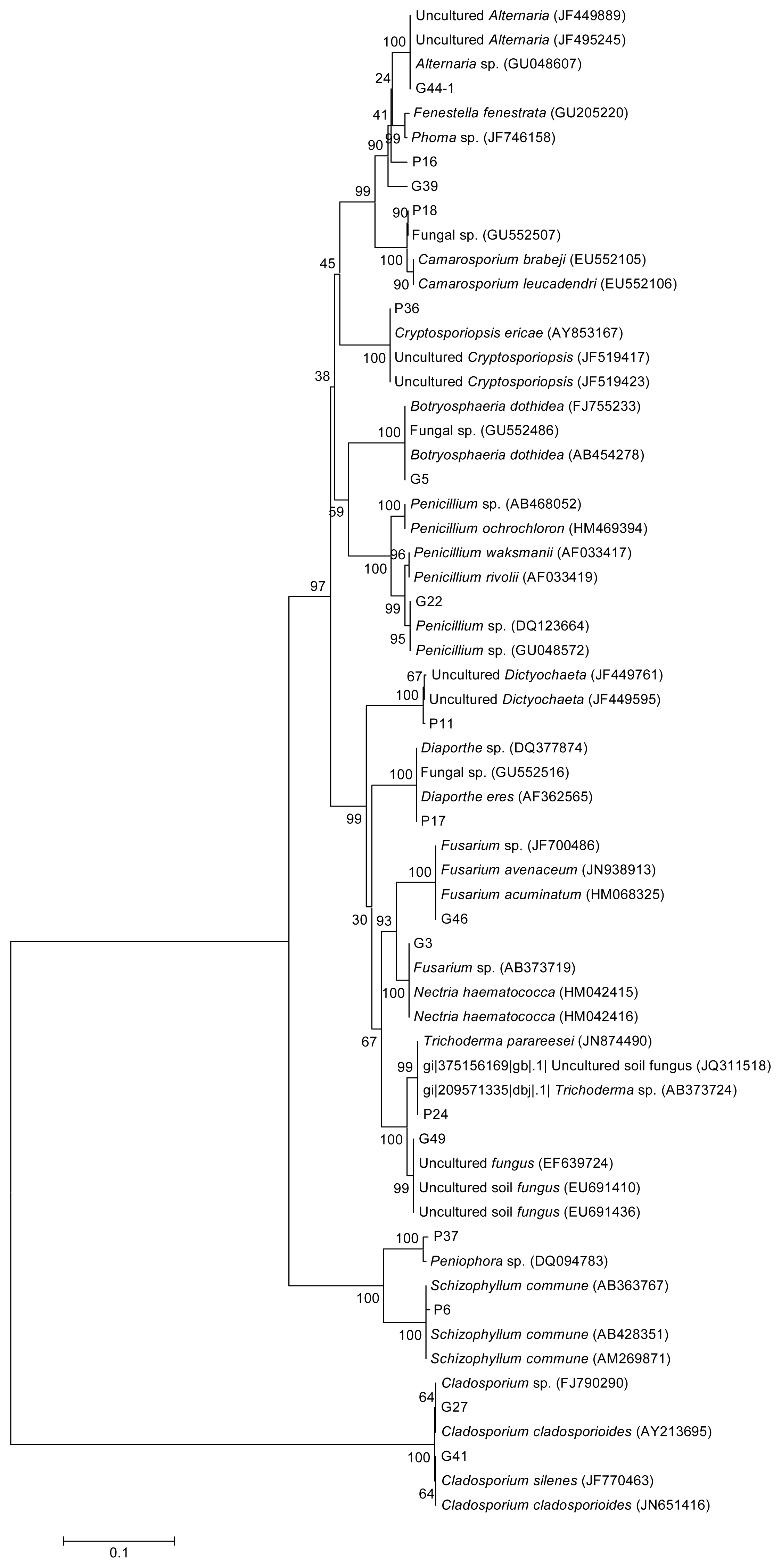
2.1. Strains Identification and Phylogenetic Analysis</title>
<p>Ninety-six strains were isolated. Genomic DNA was extracted and the 28S rDNA D1/D2 region was amplified and sequenced. The obtained sequences were compared with those in the GenBank database, and the results are shown in <xref ref-type="table" rid="t1-ijms-13-16255">Table 1</xref>.</p>
<p>A phylogenetic tree built from the 28S rDNA sequences is shown in <xref ref-type="fig" rid="f1-ijms-13-16255">Figure 1</xref>. From <xref ref-type="table" rid="t1-ijms-13-16255">Table 1</xref> and <xref ref-type="fig" rid="f1-ijms-13-16255">Figure 1</xref>, the identified fungi included: <italic>Alternaria</italic>, <italic>Botryosphaeria</italic>, <italic>Camarosporium</italic>, <italic>Cryptosporiopsis</italic>, <italic>Diaporthe</italic>, <italic>Dictyochaeta</italic>, <italic>Penicillium</italic>, <italic>Fusarium</italic>, <italic>Nectria</italic>, <italic>Peniophora</italic>, <italic>Schizophyllum</italic>, <italic>Cladosporium</italic> and <italic>Trichoderma</italic>. The most abundant genera were <italic>Diaporthe</italic> and <italic>Alternaria</italic> with 25 and 12.5% of the total number of isolates, respectively. G49 was not identified because its sequence was significantly similar to unknown fungal sequences in the GenBank database.</p>
<p><italic>Diaporthe</italic> and <italic>Altenaria</italic> were found to be the predominant genera, a finding that is different from previous studies showing that <italic>Strumella</italic>, <italic>Rhizopycnis</italic> and <italic>Entrophospora</italic> were the most abundant taxa in <italic>A. elata</italic>[<xref ref-type="bibr" rid="b11-ijms-13-16255">11</xref>]. A possible reason for this discrepancy is that the <italic>A. elata</italic> specimens were derived from different regions [<xref ref-type="bibr" rid="b17-ijms-13-16255">17</xref>].</p>
<p><italic>Diaporthe</italic> are endophytic fungi that grow in several of plant species and have been shown to produced different secondary metabolites. For example <italic>Diaporthe</italic> sp. isolated from <italic>Espeletia</italic> sp. can inhibit the growth of <italic>Phytophthora infestans</italic>, a plant pathogen [<xref ref-type="bibr" rid="b18-ijms-13-16255">18</xref>]. While <italic>Diaporthe</italic> sp. from <italic>Curcuma longa</italic> can convert curcumin into colorless hydroderivatives. Curcumin has a potent antioxidant effect. However, the distinct yellow color limits its use. This conversion may expand its application [<xref ref-type="bibr" rid="b19-ijms-13-16255">19</xref>]. <italic>Diaporthe</italic> sp. P133, isolated from <italic>Pandanus amaryllifolius</italic>, can secrete benzopyranones, which inhibit a virulent strain of <italic>Mycobacterium tuberculosis</italic>[<xref ref-type="bibr" rid="b20-ijms-13-16255">20</xref>]. <italic>Diaporthe phaseolorum</italic> isolated from mangrove forest can produce the antibacterial agent 3-hydroxypropionic acid [<xref ref-type="bibr" rid="b21-ijms-13-16255">21</xref>]. A new varied species <italic>Alternaria alternata</italic> from the bark of 200-year-old <italic>Taxus cuspidate</italic> could produce taxoids of type III with the anti-neoplastic action [<xref ref-type="bibr" rid="b22-ijms-13-16255">22</xref>]. <italic>Alternaria</italic> sp. isolated from <italic>Brassica juncea</italic> have demonstrated potential applications in biofuel feedback [<xref ref-type="bibr" rid="b23-ijms-13-16255">23</xref>]. To the best of our knowledge, there are no reports concerning <italic>Diaporthe</italic> and <italic>Altenaria</italic> isolated from <italic>A. elata</italic>.</p></sec>
<sec sec-type="methods">
<title>
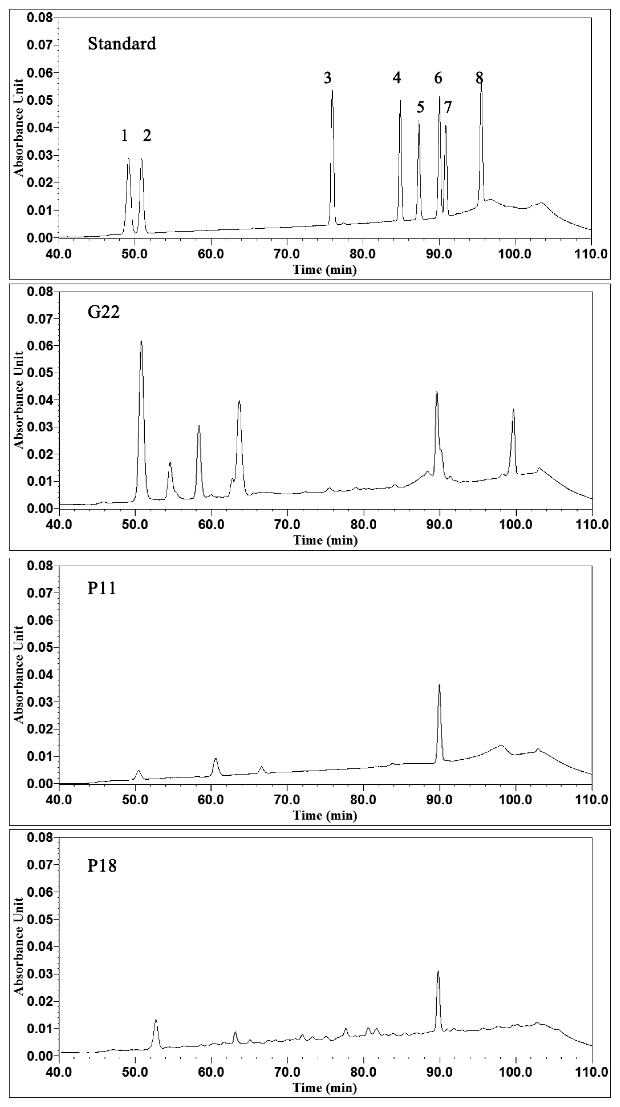
2.2. Analysis of Triterpenoid Saponins and Antimicrobial Activity</title>
<p>The concentration of triterpenoid saponin in the representative isolate from each group (<xref ref-type="table" rid="t1-ijms-13-16255">Table 1</xref>) is shown in <xref ref-type="table" rid="t2-ijms-13-16255">Table 2</xref>. The highest concentration of saponins was found in G22 (2.049 mg/mL), and this concentration is significantly higher than the level observed in P11 and P18 (<italic>p</italic> &lt; 0.05). According to the results of phylogenetic analysis, G22 was identified as a <italic>Penicillium</italic> sp., P11 was identified as a <italic>Dictyochaeta</italic> sp., and P18 was identified as <italic>Camarosporium</italic> sp. The saponin concentrations among the strains of the same genus, such as G22 (2.049 mg/mL) and P23 (0.049 mg/mL), were significantly different (<italic>p</italic> &lt; 0.05).</p>
<p>The growth-promoting factors and metabolites produced by endophytic fungi have been widely investigated and applied in both medicine and agriculture. The most notable substance produced by host endophytic fungi is taxol, a mitotic inhibitor used in cancer chemotherapy, and was originally produced by the yew tree [<xref ref-type="bibr" rid="b9-ijms-13-16255">9</xref>]. Saponins produced by <italic>A. elata</italic> have multiple therapeutic values. The culture filtrates of endophytic fungi were analyzed to identify endophytes that produce triterpenoid saponins. It is well known that <italic>Penicillium</italic> is the source of penicillin, and recent studies show that endophytic <italic>Penicillium</italic> sp. also has the capacity to secrete anti-tumor substances [<xref ref-type="bibr" rid="b24-ijms-13-16255">24</xref>,<xref ref-type="bibr" rid="b25-ijms-13-16255">25</xref>] and gibberellin [<xref ref-type="bibr" rid="b26-ijms-13-16255">26</xref>,<xref ref-type="bibr" rid="b27-ijms-13-16255">27</xref>]. In this study, G22 (<italic>Penicillium</italic> sp.) has the high capacity to produce triterpenoid saponins.</p>
<p>From <xref ref-type="table" rid="t3-ijms-13-16255">Table 3</xref>, G22 exhibited antibacterial activity against Gram-positive bacterium <italic>Staphylococcus aureus</italic> ACCC10499. P11 inhibited the growth of <italic>S. aureus</italic> ACCC10499, <italic>Rhizoctonia solani</italic> ACCC36233 and <italic>Fusarium sporotrichioides</italic>. At the same time, it expressed strong inhibition to <italic>Klebsiella pneumoniae</italic> ACCC10498. P18 also showed inhibition to <italic>K. pneumoniae</italic> ACCC10498.</p></sec>
<sec>
<title>2.3. Ginsenosides Analyses</title>
<p>To further analyze the composition of saponins, the culture filtrates of G22, P11, and P18, underwent HPLC. As a reference, eight ginsenoside standards were also analyzed. According to the spectra, which are shown in <xref ref-type="fig" rid="f2-ijms-13-16255">Figure 2</xref>, G22, P11 and P18 all produced detectable concentrations of saponins. Ginsenoside Rb2 was detected in the G22, P11, and P18 culture filtrates. More peaks were observed in the G22 culture filtrate. Ginsenoside Re was also detected in the G22 culture filtrate. These results indicate that the three strains have the capacity to produce ginsenosides, especially for G22. Additionally, G22 inhibited <italic>S. aureus</italic> growth (<xref ref-type="table" rid="t3-ijms-13-16255">Table 3</xref>). These results indicate that G22 has a great potential for the further detailed study.</p></sec></sec>
<sec>
<title>3. Experimental Section</title>
<sec>
<title>3.1. Sampling and Isolation</title>
<p>Wild <italic>A. elata</italic> plants (5 years old) were sampled from the Xiaoxing’anling area in Northeast China. The <italic>A. elata</italic> roots were immediately placed in sterile plastic bags and stored at 4 °C. The endophytes were isolated within 48 h of collection. Before disinfection, the plant samples were thoroughly washed under running tap water for 10 h. The roots were surface-disinfected with 70% (<italic>v</italic>/<italic>v</italic>) ethanol for 0.5–1 min, 5% NaOCl for 5–10 min, 70% (<italic>v</italic>/<italic>v</italic>) ethanol for 0.5–1 min and burning for 10–30 s. The samples were subsequently rinsed with sterile water, and the outer tissue was removed with a sterile scalpel. Small pieces (0.5 × 0.5 cm) of <italic>A. elata</italic> were placed in Petri dishes containing malt extract agar (Oxoid-Unipath Ltd., Hampshire, UK), Czapeck agar (Oxoid-Unipath Ltd., Hampshire, UK), or potato dextrose agar (Oxoid-Unipath Ltd., Hampshire, UK), and were incubated at 28 °C for seven days. Following the incubation, single colonies of distinctive morphotypes were isolated on the basis of their morphological characteristics and appearance. The colonies were subsequently re-isolated by plating on PDA and incubating at 28 °C for 24–48 h to obtain pure cultures. All of the isolates were vacuum freeze-dried and deposited in the collection of the College of Life Sciences, Northeast Forestry University.</p></sec>
<sec>
<title>3.2. DNA Extraction and PCR Amplification of the 28S rRNA Gene</title>
<p>F ungal genomic DNA was extracted using the EZNA Fungal DNA Mini Kit (OMEGA, USA) according to the manufacturer’s instructions. The 50 μL PCR mixtures contained 15 ng of template DNA, 1× PCR buffer (Mg<sup>2+</sup> free), 0.16 mM of each dNTP, 1.5 mM MgCl<sub>2</sub>, 0.45 μM of each primer, and 1 U of Takara rTaq DNA polymerase (Takara, Japan). The primers employed for the amplification of the D1/D2 region of the fungal 28S rRNA gene were NL1 (5′-GCATATCAATAAGCGGAGGAAAAG-3′) and NL4 (5′-GGTCCGTGTTTCAAGACGG-3′) [<xref ref-type="bibr" rid="b28-ijms-13-16255">28</xref>]. The thermocycling program consisted of initial DNA denaturation at 95 °C for 5 min followed by 30 cycles of denaturation at 95 °C for 1 min, annealing at 52 °C for 45 s, and elongation at 72 °C for 1 min 30 s, ending with a final elongation step at 72 °C for 6 min [<xref ref-type="bibr" rid="b29-ijms-13-16255">29</xref>].</p>
<p>The PCR amplification products were separated by electrophoresis through 1% (<italic>w</italic>/<italic>v</italic>) agarose gels, stained with ethidium bromide and visually examined under UV light. The PCR products were purified using the Agarose Gel DNA Extraction Kit (Takara, Japan) and sequenced by Sangon Biotech (Shanghai, China).</p></sec>
<sec sec-type="methods">
<title>3.3. Phylogenetic Analysis and Nucleotide Sequence Accession Numbers</title>
<p>The sequences generated in this study were compared with those in GenBank [<xref ref-type="bibr" rid="b30-ijms-13-16255">30</xref>]; those sequences with ≥99% similarity to the 28S rDNA D1/D2 regions (approximately 600 bp) were considered to belong to identical genera and were included in the alignment. A neighbor-joining tree was constructed using MEGA 5.0 software [<xref ref-type="bibr" rid="b31-ijms-13-16255">31</xref>]. The number of bootstrap replications was 1000. The sequences were deposited in GenBank under the accession numbers listed in <xref ref-type="table" rid="t1-ijms-13-16255">Table 1</xref>.</p></sec>
<sec>
<title>3.4. Determination of Triterpenoid Saponins</title>
<p>Each isolate was inoculated into 100 mL of PDA liquid medium (250 mL flask) and stirred at 150 rpm at 28 °C for two weeks. After ultrasonication, the supernatant was separated from the cell debris by centrifugation at 4000× <italic>g</italic> for 20 min. A 20 mL aliquot of the supernatant was poured into a 50 mL centrifuge tube (Corning Inc., Corning, NY, USA), and 20 mL of ethyl acetate was added to the same tube. After mixing, ultrasonication and incubation for 5 min, 5 mL of the supernatant was evaporated to dryness under a vacuum at 50 °C. The residue was dissolved in 2 mL of methanol. The methanol solutions were centrifuged at 4000× <italic>g</italic> for 10 min, and the supernatants were used for subsequent analysis of the total saponin content.</p>
<p>The measurement of the total extracted saponins was based on a color reaction of the acid-hydrolysis products of the saponins (<italic>i.e.</italic>, sapogenins) with vanillin. In total, 5 mL of the supernatant was added to a test tube and evaporated at 60 °C in a water bath. The residue was dissolved in 0.2 mL of 5% vanillin, mixed with 0.8 mL of perchloric acid, incubated in a 60 °C water bath for 15 min and quickly cooled in ice water. The concentration (mg/mL) of saponins in the reaction sample was determined using a spectrophotometer at 560 nm and comparing the readings against a calibration curve established with an oleanolic acid standard (National Institute for the Control of Pharmaceutical and Biological Products, Beijing, China) [<xref ref-type="bibr" rid="b32-ijms-13-16255">32</xref>].</p></sec>
<sec>
<title>3.5. Antimicrobial Activity of the Representative Strains</title>
<p>The antimicrobial activity of typical strains after 14-day cultivation against 8 microorganisms (listed in <xref ref-type="table" rid="t3-ijms-13-16255">Table 3</xref>) was assessed by the agar diffusion method [<xref ref-type="bibr" rid="b33-ijms-13-16255">33</xref>]. Three 6 mm wells were made in each disk. With the exception of <italic>Fusarium sporotrichioides</italic> (isolated in our lab), the strains were purchased from the Agricultural Culture Collection of China (ACCC). Streptomycin sulfate (5 mg/well), amoxicillin (5 mg/well) and itraconazole (4.4 mg/well) were used as positive antimicrobial controls. The activity of the extracts was estimated from the diameter (mm) of the zone of inhibition.</p></sec>
<sec>
<title>3.6. Ginsenosides Analyses</title>
<p>One hundred milliliters of ethyl acetate was added to 100 mL liquid culture. Following 30 min of agitation at 160 rpm and ultrasonication at 50 °C, the supernatant was separated from the cell debris by centrifugation at 4000× <italic>g</italic> for 30 min. After evaporation, the pellet was dissolved in 5 mL of methanol, followed by filtration through a SepPak C-18 Cartridge (Waters, Milford, MA, USA). The following water/acetonitrile gradient system was employed during HPLC analysis: 0 min, 18% acetonitrile and 82% water; 40 min, 18% acetonitrile and 82% water; 50 min, 22% acetonitrile and 78% water; 70 min, 28% acetonitrile and 72% water; 100 min, 38% acetonitrile and 62% water; and 110 min, 18% acetonitrile and 82% water.</p></sec></sec>
<sec sec-type="conclusions">
<title>4. Conclusions</title>
<p>This study focused on the diversity of endophytic fungi from <italic>A. elata</italic> in Northeast China for the first time. Ninety-six strains were isolated. They were belonged to 12 genus respectively. The most abundant genera were <italic>Diaporthe</italic> and <italic>Alteraria</italic> represented by 25 and 12.5% of the isolates respectively. The analysis from saponins showed that many isolated fungi had the capacity to produce saponins. The highest concentration of saponins was found in G22 (<italic>Penicillium</italic> sp., 2.049 mg/mL), Ginsenoside Rb2 and Re were detected in the G22.This result indicate that G22 has the capacity to produce ginsenosides and consequently has application potentials.</p></sec></body>
<back>
<ack>
<title>Acknowledgments</title>
<p>This study was supported by the Fund of “The National Forestry Public Welfare Industry Targeted Research Fund” (20100400703) and “The Daqing Technology Innovation Program” (SCX2010-08, SCYH-2011-95) and “The Daqing High-Tech Zone Special Fund” (DQGX10ZS006).</p></ack>
<ref-list>
<title>References</title>
<ref id="b1-ijms-13-16255"><label>1</label><citation citation-type="journal"><person-group person-group-type="author"><name><surname>Huang</surname><given-names>W.Y.</given-names></name><name><surname>Cai</surname><given-names>Y.Z.</given-names></name><name><surname>Hyde</surname><given-names>K.D.</given-names></name><name><surname>Corke</surname><given-names>H.</given-names></name><name><surname>Sun</surname><given-names>M.</given-names></name></person-group><article-title>Biodiversity of endophytic fungi associated with 29 traditional Chinese medicinal plants</article-title><source>Fungal Divers</source><year>2008</year><volume>33</volume><fpage>61</fpage><lpage>75</lpage></citation></ref>
<ref id="b2-ijms-13-16255"><label>2</label><citation citation-type="journal"><person-group person-group-type="author"><name><surname>Hyde</surname><given-names>K.D.</given-names></name><name><surname>Soytong</surname><given-names>K.</given-names></name></person-group><article-title>The fungal endophyte dilemma</article-title><source>Fungal Divers</source><year>2008</year><volume>33</volume><fpage>163</fpage><lpage>173</lpage></citation></ref>
<ref id="b3-ijms-13-16255"><label>3</label><citation citation-type="journal"><person-group person-group-type="author"><name><surname>Koukol</surname><given-names>O.</given-names></name></person-group><article-title>New species of <italic>Chalara</italic> occupying coniferous needles</article-title><source>Fungal Divers</source><year>2011</year><volume>49</volume><fpage>75</fpage><lpage>91</lpage><pub-id pub-id-type="doi">10.1007/s13225-011-0092-2</pub-id></citation></ref>
<ref id="b4-ijms-13-16255"><label>4</label><citation citation-type="journal"><person-group person-group-type="author"><name><surname>Li</surname><given-names>H.</given-names></name><name><surname>Shen</surname><given-names>M.</given-names></name><name><surname>Zhou</surname><given-names>Z.</given-names></name><name><surname>Li</surname><given-names>T.</given-names></name><name><surname>Wei</surname><given-names>Y.</given-names></name><name><surname>Lin</surname><given-names>L.</given-names></name></person-group><article-title>Diversity and cold adaptation of endophytic fungi from five dominant plant species collected from the Baima Snow Mountain, Southwest China</article-title><source>Fungal Divers</source><year>2012</year><volume>54</volume><fpage>79</fpage><lpage>86</lpage><pub-id pub-id-type="doi">10.1007/s13225-012-0153-1</pub-id></citation></ref>
<ref id="b5-ijms-13-16255"><label>5</label><citation citation-type="journal"><person-group person-group-type="author"><name><surname>Alberton</surname><given-names>O.</given-names></name><name><surname>Kuyper</surname><given-names>T.W.</given-names></name><name><surname>Summerbell</surname><given-names>R.C.</given-names></name></person-group><article-title>Dark septate root endophytic fungi increase growth of Scots pine seedlings under elevated CO<sub>2</sub> through enhanced nitrogen use efficiency</article-title><source>Plant Soil</source><year>2010</year><volume>328</volume><fpage>459</fpage><lpage>470</lpage><pub-id pub-id-type="doi">10.1007/s11104-009-0125-8</pub-id></citation></ref>
<ref id="b6-ijms-13-16255"><label>6</label><citation citation-type="journal"><person-group person-group-type="author"><name><surname>Saikkonen</surname><given-names>K.</given-names></name><name><surname>Saari</surname><given-names>S.</given-names></name><name><surname>Helander</surname><given-names>M.</given-names></name></person-group><article-title>Defensive mutualism between plants and endophytic fungi?</article-title><source>Fungal Divers</source><year>2010</year><volume>41</volume><fpage>101</fpage><lpage>113</lpage><pub-id pub-id-type="doi">10.1007/s13225-010-0023-7</pub-id></citation></ref>
<ref id="b7-ijms-13-16255"><label>7</label><citation citation-type="book"><person-group person-group-type="author"><name><surname>Osono</surname><given-names>T.</given-names></name><name><surname>Hirose</surname><given-names>D</given-names></name></person-group><article-title>Ecology of endophytic fungi associated with leaf litter decomposition</article-title><source>Applied Mycology</source><person-group person-group-type="editor"><name><surname>Rai</surname><given-names>M.</given-names></name><name><surname>Bridge</surname><given-names>P.D.</given-names></name></person-group><publisher-name>CAB International</publisher-name><publisher-loc>Cambridge, MA, USA</publisher-loc><year>2009</year><fpage>92</fpage><lpage>109</lpage></citation></ref>
<ref id="b8-ijms-13-16255"><label>8</label><citation citation-type="journal"><person-group person-group-type="author"><name><surname>Schulz</surname><given-names>B.</given-names></name><name><surname>Boyle</surname><given-names>C.</given-names></name><name><surname>Draeger</surname><given-names>S.</given-names></name><name><surname>Römmert</surname><given-names>A.</given-names></name><name><surname>Krohn</surname><given-names>K.</given-names></name></person-group><article-title>Endophyticfungi: A source of novel biologically active secondary metabolites</article-title><source>Mycol. Res</source><year>2002</year><volume>106</volume><fpage>996</fpage><lpage>1004</lpage><pub-id pub-id-type="doi">10.1017/S0953756202006342</pub-id></citation></ref>
<ref id="b9-ijms-13-16255"><label>9</label><citation citation-type="journal"><person-group person-group-type="author"><name><surname>Rivera-Orduña</surname><given-names>F.N.</given-names></name><name><surname>Suarez-sanchez</surname><given-names>R.A.</given-names></name><name><surname>Flores-Bustamante</surname><given-names>Z.R.</given-names></name><name><surname>Gracida-Rodriguez</surname><given-names>J.N.</given-names></name><name><surname>Flores-Cotera</surname><given-names>L.B.</given-names></name></person-group><article-title>Diversity of endophytic fungi of <italic>Taxus globosa</italic> (Mexican yew)</article-title><source>Fungal Divers</source><year>2011</year><volume>47</volume><fpage>65</fpage><lpage>74</lpage><pub-id pub-id-type="doi">10.1007/s13225-010-0045-1</pub-id></citation></ref>
<ref id="b10-ijms-13-16255"><label>10</label><citation citation-type="journal"><person-group person-group-type="author"><name><surname>Kusari</surname><given-names>S.</given-names></name><name><surname>Verma</surname><given-names>V.C.</given-names></name><name><surname>Lamshoeft</surname><given-names>M.</given-names></name><name><surname>Spiteller</surname><given-names>M.</given-names></name></person-group><article-title>An endophytic fungus from <italic>Azadirachta indica</italic> A. Juss. that produces azadirachtin</article-title><source>World J. Microbiol. Biotechnol</source><year>2012</year><volume>28</volume><fpage>1287</fpage><lpage>1294</lpage><pub-id pub-id-type="doi">10.1007/s11274-011-0876-2</pub-id><pub-id pub-id-type="pmid">22805849</pub-id></citation></ref>
<ref id="b11-ijms-13-16255"><label>11</label><citation citation-type="journal"><person-group person-group-type="author"><name><surname>Paul</surname><given-names>N.C.</given-names></name><name><surname>Kim</surname><given-names>W.K.</given-names></name><name><surname>Woo</surname><given-names>S.K.</given-names></name><name><surname>Park</surname><given-names>S.</given-names></name><name><surname>Yu</surname><given-names>S.H.</given-names></name></person-group><article-title>Fungal endophytes in roots of <italic>Aralia</italic> species and their antifungal activity</article-title><source>Plant Pathol. J</source><year>2007</year><volume>23</volume><fpage>287</fpage><lpage>294</lpage><pub-id pub-id-type="doi">10.5423/PPJ.2007.23.4.287</pub-id></citation></ref>
<ref id="b12-ijms-13-16255"><label>12</label><citation citation-type="journal"><person-group person-group-type="author"><name><surname>Sakai</surname><given-names>S.</given-names></name><name><surname>Katsumata</surname><given-names>M.</given-names></name><name><surname>Satoh</surname><given-names>Y.</given-names></name><name><surname>Nagasao</surname><given-names>M.</given-names></name><name><surname>Miyakoshi</surname><given-names>M.</given-names></name><name><surname>Ida</surname><given-names>Y.</given-names></name><name><surname>Shoji</surname><given-names>J.</given-names></name></person-group><article-title>Oleanolic acid saponins from root bark of <italic>Aralia elata</italic></article-title><source>Phytochemistry</source><year>1994</year><volume>35</volume><fpage>1319</fpage><lpage>1324</lpage><pub-id pub-id-type="doi">10.1016/S0031-9422(00)94846-5</pub-id><pub-id pub-id-type="pmid">7764823</pub-id></citation></ref>
<ref id="b13-ijms-13-16255"><label>13</label><citation citation-type="journal"><person-group person-group-type="author"><name><surname>Mujoo</surname><given-names>K.</given-names></name><name><surname>Haridas</surname><given-names>V.</given-names></name><name><surname>Hoffmann</surname><given-names>J.J.</given-names></name><name><surname>Wächter</surname><given-names>G.A.</given-names></name><name><surname>Hutter</surname><given-names>L.K.</given-names></name><name><surname>Lu</surname><given-names>Y.</given-names></name><name><surname>Blake</surname><given-names>M.E.</given-names></name><name><surname>Jayatilake</surname><given-names>G.S.</given-names></name><name><surname>Bailey</surname><given-names>D.</given-names></name><name><surname>Mills</surname><given-names>G.B.</given-names></name><etal/></person-group><article-title>Triterpenoid saponins from <italic>Acacia victoriae</italic> (Bentham) decrease tumor cell proliferation and induce apoptosis</article-title><source>Cancer Res</source><year>2001</year><volume>61</volume><fpage>5486</fpage><lpage>5490</lpage><pub-id pub-id-type="pmid">11454696</pub-id></citation></ref>
<ref id="b14-ijms-13-16255"><label>14</label><citation citation-type="journal"><person-group person-group-type="author"><name><surname>Fujimoto</surname><given-names>J.</given-names></name><name><surname>Sakaguchi</surname><given-names>H.</given-names></name><name><surname>Alki</surname><given-names>I.</given-names></name><name><surname>Toyoki</surname><given-names>H.</given-names></name><name><surname>Khatun</surname><given-names>S.</given-names></name><name><surname>Tamaya</surname><given-names>T.</given-names></name></person-group><article-title>Inhibitory effect of ginsenoside-Rb2 on invasiveness of uterine endometrial cancer cells to the basement membrane</article-title><source>Eur. J. Gynaecol. Oncol</source><year>2001</year><volume>22</volume><fpage>339</fpage><lpage>341</lpage><pub-id pub-id-type="pmid">11766734</pub-id></citation></ref>
<ref id="b15-ijms-13-16255"><label>15</label><citation citation-type="journal"><person-group person-group-type="author"><name><surname>Zhao</surname><given-names>Y.-L.</given-names></name><name><surname>Cai</surname><given-names>G.-M.</given-names></name><name><surname>Hong</surname><given-names>X.</given-names></name><name><surname>Shan</surname><given-names>L.-M.</given-names></name><name><surname>Xiao</surname><given-names>X.-H.</given-names></name></person-group><article-title>Anti-hepatitis B virus activities of triterpenoid saponin compound from <italic>Potentilla anserine</italic> L</article-title><source>Phytomedicine</source><year>2008</year><volume>15</volume><fpage>253</fpage><lpage>258</lpage><pub-id pub-id-type="doi">10.1016/j.phymed.2008.01.005</pub-id><pub-id pub-id-type="pmid">18337074</pub-id></citation></ref>
<ref id="b16-ijms-13-16255"><label>16</label><citation citation-type="journal"><person-group person-group-type="author"><name><surname>Sen</surname><given-names>S.</given-names></name><name><surname>Chakraborty</surname><given-names>R.</given-names></name><name><surname>De</surname><given-names>B.</given-names></name><name><surname>Mazumder</surname><given-names>J.</given-names></name></person-group><article-title>Plants and phytochemicals for peptic ulcer: An overview</article-title><source>Pharmacognosy Rev</source><year>2009</year><volume>3</volume><fpage>270</fpage><lpage>279</lpage></citation></ref>
<ref id="b17-ijms-13-16255"><label>17</label><citation citation-type="journal"><person-group person-group-type="author"><name><surname>Davis</surname><given-names>E.C.</given-names></name><name><surname>Shaw</surname><given-names>A.J.</given-names></name></person-group><article-title>Biogeographic and phylogenetic patterns in diversity of liverwort-associated endophytes</article-title><source>Am. J. Bot</source><year>2008</year><volume>95</volume><fpage>914</fpage><lpage>924</lpage><pub-id pub-id-type="doi">10.3732/ajb.2006463</pub-id><pub-id pub-id-type="pmid">21632414</pub-id></citation></ref>
<ref id="b18-ijms-13-16255"><label>18</label><citation citation-type="journal"><person-group person-group-type="author"><name><surname>Prada</surname><given-names>H.</given-names></name><name><surname>Avila</surname><given-names>L.</given-names></name><name><surname>Sierra</surname><given-names>R.</given-names></name><name><surname>Bernal</surname><given-names>A.</given-names></name><name><surname>Restrepo</surname><given-names>S.</given-names></name></person-group><article-title>Morphological and molecular characterization of the antagonistic interaction between the endophyte <italic>Diaporthe</italic> sp. isolated from frailejon (<italic>Espeletia</italic> sp.) and the plant pathogen <italic>Phytophthora infestans</italic></article-title><source>Rev. Iberoam. Micol</source><year>2009</year><volume>26</volume><fpage>198</fpage><lpage>201</lpage><pub-id pub-id-type="doi">10.1016/j.riam.2009.01.002</pub-id><pub-id pub-id-type="pmid">19635440</pub-id></citation></ref>
<ref id="b19-ijms-13-16255"><label>19</label><citation citation-type="journal"><person-group person-group-type="author"><name><surname>Maehara</surname><given-names>S.</given-names></name><name><surname>Ikeda</surname><given-names>M.</given-names></name><name><surname>Haraguchi</surname><given-names>H.</given-names></name><name><surname>Kitamura</surname><given-names>C.</given-names></name><name><surname>Nagoe</surname><given-names>T.</given-names></name><name><surname>Ohashi</surname><given-names>K.</given-names></name><name><surname>Shibuya</surname><given-names>H.</given-names></name></person-group><article-title>Microbial conversion of curcumin into colorless hydroderivatives by the endophytic fungus <italic>Diaporthe</italic> sp. associated with <italic>Curcuma longa</italic></article-title><source>Chem. Pharm. Bull</source><year>2011</year><volume>59</volume><fpage>1042</fpage><lpage>1044</lpage><pub-id pub-id-type="doi">10.1248/cpb.59.1042</pub-id><pub-id pub-id-type="pmid">21804251</pub-id></citation></ref>
<ref id="b20-ijms-13-16255"><label>20</label><citation citation-type="journal"><person-group person-group-type="author"><name><surname>Bungihan</surname><given-names>M.E.</given-names></name><name><surname>Tan</surname><given-names>M.A.</given-names></name><name><surname>Kitajima</surname><given-names>M.</given-names></name><name><surname>Kogure</surname><given-names>N.</given-names></name><name><surname>Franzblau</surname><given-names>S.G.</given-names></name><name><surname>Dela Cruz</surname><given-names>T.E.</given-names></name><name><surname>Takayama</surname><given-names>H.</given-names></name><name><surname>Nonato</surname><given-names>M.G.</given-names></name></person-group><article-title>Bioactive metabolites of <italic>Diaporthe</italic> sp. P133, an endophytic fungus isolated from <italic>Pandanus amaryllifolius</italic></article-title><source>J. Nat. Med</source><year>2011</year><volume>65</volume><fpage>606</fpage><lpage>609</lpage><pub-id pub-id-type="doi">10.1007/s11418-011-0518-x</pub-id><pub-id pub-id-type="pmid">21394664</pub-id></citation></ref>
<ref id="b21-ijms-13-16255"><label>21</label><citation citation-type="journal"><person-group person-group-type="author"><name><surname>Sebastianes</surname><given-names>F.L.S.</given-names></name><name><surname>Cabedo</surname><given-names>N.</given-names></name><name><surname>Aouad</surname><given-names>N.E.</given-names></name><name><surname>Valente</surname><given-names>A.M.M.P.</given-names></name><name><surname>Lacava</surname><given-names>P.T.</given-names></name><name><surname>Azevedo</surname><given-names>J.L.</given-names></name><name><surname>Pizzirani-Kleiner</surname><given-names>A.A.</given-names></name><name><surname>Cortes</surname><given-names>D</given-names></name></person-group><article-title>3-Hydroxypropionic acid as an antibacteria agent from endophytic fungi <italic>Diaporthe phaseolorum</italic></article-title><source>Curr. Microbiol.</source><year>2012</year><pub-id pub-id-type="doi">10.1007/s002834-012-0206-4</pub-id></citation></ref>
<ref id="b22-ijms-13-16255"><label>22</label><citation citation-type="journal"><person-group person-group-type="author"><name><surname>Xiang</surname><given-names>Y.</given-names></name><name><surname>Lu</surname><given-names>A.</given-names></name><name><surname>Wu</surname><given-names>W.</given-names></name></person-group><article-title>Identification of <italic>Taxus cuspidata</italic> sieb. et Zucc. endophytic fungi-new species, species known and their metabolite</article-title><source>J. For. Res</source><year>2003</year><volume>14</volume><fpage>290</fpage><lpage>294</lpage><pub-id pub-id-type="doi">10.1007/BF02857856</pub-id></citation></ref>
<ref id="b23-ijms-13-16255"><label>23</label><citation citation-type="journal"><person-group person-group-type="author"><name><surname>Dey</surname><given-names>P.</given-names></name><name><surname>Banerjee</surname><given-names>J.</given-names></name><name><surname>Maiti</surname><given-names>M.K.</given-names></name></person-group><article-title>Comparative lipid profiling of two endophytic fungal isolates—<italic>Colletotrichum</italic> sp. and <italic>Alternaria</italic> sp. having potential utilities as biodiesel feedstock</article-title><source>Bioresource Technol</source><year>2011</year><volume>102</volume><fpage>5815</fpage><lpage>5823</lpage><pub-id pub-id-type="doi">10.1016/j.biortech.2011.02.064</pub-id></citation></ref>
<ref id="b24-ijms-13-16255"><label>24</label><citation citation-type="journal"><person-group person-group-type="author"><name><surname>Chen</surname><given-names>G.</given-names></name><name><surname>Zhu</surname><given-names>Y.</given-names></name><name><surname>Wang</surname><given-names>H.Z.</given-names></name><name><surname>Wang</surname><given-names>S.J.</given-names></name><name><surname>Zhang</surname><given-names>R.Q.</given-names></name></person-group><article-title>The metabolites of a mangrove endophytic fungus, <italic>Penicillium thomi</italic></article-title><source>J. Asian Nat. Prod. Res</source><year>2007</year><volume>9</volume><fpage>159</fpage><lpage>164</lpage><pub-id pub-id-type="doi">10.1080/10286020500480423</pub-id><pub-id pub-id-type="pmid">17454313</pub-id></citation></ref>
<ref id="b25-ijms-13-16255"><label>25</label><citation citation-type="journal"><person-group person-group-type="author"><name><surname>Guo</surname><given-names>Z.</given-names></name><name><surname>Cheng</surname><given-names>F.</given-names></name><name><surname>Zou</surname><given-names>K.</given-names></name><name><surname>Wang</surname><given-names>J.</given-names></name><name><surname>She</surname><given-names>Z.</given-names></name><name><surname>Lin</surname><given-names>Y.</given-names></name></person-group><article-title>Secondary metabolites from the mangrove endophytic fungus <italic>Penicillium</italic> sp. (SBE-8)</article-title><source>Nat. Prod. Commun</source><year>2009</year><volume>4</volume><fpage>1481</fpage><lpage>1483</lpage><pub-id pub-id-type="pmid">19967978</pub-id></citation></ref>
<ref id="b26-ijms-13-16255"><label>26</label><citation citation-type="journal"><person-group person-group-type="author"><name><surname>Hamayun</surname><given-names>M.</given-names></name><name><surname>Khan</surname><given-names>S.A.</given-names></name><name><surname>Iqbal</surname><given-names>I.</given-names></name><name><surname>Ahmad</surname><given-names>B.</given-names></name><name><surname>Lee</surname><given-names>I.J.</given-names></name></person-group><article-title>Isolation of a gibberellin-producing fungus (<italic>Penicillium</italic> sp. MH7) and growth promotion of Crown daisy (<italic>Chrysanthemum coronarium</italic>)</article-title><source>J. Microbiol. Biotechnol</source><year>2010</year><volume>20</volume><fpage>202</fpage><lpage>207</lpage><pub-id pub-id-type="pmid">20134253</pub-id></citation></ref>
<ref id="b27-ijms-13-16255"><label>27</label><citation citation-type="journal"><person-group person-group-type="author"><name><surname>Ahmad</surname><given-names>N.</given-names></name><name><surname>Hamayun</surname><given-names>M.</given-names></name><name><surname>Khan</surname><given-names>S.A.</given-names></name><name><surname>Khan</surname><given-names>A.L.</given-names></name><name><surname>Lee</surname><given-names>I.J.</given-names></name><name><surname>Shin</surname><given-names>D.H.</given-names></name></person-group><article-title>Gibberellin-producing endophytic fungi isolated from <italic>Monochoria vaginalis</italic></article-title><source>J. Microbiol. Biotechnol</source><year>2011</year><volume>20</volume><fpage>1744</fpage><lpage>1749</lpage></citation></ref>
<ref id="b28-ijms-13-16255"><label>28</label><citation citation-type="journal"><person-group person-group-type="author"><name><surname>Redecke</surname><given-names>D.</given-names></name></person-group><article-title>Specific PCR primers to identify arbuscular mycorrhizal fungi within colonized roots</article-title><source>Mycorrhiza</source><year>2000</year><volume>10</volume><fpage>73</fpage><lpage>80</lpage><pub-id pub-id-type="doi">10.1007/s005720000061</pub-id></citation></ref>
<ref id="b29-ijms-13-16255"><label>29</label><citation citation-type="journal"><person-group person-group-type="author"><name><surname>Yang</surname><given-names>H.</given-names></name><name><surname>Wu</surname><given-names>H.</given-names></name><name><surname>Wang</surname><given-names>X.</given-names></name><name><surname>Cui</surname><given-names>Z.</given-names></name><name><surname>Li</surname><given-names>Y.</given-names></name></person-group><article-title>Selection and characteristics of a switchgrass-colonizing microbial community to produce extracellular cellulases and xylanases</article-title><source>Bioresour. Technol</source><year>2011</year><volume>102</volume><fpage>3546</fpage><lpage>3550</lpage><pub-id pub-id-type="doi">10.1016/j.biortech.2010.09.009</pub-id><pub-id pub-id-type="pmid">20933405</pub-id></citation></ref>
<ref id="b30-ijms-13-16255"><label>30</label><citation citation-type="web"><collab>GenBank</collab><source>Basic Local Alignment Search Tool</source><comment>Available online: <ext-link xlink:href="http://blast.ncbi.nlm.nih.gov/Blast.cgi" ext-link-type="uri">http://blast.ncbi.nlm.nih.gov/Blast.cgi</ext-link></comment><access-date>accessed on 12 June 2012</access-date></citation></ref>
<ref id="b31-ijms-13-16255"><label>31</label><citation citation-type="journal"><person-group person-group-type="author"><name><surname>Tamura</surname><given-names>K.</given-names></name><name><surname>Peterson</surname><given-names>D.</given-names></name><name><surname>Peterson</surname><given-names>N.</given-names></name><name><surname>Stecher</surname><given-names>G.</given-names></name><name><surname>Nei</surname><given-names>M.</given-names></name><name><surname>Kumar</surname><given-names>S.</given-names></name></person-group><article-title>MEGA5: Molecular evolutionary genetics analysis using maximum likehood, evolutionary distance, and maximum parsimony methods</article-title><source>Mol. Biol. Evol</source><year>2011</year><volume>28</volume><fpage>2731</fpage><lpage>2739</lpage><pub-id pub-id-type="doi">10.1093/molbev/msr121</pub-id><pub-id pub-id-type="pmid">21546353</pub-id></citation></ref>
<ref id="b32-ijms-13-16255"><label>32</label><citation citation-type="journal"><person-group person-group-type="author"><name><surname>Liu</surname><given-names>H.G.</given-names></name><name><surname>Li</surname><given-names>T.</given-names></name><name><surname>Zhao</surname><given-names>Y.L.</given-names></name><name><surname>Zhang</surname><given-names>J.</given-names></name><name><surname>Wang</surname><given-names>Y.Z.</given-names></name></person-group><article-title>Determination of some metabolites of <italic>Cordyceps sobolifera</italic></article-title><source>Afr. J. Microbiol. Res</source><year>2011</year><volume>5</volume><fpage>5518</fpage><lpage>5522</lpage></citation></ref>
<ref id="b33-ijms-13-16255"><label>33</label><citation citation-type="journal"><person-group person-group-type="author"><name><surname>Hormazabal</surname><given-names>E.</given-names></name><name><surname>Piontelli</surname><given-names>E.</given-names></name></person-group><article-title>Endophytic fungi from chilean native gymnosperms: Antimicrobial activity against human and phytopathogenic fungi</article-title><source>World J. Microbiol. Biotechnol</source><year>2009</year><volume>25</volume><fpage>813</fpage><lpage>819</lpage><pub-id pub-id-type="doi">10.1007/s11274-008-9953-6</pub-id></citation></ref></ref-list>
<sec sec-type="display-objects">
<title>Figures and Tables</title>
<fig id="f1-ijms-13-16255" position="float">
<label>Figure 1</label>
<caption>
<p>A 28S rDNA sequence-based phylogenetic tree constructed using the neighbor-joining method. Scale bar represents 10% estimated sequence divergence. Numbers in the branches indicate bootstrap values (percentages for 1000 replicates).</p></caption>
<graphic xlink:href="ijms-13-16255f1.gif"/></fig>
<fig id="f2-ijms-13-16255" position="float">
<label>Figure 2</label>
<caption>
<p>High-performance liquid chromatography (HPLC) spectra of the ginsenoside standards and the culture filtrates of the representative strains. 1, Rg1; 2, Re; 3, Rf; 4, Rb1; 5, Rc; 6, Rb2; 7, Rb3; 8, Rd.</p></caption>
<graphic xlink:href="ijms-13-16255f2.gif"/></fig>
<table-wrap id="t1-ijms-13-16255" position="float">
<label>Table 1</label>
<caption>
<p>Similarity between the isolates and closest species in GenBank.</p></caption>
<table frame="hsides" rules="groups">
<thead>
<tr>
<th align="left" valign="bottom">Strain ID (Strains)</th>
<th align="left" valign="bottom">Accession no.</th>
<th align="left" valign="bottom">Closest species (Accession no.)</th>
<th align="left" valign="bottom">Coverage (%)</th></tr></thead>
<tbody>
<tr>
<td align="left" valign="top">P17 (24)</td>
<td align="left" valign="top">JQ807963</td>
<td align="left" valign="top">Fungal sp. (GU552516)</td>
<td align="left" valign="top">100</td></tr>
<tr>
<td align="left" valign="top"/>
<td align="left" valign="top"><italic>Diaporthe</italic> sp. (DQ377874)</td>
<td align="left" valign="top">100</td></tr>
<tr>
<td align="left" valign="top"/>
<td align="left" valign="top"><italic>Diaporthe eres</italic> (AF362565)</td>
<td align="left" valign="top">100</td></tr>
<tr>
<td align="left" valign="top">G44-1 (12)</td>
<td align="left" valign="top">JQ807974</td>
<td align="left" valign="top">Uncultured <italic>Alternaria</italic> (JF495245)</td>
<td align="left" valign="top">100</td></tr>
<tr>
<td align="left" valign="top"/>
<td align="left" valign="top">Uncultured <italic>Alternaria</italic> (JF449889)</td>
<td align="left" valign="top">100</td></tr>
<tr>
<td align="left" valign="top"/>
<td align="left" valign="top"><italic>Alternaria</italic> sp. (GU048607)</td>
<td align="left" valign="top">100</td></tr>
<tr>
<td align="left" valign="top">G22 (8)</td>
<td align="left" valign="top">JQ807910</td>
<td align="left" valign="top"><italic>Penicillium</italic> sp. (GU048572)</td>
<td align="left" valign="top">100</td></tr>
<tr>
<td align="left" valign="top"/>
<td align="left" valign="top"><italic>Penicillium</italic> sp. (DQ123664)</td>
<td align="left" valign="top">100</td></tr>
<tr>
<td align="left" valign="top"/>
<td align="left" valign="top"><italic>Penicillium rivolii</italic> (GU033419)</td>
<td align="left" valign="top">99.3</td></tr>
<tr>
<td align="left" valign="top">G27 (8)</td>
<td align="left" valign="top">JQ807969</td>
<td align="left" valign="top"><italic>Cladosporium</italic> sp. (FJ790290)</td>
<td align="left" valign="top">100</td></tr>
<tr>
<td align="left" valign="top"/>
<td align="left" valign="top"><italic>Cladosporium cladosporioides</italic> (AY213695)</td>
<td align="left" valign="top">100</td></tr>
<tr>
<td align="left" valign="top"/>
<td align="left" valign="top"><italic>Passalora fulva</italic> (AB100653)</td>
<td align="left" valign="top">100</td></tr>
<tr>
<td align="left" valign="top">P6 (6)</td>
<td align="left" valign="top">JQ807984</td>
<td align="left" valign="top"><italic>Schizophyllum commune</italic> (AM269871)</td>
<td align="left" valign="top">99.8</td></tr>
<tr>
<td align="left" valign="top"/>
<td align="left" valign="top"><italic>Schizophyllum commune</italic> (AB428351)</td>
<td align="left" valign="top">99.8</td></tr>
<tr>
<td align="left" valign="top"/>
<td align="left" valign="top"><italic>Schizophyllum commune</italic> (AB363767)</td>
<td align="left" valign="top">99.8</td></tr>
<tr>
<td align="left" valign="top">P11(5)</td>
<td align="left" valign="top">JQ807967</td>
<td align="left" valign="top">Uncultured <italic>Dictyochaeta</italic> (JF449595)</td>
<td align="left" valign="top">99.3</td></tr>
<tr>
<td align="left" valign="top"/>
<td align="left" valign="top">Uncultured <italic>Dictyochaeta</italic> (JF449592)</td>
<td align="left" valign="top">98.8</td></tr>
<tr>
<td align="left" valign="top"/>
<td align="left" valign="top"><italic>Dictyochaeta simplex</italic> (AF178559)</td>
<td align="left" valign="top">98</td></tr>
<tr>
<td align="left" valign="top">P18(5)</td>
<td align="left" valign="top">JQ807982</td>
<td align="left" valign="top">Fungal sp. (GU552507)</td>
<td align="left" valign="top">100</td></tr>
<tr>
<td align="left" valign="top"/>
<td align="left" valign="top"><italic>Camarosporium leucadendri</italic> (EU552106)</td>
<td align="left" valign="top">99.7</td></tr>
<tr>
<td align="left" valign="top"/>
<td align="left" valign="top"><italic>Camarosporium brabeji</italic> (EU552105)</td>
<td align="left" valign="top">99.7</td></tr>
<tr>
<td align="left" valign="top">G3 (5)</td>
<td align="left" valign="top">JQ807935</td>
<td align="left" valign="top"><italic>Nectria haematococca</italic> (HM042416)</td>
<td align="left" valign="top">100</td></tr>
<tr>
<td align="left" valign="top"/>
<td align="left" valign="top"><italic>Nectria haematococca</italic> (AB373719)</td>
<td align="left" valign="top">100</td></tr>
<tr>
<td align="left" valign="top">G41 (4)</td>
<td align="left" valign="top">JQ807972</td>
<td align="left" valign="top"><italic>Cladosporium cladosporioides</italic> (JN651416)</td>
<td align="left" valign="top">100</td></tr>
<tr>
<td align="left" valign="top"/>
<td align="left" valign="top"><italic>Cladosporium silenes</italic> (JF770463)</td>
<td align="left" valign="top">100</td></tr>
<tr>
<td align="left" valign="top"/>
<td align="left" valign="top">Uncultured <italic>Cladosporium</italic> (JF449832)</td>
<td align="left" valign="top">100</td></tr>
<tr>
<td align="left" valign="top">G46 (4)</td>
<td align="left" valign="top">JQ807956</td>
<td align="left" valign="top"><italic>Fusarium avenaceum</italic> (JN938913)</td>
<td align="left" valign="top">100</td></tr>
<tr>
<td align="left" valign="top"/>
<td align="left" valign="top"><italic>Fusarium</italic> sp. (JF700486)</td>
<td align="left" valign="top">100</td></tr>
<tr>
<td align="left" valign="top"/>
<td align="left" valign="top"><italic>Fusarium avenaceum</italic> (HM068325)</td>
<td align="left" valign="top">100</td></tr>
<tr>
<td align="left" valign="top">P37 (3)</td>
<td align="left" valign="top">JQ807942</td>
<td align="left" valign="top"><italic>Peniophora</italic> sp. (HM595610)</td>
<td align="left" valign="top">99.8</td></tr>
<tr>
<td align="left" valign="top"/>
<td align="left" valign="top"><italic>Peniophoraceae</italic> sp. (AB576771)</td>
<td align="left" valign="top">99.8</td></tr>
<tr>
<td align="left" valign="top"/>
<td align="left" valign="top"><italic>Peniophoraceae</italic> sp. (DQ094783)</td>
<td align="left" valign="top">99.8</td></tr>
<tr>
<td align="left" valign="top">P24(3)</td>
<td align="left" valign="top">JQ807922</td>
<td align="left" valign="top">Uncultured soil <italic>fungus</italic> (JQ311518)</td>
<td align="left" valign="top">99.5</td></tr>
<tr>
<td align="left" valign="top"/>
<td align="left" valign="top"><italic>Trichoderma parareesei</italic> (JN874490)</td>
<td align="left" valign="top">99.3</td></tr>
<tr>
<td align="left" valign="top"/>
<td align="left" valign="top"><italic>Trichoderma</italic> sp. (AB373724)</td>
<td align="left" valign="top">99.1</td></tr>
<tr>
<td align="left" valign="top">P16 (3)</td>
<td align="left" valign="top">JQ807976</td>
<td align="left" valign="top">Uncultured <italic>Pleosporales</italic> (JF691161)</td>
<td align="left" valign="top">100</td></tr>
<tr>
<td align="left" valign="top"/>
<td align="left" valign="top">Uncultured <italic>Epicoccum</italic> (JF449817)</td>
<td align="left" valign="top">100</td></tr>
<tr>
<td align="left" valign="top"/>
<td align="left" valign="top">Uncultured <italic>Epicoccum</italic> (JF449816)</td>
<td align="left" valign="top">100</td></tr>
<tr>
<td align="left" valign="top">P36 (2)</td>
<td align="left" valign="top">JQ807968</td>
<td align="left" valign="top">Uncultured <italic>Cryptosporiopsis</italic> (JF519417)</td>
<td align="left" valign="top">100</td></tr>
<tr>
<td align="left" valign="top"/>
<td align="left" valign="top">Uncultured <italic>Cryptosporiopsis</italic> (JF519423)</td>
<td align="left" valign="top">99.8</td></tr>
<tr>
<td align="left" valign="top"/>
<td align="left" valign="top"><italic>Cryptosporiopsis ericae</italic> (AY853167)</td>
<td align="left" valign="top">99.8</td></tr>
<tr>
<td align="left" valign="top">G5 (2)</td>
<td align="left" valign="top">JQ807983</td>
<td align="left" valign="top">Fungal sp. (GU552486)</td>
<td align="left" valign="top">100</td></tr>
<tr>
<td align="left" valign="top"/>
<td align="left" valign="top"><italic>Botryosphaeria dothidea</italic> (AB454278)</td>
<td align="left" valign="top">100</td></tr>
<tr>
<td align="left" valign="top"/>
<td align="left" valign="top"><italic>Botryosphaeria dothidea</italic> (FJ755233)</td>
<td align="left" valign="top">100</td></tr>
<tr>
<td align="left" valign="top">G49 (1)</td>
<td align="left" valign="top">JQ807918</td>
<td align="left" valign="top">Uncultured soil <italic>fungus</italic> (EU691410)</td>
<td align="left" valign="top">100</td></tr>
<tr>
<td align="left" valign="top"/>
<td align="left" valign="top">Uncultured soil <italic>fungus</italic> (EU691436)</td>
<td align="left" valign="top">100</td></tr>
<tr>
<td align="left" valign="top"/>
<td align="left" valign="top">Uncultured <italic>fungus</italic> (EF639724)</td>
<td align="left" valign="top">100</td></tr>
<tr>
<td align="left" valign="top">G39 (1)</td>
<td align="left" valign="top">JQ807981</td>
<td align="left" valign="top"><italic>Corynespora smithii</italic> (GU323201)</td>
<td align="left" valign="top">94.6</td></tr>
<tr>
<td align="left" valign="top"/>
<td align="left" valign="top"><italic>Fenestella fenestrate</italic> (GU205220)</td>
<td align="left" valign="top">94.6</td></tr>
<tr>
<td align="left" valign="top"/>
<td align="left" valign="top"><italic>Phoma</italic> sp. (JF746158)</td>
<td align="left" valign="top">94.3</td></tr></tbody></table></table-wrap>
<table-wrap id="t2-ijms-13-16255" position="float">
<label>Table 2</label>
<caption>
<p>Triterpenoid saponin production of the representative isolate from each group.</p></caption>
<table frame="hsides" rules="groups">
<thead>
<tr>
<th align="center" valign="top">Isolate ID</th>
<th align="center" valign="top">Mean ± Stdev (mg/mL) (<italic>p</italic> &lt; 0.05)</th>
<th align="center" valign="top">Expected species</th></tr></thead>
<tbody>
<tr>
<td align="center" valign="top">G22</td>
<td align="center" valign="top">2.049 ± 0.044 a</td>
<td align="left" valign="top"><italic>Penicillium</italic> sp.</td></tr>
<tr>
<td align="center" valign="top">P11</td>
<td align="center" valign="top">0.162 ± 0.004 b</td>
<td align="left" valign="top"><italic>Dictyochaeta</italic> sp.</td></tr>
<tr>
<td align="center" valign="top">P18</td>
<td align="center" valign="top">0.156 ± 0.006 b</td>
<td align="left" valign="top"><italic>Camarosporium leucadendri</italic></td></tr>
<tr>
<td align="center" valign="top">G27</td>
<td align="center" valign="top">0.131 ± 0.003 c</td>
<td align="left" valign="top"><italic>Cladosporium</italic> sp.</td></tr>
<tr>
<td align="center" valign="top">P6</td>
<td align="center" valign="top">0.120 ± 0.003 cd</td>
<td align="left" valign="top"><italic>Schizophyllum commune</italic></td></tr>
<tr>
<td align="center" valign="top">G49</td>
<td align="center" valign="top">0.113 ± 0.005 cd</td>
<td align="left" valign="top">Uncultured soil <italic>fungus</italic></td></tr>
<tr>
<td align="center" valign="top">P37</td>
<td align="center" valign="top">0.109 ± 0.006 d</td>
<td align="left" valign="top"><italic>Peniophora</italic> sp.</td></tr>
<tr>
<td align="center" valign="top">P17</td>
<td align="center" valign="top">0.084 ± 0.005 e</td>
<td align="left" valign="top"><italic>Diaporthe</italic> sp.</td></tr>
<tr>
<td align="center" valign="top">P16</td>
<td align="center" valign="top">0.066 ± 0.005 ef</td>
<td align="left" valign="top"><italic>Epicoccum</italic> sp.</td></tr>
<tr>
<td align="center" valign="top">G41</td>
<td align="center" valign="top">0.066 ± 0.006 efg</td>
<td align="left" valign="top"><italic>Cladosporium cladosporioides</italic></td></tr>
<tr>
<td align="center" valign="top">G46</td>
<td align="center" valign="top">0.065 ± 0.003 efg</td>
<td align="left" valign="top"><italic>Fusarium avenaceum</italic></td></tr>
<tr>
<td align="center" valign="top">P24</td>
<td align="center" valign="top">0.060 ± 0.006 fg</td>
<td align="left" valign="top"><italic>Trichoderma parareesei</italic></td></tr>
<tr>
<td align="center" valign="top">G44-1</td>
<td align="center" valign="top">0.059 ± 0.002 fg</td>
<td align="left" valign="top"><italic>Alternaria</italic> sp.</td></tr>
<tr>
<td align="center" valign="top">G39</td>
<td align="center" valign="top">0.048 ± 0.001 gh</td>
<td align="left" valign="top"><italic>Corynespora smithii</italic></td></tr>
<tr>
<td align="center" valign="top">G5</td>
<td align="center" valign="top">0.038 ± 0.004 h</td>
<td align="left" valign="top"><italic>Botryosphaeria dothidea</italic></td></tr>
<tr>
<td align="center" valign="top">G3</td>
<td align="center" valign="top">0.032 ± 0.005 h</td>
<td align="left" valign="top"><italic>Nectria haematococca</italic></td></tr>
<tr>
<td align="center" valign="top">P36</td>
<td align="center" valign="top">0.030 ± 0.002 h</td>
<td align="left" valign="top"><italic>Cryptosporiopsis</italic> sp.</td></tr></tbody></table></table-wrap>
<table-wrap id="t3-ijms-13-16255" position="float">
<label>Table 3</label>
<caption>
<p>Antimicrobial activity of representative endophytic fungi strains.</p></caption>
<table frame="hsides" rules="groups">
<thead>
<tr>
<th align="center" valign="middle" rowspan="3">Test strains</th>
<th colspan="3" align="center" valign="top">Representative endophytic strains</th></tr>
<tr>
<th colspan="3" align="left" valign="top">
<hr/></th></tr>
<tr>
<th align="center" valign="top">G22</th>
<th align="center" valign="top">P11</th>
<th align="center" valign="top">P18</th></tr></thead>
<tbody>
<tr>
<td align="left" valign="top"><italic>Staphylococcus aureus</italic> ACCC10499</td>
<td align="center" valign="top">++</td>
<td align="center" valign="top">+</td>
<td align="center" valign="top">−</td></tr>
<tr>
<td align="left" valign="top"><italic>Bacillus subtitis</italic> ACCC10243</td>
<td align="center" valign="top">−</td>
<td align="center" valign="top">−</td></tr>
<tr>
<td align="left" valign="top"><italic>Klebsiella pneumoniae</italic> ACCC10498</td>
<td align="center" valign="top">−</td>
<td align="center" valign="top">+++</td>
<td align="center" valign="top">+</td></tr>
<tr>
<td align="left" valign="top"><italic>Pseudomonas aeruginosa</italic> ACCC10500</td>
<td align="center" valign="top">−</td>
<td align="center" valign="top">−</td></tr>
<tr>
<td align="left" valign="top"><italic>Phytophthora cactorum</italic> ACCC36421</td>
<td align="center" valign="top">−</td>
<td align="center" valign="top">−</td></tr>
<tr>
<td align="left" valign="top"><italic>Rhizoctonia solani</italic> ACCC36233</td>
<td align="center" valign="top">−</td>
<td align="center" valign="top">+</td>
<td align="center" valign="top">−</td></tr>
<tr>
<td align="left" valign="top"><italic>Aspergillus niger</italic> ACCC30005</td>
<td align="center" valign="top">−</td>
<td align="center" valign="top">−</td></tr>
<tr>
<td align="left" valign="top"><italic>Fusarium sporotrichioides</italic></td>
<td align="center" valign="top">−</td>
<td align="center" valign="top">+</td>
<td align="center" valign="top">−</td></tr></tbody></table>
<table-wrap-foot><fn id="tfn1-ijms-13-16255">
<p>(−) no inhibition; (+) presence of a zone of growth inhibition +++ width of growth inhibition zone &gt; 10 mm, ++ 5–10 mm, + 1–5 mm.</p></fn></table-wrap-foot></table-wrap></sec></back></article>
