Article

Interaction of Human Serum Album and C60 Aggregates in Solution

State Key Laboratory of Environmental Chemistry and Ecotoxicology, Research Center for Eco-Environmental Sciences, Chinese Academy of Sciences, Beijing 10085, China
*
Author to whom correspondence should be addressed.
Int. J. Mol. Sci. 2011, 12(8), 4964-4974; https://doi.org/10.3390/ijms12084964
Submission received: 30 March 2011 / Revised: 12 July 2011 / Accepted: 27 July 2011 / Published: 4 August 2011
(This article belongs to the Special Issue Bioactive Nanoparticles (special issue))

Abstract

:
An important property of C60 in aquatic ecotoxicology is that it can form stable aggregates with nanoscale dimensions, namely nC60. Aggregation allows fullerenes to remain suspended for a long time, and the reactivity of individual C60 is substantially altered in this aggregate form. Herein, we investigated the interaction of nC60 and human serum album (HSA) using the methods of fluorescence, fluorescence dynamics, circular dichroism (CD), and site marker competitive experiments. We proposed a binding model consistent with the available experimental results for the interactions of nC60 with HSA. During the interaction process, the structure and conformation of HSA were affected, leading to functional changes of drug binding sites of HSA.

Graphical Abstract

1. Introduction

Since the discovery of fullerene (C60) in 1985 [1], concerns have been raised about the proposed applications of C60 and its derivatives in biology and pharmacology [2,3]. These potential applications include items as varied as antiviral, anticancer, or antioxidant agents [4], and drug delivery vehicles [5,6]. Despite the broad range of research focused on the application potential of fullerenes, their toxicological and environmental effects are still not well known [7]. With an octanol-water partition coefficient log KOW of 6.67 and a solubility of less than 10−9 mg/L, pristine fullerene is poorly soluble in water [8]. For C60 fullerene, its interaction with proteins has been poorly studied due in particular to its low solubility in water, and hence much research has focused on the interactions between its water-soluble derivatives and some proteins such as HIV protease [9,10], a fullerene-specific antibody [11], human serum albumin (HSA) [12] and bovine serum albumin (BSA) [13].
An important property of C60 in aquatic ecotoxicology is its acquisition of charge and ability to form relatively stable clusters (referred to as nC60) in aqueous systems, by means of natural processes like water flow and mixing, as well as by vigorous stirring in the laboratory [1416]. Aggregation allows fullerenes to remain suspended for weeks or months [17], this aspect of nC60 may provide a more complete understanding of how it will behave in natural systems or in organisms.
In previous work, BSA was found to adopt a more flexible conformational state on the boundary surface of gold nanoparticles as a result of the conformational changes in the bioconjugates [18]. It was also demonstrated that C60 nanoparticles can be stabilized by nonspecific adsorption of HSA and remain well dispersed even in the physiological environment [19]. Therefore, for further understanding of the transportation of nC60 fullerene in the bodies and the resulting related bio-effects, the interaction of nC60 fullerene with proteins is crucial and must be ascertained. In this study, the interaction between human serum albumin and nC60 in aqueous solution was investigated by fluorescence, fluorescence dynamics, circular dichroism (CD), and binding site marker competitive experiments.

2. Results and Discussion

2.1 The Fluorescence Quenching of HSA by nC60

Quenching refers to any process which decreases the fluorescence intensity of a given substance. A variety of processes can result in quenching, such as excited state reactions, energy transfer, complex-formation and collisional quenching. Figure 1 shows the fluorescence spectra of HSA in the absence and the presence of nC60 in phosphate buffer with an excitation wavelength of 280 nm. HSA displayed the maximum emission at a wavelength of 343 nm. The fluorescence intensity of HSA decreased with increasing concentration of nC60 and the maximum emission wavelength was slightly red shifted. It is known that the shift of maximum emission wavelength corresponded to a polarity change around the chromophore residues. Red shift indicates that the microenvironment of fluorophors in HSA is changed after addition of nC60.

2.2. Quenching Mechanism of nC60 to HSA

For dynamic quenching, the quenching is an additional process that deactivates the excited state besides radiative emission through the collision between the quencher and fluorophore. As for HSA, the dependence of the emission intensity on quencher concentration [Q] is given by the Stern-Volmer equation:
F 0 / F = 1 + K q τ 0 [ Q ] = 1 + K S V [ Q ]
where F and F0 are the fluorescence intensity in the absence and in the presence of quencher, respectively; τ0 is the lifetime of HSA; Kq is the bimolecular rate constant for the dynamic reaction of the quencher with the fluorophore; Ksv is called the Stern-Volmer constant.
For static quenching, the binding of a quencher (Q) to a protein (M) can be described with the following reaction:
nQ + M MQ n
Then the binding constant (K) is given by
K = [ MQ n ] [ M ] [ Q ] n
The relation between the fluorescence intensity and concentration of quencher [ Q ] is given by
[ M ] 0 = [ M ] + [ MQ n ]
F 0 F F = [ M ] 0 [ M ] [ M ] = [ MQ n ] [ M ] = K [ Q ] n
Rearrange Equation (4)
F 0 F = 1 + K [ Q ] n
The quenching data were presented as a plot of F 0 F versus [Q], and the Stern-Volmer plot is an upward curvature, concave towards the y-axis, as shown in Figure 2A.
This result indicates that HSA can be quenched both by collisions and by complex formation with C60 nanoparticles. Then the fractional fluorescence remaining ( F 0 F ) is given by
F F 0 = f ( 1 + K SV [ Q ] )
where f is the fraction not complexed, and (1 + KSV[Q]) is the fraction not quenched by collisional encounters.
According to the definition, f can be obtained from Equation (5), and then rearrangement of Equation (6) yields:
( F 0 F 1 ) = K SV [ Q ] + K [ Q ] n + K SV K [ Q ] n + 1
The nonlinear fitting analysis was performed by plotting ( F 0 F 1 ) against Q (Figure 2B). From the Stern-Volmer plot, the value of KSV was 6.56 × 10⁴ L·mol−1. The Kq was easily calculated according to Equation (1), 6.56 × 10¹² L·mol−1·s−1, which is much higher than the maximal collided dynamic quenching constant (2.0 × 10¹⁰ L·mol−1·s−1) [20]. This result indicates that the fluorescence quenching of HSA by the addition of nC60 is mainly caused by static quenching. There is non-linearity obtained from Stern-Volmer when the nC60 concentration is lower than 11.30 μM. When nC60 concentration is higher than 11.30 μM, the intrinsic fluorescence of HSA is significantly quenched. The plot appears to be an upward curvature with increasing nC60 concentration, which is a characteristic feature of mixed quenching. This suggests that it is not a single quenching mechanism that exists in the binding process. The mechanism of HSA quenching caused by water-soluble pristine nC60 is different to that caused by water-soluble nC60 derivative [12].

2.3. The Conformation Study of HSA

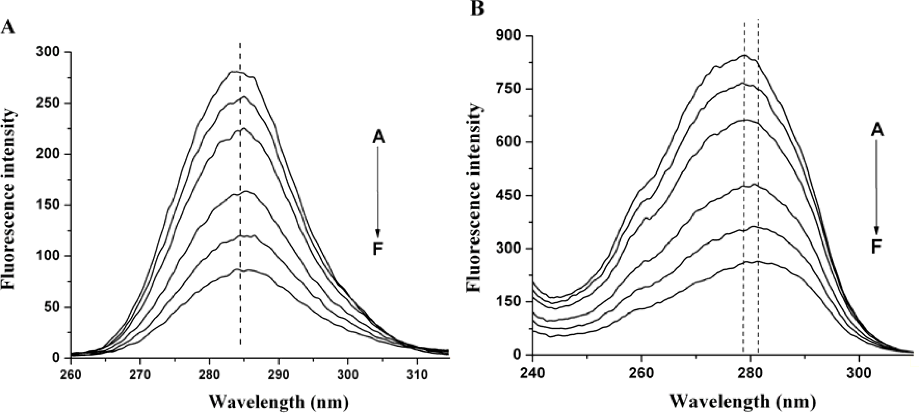
Synchronous fluorescence spectroscopy is a common method used to provide information about conformational changes of protein since the possible shift of maximum emission wavelength is related to the polarity of the environment. The synchronous fluorescence spectra of HSA-nC60 system are shown in Figure 3. The maximum emission wavelength of tyrosine residues has a small red shift (from 283 nm to 285 nm) when Δλ = 15 nm, indicating that there are some changes in the environment of the tyrosine residues. The maximum emission wavelength of tryptophan residues red shifts from 279.5 nm to 281.5 nm when Δλ = 60 nm (Figure 3B). This suggests that there is a less hydrophobic or more polar environment change around the tyrosine residues and tryptophan residues. This may be ascribed to the fact that the hydrophobic amino acid structure surrounding tryptophan residues in HSA tends to collapse slightly, resulting in tyrosine residues and tryptophan residues being more exposed to the aqueous phase.
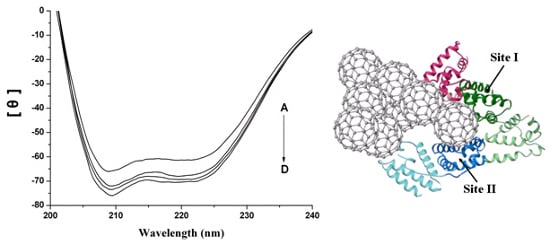
Circular dichroism spectra can sensitively monitor conformation changes in the protein upon interaction with the ligand. In this experiment, the calculated α-helicity content of native HSA solution is 54.9%, and with the addition of nC60 to the native HSA solution, the α-helicity content of HSA increased to 59.8%, 61.2% and 62.0% (Figure 4). Apparently, the higher the added nC60 concentration, the more the α-helicity content. The increase of α-helicity content indicated that the binding of HSA and nC60 induces protein folding, which results in some hydrophobic regions becoming more compact.

2.4. Interaction between HSA and nC60

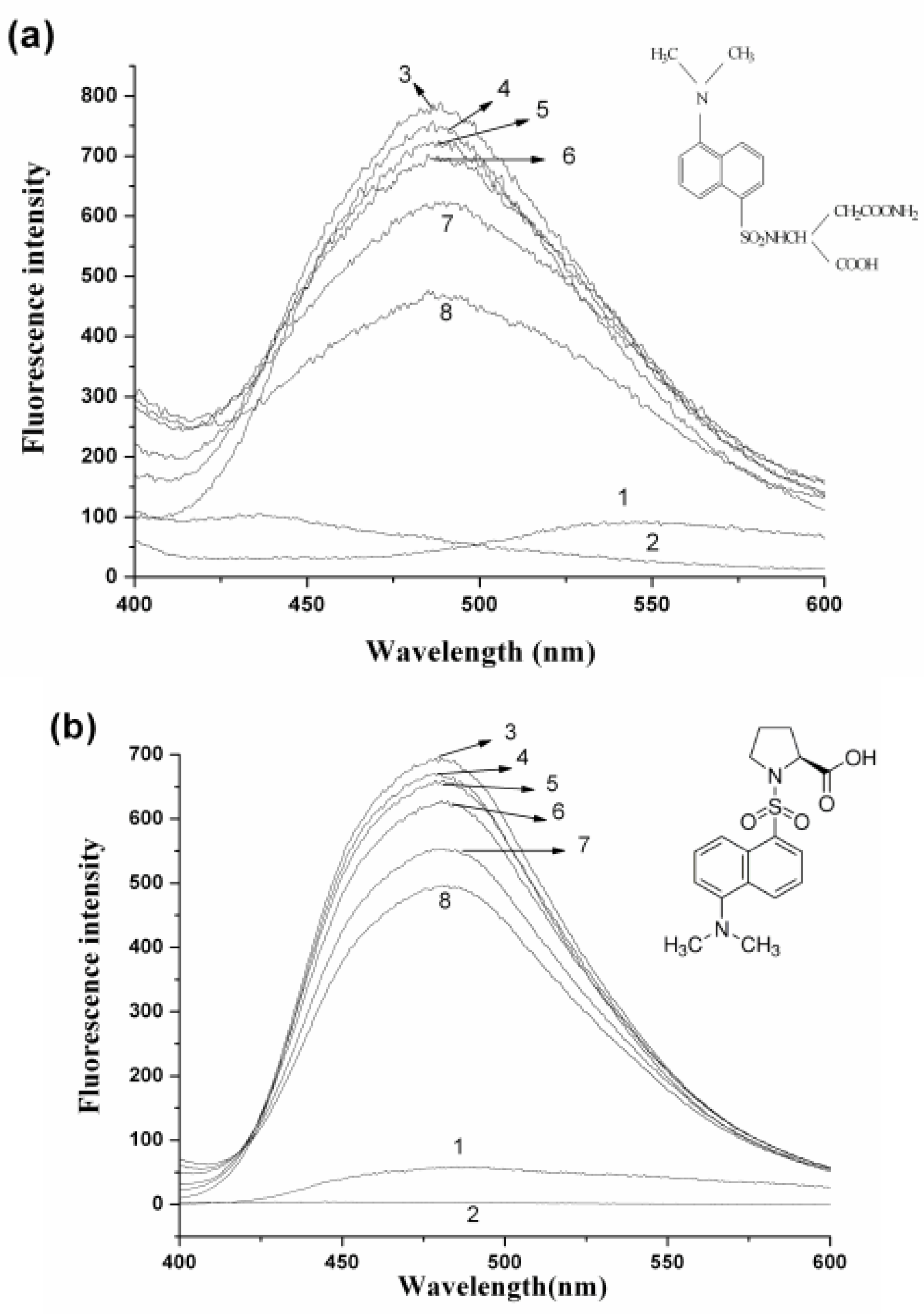
It is well known that HSA has two major drug binding sites, site I and site II, which are located in the hydrophobic pocket of sub-domain IIA and sub-domain IIIA, respectively. In order to identify the nC60-binding site on HSA, two probes were used. One probe of HSA is dansylamide (DNSA), whose binding site is located in the region of sub-domain IIA (sudlow site I); another probe, dansylproline (DP) is bound to HSA in the sub-domain IIIA (sudlow site II) [21,22]. During the experiment, nC60 was gradually added to the solution of HSA with site probes held in equimolar concentrations (20 μM). No absorption of these two probes to nanoparticles of C60 was observed in capillary electrophoresis analysis (data not shown). As shown in Figure 5, only the probes or HSA alone have no fluorescence signal within the wavelengths 400–600 nm. When the probes and HSA are mixed together, the DNSA-HSA complex has a maximum emission wavelength at 487 nm (excitation wavelength 350 nm) and DP-HSA has a maximum emission wavelength at 480 nm (excitation wavelength 375 nm). With the addition of nC60 to the HSA-probe solution, the fluorescence intensity of the HSA-probe complex was significantly lower than that without nC60, indicating that the binding of DNSA/DP to HSA was affected by adding nC60. The fluorescence intensity of DNSA-HSA was lower than that of DP-HSA, indicating that nC60 could more strongly influence the binding of DNSA to HSA than that of NP to HSA.

2.5. Representation of Interaction of HSA and nC60

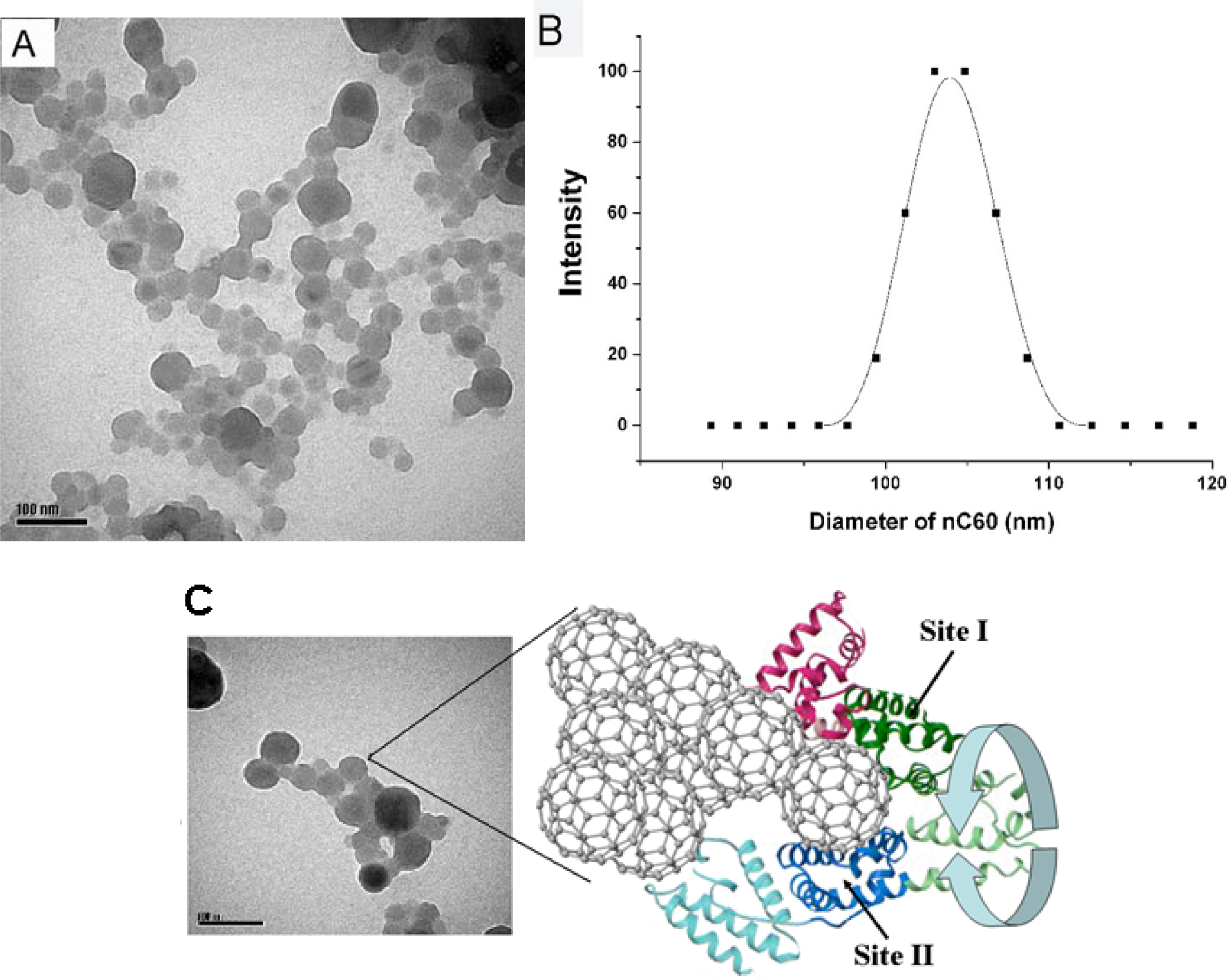
Our results above indicate that the fluorescence quenching and conformational changes of HSA can be attributed to the interaction between HSA and nC60. C60 fullerene has also been reported to be bound to the subdomain IIA of HSA using time-resolved fluorescence decay experiments, docking calculations, and binding site alignment methods [13,23]. Here we have employed several approaches in order to address the issue of fullerene interactions with proteins. What needs to be especially pointed out is that it is difficult to incorporate nC60 into the binding pocket because the size of nC60 is larger than protein molecules. The hydrodynamic diameter of the nC60 nanoparticles was determined to be from 50 nm to 110 nm by TEM and DLS, respectively (Figure 6A, B). Ganazzoli et al. reported atomistic computer simulations of some albumin subdomains on a hydrophobic graphite surface [24]. According to these simulations, we proposed a binding model to describe the interaction between HSA and nC60 (Figure 6C). In this model, HSA is adsorbed on the surface of nC60 aggregate which leads to the fluorescence quenching and conformational changes of HSA. The interaction sites were presumed to be near to Site I and Site II of HSA; following the structure and conformation of HSA was affected during adsorption. In our previous study, we have observed that HSA could compete for the surface of nC60 with functional enzymes [25].

3. Experimental

3.1. Materials

C60 (99.5% purity) was obtained from Sigma Chemical Company, St. Louis, USA. HSA (98% purity, essentially fatty acid and globulin free) was purchased from NEB (USA). All solutions were prepared in ultrapure water with the resistivity of 18.2 MΩ·cm (ALGA system, UK). Phosphate buffer (10 mM, pH = 7.4) was prepared with chemicals of analytical pure grade, obtained from commercial sources, and filtered with a 0.22-μmm filter (Millipore, Bedford, MA, USA). All other materials were of analytical pure grade.

3.2. Preparation and Characterization of Water-Soluble nC60

C60 suspensions were prepared according to the method described previously [26]. Ten milligrams of C60 was completely dissolved in 10 mL toluene. Then 2.5 mL C60 toluene solution was added into 50 mL water. After ultrasonic treatment for approximately 4 h, toluene was allowed to evaporate under vacuum for at least 1 h. The mean hydrodynamic diameter of the nC60 nanoparticles was determined by transmission electron microscopy (TEM) and dynamic light scattering (DLS) (BI-20OSM laser light scattering spectrometer, Brookhaven, USA).

3.3. Methods

Absorption spectra were recorded on a UV-2102PCS UV-Vis spectrophotometer (UNICO, USA) equipped with a 1.0 cm quartz cuvette at room temperature. The wavelength range was recorded from 200 to 500 nm.
All fluorescence emission spectra were recorded in the range of 300–450 nm on a LS-55 spectrofluorimeter (PE, USA) equipped with 100 μL quartz cells. The bandwidth of excitation and emission (3 nm) and scan speed (300 nm/min) were kept constant within each data set. Fluorescence intensity at 343 nm was used for calculation in Stern-Volmer equation. All measurements were performed at room temperature (∼25 °C).
Binding site marker competitive experiments were carried out by the fluorescence titration methods. The concentration of HSA and dansylamide/dansylproline (DNSA/DP) were all kept at 10 μM. Then nC60 was gradually titrated into the HSA-DNSA/DP systems. Fluorescence spectra were recorded with an excitation wavelength of 370 nm (for DNSA) and 375 nm (for DNP) in the range of 400–600 nm.
Circular dichroism (CD) spectroscopy was performed on a J-810 spectropolarimeter (Jasco Co., Japan) over a wavelength of 200–260 nm. The scanning speed was set at 500 nm/min. Each spectrum was the average of three successive scans and appropriate buffer solutions running under the same conditions were taken as blank and their contributions were subtracted from the experimental spectra.

4. Conclusions

We observed a quenching of fluorescence of HSA in the presence of nC60, and fluorescence quenching mechanism was investigated by the Stern-Volmer equation which showed a characteristic feature for the combined fluorescence quenching. The binding of HSA to nC60 increases the polar environment of the Trp residue, resulting in a red shift in the fluorescence spectra. The CD results showed that the percentage of α-helicity of HSA increased, revealing that the protein becomes more compact upon association with nC60. The results indicate that the fluorescence quenching and conformational changes of HSA can be attributed to the interaction between HSA and nC60. In the binding site marker competitive experiment, the binding of DNSA/DP to HSA was affected by adding nC60. We propose that HSA interacted with aggregate nC60 surface, and led to the functional changes of drug binding sites of HSA.

Acknowledgments

This research was supported by grants from the National Natural Science Foundation of China (Nos. 21077116, 20921063, 20890112, 20805057), and the National Basic Research Program of China (Nos. 09CB421605, 10CB933502) to M. Song and H. Wang.

References

  1. Kroto, HW; Heath, JR; O’Brien, SC; Curl, RF; Smalley, RE. C60-Buckminsterfullerene. Nature 1985, 318, 162–163. [Google Scholar]
  2. Wilson, LJ. Medical applications of fullerenes and metallofullerenes. Electrochem. Soc. Interface 1999, 8, 24–28. [Google Scholar]
  3. Da Ros, T; Prato, M. Medicinal chemistry with fullerenes and fullerene derivatives. Chem. Commun 1999, 8, 663–669. [Google Scholar]
  4. Bakry, R; Vallant, RM; Najam-ul-Haq, M; Rainer, M; Szabo, Z; Huck, CW; Bonn, GK. Medicinal applications of fullerenes. Int. J. Nanomed 2007, 2, 639–649. [Google Scholar]
  5. Ashcroft, JM; Tsyboulski, DA; Hartman, KB; Zakharian, TY; Marks, JW; Weisman, RB; Rosenblum, MG; Wilson, LJ. Fullerene (C60) immunoconjugates: Interaction of water-soluble C60 derivatives with the murine anti-gp240 melanoma antibody. Chem. Commun 2006, 28, 3004–3006. [Google Scholar]
  6. Zakharian, TY; Seryshev, A; Sitharaman, B; Gilbert, BE; Knight, V; Wilson, LJ. A fullerene-paclitaxel chemotherapeutic: Synthesis, characterization, and study of biological activity in tissue culture. J. Am. Chem. Soc 2005, 127, 12508–12509. [Google Scholar]
  7. Jia, G; Wang, H; Yan, L; Wang, X; Pei, R; Yan, T; Zhao, Y; Guo, X. Cytotoxicity of carbon nanomaterials: Single-wall nanotube, multi-wall nanotube and fullerene. Environ. Sci. Technol 2005, 39, 1378–1383. [Google Scholar]
  8. Heymann, D. Solubillity of fullerenes C60 and C70 in seven normal alcohols and their deduced solubility in water. Fullerene Sci. Technol 1996, 4, 509–515. [Google Scholar]
  9. Friedman, SH; DeCamp, DL; Sijbesma, R; Srdanov, G; Wudl, F; Kenyon, GL. Inhibition of the HIV-1 protease by fullerene derivatives: Model building studies and experimental verification. J. Am. Chem. Soc 1993, 115, 6506–6509. [Google Scholar]
  10. Marcorin, GL; Da Ros, T; Castellano, S; Stefancich, G; Bonin, I; Miertus, S; Prato, M. Design and synthesis of novel [60]fullerene derivatives as potential HIV aspartic protease inhibitors. Org. Lett 2000, 2, 3955–3958. [Google Scholar]
  11. Braden, BC; Goldbaum, FA; Chen, BX; Kirschner, AN; Wilson, SR; Erlanger, BF. X-ray crystal structure of an anti-Buckminsterfullerene antibody fab fragment: Biomolecular recognition of C(60). Proc. Natl. Acad. Sci. USA 2000, 97, 12193–12197. [Google Scholar]
  12. Zhang, XF; Shu, CY; Xie, L; Wang, CR; Zhang, YZ; Xiang, JF; Li, L; Tang, YL. protein conformation changes induced by a novel organophosphate-containing water-soluble derivative of a C60 fullerene nanoparticle. J. Phys. Chem. C 2007, 111, 14327–14333. [Google Scholar]
  13. Belgorodsky, B; Fadeev, L; Ittah, V; Benyamini, H; Zelner, S; Huppert, D; Kotlyar, AB; Gozin, M. Formation and characterization of stable human serum albumin-tris-malonic acid [C60]fullerene complex. Bioconjug. Chem 2005, 16, 1058–1062. [Google Scholar]
  14. Brant, J; Lecoanet, H; Wiesner, MR. Aggregation and deposition characteristics of fullerene nanoparticles in aqueous systems. J. Nanopart. Res 2005, 7, 545–553. [Google Scholar]
  15. Scharff, P; Risch, K; Carta-Abelmann, L; Dmytruk, IM; Bilyi, MM; Golub, OA; Khavryuchenko, AV; Buzaneva, EV; Aksenov, VL; Avdeev, MV; et al. Structure of C60 fullerene in water: Spectroscopic data. Carbon 2004, 42, 1203–1206. [Google Scholar]
  16. Chen, X; Kan, AT; Tomson, MB. Naphtalene adsorption and desorption from aqueous C60 fullerene. J. Chem. Eng. Data 2004, 49, 675–683. [Google Scholar]
  17. Deguchi, S; Alargova, RG; Tsuji, K. Stable dispersions of fullerenes C60 and C70 in water. Preparation and characterization. Langmuir 2001, 17, 6013–1017. [Google Scholar]
  18. Shang, L; Wang, Y; Jiang, J; Dong, S. pH-dependent protein conformational changes in albumin: gold nanoparticle bioconjugates: A spectroscopic study. Langmuir 2007, 23, 2714–2721. [Google Scholar]
  19. Deguchi, S; Yamazaki, T; Mukai, S; Usami, R; Horikoshi, K. Stabilization of C60 nanoparticles by protein adsorption and its implications for toxicity studies. Chem. Res. Toxicol 2007, 20, 854–858. [Google Scholar]
  20. Lakowicz, JR; Weber, G. Quenching of fluorescence by oxygen. Probe for structural fluctuations in macromolecules. Biochemistry 1973, 21, 4161–4170. [Google Scholar]
  21. Sudlow, G; Birkett, DJ; Wade, DN. The Characterization of two specific drug binding sites on human serum albumin. Mol. Pharmacol 1975, 11, 824–832. [Google Scholar]
  22. Chuang, VT; Kuniyasu, A; Nakayama, H; Matsushita, Y; Hirono, S; Otagiri, M. Helix 6 of subdomain III A of human serum albumin is the region primarily photolabeled by ketoprofen, an arylpropionic acid NSAID containing a benzophenone moiety. Biochim. Biophys. Acta 1999, 1434, 18–30. [Google Scholar]
  23. Benyamini, H; Shulman-Peleg, A; Wolfson, HJ; Belgorodsky, B; Fadeev, L; Gozin, M. Interaction of C60-Fullerene and Carboxyfullerene with proteins: Docking and binding site alignment. Bioconjug. Chem 2006, 17, 378–386. [Google Scholar]
  24. Raffaini, G; Ganazzoli, F. Simulation study of the interaction of some albumin subdomains with a flat graphite surface. Langmuir 2003, 19, 3403–3412. [Google Scholar]
  25. Song, M; Jiang, G; Yin, J; Wang, H. Inhibition of Taq polymerase activity by pristine fullerene nanoparticles can be mitigated by abundant proteins. Chem. Commun 2010, 46, 1404–1406. [Google Scholar]
  26. Andrievsky, GV; Kosevich, MV; Vovk, OM; Shelkovsky, VS; Vashchenko, LA. On the production of an aqueous colloidal solution of fullerenes. J Chem Soc Chem Commun 1995, 1281–1282. [Google Scholar]
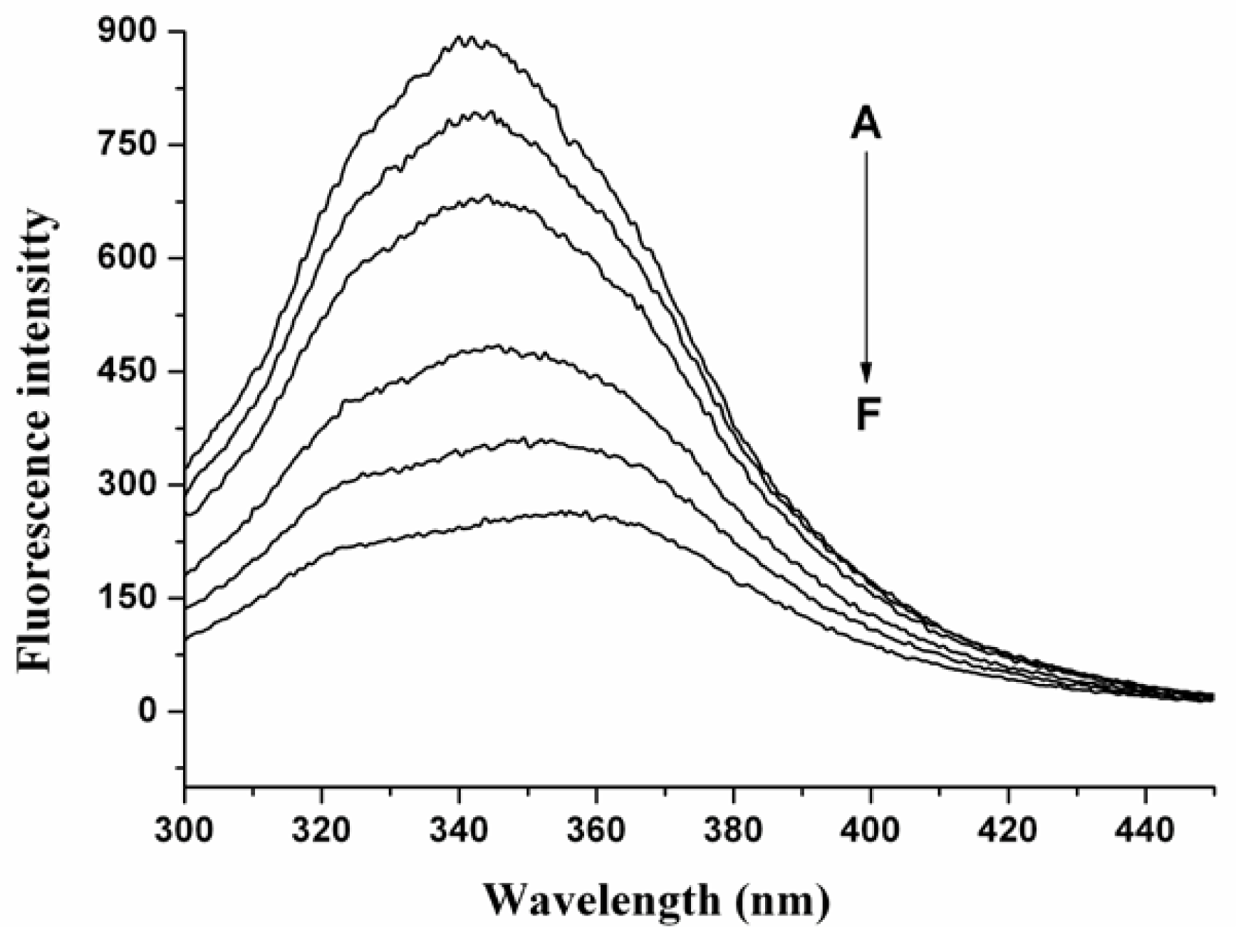
Figure 1. Fluorescence spectra of human serum album (HSA) in the presence of nC60 aggregates. HSA concentration was 20 μM. The concentration of C60 (from A to F) was 0, 1.41, 2.83, 5.66, 8.48, and 11.30 μM, respectively (pH = 7.4; ex = 280 nm).
Ijms 12 04964f1
Figure 2. (A) The Stern-Volmer plot for the fluorescence quenching of HSA by nC60; (B) Nonlinear fitting analysis performed by plotting (F0/F-1) against [Q].
Ijms 12 04964f2
Figure 3. Synchronous fluorescence spectra of HSA in the presence of nC60. (A) Δλ = 15 nm; (B) Δλ = 60 nm. HSA concentration is 20 μM. The concentration of C60 (from A to F) was 0, 1.41, 2.83, 5.66, 8.48, 11.30 μM, respectively.
Ijms 12 04964f3
Figure 4. The circular dichroism (CD) spectra of the HSA-nC60 system. Concentration of HSA was 7.3 μM; Concentration of nC60 (from A to D) was 0, 3.38, 5.63, 11.8 μM, respectively.
Ijms 12 04964f4
Figure 5. Effect of nC60 on binding site probe-HSA system. Concentrations of HSA and probes (DNSA and DP) were 20 μM. (a) 1: HSA only; 2: DNSA only; Ex = 350 nm; (b) 1: HSA only, 2: DP only; ex = 375 nm. Concentrations of C60 (from 3 to 8) were 0, 1.41, 2.83, 5.66, 8.48, 11.30 μM, respectively. The inserts correspond to the molecular structure of binding site probes.
Ijms 12 04964f5
Figure 6. (A) Transmission electron microscopy (TEM) image of nC60; (B) The mean hydrodynamic diameter of the nC60 measured by dynamic light scattering (DLS); (C) Schematic representation of HSA interacting with aggregate nC60 surface.
Ijms 12 04964f6