Communication

Mechanism of Sphingosine 1-Phosphate- and Lysophosphatidic Acid-Induced Up-Regulation of Adhesion Molecules and Eosinophil Chemoattractant in Nerve Cells

1
Department of Medicine, Royal College of Surgeons in Ireland, Beaumont Hospital, Dublin 9, Ireland
2
Department of Dermatology, University of Utah, Salt Lake City, UT 84132, USA
*
Author to whom correspondence should be addressed.
Int. J. Mol. Sci. 2011, 12(5), 3237-3249; https://doi.org/10.3390/ijms12053237
Submission received: 8 March 2011 / Revised: 5 May 2011 / Accepted: 9 May 2011 / Published: 17 May 2011
(This article belongs to the Section Biochemistry)

Abstract

:
The lysophospholipids sphingosine 1-phosphate (S1P) and lysophosphatidic acid (LPA) act via G-protein coupled receptors S1P1–5 and LPA1–3 respectively, and are implicated in allergy. Eosinophils accumulate at innervating cholinergic nerves in asthma and adhere to nerve cells via intercellular adhesion molecule-1 (ICAM-1). IMR-32 neuroblastoma cells were used as an in vitro cholinergic nerve cell model. The Gi coupled receptors S1P1, S1P3, LPA1, LPA2 and LPA3 were expressed on IMR-32 cells. Both S1P and LPA induced ERK phosphorylation and ERK- and Gi-dependent up-regulation of ICAM-1 expression, with differing time courses. LPA also induced ERK- and Gi-dependent up-regulation of the eosinophil chemoattractant, CCL-26. The eosinophil granule protein eosinophil peroxidase (EPO) induced ERK-dependent up-regulation of transcription of S1P1, LPA1, LPA2 and LPA3, providing the situation whereby eosinophil granule proteins may enhance S1P- and/or LPA- induced eosinophil accumulation at nerve cells in allergic conditions.

1. Introduction

In animal models of asthma, and in many asthma patients, antigen inhalation results in inhibition of muscarinic M2 receptors on parasympathetic nerves, leading to acetylcholine release and hence mucous secretion and contraction of bronchial smooth muscle [1].
Eosinophilia is a hallmark of allergic diseases such as asthma and allergic rhinitis [2]. Eosinophils accumulate at innervating cholinergic nerves in human and animal models of allergic conditions, contributing to nerve hyperresponsiveness due to eosinophil major basic protein (MBP)-mediated M2 antagonism [3,4]. Eosinophil accumulation at airway nerves is mediated by eosinophil CCR3 receptors [5]. We have employed the neuroblastoma cell line IMR-32 as a useful in vitro model of cholinergic nerve cell function. Differentiated IMR-32 cells express neuronal muscarinic M2 receptors and release acetylcholine in response to eosinophil adhesion via nerve intercellular adhesion molecule-1 (ICAM-1) and vascular cell adhesion molecule-1 (VCAM-1) [68]. Adhesion results in both the release of eosinophil granule proteins and the generation of signaling events and phenotypic changes within the nerve cells [6,913]. Released eosinophil granule proteins [6] induce further nerve cell signaling and protection from apoptosis [12]. Thus, eosinophils influence nerve function due to direct contact via ICAM-1 and VCAM-1 and consequent release of granule proteins.
S1P and LPA are bioactive phospholipids, which exert many of their effects via G-protein coupled S1P receptors S1P1, S1P2, S1P3, S1P4 and S1P5 and LPA receptors LPA1, LPA2, LPA3 and the structurally distant LPA4 respectively [1416] as well as LPA5 which has a distinct expression pattern suggesting specific physiological functions [17]. S1P and LPA have both been implicated in various aspects of neuronal function and activity and neuronal cell intracellular signaling, including cytoskeletal reorganization, acetylcoholine release, apoptosis and neurite outgrowth or retraction [1821].
A role for both S1P and LPA and their receptors has also been postulated in airway inflammatory responses, for example in allergy (reviewed [22,23]. Both S1P and LPA levels in bronchoalveolar lavage fluid are significantly increased 18–24 h following segmental allergen challenge in allergy patients and S1P promotes airway remodeling [24,25]. S1P is released primarily from activated platelets and activated mast cells [26]. S1P promotes airway remodeling [24] and we and others have shown that eosinophils express functional S1P receptors [27,28]. In vitro and animal studies have indicated that LPA induces eosinophil and neutrophil migration [2931]. LPA also induces mast cell chemokine and histamine release [32,33]. Both S1P and LPA up-regulate intercellular adhesion molecule-1 (ICAM-1) in HUVECs [34]. Importantly in the context of allergy, S1P up-regulates ICAM-1 in human alveolar epithelial cells [3537].
We hypothesized that cholinergic nerve cells would express receptors for S1P and LPA and that ligand binding to these receptors would activate intracellular signaling pathways in the nerve cells leading to transcriptional up-regulation of ICAM-1 and of members of the eosinophil eotaxin chemoattractant family. This would further implicate S1P and/or LPA in allergic symptoms by enhancing eosinophil accumulation at and adhesion to nerve cells and hence to nerve cell hyperreactivity.

2. Results and Discussion

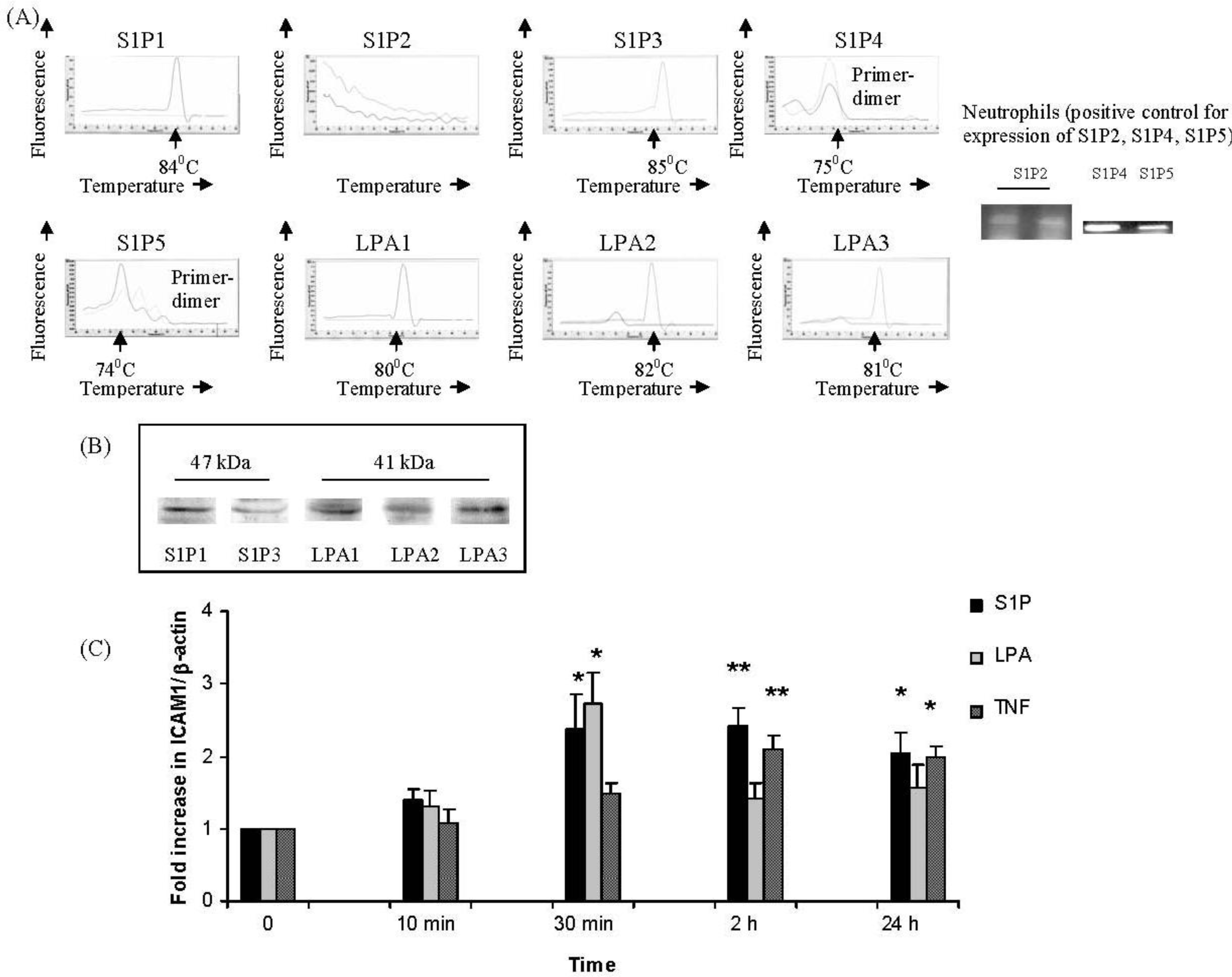
2.1. IMR32 Cells Express S1P1, S1P3 and LPA1–3; S1P and LPA Induce Up-Regulation of ICAM-1 Transcription

Given that there is both an increased level and defined source of both LPA and SIP after allergen challenge we investigated the role of these lysophospholipids in an in vitro model of eosinophil/nerve cell interactions. We and others have used butyrate-differentiated IMR-32 cells as a model of cholinergic nerve function in a variety of studies [611,13,38,39]. They are considered to be a convenient in vitro model for studying aspects of neurobiology at the molecular and cell level.
Expression of S1P receptors S1P1 and S1P3 (Figure 1A) and of LPA1–3 (Figure 1B) was confirmed in IMR32 cells maintained in proliferation or differentiation medium by real-time PCR. No expression of S1P2, S1P4 or S1P5 was detected after 40 cycles of PCR in any cells (Figure 1A); as a positive control, expression of all three of these receptors was readily detected on neutrophils (Figure 1A). All further experiments were in differentiating IMR32 cells, as these cells display a strongly cholinergic phenotype in differentiation medium [8,24,38]. We confirmed by Western blotting that, identically to the cDNA expression profile, differentiating IMR32 cells expressed S1P1, S1P3 (Figure 1B) and LPA1–3 proteins (Figure 1B), but not S1P2, S1P4 or S1P5. We determined by real time PCR that both S1P (Figure 1C) and LPA (Figure 1C) increased expression of ICAM-1 two- to three-fold over baseline levels, a similar order of magnitude as the positive control stimulus, TNF- (Figure 1C). However, up-regulation of ICAM-1 expression was induced more transiently by LPA (Figure 1C), compared to S1P which induced a sustained up-regulation between 30 min to 24 h (Figure 1C), similar to TNF- (Figure 1C).

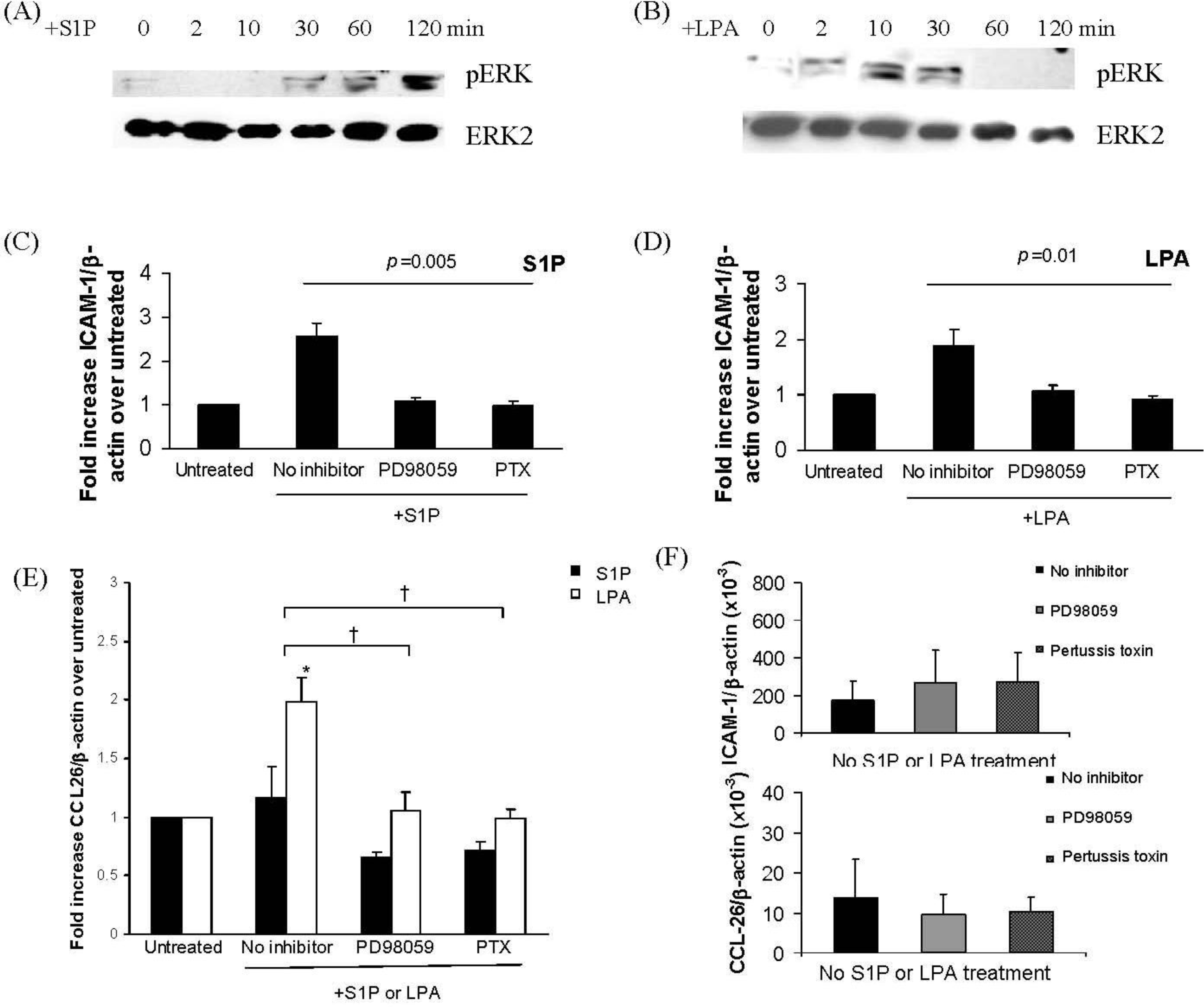
2.2. S1P and LPA Induce ERK Activation in IMR32 Cells with Different Time Courses with Consequenct Gi-Protein Coupled and ERK-Dependent Up-Regulation of ICAM-1 and/or CCL-26

To determine the intracellular signaling mechanisms that could mediate S1P- and/or LPA-induced up-regulation of ICAM-1 on IMR32 cells, activation of the ERK MAP kinase was examined. Both S1P (Figure 2A) and LPA (Figure 2B) induced ERK phosphorylation but with different time courses. S1P induced activation of ERK was observable at 30 min of stimulation and remained robust at 1–2 h (Figure 2A) while LPA induced a more rapid ERK activation which had fallen to baseline levels between 30 and 60 min of stimulation (Figure 2B). To further determine intracellular mediators involved in S1P- and LPA-induced ICAM-1 up-regulation, IMR32 cells were incubated with S1P (1 μM, 2 h) or LPA (1 μM, 30 min). These treatment times were chosen based on the time course for ICAM-1 induction (Figure 1C) which suggested that these were the optimal times for S1P and LPA respectively. Cells were pre-treated with either the MEK inhibitor PD98059, which prevents ERK phosphorylation or with pertussis toxin (PTX), to inhibit the Gi protein, to which all the receptors S1P1, S1P3 and LPA1–3 can couple. S1P-induced (Figure 2C) or LPA-induced (Figure 2D) up-regulation of ICAM-1 was measured by real-time PCR and compared between cells pre-treated with PD98059 or PTX and non-pre-treated cells. This revealed that up-regulation of ICAM-1 by both S1P and by LPA was dependent on both ERK phosphorylation and on Gi coupling. This is consistent with the recently demonstrated mechanism of S1P-induced up-regulation of ICAM-1 in airway epithelial cells [34].
Real-time PCR was also used to monitor expression of the eosinophil chemoattractant CCL26 (eotaxin-3), normalized to β-actin expression (Figure 2E). Results indicated that at the time points shown, LPA but not S1P up-regulated CCL26 expression (Figure 2E); we have also observed no up-regulation of CCL26 by S1P at other time points (data not shown). LPA-induced CCL26 up-regulation was dependent on ERK phosphorylation and on Gi coupling (Figure 2E). This is the first time that LPA but not S1P has been shown to up-regulate expression of an eosinophil chemoattractant, CCL26. These results imply that further intracellular mediators are involved in CCL26 expression, which are activated by LPA but not S1P, for example the transcription factor STAT-6 [40].
Our previous studies have shown that eosinophils accumulate at cholinergic nerves in human and animal models of allergic conditions such as asthma and rhinitis [41]. In vitro, eosinophils adhere to primary nerve cells and to differentiated IMR-32 cells via the adhesion molecules ICAM-1 and VCAM-1, with implications for nerve cell cholinergic phenotype and survival [813,38]. In a guinea pig model of asthma, accumulation of eosinophils at nerves was significantly decreased by treatment with CCR3 antagonist prior to antigen inhalation [5]. CCR3 antagonism also consequently reduced antigen-associated loss of M2 receptor function [5]. LPA induced up-regulation of expression of CCL26 message in differentiated IMR-32 cells; CCL26 (eotaxin-3) is a CCR3 agonist, which is up-regulated in asthmatics following allergen challenge [42] CCL26 rather than CCL11 (eotaxin-1) or CCL24 (eotaxin-2) is therefore suggested to be responsible for continuing eosinophil recruitment at later stages following allergen challenge. Furthermore, both S1P and LPA induced up-regulation of ICAM-1. Thus S1P and LPA induce conditions whereby eosinophil migration and adhesion to nerve cells would be favoured. In the context of allergy, this suggests a previously unrecognized role for these lysophospholipids in promotion of eosinophil interaction with nerves and hence nerve cell hyperreactivity, with its associated bronchoconstriction.

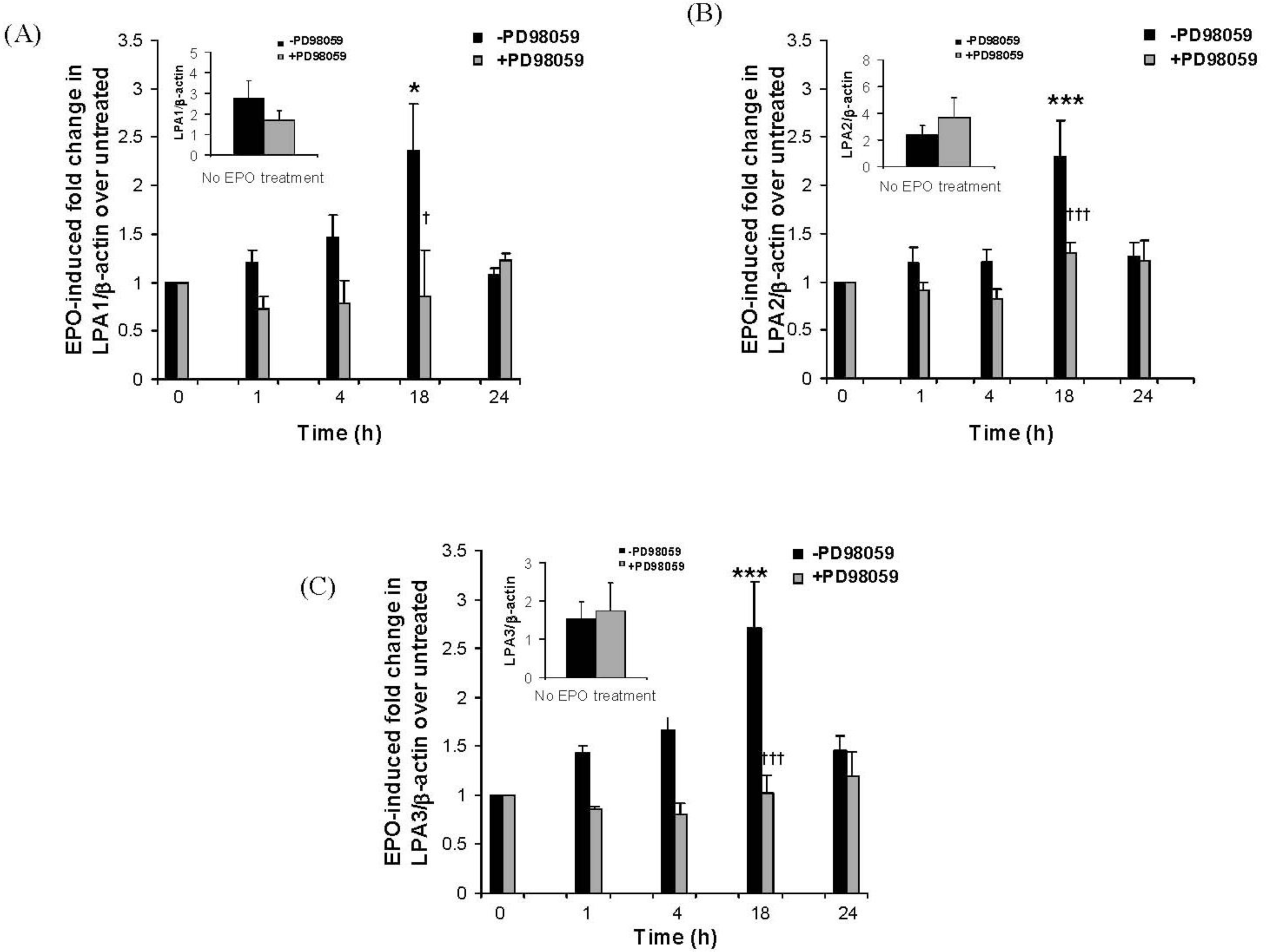
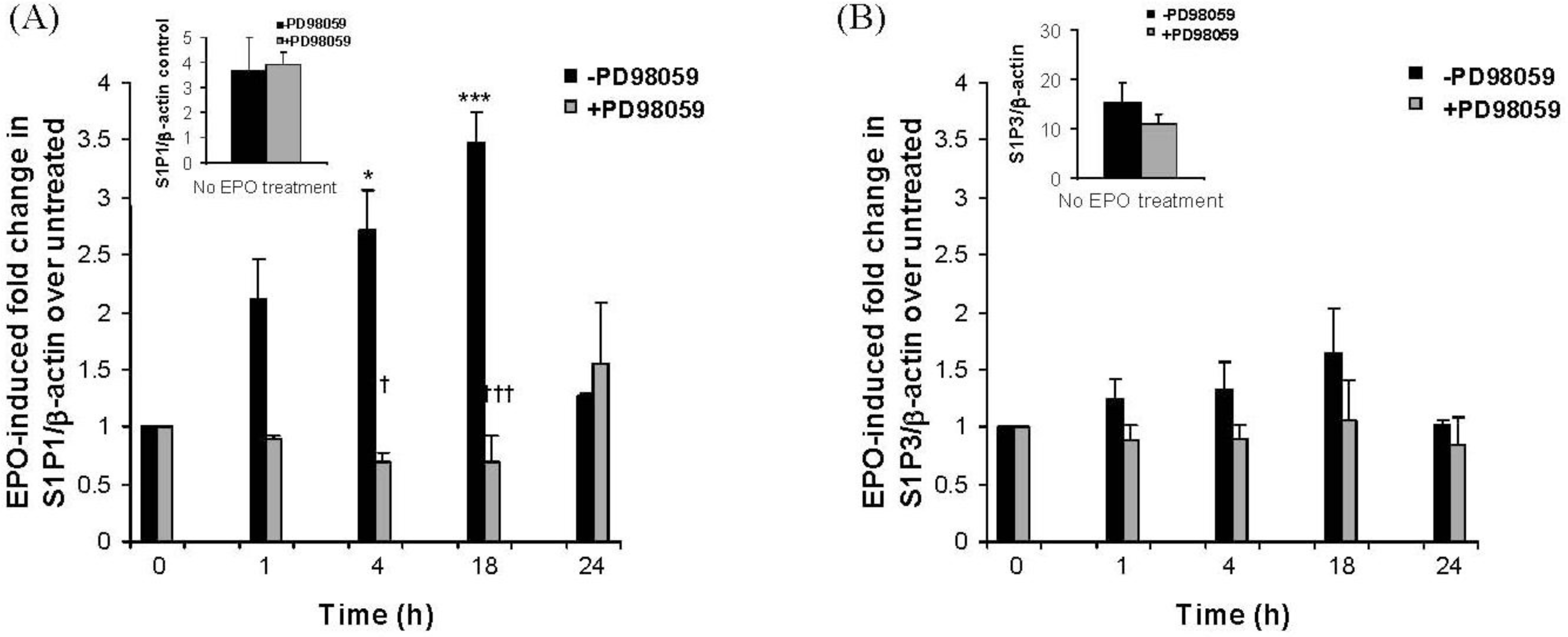
2.3. The Eosinophil Granule Protein EPO Induces Up-Regulation of S1P1, LPA1, LPA2 and LPA3 Receptors Expression

We have previously shown that eosinophil adhesion to IMR32 cells induces eosinophil degranulation and release of granule proteins, including eosinophil peroxidase (EPO) [810]. To determine whether released eosinophil granule protein could increase expression of IMR32 S1P or LPA receptors, nerve cells were treated with eosinophil peroxidase (EPO) (1 μg/mL) for 1, 4, 18 or 24 h in the presence or absence of PD98059. Real-time PCR was used to monitor the expression of S1P1 and S1P3 receptors (Figure 3) or LPA1, LPA2 and LPA3 receptors (Figure 4). EPO significantly enhanced expression of S1P1 by approximately 3–4 fold between 4–18 h of stimulation (Figure 3A). This up-regulation was dependent on ERK activation as it was abolished in the presence of PD98059. S1P3 expression was unaffected by EPO (Figure 3B). LPA1 (Figure 4A), LPA2 (Figure 4B) and LPA3 (Figure 4C) expression were all raised approximately 3-fold at 18 h of stimulation, then fell back to control levels by 24 h.
These results imply that S1P and LPA, acting via their nerve cell receptors, can promote eosinophil recruitment and adhesion and that subsequently, eosinophil adhesion and the resulting degranulation can release a factor, namely EPO, which up-regulates the LPA and S1P receptors responsible for this effect and hence perpetuate or prolong it.
In light of the results of our study, it would be of interest to determine whether antagonism of individual S1P and/or LPA receptors reduces accumulation at and/or adhesion of eosinophils to nerves in animal models of asthma, similarly to CCR3 antagonism. Up-regulation of ICAM-1 by both S1P and LPA was dependent on Gi-coupling, as was LPA-induced up-regulation of CCL26. All the receptors IMR-32-expressed receptors, S1P1, S1P3, LPA1, LPA2 and LPA3, couple to Gi. Definitive receptor identification awaits further studies utilizing receptor-specific ligands or multiple siRNA technology, which was considered impractical for this work given the abundance of the specific receptors.

3. Experimental Section

IMR32 nerve cell culture: The human cholinergic neuroblastoma cell line IMR32 was depleted of fibroblasts, as described previously [10]. The cells were maintained in culture in proliferation medium (DMEM Plus Glutamax, 5% FCS, 100 U/mL penicillin/streptomycin; GIBCO® Invitrogen, Paisley, UK) at 37 °C in an atmosphere of 5% CO2. On achieving confluence, cells were plated at a density of 5 × 10⁵/well in 6-well cell culture dishes and grown in proliferation medium for 48 h. Proliferation media was then replaced by differentiation medium (DMEM Plus Glutamax, 2% FCS, 2 mM sodium butyrate (Sigma, Poole, UK), 100 U/mL penicillin/streptomycin) and cells were used for experimentation after a further 6 days. In some experiments, individual wells were maintained in proliferation medium in parallel with the differentiating cells.
mRNA Analysis: Total RNA was isolated from the cells with TRI reagent™ (Sigma), according to the manufacturer’s instructions. For both quantitative LightCycler PCRs (Roche, Mannheim, Germany) and semiquantitative RT-PCRs, 1 μg of RNA was reverse transcribed with AMV reverse transcriptase and oligo-dT primer using a 1st strand cDNA synthesis kit (Roche), according to the manufacturer’s instructions.
Semi-quantitative RT-PCR analysis of cDNA preparations was carried out in 50 μL reactions with Taq-DNA polymerase for S1P1–5 or LPA1–3, using the primers sets (MWG Biotech/Eurofins AG, Ebersberg, Germany) outlined in Table 1. PCR conditions were: 94 °C, 4 min (1 cycle); 94 °C, 1.5 min, 54 °C, 1.5 min, 72 °C, 2 min (25–40 cycles); 72 °C, 10 min (1 cycle). PCR products were separated by 1.5% agarose gel electrophoresis and photographed under UV illumination. 40 cycles of PCR were carried out to verify absence of S1P2, S1P4 and S1P5.
Quantitative RT-PCR analysis was carried out on the LightCycler (Roche) using fast start Taq DNA polymerase containing the double-stranded DNA binding dye SYBR Green 1 as in [38]. Primers are shown in Table 1. The samples were denatured at 95 °C for 15 min followed by 35–40 cycles of denaturation, annealing and extension at 95 °C for 15 s, 55 °C for 25 s, and 72 °C for 11 s (ICAM-1, CCL26, S1P1, S1P2, S1P3, S1P4, S1P5, LPA1, LPA2, LPA3), or 95 °C for 15 s, 55 °C for 20 s, and 72 °C for 20 s (-actin). Characteristic melting curves were obtained at the end of amplification by cooling the samples to 65 °C for 15 s followed by further cooling to 40 °C for 30 s. Serial 10-fold dilutions were prepared from individual PCR products, which were then used as standards to plot against the unknown samples. Quantification of data was analyzed using the LightCycler analysis software, and values were normalized to the level of β-actin expression for each sample on the same template cDNA. Results for induction over baseline values of ICAM-1 or CCL26 expression by S1P (1 μM) or LPA (1 μM) were obtained; in some experiments fold induction was compared between untreated, differentiated IMR32 cells versus cells pre-treated overnight with the MEK/ERK inhibitor PD98059 (Sigma) (50 μM) or with pertussis toxin (Sigma) (0.1 μg/mL). Results for induction over baseline values of S1P1, S1P3, LPA1, LPA2 or LPA3 by EPO (1 μg/mL, various time points) were also compared between untreated, differentiated IMR32 cells versus cells pre-treated overnight with the MEK/ERK inhibitor PD98059 (50 μM).
Nuclear and cytoplasmic protein preparation: IMR-32 cells (5 × 10⁵) were differentiated for 6 days with sodium butyrate as described above and then incubated with LPA or S1P for various time periods from 2 min to 2 h. Nuclear and cytoplasmic extracts were isolated from IMR-32 cells, essentially as in [10]. Protein concentration was established by the Bradford method [43] and nuclear and cytoplasmic extracts stored at −80 °C.
Western blotting: Total cell protein was extracted from IMR32 cells using Tri Reagent, according to the manufacturer’s instructions and quantified according to the Bradford method [43]. Total cell protein (S1P1–5, LPA1–3 probing) or cytoplasmic protein (ERK probing) (10 μg) was subjected to Western blotting as previously described [38] Membranes were incubated in blocking buffer (Dulbecco’s PBS (Invitrogen) containing 0.2% (w/v) I-block and 0.1% (v/v) Tween-20) for 1 h at room temperature then incubated for 2 h in blocking buffer containing the individual respective HRP-conjugated primary antibody (Santa Cruz, CA, USA) (1:500 for each). Following six 5-min washes in washing buffer (PBS pH 7.4, 0.1% (v/v) Tween-20) membranes were incubated for 1 h in blocking buffer containing a dilution of the appropriate secondary antibody (1:10,000 dilution). Membranes were then washed 6 times for 5 min each and exposed to SuperSignal West Pico Chemiluminescent Substrate (Pierce Technology Inc., Rockford, IL, USA) for Western Blotting for 5 min at room temperature. Blots were then exposed to X-OMAT light sensitive film to obtain an image.
Statistical analysis: Values are expressed as mean SEM. The statistical significance of differences between proliferating and differentiating cells was evaluated by two-tail Student’s t-test or by ANOVA, as appropriate. The Graphpad Instat program was used. A p-value of 0.05 or less was taken as significant.

4. Conclusion

Eosinophils accumulate at cholinergic nerves at sites of inflammation in asthma and allergic rhinitis. We have previously shown that eosinophils adhere to nerve cells via ICAM-1 [6,7]. In this study we have shown that the lysophospholipid LPA induces transcriptional up-regulation of expression of the eosinophil chemoattractant CCL26, which is up-regulated in asthmatics following allergen challenge [42] and is suggested to be responsible for continuing eosinophil recruitment at later stages following allergen challenge. Furthermore, both LPA and another lysophospholipid, S1P, induce up-regulation of ICAM-1. We and others have previously shown that S1P can also induce a direct chemoattractive effect on eosinophils [27,28]. Thus both S1P and LPA induce conditions whereby eosinophil migration and adhesion to nerve cells would be favored. In the context of allergy, this suggests a previously unrecognized role for these lysophospholipids in promotion of eosinophil interaction with nerves and hence nerve cell hyperreactivity, with its associated bronchoconstriction. Furthermore, the eosinophil granule protein EPO, induced up-regulation of the S1P receptor S1P1 and the LPA receptors LPA1–3. We have previously shown EPO to be released from eosinophils upon their adhesion to nerve cells via ICAM-1. Hence, the potential exists for a perpetuation of the S1P and LPA driven effects on eosinophil and nerve interaction as the EPO released from eosinophils attached to nerve cells could cause up-regulation of the S1P and LPA receptors, hence resulting in more S1P and LPA binding, more up-regulation of ICAM-1 and CCL-26 and more eosinophil accumulation.

References

  1. Fryer, AD; Jacoby, DB. Plasticity of cholinergic and tachykinergic nerves: The convergence of the twain. Am. J. Physiol. Lung Cell. Mol. Physiol 2002, 283, L907–L908. [Google Scholar]
  2. Rothenberg, ME; Hogan, SP. The eosinophil. Annu. Rev. Immunol 2006, 24, 147–174. [Google Scholar]
  3. Evans, CM; Fryer, AD; Jacoby, DB; Gleich, GJ; Costello, RW. Pretreatment with antibody to eosinophil major basic protein prevents hyperresponsiveness by protecting neuronal M2 muscarinic receptors in antigen-challenged guinea pigs. J. Clin. Invest 1997, 100, 2254–2262. [Google Scholar]
  4. Sheahan, P; Walsh, RM; Walsh, MA; Costello, RW. Induction of nasal hyper-responsiveness by allergen challenge in allergic rhinitis: the role of afferent and efferent nerves. Clin. Exp. Allergy 2005, 35, 45–51. [Google Scholar]
  5. Fryer, AD; Stein, LH; Nie, Z; Curtis, DE; Evans, CM; Hodgson, ST; Jose, PJ; Belmonte, KE; Fitch, E; Jacoby, DB. Neuronal eotaxin and the effects of CCR3 antagonist on airway hyperreactivity and M2 receptor dysfunction. J. Clin. Invest 2006, 116, 228–236. [Google Scholar]
  6. Kingham, PJ; McLean, WG; Sawatzky, DA; Walsh, MT; Costello, RW. Adhesion-dependent interactions between eosinophils and cholinergic nerves. Am. J. Physiol. Lung Cell. Mol. Physiol 2002, 282, L1229–L1238. [Google Scholar]
  7. Sawatzky, DA; Kingham, PJ; Court, E; Kumaravel, B; Fryer, AD; Jacoby, DB; McLean, WG; Costello, RW. Eosinophil adhesion to cholinergic nerves via ICAM-1 and VCAM-1 and associated eosinophil degranulation. Am. J. Physiol. Lung Cell. Mol. Physiol 2002, 282, L1279–L1288. [Google Scholar]
  8. Sawatzky, DA; Kingham, PJ; Durcan, N; McLean, WG; Costello, RW. Eosinophil-induced release of acetylcholine from differentiated cholinergic nerve cells. Am. J. Physiol. Lung Cell. Mol. Physiol 2003, 285, L1296–L1304. [Google Scholar]
  9. Kingham, PJ; McLean, WG; Walsh, MT; Fryer, AD; Gleich, GJ; Costello, RW. Effects of eosinophils on nerve cell morphology and development: The role of reactive oxygen species and p38 MAP kinase. Am. J. Physiol. Lung Cell. Mol. Physiol 2003, 285, L915–L924. [Google Scholar]
  10. Walsh, MT; Curran, DR; Kingham, PJ; Morgan, RK; Durcan, N; Gleich, GJ; McLean, WG; Costello, RW. Effect of eosinophil adhesion on intracellular signaling in cholinergic nerve cells. Am. J. Respir. Cell Mol. Biol 2004, 30, 333–341. [Google Scholar]
  11. Morgan, RK; Kingham, PJ; Walsh, MT; Curran, DC; Durcan, N; McLean, WG; Costello, RW. Eosinophil adhesion to cholinergic IMR-32 cells protects against induced neuronal apoptosis. J. Immunol 2004, 173, 5963–5970. [Google Scholar]
  12. Morgan, RK; Costello, RW; Durcan, N; Kingham, PJ; Gleich, GJ; McLean, WG; Walsh, MT. Diverse effects of eosinophil cationic granule proteins on IMR-32 nerve cell signaling and survival. Am. J. Respir. Cell Mol. Biol 2005, 33, 169–177. [Google Scholar]
  13. Curran, DR; Morgan, RK; Kingham, PJ; Durcan, N; McLean, WG; Walsh, MT; Costello, RW. Mechanism of eosinophil induced signaling in cholinergic IMR-32 cells. Am. J. Physiol. Lung Cell. Mol. Physiol 2005, 288, L326–L332. [Google Scholar]
  14. Meyer zu Heringdorf, D; Jakobs, KH. Lysophospholipid receptors: Signalling, pharmacology and regulation by lysophospholipid metabolism. Biochim. Biophys. Acta 2007, 1768, 923–940. [Google Scholar]
  15. Noguchi, K; Ishii, S; Shimizu, T. Identification of p2y9/GPR23 as a novel G protein-coupled receptor for lysophosphatidic acid, structurally distant from the Edg family. J. Biol. Chem 2003, 278, 25600–25606. [Google Scholar]
  16. Rosen, H; Goetzl, EJ. Sphingosine 1-phosphate and its receptors: An autocrine and paracrine network. Nat. Rev. Immunol 2005, 5, 560–570. [Google Scholar]
  17. Lee, CW; Rivera, R; Gardell, S; Dubin, AE; Chun, J. GPR92 as a new G12/13- and Gq-coupled lysophosphatidic acid receptor that increases cAMP, LPA5. J. Biol. Chem 2006, 281, 23589–23597. [Google Scholar]
  18. Buccoliero, R; Futerman, AH. The roles of ceramide and complex sphingolipids in neuronal cell function. Pharmacol. Res 2003, 47, 409–419. [Google Scholar]
  19. Colombaioni, L; Garcia-Gil, M. Sphingolipid metabolites in neural signalling and function. Brain Res. Rev 2004, 46, 328–355. [Google Scholar]
  20. Fukushima, N. LPA in neural cell development. J. Cell. Biochem 2004, 92, 993–1003. [Google Scholar]
  21. Fukushima, N; Shano, S; Moriyama, R; Chun, J. Lysophosphatidic acid stimulates neuronal differentiation of cortical neuroblasts through the LPA(1)-G(i/o) pathway. Neurochem. Int 2007, 50, 302–307. [Google Scholar]
  22. Oskeritzian, CA; Milstien, S; Spiegel, S. Sphingosine-1-phosphate in allergic responses, asthma and anaphylaxis. Pharmacol. Ther 2007, 115, 390–399. [Google Scholar]
  23. Zhao, Y; Natarajan, V. Lysophosphatidic acid signaling in airway epithelium: Role in airway inflammation and remodeling. Cell. Signal 2009, 21, 367–377. [Google Scholar]
  24. Ammit, AJ; Hastie, AT; Edsall, LC; Hoffman, RK; Amrani, Y; Krymskaya, VP; Kane, SA; Peters, SP; Penn, RB; Spiegel, S; et al. Sphingosine 1-phosphate modulates human airway smooth muscle cell functions that promote inflammation and airway remodeling in asthma. FASEB J 2001, 15, 1212–1214. [Google Scholar]
  25. Georas, SN; Berdyshev, E; Hubbard, W; Gorshkova, IA; Usatyuk, PV; Saatian, B; Myers, AC; Williams, MA; Xiao, HQ; Liu, M; et al. Lysophosphatidic acid is detectable in human bronchoalveolar lavage fluids at baseline and increased after segmental allergen challenge. Clin. Exp. Allergy 2007, 37, 311–322. [Google Scholar]
  26. Goetzl, EJ; Wang, W; McGiffert, C; Huang, MC; Graler, MH. Sphingosine 1-phosphate and its G protein-coupled receptors constitute a multifunctional immunoregulatory system. J. Cell. Biochem 2004, 92, 1104–1114. [Google Scholar]
  27. Mackle, T; Gendy, SS; Walsh, M; McConn-Walsh, R; Costello, RW; Walsh, MT. Role of sphingosine 1-phosphate receptor expression in eosinophils of patients with allergic rhinitis, and effect of topical nasal steroid treatment on this receptor expression. J. Laryngol. Otol 2008, 122, 1309–1317. [Google Scholar]
  28. Roviezzo, F; Del Galdo, F; Abbate, G; Bucci, M; D’Agostino, B; Antunes, E; De Dominicis, G; Parente, L; Rossi, F; Cirino, G; De Palma, R. Human eosinophil chemotaxis and selective in vivo recruitment by sphingosine 1-phosphate. Proc. Natl. Acad. Sci. USA 2004, 101, 11170–11175. [Google Scholar]
  29. Rahaman, M; Costello, RW; Belmonte, KE; Gendy, SS; Walsh, MT. Neutrophil sphingosine 1-phosphate and lysophosphatidic acid receptors in pneumonia. Am. J. Respir. Cell Mol. Biol 2006, 34, 233–241. [Google Scholar]
  30. Idzko, M; Laut, M; Panther, E; Sorichter, S; Durk, T; Fluhr, JW; Herouy, Y; Mockenhaupt, M; Myrtek, D; Elsner, P; Norgauer, J. Lysophosphatidic acid induces chemotaxis, oxygen radical production, CD11b up-regulation, Ca2+ mobilization, and actin reorganization in human eosinophils via pertussis toxin-sensitive G proteins. J. Immunol 2004, 172, 4480–4485. [Google Scholar]
  31. Hashimoto, T; Ohata, H; Honda, K. Lysophosphatidic acid (LPA) induces plasma exudation and histamine release in mice via LPA receptors. J. Pharmacol. Sci 2006, 100, 82–87. [Google Scholar]
  32. Lin, DA; Boyce, JA. IL-4 regulates MEK expression required for lysophosphatidic acid-mediated chemokine generation by human mast cells. J. Immunol 2005, 175, 5430–5438. [Google Scholar]
  33. Hashimoto, T; Ohata, H; Momose, K; Honda, K. Lysophosphatidic acid induces histamine release from mast cells and skin fragments. Pharmacology 2005, 75, 13–20. [Google Scholar]
  34. Milara, J; Mata, M; Mauricio, MD; Donet, E; Morcillo, EJ; Cortijo, J. Sphingosine-1-phosphate increases human alveolar epithelial IL-8 secretion, proliferation and neutrophil chemotaxis. Eur. J. Pharmacol 2009, 609, 132–139. [Google Scholar]
  35. Lee, H; Lin, CI; Liao, JJ; Lee, YW; Yang, HY; Lee, CY; Hsu, HY; Wu, HL. Lysophospholipids increase ICAM-1 expression in HUVEC through a Gi- and NF-kappaB-dependent mechanism. Am. J. Physiol. Cell Physiol 2004, 287, C1657–C1666. [Google Scholar]
  36. Lin, CI; Chen, CN; Lin, PW; Chang, KJ; Hsieh, FJ; Lee, H. Lysophosphatidic acid regulates inflammation-related genes in human endothelial cells through LPA1 and LPA3. Biochem. Biophys. Res. Commun 2007, 363, 1001–1008. [Google Scholar]
  37. Lin, CI; Chen, CN; Lin, PW; Lee, H. Sphingosine 1-phosphate regulates inflammation-related genes in human endothelial cells through S1P1 and S1P3. Biochem. Biophys. Res. Commun 2007, 355, 895–901. [Google Scholar]
  38. Durcan, N; Costello, RW; McLean, WG; Blusztajn, J; Madziar, B; Fenech, AG; Hall, IP; Gleich, GJ; McGarvey, L; Walsh, MT. Eosinophil-mediated cholinergic nerve remodeling. Am. J. Respir. Cell Mol. Biol 2006, 34, 775–786. [Google Scholar]
  39. Kingham, PJ; Costello, RW; McLean, WG. Eosinophil and airway nerve interactions. Pulm. Pharmacol. Ther 2003, 16, 9–13. [Google Scholar]
  40. Blanchard, C; Durual, S; Estienne, M; Emami, S; Vasseur, S; Cuber, JC. Eotaxin-3/CCL26 gene expression in intestinal epithelial cells is up-regulated by interleukin-4 and interleukin-13 via the signal transducer and activator of transcription 6. Int. J. Biochem. Cell Biol 2005, 37, 2559–2573. [Google Scholar]
  41. Costello, RW; Schofield, BH; Kephart, GM; Gleich, GJ; Jacoby, DB; Fryer, AD. Localization of eosinophils to airway nerves and effect on neuronal M2 muscarinic receptor function. Am. J. Physiol 1997, 273, L93–L103. [Google Scholar]
  42. Berkman, N; Ohnona, S; Chung, FK; Breuer, R. Eotaxin-3 but not eotaxin gene expression is upregulated in asthmatics 24 hours after allergen challenge. Am. J. Respir. Cell Mol. Biol 2001, 24, 682–687. [Google Scholar]
  43. Bradford, MM. A rapid and sensitive method for the quantitation of microgram quantities of protein utilizing the principle of protein-dye binding. Anal. Biochem 1976, 72, 248–254. [Google Scholar]
Figure 1. S1P and LPA receptors in IMR-32 cells and S1P- or LPA- induced ICAM-1 transcription. IMR32 cells were plated in 6-well tissue culture dishes in differentiation medium (5 × 10⁵ per well) then harvested for: (A) RNA and cDNA preparation and semi-quantitative or quantitative PCR using primers for (A) S1P1–5 or LPA1–3 or (B) total protein preparation and Western blotting for receptors S1P1, S1P3 or LPA1–3. (C) IMR32 cells in differentiation medium were treated with: S1P (1 μM), LPA (1 μM) or TNF (10 ng/mL) for the indicated times, then harvested for RNA and cDNA preparation and real-time PCR using primers for ICAM-1 or β-actin. Results are expressed as fold increase in ICAM-1/β-actin ratio over non-EPO treated (0) cells (set to unity). Mean ± SEM; * p < 0.05, **p < 0.01, significantly increased versus untreated cells.
Ijms 12 03237f1
Figure 2. S1P and LPA induce Gi-mediated ERK phosphorylation and differential up-regulation of ICAM-1 and CCL-26. IMR32 cells in differentiation medium were treated with: S1P (A, C, E) (1 μM) or LPA (1 μM) (B, D, E) for the indicated times. Cells were harvested for cytoplasmic protein and subjected to Western blotting with antibody to phosphor-ERK (A, B, top panels) then ERK2 (A, B, bottom panels) for normalization Blots shown are representative of three similar experiments. Real-time PCR was carried out on cDNA from cells stimulated with S1P or LPA (both 1 μM) in the presence or absence of pre-treatment overnight with the MEK/ERK inhibitor PD98059 (50 μM) or the Gi protein coupling inhibitor, pertussis toxin (PTX) (0.1 μg/mL) (C, D, E), using primers for ICAM-1 (C, D) or CCL-26 (E) or β-actin (C, D, E). Results are expressed as fold increase in ICAM-1/β-actin or CCL-26/β-actin ratio over untreated cells. Mean ± SEM, n = 4; * p < 0.05, LPA-induced fold increase over untreated; p < 0.05, PD98059 or PTX-induced reduction in LPA-mediated CCL26 up-regulation. Panel F shows that the baseline absolute ratio of ICAM-1 or CCL-26 to β-actin is not significantly different in the presence of either inhibitor pre-treatment.
Ijms 12 03237f2
Figure 3. Eosinophil peroxidase induces ERK-dependent transcriptional up-regulation of S1P1 but not S1P3 in IMR32 cells. IMR32 cells in differentiation medium were pre-treated or not overnight with PD98059 (50 μM). Cells were then treated with eosinophil peroxidase (EPO) (1 μg/mL) for the indicated times then harvested for RNA and cDNA preparation and real-time PCR using primers for: (A) S1P1 or (B) S1P3 or β-actin (A and B). Results are expressed as EPO-induced fold increase in S1P receptor/β-actin ratio over non-EPO treated cells harvested at the same time as the 24 hour time point, which are set to unity. Inserts show absolute values of S1P receptor versus β-actin in non-EPO-treated cells, cultured in the presence or absence of PD98059 pre-treatment, and harvested at the same time as the 24 h time point cells. Mean ± SEM; * p < 0.05, *** p < 0.001, EPO-induced fold increase in S1P1 over untreated; † p < 0.05, ††† p < 0.001, PD98059-induced reduction in EPO-mediated S1P1 up-regulation.
Ijms 12 03237f3
Figure 4. Eosinophil peroxidase induces ERK-dependent transcriptional up-regulation of LPA1, LPA2 and LPA3 in IMR32 cells. IMR32 cells in differentiation medium were pre-treated or not overnight with PD98059 (50 M). Cells were then treated with eosinophil peroxidase (EPO) (1 μg/mL) for the indicated times then harvested for RNA and cDNA preparation and real-time PCR using primers for: (A) LPA1 or (B) LPA2 or (C) LPA3 or β-actin (A, B and C). Results are expressed as EPO-induced fold increase in LPA receptor/β-actin ratio over non-EPO treated cells harvested at the same time as the 24 h time point, which are set to unity. Inserts show baseline absolute values of LPA receptor versus β-actin in non-EPO treated cells, cultured in the presence or absence of PD98059 pre-treatment, and harvested at the same time as the 24 hour time point cells. Mean ± SEM; * p < 0.05, *** p < 0.001, EPO-induced fold increase in LPA receptor over untreated; † p < 0.05, ††† p < 0.001, PD98059-induced reduction in EPO-mediated S1P1 up-regulation.
Ijms 12 03237f4
Table 1. Primers used in this study.
GenePrimer SequenceAmplicon Length (Base Pairs)
S1P15′-ATCGTCCTGAGCGTCTTCAT-3′ (forward)
5′-CCAGGAAGTACTCCGCTCTG-3′ (reverse)
95
S1P25′-CCAAGCATTATGTGCTGTGC-3′ (forward)
5′-CAGAAGGAGGATGCTGAAGG-3′ (reverse)
186
S1P35′-ACCAGTACGTGGGGAAGTTG-3′ (forward)
5′-GGCAATCAAAACCATCAGGT-3′ (reverse)
105
S1P45′-CCAAGCGCTACATCCTCTTC-3 (forward)
5′-CAGAGGTTGGAGCCAAAGAC-3′ (reverse)
221
S1P55′-ACAACTACACCGGCAAGCTC-3′ (forward)
5′-GCCCCGACAGTAGGATGTT-3′ (reverse)
218
LPA15′-ATTTCACAGCCCCAGTTCAC-3′ (forward)
5′-CACCAGCTTGCTGACTGTGT-3′ (reverse)
106
LPA25′-CTGCTCCTGGATGGTTTAGG-3′ (forward)
5′-CTCGGCAAGAGTACACAGCA-3′ (reverse)
95
LPA35′-TTGCCTCTGCAACATCTCTG-3′ (forward)
5′-ATGATGAGGAAGGCCATGAG-3 (reverse)
82
ICAM-15′-CAAGGCCTCAGTCAGTGTGA-3′ (forward)
5′-CCTCTGGCTTCGTCAGAATC-3′ (reverse)
129
CCL265′-CCTCCTGAGTCTCCACCTTG-3′ (forward)
5′-TGGGAGCAGCTGTTACTGGT-3′ (reverse)
115
-actin5′-GGACTTCGAGCAAGAGATGG-3′ (forward)
5′-AGGAAGGAAGGCTGGAAGAG-3′ (reverse)
118

