In Vitro Response of Retinal Pigment Epithelial Cells Exposed to Chitosan Materials Prepared with Different Cross-Linkers

Abstract
:1. Introduction
2. Results and Discussion
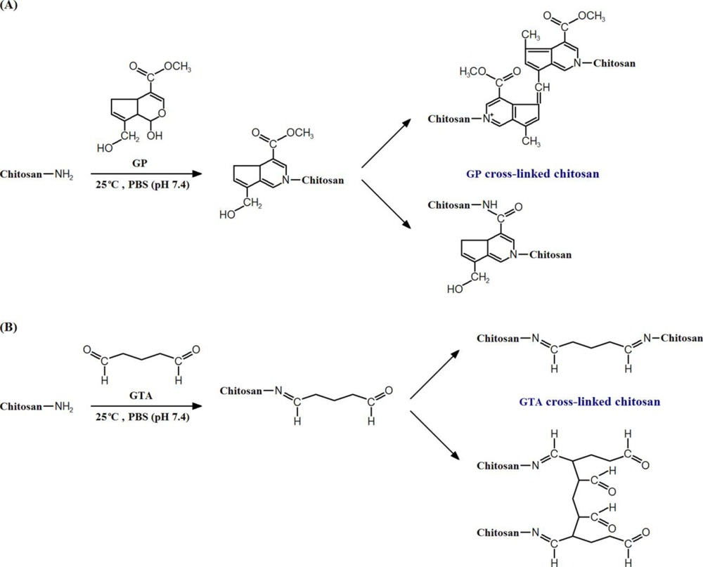
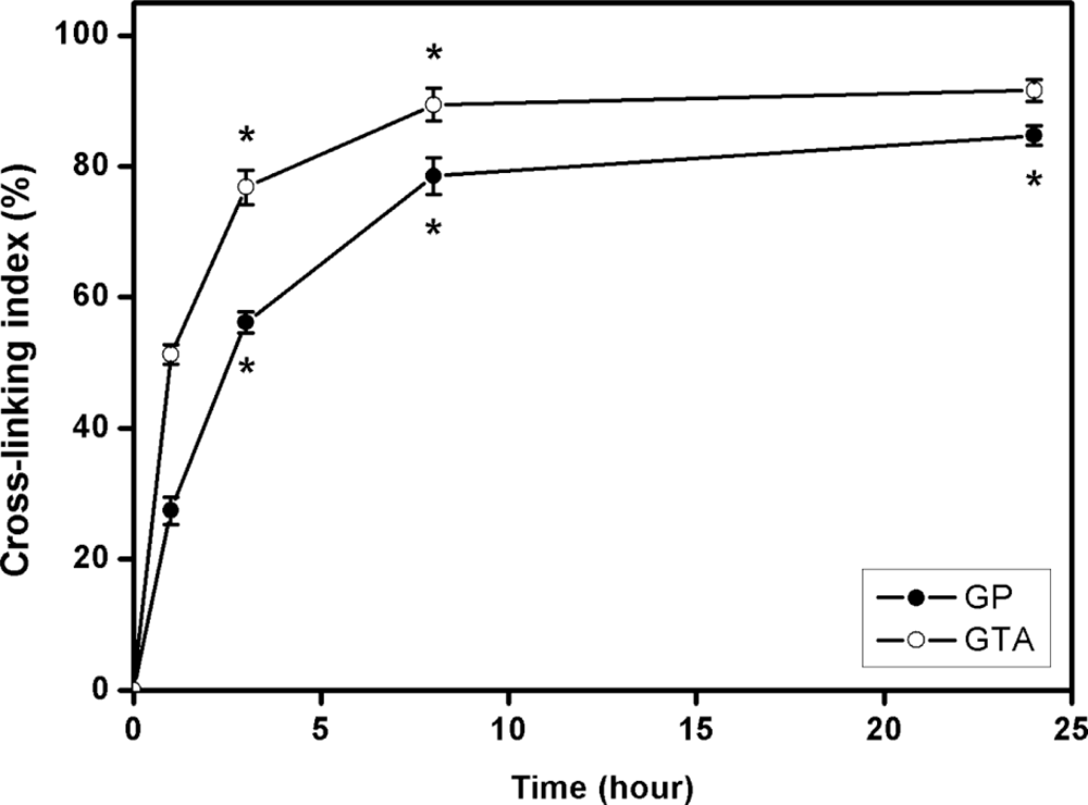
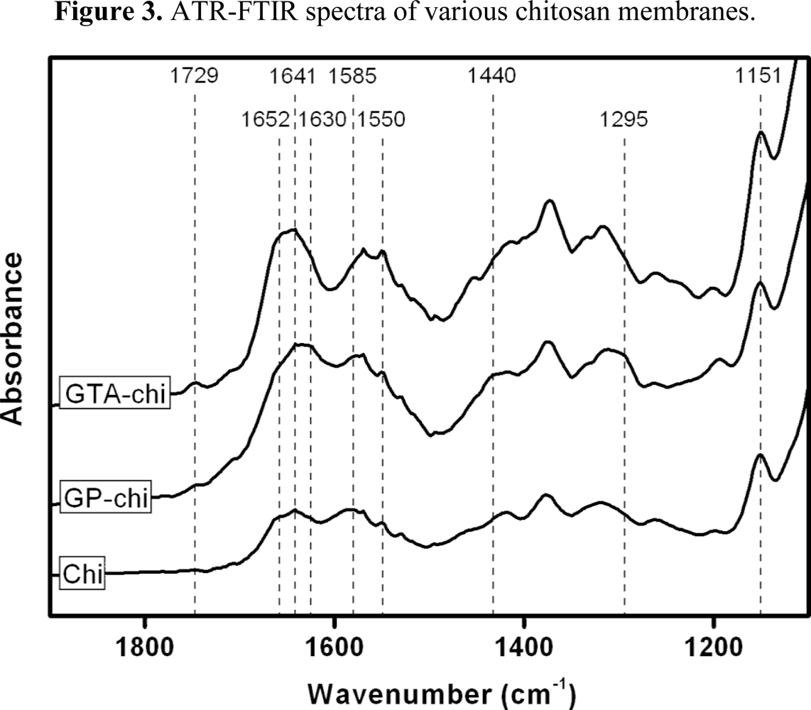
2.1. Preparation of GP or GTA Cross-Linked Chitosan Membranes
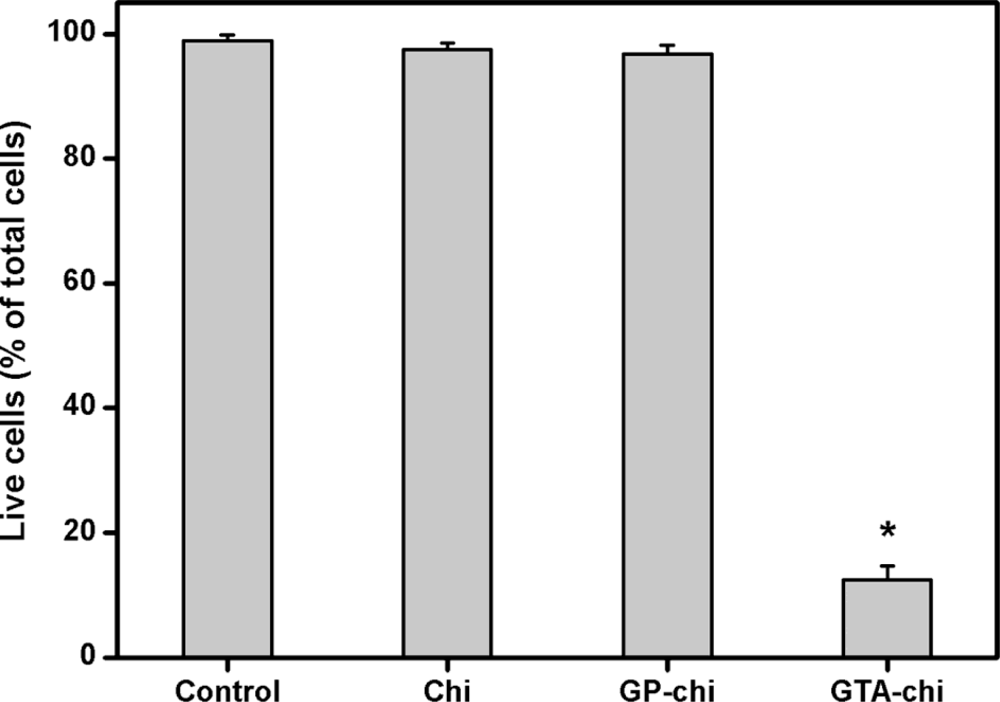
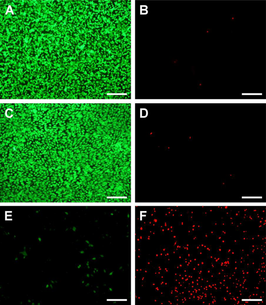
2.2. Cell Viability Assays
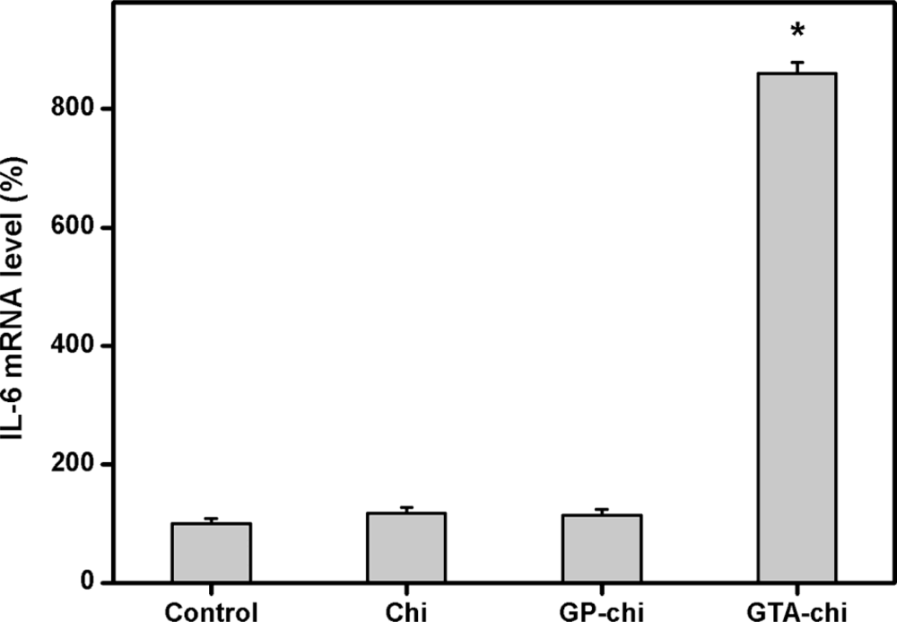
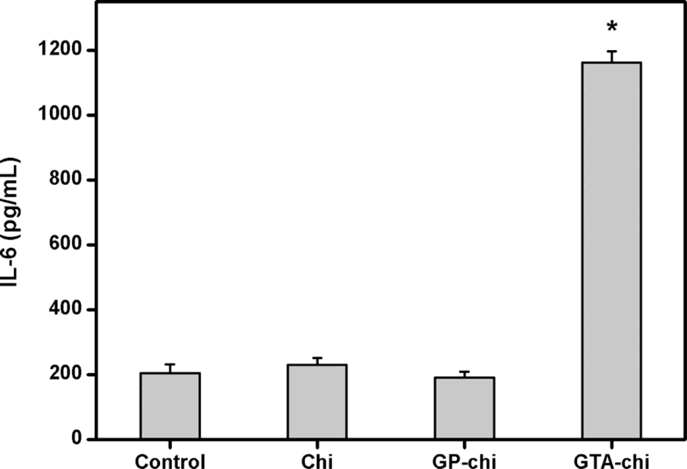
2.3. Pro-Inflammatory Gene and Cytokine Expression Analyses
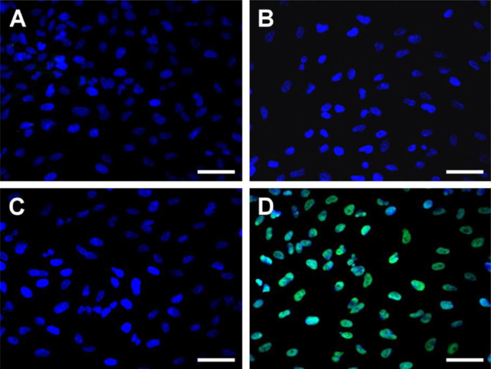
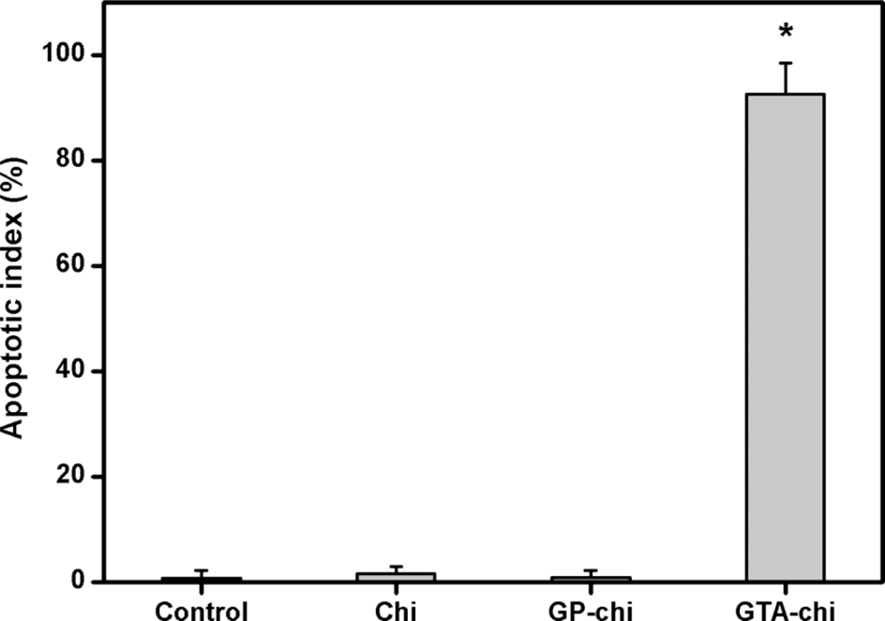
2.4. Apoptosis Assays
3. Experimental Section
3.1. Materials
3.2. Preparation of GP or GTA Cross-Linked Chitosan Membranes
3.3. Human RPE Cell Line Cultures
3.4. Cell Viability Assays
3.5. Pro-Inflammatory Gene and Cytokine Expression Analyses
3.6. Apoptosis Assays
3.7. Statistics
4. Conclusions
Acknowledgments
References
- Jager, RD; Mieler, WF; Miller, JW. Age-related macular degeneration. N. Engl. J. Med 2008, 358, 2606–2617. [Google Scholar]
- Hsiue, GH; Lai, JY; Lin, PK. Absorbable sandwich-like membrane for retinal-sheet transplantation. J. Biomed. Mater. Res 2002, 61, 19–25. [Google Scholar]
- Lai, JY; Lin, PK; Hsiue, GH; Cheng, HY; Huang, SJ; Li, YT. Low Bloom strength gelatin as a carrier for potential use in retinal sheet encapsulation and transplantation. Biomacromolecules 2009, 10, 310–319. [Google Scholar]
- Lai, JY; Li, YT. Evaluation of cross-linked gelatin membranes as delivery carriers for retinal sheets. Mater. Sci. Eng. C-Biomimetic Supramol. Syst 2010, 30, 677–685. [Google Scholar]
- Ideta, R; Tasaka, F; Jang, WD; Nishiyama, N; Zhang, GD; Harada, A; Yanagi, Y; Tamaki, Y; Aida, T; Kataoka, K. Nanotechnology-based photodynamic therapy for neovascular disease using a supramolecular nanocarrier loaded with a dendritic photosensitizer. Nano Lett 2005, 5, 2426–2431. [Google Scholar]
- Nishiyama, N; Iriyama, A; Jang, WD; Miyata, K; Itaka, K; Inoue, Y; Takahashi, H; Yanagi, Y; Tamaki, Y; Koyama, H; Kataoka, K. Light-induced gene transfer from packaged DNA enveloped in a dendrimeric photosensitizer. Nat. Mater 2005, 4, 934–941. [Google Scholar]
- Sashiwa, H; Yajima, H; Aiba, S. Synthesis of a chitosan-dendrimer hybrid and its biodegradation. Biomacromolecules 2003, 4, 1244–1249. [Google Scholar]
- Jayakumar, R; Prabaharan, M; Nair, SV; Tamura, H. Novel chitin and chitosan nanofibers in biomedical applications. Biotechnol. Adv 2010, 28, 142–150. [Google Scholar]
- Mi, FL; Tan, YC; Liang, HF; Sung, HW. In vivo biocompatibility and degradability of a novel injectable-chitosan-based implant. Biomaterials 2002, 23, 181–191. [Google Scholar]
- De Souza, R; Zahedi, P; Allen, CJ; Piquette-Miller, M. Biocompatibility of injectable chitosan-phospholipid implant systems. Biomaterials 2009, 30, 3818–3824. [Google Scholar]
- Felt, O; Furrer, P; Mayer, JM; Plazonnet, B; Buri, P; Gurny, R. Topical use of chitosan in ophthalmology: tolerance assessment and evaluation of precorneal retention. Int. J. Pharm 1999, 180, 185–193. [Google Scholar]
- de la Fuente, M; Raviña, M; Paolicelli, P; Sanchez, A; Seijo, B; Alonso, MJ. Chitosan-based nanostructures: a delivery platform for ocular therapeutics. Adv. Drug Deliv. Rev 2010, 62, 100–117. [Google Scholar]
- Ma, DHK; Lai, JY; Cheng, HY; Tsai, CC; Yeh, LK. Carbodiimide cross-linked amniotic membranes for cultivation of limbal epithelial cells. Biomaterials 2010, 31, 6647–6658. [Google Scholar]
- Lai, JY; Li, YT. Functional assessment of cross-linked porous gelatin hydrogels for bioengineered cell sheet carriers. Biomacromolecules 2010, 11, 1387–1397. [Google Scholar]
- Lu, PL; Lai, JY; Ma, DHK; Hsiue, GH. Carbodiimide cross-linked hyaluronic acid hydrogels as cell sheet delivery vehicles: characterization and interaction with corneal endothelial cells. J. Biomater. Sci.-Polym. Ed 2008, 19, 1–18. [Google Scholar]
- Lai, JY; Ma, DHK; Cheng, HY; Sun, CC; Huang, SJ; Li, YT; Hsiue, GH. Ocular biocompatibility of carbodiimide cross-linked hyaluronic acid hydrogels for cell sheet delivery carriers. J. Biomater. Sci.-Polym. Ed 2010, 21, 359–376. [Google Scholar]
- Nishi, C; Nakajima, N; Ikada, Y. In vitro evaluation of cytotoxicity of diepoxy compounds used for biomaterial modification. J. Biomed. Mater. Res 1995, 29, 829–834. [Google Scholar]
- Risbud, M; Hardikar, A; Bhonde, R. Chitosan-polyvinyl pyrrolidone hydrogels as candidate for islet immunoisolation: in vitro biocompatibility evaluation. Cell Transplant 2000, 9, 25–31. [Google Scholar]
- Mi, FL; Huang, CT; Liang, HF; Chen, MC; Chiu, YL; Chen, CH; Sung, HW. Physicochemical, antimicrobial, and cytotoxic characteristics of a chitosan film cross-linked by a naturally occurring cross-linking agent, aglycone geniposidic acid. J. Agric. Food Chem 2006, 54, 3290–3296. [Google Scholar]
- Silva, SS; Motta, A; Rodrigues, MT; Pinheiro, AFM; Gomes, ME; Mano, JF; Reis, RL; Migliaresi, C. Novel genipin-cross-linked chitosan/silk fibroin sponges for cartilage engineering strategies. Biomacromolecules 2008, 9, 2764–2774. [Google Scholar]
- Yin, L; Zhao, X; Cui, L; Ding, J; He, M; Tang, C; Yin, C. Cytotoxicity and genotoxicity of superporous hydrogel containing interpenetrating polymer networks. Food Chem. Toxicol 2009, 47, 1139–1145. [Google Scholar]
- Zhang, K; Qian, Y; Wang, H; Fan, L; Huang, C; Yin, A; Mo, X. Genipin-crosslinked silk fibroin/hydroxybutyl chitosan nanofibrous scaffolds for tissue-engineering application. J. Biomed. Mater. Res. A 2010, 95, 870–881. [Google Scholar]
- Lai, JY. Biocompatibility of chemically cross-linked gelatin hydrogels for ophthalmic use. J. Mater. Sci.-Mater. Med 2010, 21, 1899–1911. [Google Scholar]
- Butler, MF; Ng, YF; Pudney, PDA. Mechanism and kinetics of the crosslinking reaction between biopolymers containing primary amine groups and genipin. J. Polym. Sci. Pol. Chem 2003, 41, 3941–3953. [Google Scholar]
- Liang, HC; Chang, WH; Lin, KJ; Sung, HW. Genipin-crosslinked gelatin microspheres as a drug carrier for intramuscular administration: in vitro and in vivo studies. J. Biomed. Mater. Res. A 2003, 65, 271–282. [Google Scholar]
- Sung, HW; Chang, Y; Liang, IL; Chang, WH; Chen, YC. Fixation of biological tissues with a naturally occurring crosslinking agent: fixation rate and effects of pH, temperature, and initial fixative concentration. J. Biomed. Mater. Res 2000, 52, 77–87. [Google Scholar]
- Chiono, V; Pulieri, E; Vozzi, G; Ciardelli, G; Ahluwalia, A; Giusti, P. Genipin-crosslinked chitosan/gelatin blends for biomedical applications. J. Mater. Sci.-Mater. Med 2008, 19, 889–898. [Google Scholar]
- Sundararaghavan, HG; Monteiro, GA; Lapin, NA; Chabal, YJ; Miksan, JR; Shreiber, DI. Genipin-induced changes in collagen gels: correlation of mechanical properties to fluorescence. J. Biomed. Mater. Res. A 2008, 87, 308–320. [Google Scholar]
- Schiffman, JD; Schauer, CL. Cross-linking chitosan nanofibers. Biomacromolecules 2007, 8, 594–601. [Google Scholar]
- Lai, JY. The role of bloom index of gelatin on the interaction with retinal pigment epithelial cells. Int. J. Mol. Sci 2009, 10, 3442–3456. [Google Scholar]
- Amaral, IF; Granja, PL; Barbosa, MA. Chemical modification of chitosan by phosphorylation: an XPS, FT-IR and SEM study. J. Biomater. Sci.-Polym. Ed 2005, 16, 1575–1593. [Google Scholar]
- VandeVord, PJ; Matthew, HWT; DeSilva, SP; Mayton, L; Wu, B; Wooley, PH. Evaluation of the biocompatibility of a chitosan scaffold in mice. J. Biomed. Mater. Res 2002, 59, 585–590. [Google Scholar]
- Keong, LC; Halim, AS. In vitro models in biocompatibility assessment for biomedical-grade chitosan derivatives in wound management. Int. J. Mol. Sci 2009, 10, 1300–1313. [Google Scholar]
- Lai, JY; Lu, PL; Chen, KH; Tabata, Y; Hsiue, GH. Effect of charge and molecular weight on the functionality of gelatin carriers for corneal endothelial cell therapy. Biomacromolecules 2006, 7, 1836–1844. [Google Scholar]
- Chatelet, C; Damour, O; Domard, A. Influence of the degree of acetylation on some biological properties of chitosan films. Biomaterials 2001, 22, 261–268. [Google Scholar]
- Koo, HJ; Song, YS; Kim, HJ; Lee, YH; Hong, SM; Kim, SJ; Kim, BC; Jin, C; Lim, CJ; Park, EH. Antiinflammatory effects of genipin, an active principle of gardenia. Eur. J. Pharmacol 2004, 495, 201–208. [Google Scholar]
- Hsiue, GH; Lai, JY; Chen, KH; Hsu, WM. A novel strategy for corneal endothelial reconstruction with a bioengineered cell sheet. Transplantation 2006, 81, 473–476. [Google Scholar]
- Lai, JY; Hsiue, GH. Functional biomedical polymers for corneal regenerative medicine. React. Funct. Polym 2007, 67, 1284–1291. [Google Scholar]
- Lai, JY; Chen, KH; Hsiue, GH. Tissue-engineered human corneal endothelial cell sheet transplantation in a rabbit model using functional biomaterials. Transplantation 2007, 84, 1222–1232. [Google Scholar]
- Lu, PL; Lai, JY; Tabata, Y; Hsiue, GH. A methodology based on the “anterior chamber of rabbit eyes” model for noninvasively determining the biocompatibility of biomaterials in an immune privileged site. J. Biomed. Mater. Res. A 2008, 86, 108–116. [Google Scholar]
- Gough, JE; Scotchford, CA; Downes, S. Cytotoxicity of glutaraldehyde crosslinked collagen/poly(vinyl alcohol) films is by the mechanism of apoptosis. J. Biomed. Mater. Res 2002, 61, 121–130. [Google Scholar]
- Marczak, A; Jóźwiak, Z. Damage to the cell antioxidative system in human erythrocytes incubated with idarubicin and glutaraldehyde. Toxicol. in Vitro 2009, 23, 1188–1194. [Google Scholar]
- Lai, JY; Chen, KH; Hsu, WM; Hsiue, GH; Lee, YH. Bioengineered human corneal endothelium for transplantation. Arch. Ophthalmol 2006, 124, 1441–1448. [Google Scholar]


© 2010 by the authors; licensee MDPI, Basel, Switzerland. This article is an open-access article distributed under the terms and conditions of the Creative Commons Attribution license (http://creativecommons.org/licenses/by/3.0/).
Share and Cite
Lai, J.-Y.; Li, Y.-T.; Wang, T.-P. In Vitro Response of Retinal Pigment Epithelial Cells Exposed to Chitosan Materials Prepared with Different Cross-Linkers. Int. J. Mol. Sci. 2010, 11, 5256-5272. https://doi.org/10.3390/ijms11125256
Lai J-Y, Li Y-T, Wang T-P. In Vitro Response of Retinal Pigment Epithelial Cells Exposed to Chitosan Materials Prepared with Different Cross-Linkers. International Journal of Molecular Sciences. 2010; 11(12):5256-5272. https://doi.org/10.3390/ijms11125256
Chicago/Turabian StyleLai, Jui-Yang, Ya-Ting Li, and Tsu-Pin Wang. 2010. "In Vitro Response of Retinal Pigment Epithelial Cells Exposed to Chitosan Materials Prepared with Different Cross-Linkers" International Journal of Molecular Sciences 11, no. 12: 5256-5272. https://doi.org/10.3390/ijms11125256