Article

Screening of Molecular Virulence Markers in Staphylococcus aureus and Pseudomonas aeruginosa Strains Isolated from Clinical Infections

1
National Institute for Research in Microbiology and Immunology, Cantacuzino, Spl. Independentei 103, cod 060631, Bucharest 060101, Romania
2
Department of Microbiology, Faculty of Biology, University of Bucharest, Aleea Portocalelor 1-3, Bucharest 060101, Romania
3
Institute for Cardiovascular Diseases Prof. C.C. Iliescu, Bucharest 060101, Romania
*
Author to whom correspondence should be addressed.
Int. J. Mol. Sci. 2010, 11(12), 5273-5291; https://doi.org/10.3390/ijms11125273
Submission received: 12 November 2010 / Revised: 2 December 2010 / Accepted: 3 December 2010 / Published: 21 December 2010
(This article belongs to the Special Issue Advances in Molecular Diagnostics)

Abstract

:
Staphylococcus (S.) aureus and Pseudomonas (Ps.) aeruginosa are two of the most frequently opportunistic pathogens isolated in nosocomial infections, responsible for severe infections in immunocompromised hosts. The frequent emergence of antibiotic-resistant S. aureus and Ps. aeruginosa strains has determined the development of new strategies in order to elucidate the different mechanisms used by these bacteria at different stages of the infectious process, providing the scientists with new procedures for preventing, or at least improving, the control of S. aureus and Ps. aeruginosa infections. The purpose of this study was to characterize the molecular markers of virulence in S. aureus and Ps. aeruginosa strains isolated from different clinical specimens. We used multiplex and uniplex PCR assays to detect the genes encoding different cell-wall associated and extracellular virulence factors, in order to evaluate potential associations between the presence of putative virulence genes and the outcome of infections caused by these bacteria. Our results demonstrate that all the studied S. aureus and Ps. aeruginosa strains synthesize the majority of the investigated virulence determinants, probably responsible for different types of infections.

1. Introduction

S. aureus is a versatile human pathogen with the ability to cause a large spectrum of human diseases, ranging from skin lesions (abscesses, impetigo) to invasive and more serious infections (osteomyelitis, septic arthritis, pneumonia, endocarditis). The ability of S. aureus to cause disease has been attributed to an impressive spectrum of cell-wall-associated (protein A, clumping factors, fibronectin binding proteins, and other adhesive matrix molecules) factors, and extracellular toxins (coagulase, hemolysins, enterotoxins, toxic-shock syndrome toxin 1, exfoliative toxins, and Panton-Valentine leukocidin) as virulence determinants. Excepting the toxemic syndromes determined by toxic-shock syndrome toxin 1 (TSST-1), scalded skin syndrome as a result of epidermolytic toxins activity, and food poisoning as a consequence of ingestion of preformed enterotoxins, the pathogenesis of S. aureus is the result of interaction between a variety of host factors and the bacterial virulence determinants [1]. The coordinated expression of S. aureus virulence factors is regulated by a complex network including the quorum-sensing system agr and the well characterized virulence gene regulators. The specific role and/or importance of a virulence factor in S. aureus pathogenesis may vary from one infection type to another. When considering that a high number of S. aureus strains are more difficult to be treated due to their multiple resistance to antibiotics, the elucidation of S. aureus pathogenesis at the molecular level becomes imperative in the fight against this important human pathogen, for the purpose of finding new therapeutic strategies.
Ps. aeruginosa is an opportunistic pathogen responsible for chronic lung infection in cystic fibrosis patients, as well as for nosocomial infections in immunocompromised hosts. This bacterium determines many kinds of infections: urinary tract infections, respiratory system infections, dermatitis, soft tissue infections, bacteremia, bone and joint infections, gastrointestinal infections and a variety of systemic infections, particularly in immunosuppressed patients with severe burns, cancer and AIDS. In patients with cystic fibrosis especially disposed to Ps. aeruginosa infections, these bacteria are responsible for high rates of morbidity and mortality [2].
The pathogenesis of Ps. aeruginosa infections is multifactorial, as suggested by the large number of cell-associated and extracellular virulence determinants of these bacteria. The first step in Ps. aeruginosa infections is the colonization of the altered epithelium. Adherence of Ps. aeruginosa to the epithelium is mediated by fimbriae, type 4 pili and flagella [2]. After colonization, Ps. aeruginosa produces several extracellular virulence factors (alkaline protease and staphylolytic protease, elastase, protease IV, heat-labile and heat-stable hemolysins, phospholipases C and exotoxins A, S, T, U, Y), responsible for extensive tissue damage, bloodstream invasion, and dissemination [3]. Many of the extracellular virulence factors are controlled by cell-to-cell signaling systems [2]. These regulatory systems enable Ps. aeruginosa to synthesize virulence factors in a coordinated, cell-density-dependent manner that can allow the bacteria to surpass/exceed the host defense mechanisms. Interference with the virulence factor production dependent of the cell-to-cell signaling system is a promising therapeutic approach for decreasing the rate of morbidity and mortality caused by Ps. aeruginosa colonization and infections [2].
The purpose of this study was to characterize the molecular markers of virulence in S. aureus and Ps. aeruginosa strains isolated from different clinical specimens, in order to evaluate potential associations between the presence of putative virulence genes and the outcome of infections caused by these bacteria.

2. Results and Discussion

The results of phenotypic characterization for the presence of enzymatic virulence factors among the analyzed strains are listed in Tables 1 and 2.
We have detected by PCR analysis 15 virulence genes in S. aureus, respectively 7 in Ps. aeruginosa strains isolated from infections with different clinical pictures. The results of PCR assays are presented in Figures 114.

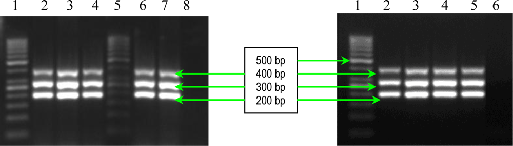
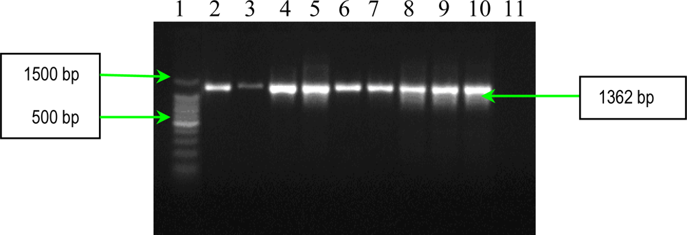
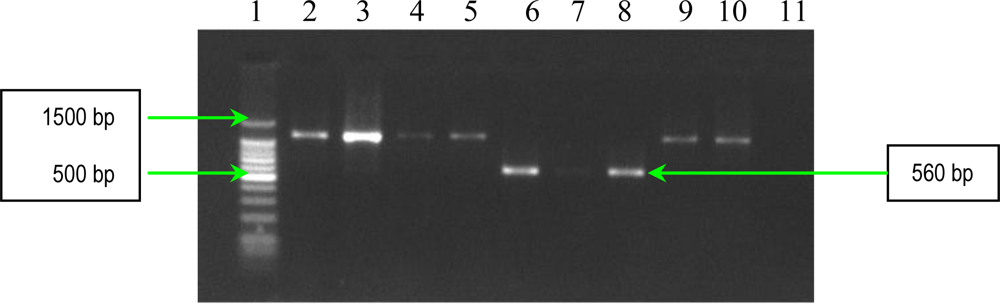
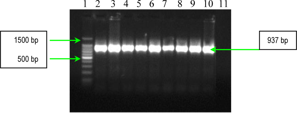
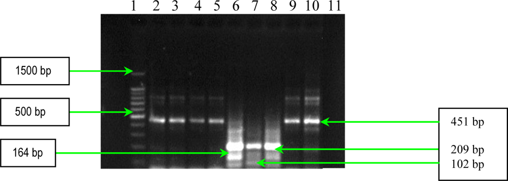
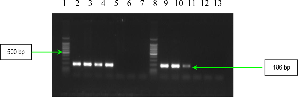
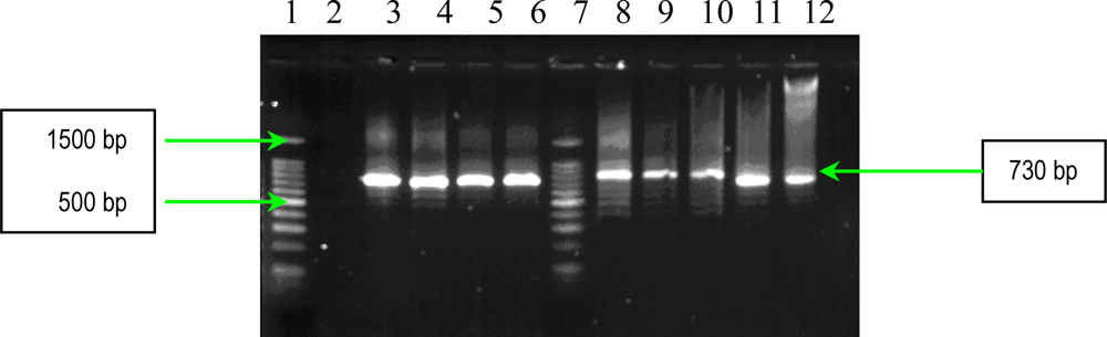
2.1. PCR Assays for S. aureus Virulence Genes Detection

The initial step in the pathogenesis of S. aureus infection is the attachment of the bacteria to the human cell surfaces and implanted devices. Adhesion of S. aureus may be mediated by specific cell-surface proteins, or be a result of interactions between the cell-surface proteins and the host proteins, such as von Willebrand factor, fibronectin, fibrinogen and collagen [4]. By these means, S. aureus can adhere directly to eukaryotic cell receptors or, alternatively, can bind to plasma-coated inserted devices. Therefore, S. aureus produces two types of adhesins. One set has a characteristic LPXTG motif that anchors the adhesin to the components of the extracellular matrix (ECM); the representatives of this kind of adhesins are grouped in the family named microbial surface components recognizing adhesive matrix molecules (MSCRAMMs). This adhesins family constitutes: protein A, the fibronectin-binding proteins (Fnbps), collagen-binding protein (Cna), elastin-binding protein, clumping factors A and B (ClfA and B). The constituents of the second set of adhesins are noncovalently anchored to the cell surface, including the fibrinogen-binding protein (Fgbp) and coagulase. Fibrinogen is the most abundant host protein in endothelial lesions. Among fibrinogen-binding proteins expressed by S. aureus on bacterial cells, there are clumping factors A and B (ClfA, ClfB) responsible for typical S. aureus clumping in plasma, promoting both adherence to fibrinogen-coated surfaces in vitro as well as endocarditis in experimental animal models in vivo [5]. ClfA and ClfB have a similar molecular organization and a high sequence similarity. However, their fibrinogen-binding domain (the A domain), with only 26% structure identity, interacts with different parts of fibrinogen, i.e., the γ-chain for ClfA and both the α- and β-chains for ClfB. Moreover, the clfA and clfB genes are differently regulated; clfA being expressed during bacterial growth, whereas clfB only during the early logarithmic phase. This aspect raises the question whether ClfA and ClfB might act in synergy to help the cells to attach more firmly to fibrinogen-coated surfaces during the bacterial growth cycle [5]. The results obtained in experimental models suggest that S. aureus strains, that specifically express receptors for fibrinogen and fibronectin, are associated with infective endocarditis, while strains producing receptors for bone sialoprotein, collagen, and fibronectin, are associated with osteomyelitis and arthritis [6].
The results of multiplex PCR1 for detection of bbp and ebpS genes, showed that S. aureus studied strains have no bbp gene, while six of them possess the ebpS gene (Figure 1). The presence of the elastin binding protein encoding gene in some S. aureus analyzed strains, emphasizes their involvement in invasive infections. The PCR2 performed for detection of four MSCRAMMs genes (fib, clfA, clfB and fnbB) showed that all S. aureus analyzed strains possess three genes (fib, clfA, clfB) responsible for encoding fibrinogen-binding protein and clumping factors A and B (Figure 2).
The results of PCR analysis concerning the presence of the fnbA gene showed that all S. aureus strains possess this gene (Figure 3). Fnbps were shown to be required for the internalization S. aureus into nonprofessional phagocytic cells. Different studies showed that efficient invasion of S. aureus is critically dependent on one of the two known forms of Fnbps, FnbpA or FnbpB. Finally, the results of PCR analysis concerning the presence of cna gene, demonstrated the polymophism of this gene among the studied S. aureus strains. Seven strains possess a 1120 bp gene, whereas two exhibited a variant of 560 bp gene (Figure 4). The results of PCR assays for adhesins genes showed that all studied S. aureus strains possess: elastin-binding protein, clumping factors A and B, fibronectin-binding protein A, fibrinogen-binding protein and collagen-binding protein. The presence of these adhesins demonstrates the involvement of the respective virulence factors in the determined S. aureus infections associated with cardiovascular devices.
Bacterial survival during an infection is a process dependent on the ability of the organism to override the host’s defense possibilities. S. aureus synthesizes a lot of toxins and exoproteins active against host defense mechanisms. Almost all bacterial strains synthesize a lot of enzymes and cytotoxins including four hemolysins (alpha, beta, gamma, and delta), nucleases, proteases, lipases, hyaluronidase, and collagenase [7]. The main function of these proteins may be to convert local host tissues into nutrients required for bacterial growth. Some strains synthesize one or more additional exoproteins, including toxic shock syndrome toxin-1 (TSST 1), the staphylococcal enterotoxins (SEA, SEB, SEC, SED, SEE, SEG, SEH, and SEI), and the exfoliative toxins A and B [8]. These toxins affect neutrophils and macrophages, gamma-hemolysin, being supplementary, is responsible for the lysis of many varieties of mammalian erythrocytes.
Coagulase is an extracellular protein, binding the prothrombin in the host to form a complex called staphylothrombin. The thrombin characteristic protease activity in the complex, determines the conversion of fibrinogen into fibrin. In this way the S. aureus cells protect themselves from the phagocytic and immune defense of the host by forming localized clotting. The result of PCR assay shows that all S. aureus analyzed strains possessed this factor (Figure 5).
Panton-Valentine leukocidin (PVL) is a bicomponent leukocidin responsible for leukocyte destruction and tissue necrosis. PVL, together with gamma-hemolysin, belongs to the recently described family of synergohymenotropic toxins. These toxins determine membrane disruptions of host defense cells and erythrocytes by the synergistic activity of two non-associated classes of secretory proteins, designated as S and F [9]. The results of multiplex PCR for detection of genes encoding these toxins showed that hlg gene was present in all analyzed S. aureus strains, whereas the luk-PV gene was absent (Figure 6). These results are in agreement with those published, that is to say virtually all strains of S. aureus synthesize gamma-hemolysin, whereas PVL is synthesized only by less than 5% of strains [10]. However, the PVL gene is frequently seen in S. aureus strains associated with necrotic lesions involving the skin, and in severe necrotic hemorrhagic pneumonia, whereas it is seldom seen in strains responsible for other infections, such as infective endocarditis and hospital-acquired staphylococcal infections.
The results of multiplex PCR assay for detection of staphylococcal enterotoxins genes (sea, seb, sec, sed and see) showed that four analyzed S. aureus strains possess the sec gene, whereas two strains possess three genes (sea, seb and see) (Figure 7). This assay offers a very specific, quick, reliable, and inexpensive alternative to conventional PCR assays used in clinical laboratories to identify various staphylococcal enterotoxin genes.
The variable presence of virulence genes in S. aureus strains isolated from blood cultures provides evidence that different bacterial factors play a redundant role in determining invasive disease in hospital units.

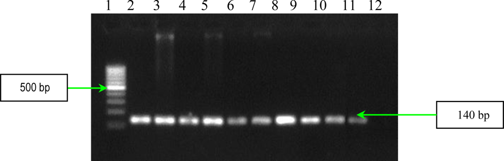
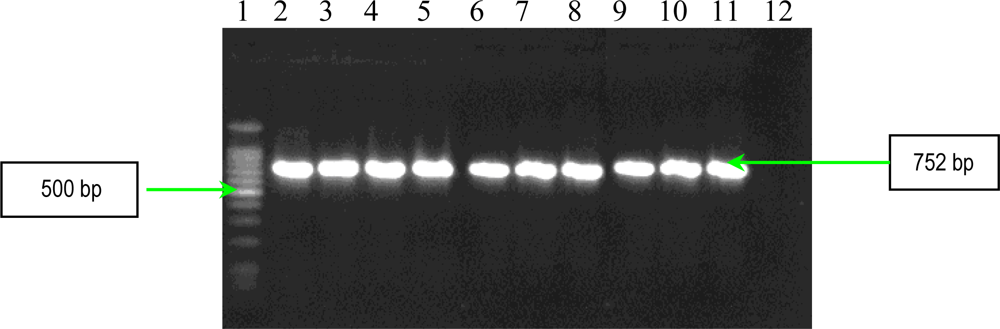
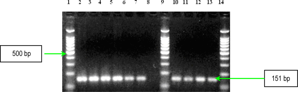
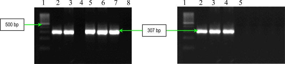
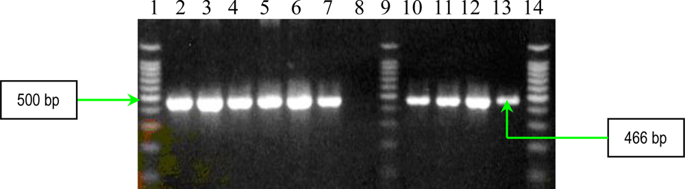
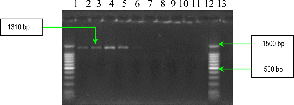
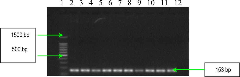
2.2. PCR Assays for Ps. aeruginosa Virulence Genes Detection

Ps. aeruginosa exhibits many virulence factors that contribute to its pathogenicity. Some of these factors help colonization, whereas others facilitate bacterial invasion. Ps. aeruginosa colonization involves a lot of factors, including fimbriae, type 4 pili, flagella, and surface polysaccharides. The ability of Ps. aeruginosa to invade tissues depends upon production of extracellular enzymes and toxins that break down physical barriers by disrupting host cell membranes and annihilating the host, as well as resistance to phagocytosis and host immune defense armamentarium. The tissue invasion by Ps. aeruginosa is promoted by the production of proteases, hemolysins and cytotoxin (leukocidin). Ps. aeruginosa synthesizes several proteases (LasB elastase, LasA staphylolytic protease, alkaline protease, and protease IV) responsible for: complement inactivation, and surfactant proteins A and D (SP-A and SP-D) degradation. These proteins play an important role in innate immunity, cleavage IgG antibodies, and IFN and TNF inactivation [11]. Elastin is a major part of human lung tissue responsible for lung expansion and contraction. Moreover, elastin is an important component of blood vessels, conferring them their resilience. The concerted activity of LasB elastase and LasA staphylolytic protease is responsible for elastolytic activity, implicated in destruction of elastin-containing human lung tissue and the pulmonary hemorrhages in invasive Ps. aeruginosa infections. The LasB elastase is a zinc metalloprotease, encoded by the lasB gene, responsible for degradation of elastin, fibrin and collagen [11]. Alkaline protease interferes with fibrin formation, implicated in fibrin lysis, and inactivates important host defense proteins, such as antibodies, complement, IFN-γ, and cytokines. Protease IV is important in the pathogenesis of P. aeruginosa-induced microbial keratitis, but little is known about its role in other clinical isolates. The virulence of protease IV in ocular infection has been attributed to the destruction of host proteins, including fibrinogen and components of the immune system. Protease IV also degrades structural proteins, such as elastin, facilitating bacterial adherence and infection [12].
Results of PCR analysis concerning the presence of genes encoding proteases, showed that all studied Ps. aeruginosa strains possess all the tested proteases (LasB elastase, alkaline protease and protease IV), that are correlated with type of infection (Figures 810).
Three other soluble proteins involved in Ps. aeruginosa invasion are represented by a rhamnolipid, two phospholipases C, haemolytic phospholipase C (PLC-H), and non-haemolytic phospholipase C (PLC-N). These factors may act synergistically to break down phospholipids (e.g., phosphatidylcholine and sphingomyelin), and contribute to invasion by means of their cytotoxic effects on neutrophils, lymphocytes and other eucaryotic cells. Rhamnolipid, a rhamnose-containing glycolipid biosurfactant, has a detergent-like structure and is considered to solubilize the phospholipids of lung surfactant, making them more accessible to cleavage by phospholipase C. The resulting loss of lung surfactant may be responsible for the atelectasis associated with chronic and acute Ps. aeruginosa lung infection. Rhamnolipid also inhibits the mucociliary transport and ciliary function of human respiratory epithelium [13]. The results of PCR analysis showed that all Ps. aeruginosa possess rhlAB gene, encoding the rhamnolipid (Figure 11). Concerning the presence of two phospholipases C, the results of PCR analysis showed that all Ps. aeruginosa strains possess plcN gene, whereas plcH gene is present only in seven of the studied strains (Figures 12 and 13). These two enzymes could work sequentially and synergistically, PLC-H would promote degradation of the erythrocyte membrane (phospholipids components of the outer leaflet: phosphatidylcholine and sphingomyelin), exposing the inner leaflet. PLC-N could then hydrolyze phospatidylserine present in the inner leaflet. The products of phospholipid hydrolysis would be digested by alkaline phosphatase, pyrophosphate being released. The experimental studies demonstrated that purified PLC-H determines vascular permeability, organ injuries, and death when injected in high doses into mice, proving that PLC-H is an important virulence factor. Some Ps. aeruginosa strains are producers of a mucoid exopolysaccharide (alginate) that forms the matrix of the Ps. aeruginosa biofilm, anchoring the cells and, in certain cases, protecting bacteria from the host defense armamentarium consisting of: lymphocytes, phagocytes, the ciliary action of the respiratory tract, antibodies and complement. At the same time these strains are less susceptible to antibiotics than their planktonic counterparts, being most often isolated from patients with cystic fibrosis and usually found in lung tissues of such individuals. The algD gene encodes GDP-mannose dehydrogenase (GMD), which acts as a rate-limiting enzyme in mucoid strains by catalyzing the conversion of GDP-mannose to GDP-mannuronic acid, thereby promoting the cell to alginate production [14]. The results of PCR analysis concerning the presence of algD gene showed that only five strains possess this gene, an aspect which demonstrates the involvement of these strains in infections with biofilm formation (Figure 14).

3. Experimental Section

3.1. Bacterial Strains

In this study nine S. aureus and ten Ps. aeruginosa strains were studied, selected from a pool of bacterial strains isolated between 2005–2007 from patients hospitalized in Fundeni Hospital, Bucharest. S. aureus strains were isolated from patients with infections (blood cultures) with associated cardiovascular devices, whereas Ps. aeruginosa strains originated from different clinical specimens (tracheo-bronchical secretions, wound secretions and sputum). The strains identification were performed with the help of conventional and API microtests and VITEK I automatic system.
Concerning the S. aureus strains, 23 virulence factors were evaluated, out of which eight (lechitinase, lipase, amylase, caseinase, DN-ase and alpha, beta and delta hemolysins) were assessed using phenotypic tests, and 15 (fibronectin binding proteins A and B, fibrinogen binding protein, clumping factors A and B, elastin binding protein, bone sialoprotein binding protein, collagen-binding protein, Panton-Valentine leukocidin, gamma-hemolysin and SEA, SEB, SEC, SED and SEE enterotoxins) were evaluated using PCR assays.
In the Ps. aeruginosa studied strains, 13 virulence factors were evaluated, out of which six (lechitinase, lipase, amylase, caseinase, DN-ase, and hemolysins) were assessed using phenotypic tests, whereas seven (elastase, alkaline protease, protease IV, rhamnolipid, hemolytic phospholipase C, non hemolytic phospholipase C and alginate) were evaluated using PCR assays.
The phenotypic characterization for the presence of enzymatic virulence factors was performed by cultivating the strains in available media for enzymes activity detection [15].
For detection of hemolysins, the strains were streaked on blood agar plates containing 5% (vol/vol) sheep blood in order to obtain isolated colonies. After incubation at 37 °C for 24 h the clear zone (total lysis of red blood cells) around the colonies was registered as positive reaction. Some S. aureus strains proved the presence of CAMP-like factor, a special hemolysin, the activity of which is revealed by streaking the bacterial studied strain on 5% sheep blood agar, at 8 mm distance perpendicularly on the β-hemolytic Staphylococcus aureus ATCC 25923 reference strain, and incubating the plate aerobically at 37 °C for 24 h. The occurrence of synergic β-haemolysis at the confluence of the two spot areas indicates the presence of CAMP-like factor.
Protease activity was determined using two different media as substrate: 15% soluble casein and starch (final concentration 1% in nutritive gelose) obtained from the Cantacuzino Institute Culture Media Laboratory. The strains were spotted and after incubation for 24 h at 37 °C, precipitation surrounding the growth area indicated casein/starch proteolysis (caseinase/amylase presence) [15].
DNA-se production was studied using DNA agar medium. The strains were spotted and after incubation for 24 h at 37 °C, a drop of HCl/1N solution was added to the spotted cultures; a clearing zone around the culture was registered as a positive reaction [15].
For lecithinase and lipase production, the cultures were spotted onto 2.5% yolk agar, and respectively on Tween 80 agar with a substrate at a final concentration of 1%, and thereafter the plates were incubated at 37 °C up to 7 days. An opaque (precipitation) zone surrounding the spot showed the lecithinase, respectively lipase production [15].
Subsequently, we used multiplex and uniplex PCR assays for the detection of genes encoding the putative’s most important virulence factors in the studied strains (coagulase, adhesins, gamma-hemolysin, enterotoxins for S. aureus strains, and elastase, alkaline protease, protease IV, rhamnolipid, alginate, haemolytic phospholipase C and non-hemolytic phospholipase C for Ps. aeruginosa strains). Chromosomal DNA was extracted from nineteen clinical strains. One colony of each strain cultured on solid medium was inoculated into 5 mL of BHI (Broth Heart Infusion) and grown overnight at 37°C with shaking. From these strain cultures, DNA extraction was performed by using Wizard DNA Genomic Purification kit (Promega, U.S.) according to the manufacturer′s recommendations. Chromosomal DNAs obtained were used as templates for all PCR experiments. The PCR reactions were carried out in an Applied Biosystems 2700 Thermal Cycler.

3.2. Detection of S. aureus Virulence Genes by PCR

3.2.1. Multiplex and Uniplex PCRs for Detection of Genes Encoding Adhesins

The detection of staphylococcal microbial surface components recognizing adhesive matrix molecules (MSCRAMMs) with the help of binding assays using purified matrix molecules, is expensive, especially due to the high cost of purified matrix. This phenotypic method has the disadvantage of displaying a redundance of chemical structure of some adhesins: two fibronectin binding proteins (FnbpA and – B) and three receptors for fibrinogen (clumping factors A and B and fibrinogen binding protein, Fib). In addition, some MSCRAMMs bind to more than one matrix molecule (for example, FnbpA binds to both fibronectin and fibrinogen) [6]. In conclusion, the genetic identification of the genes responsible for the respective adhesins synthesis is obviously necessary. PCR assays were used for detection of eight genes encoding MSCRAMMs in S. aureus strains isolated from patients with infections associated with cardiovascular devices. The nucleotide sequences of ebpS (encoding elastin binding protein), fnbB (encoding fibronectin binding protein B), fib (encoding fibrinogen binding protein), clfA and clfB (encoding clumping factors A and B), and bbp (encoding bone sialoprotein binding protein) obtained from GenBank (accession numbers U48826, X62992, X72014, Z18852, AJ224764, and Y18653, respectively) were compared and evaluated using Blast and ClustalX softwares to identify regions, unique for each gene, which have similar annealing temperatures [6]. The primers (Invitrogen, U.S.) used for two multiplex PCRs and for two uniplex PCRs, are listed in Table 3, and in Table 4. Two primer sets were prepared for multiplex PCRs: PCR1 to amplify bbp and ebpS genes, and PCR2 to amplify fnbB, fib, clfA, and clfB genes. The thermal cycling conditions for multiplex PCRs included an initial denaturation step (5 min at 94 °C) followed by 25 cycles of amplification (denaturation for 1 min at 94 °C, annealing for 1 min at 55 °C, and extension for 1 min at 72 °C) [6]. The reaction was completed after a 10 min incubation at 72 °C. Synthesized DNA fragments were detected on 1.5% agarose gels by ethidium bromide staining (Figures 1 and 2).
PCR used for detection of fnbA gene (encoding fibronectin binding protein A) had the following thermal cycling conditions: an initial denaturation step (5 min at 94 °C) followed by 30 cycles of amplification (denaturation for 1 min at 94 °C, annealing for 1 min at 50 °C, and extension for 2 min at 72 °C) [8]. The reaction was ready after a 10 min incubation at 72°C, and the amplified products were detected on 1.5% agarose gels by ethidium bromide staining (Figure 3).
The detection of cna gene (encoding collagen binding protein) was performed by PCR with the specific primers listed in Table 4 [8]. The thermal cycling conditions for cna gene amplification included an initial denaturation step (5 min at 94 °C) followed by 30 cycles of amplification (denaturation for 1 min at 94 °C, annealing for 1 min at 55 °C, and extension for 2 min at 72 °C). The reaction was finished after a 10 min incubation at 72 °C and the amplified products were detected on 1.5% agarose gels by ethidium bromide staining (Figure 4).

3.3. PCR Assays for Detection of Extracellular Proteins (Coagulase, Panton-Valentine Leukocidin and Gamma–Hemolysin)

Detection of coagulase gene of the staphylococcal studied strains was performed by PCR using the following primers: COAG-2: 5′-CGAGACCAAGATTCAACAAG-3′ and COAG-3: 5′AAAGAAAACCACTCACATCA-3′ [16]. DNA amplification was carried out in thermocycler with the following thermal profile: initial denaturation at 95 °C for 2 min followed by 30 cycles of amplification (denaturation at 95 °C for 30 s, annealing at 58 °C for 2 min, and extension at 72 °C for 4 min) and a final extension at 72 °C for 7 min [16]. Synthesized DNA fragments were detected on 1.5% agarose gels by ethidium bromide staining. A negative control (pure water) was included. The lengths of the PCR products were estimated by comparison with the 100 bp DNA ladder molecular size markers (Promega) (Figure 5).
A multiplex PCR were used for the simultaneous detection of genes encoding the Panton-Valentine leukocidin and gamma-hemolysin. Oligonucleotide primers were designed according to the published sequences of the PVL genes (GenBank accession numbers X72700 and AB006796) and the gamma-hemolysin genes (GenBank accession numbers X81586 and L01055), the former to obtain coamplification of lukS-PV and lukF-PV, and the latter to obtain coamplification of hlgC and hlgB [10] (Table 5).
The thermal cycling conditions for luk-PV and hlg genes co-amplification included an initial denaturation step (5 min at 94 °C) followed by 30 cycles of amplification (30 s of denaturation at 94 °C, 30 s of annealing at 55 °C, and 1 min extension at 72 °C). The reaction was completed with a 10 min incubation step at 72 °C. The PCR products were tested by electrophoresis on 1.5% agarose gels. Synthesized DNA fragments were detected on 1.5% agarose gels by ethidium bromide staining. A negative control (pure water) was included. The lengths of the PCR products were estimated by comparison with the 100 bp DNA ladder molecular size markers (Promega) (Figure 6).
Multiplex PCR for detection of enterotoxins SEA, SEB, SEC, SED and SEE. The reaction conditions for the multiplex PCR assay were optimized to ascertain that all of the target gene sequences were satisfactorily amplified. For this purpose, a multiplex set was used containing: 200 mM deoxynucleoside triphosphates; 10 μL of 5X Green GoTaq Flexi Reaction Buffer; 1.5 mM MgCl2; 20 pmol (each) of sea, seb, sec and see primers; 40 pmol of sed primer; 2.5 U of FlexiTaq DNA polymerase (Promega), and 10 to 1,000 ng of template DNA. The volume of this mixture was adjusted to 50 μL with sterile nuclease free water. The primers were designed to target the coding regions of the genes; measures were taken to avoid areas of homology within the enterotoxins structural genes. The primer sequences used in the multiplex PCRs are described in Table 6. DNA amplification was carried out in thermocycler with the following thermal cycling profile: an initial denaturation at 94 °C for 5 min was followed by 35 cycles of amplification (denaturation at 94 °C for 2 min, annealing at 57 °C for 2 min, and extension at 72 °C for 1 min), ending with a final extension at 72 °C for 7 min [17]. The sizes of the amplicons obtained from the studied strains corresponded to the predicted sizes (Table 6). As a negative control, sterile water was used instead of DNA, and no amplicon was observed (Figure 7).

3.4. Detection of Ps. aeruginosa Virulence Genes by PCR

The prevalence of extracellular virulence genes encoding elastase (lasB), alkaline protease (aprA), protease IV (TCF), rhamnolipid (rhlAB), alginate (algD), hemolytic phospholipase C (plcH) and non-hemolytic phospholipase C (plcN) was determined by PCR assays.
PCR for detection of elastase, alkaline protease and rhamnolipid genes. Specific oligonucleotide primers used for detection of genes encoding these virulence factors are listed in Table 7. Parameters for the amplification cycles were: an initial denaturation at 94 °C for 5 min, followed by 30 cycles of amplification (denaturation at 94 °C for 1 min, annealing at 52 °C for 1 min, and primer extension at 72 °C for 1.5 min), ending with a final extension at 72 °C for 5 min [3]. Agarose gel (1.5%) electrophoresis was used for examining the products after PCR (Figures 8, 9 and 11).
PCR for the protease IV gene detection was performed by PCR using specific primers: 5′-TATTTCGCCGACTCCCTGTA-3′ (TCF) and 5′-GAATAGACGCCGCTGAAATC-3′ (TCR) [12]. DNA amplification was carried out in thermocycler with the following thermal cycling profile: an initial denaturation at 94 °C for 5 min was followed by 34 cycles of amplification (denaturation at 94 °C for 30 s, annealing at 60 °C for 30 s, and extension at 72 °C for 2 min), ending with a final extension at 72 °C for 5 min. Synthesized PCR products were detected on 1.5% agarose gels by ethidium bromide staining (Figure 10).
PCR for alginate, hemolytic phospholipase C and non-hemolytic phospholipase C genes detection. The genes for these virulence determinants were amplified with primers selected on the basis of the published PAO1 sequence (Table 8) [13]. The amplification protocol was: 94 °C for 3 min, 30 cycles of 94 °C for 30 s, 55 °C for 1 min and 72 °C for 1 min 30 s, and 72 °C for 5 min. Each gene was amplified separately. PCR products were separated in a 1.5% agarose gel for 1 h at 100 V, stained with ethidium bromide and detected by UV transillumination. Amplified genes were identified on the basis of fragment size (Figures 1214).

4. Conclusions

Our study demonstrated that all studied S. aureus and Ps. aeruginosa strains possess several virulence determinants, many of which are redundant, responsible for the clinical evolution of different infectious processes induced by these infectious agents. All S. aureus strains possess seven genes, of which five are encoding adhesins (fibrinogen-binding protein, clumping factors A and B, fibronectin-binding protein A, collagen-binding protein), gamma-hemolysin and coagulase. The Ps. aeruginosa strains possess five genes encoding for: LasB elastase, alkaline protease, protease IV, and non-hemolytic phospholipase C. Future studies should be concentrated on validating these findings and the in vivo significance of different virulence profiles.

Acknowledgments

This work was supported by the strategic grant POSDRU/89/1.5/S/58852, Project “Postdoctoral program for training scientific researchers” co-financed by the European Social Fund within the Sectorial Operational Program Human Resources Development 2007–2013”, by the Partnership Project No. 42150/2008-2011 and by the Human Resources 135/2010 grant (Contract no. 76/29.07.2010).

References

  1. Jarraud, S; Mougel, C; Thioulouse, J; Lina, G; Meugnier, H; Forey, F; Nesme, X; Etienne, J; Vandenesch, F. Relationships between Staphylococcus aureus genetic background, virulence factors, agr groups (alleles), and human disease. Infect. Immun 2002, 70, 631–641. [Google Scholar]
  2. Delden, CV; Iglewski, BH. Cell-to-cell signaling and Pseudomonas aeruginosa infections. Emerg. Infect. Dis 1998, 4, 551–558. [Google Scholar]
  3. Zhu, H; Bandara, R; Conibear, TCR; Thuruthyil, SJ; Rice, SA; Kjelleberg, S; Givskov, M; Willcox, MDP. Pseudomonas aeruginosa with lasI quorum-sensing deficiency during corneal infection. Invest. Ophthalmol. Vis. Sci 2004, 45, 1897–1903. [Google Scholar]
  4. Foster, TJ; Hook, M. Surface protein adhesins of Staphylococcus aureus. Trends Microbiol 1998, 12, 484–488. [Google Scholar]
  5. Entenza, JM; Foster, TJ; Ni Eidhin, D; Vaudaux, P; Francioli, P; Moreillon, P. Contribution of clumping factor B to pathogenesis of experimental endocarditis due to Staphylococcus aureus. Infect. Immun 2000, 68, 5443–5446. [Google Scholar]
  6. Tristan, A; Ying, L; Bes, M; Etienne, J; Vandenesch, F; Lina, G. Use of multiplex PCR to identify Staphylococcus aureus adhesins involved in human hematogenous infections. J. Clin. Microbiol 2003, 41, 4465–4467. [Google Scholar]
  7. Dinges, MM; Orwin, PM; Schlievert, PM. Exotoxins of Staphylococcus aureus. Clin. Microbiol. Rev 2000, 13, 16–34. [Google Scholar]
  8. Peacock, SJ; Moore, CE; Justice, A; Kantzanou, M; Story, L; Mackie, K; O’Neill, G; Day, NPJ. Virulent combinations of adhesin and toxin genes in natural populations of Staphylococcus aureus. Infect. Immun 2002, 70, 4987–4996. [Google Scholar]
  9. Prevost, G; Cribier, B; Couppie, P. Panton-Valentine leucocidin and gamma-hemolysin from Staphylococcus aureus ATCC 49775 are encoded by distinct genetic loci and have different biological activities. Infect. Immun 1995, 63, 4121–4129. [Google Scholar]
  10. Lina, G; Piemont, Y; Godail-Gamot, F; Bes, M; Peter, MO; Gauduchon, V; Vandenesch, F; Etienne, J. Involvement of panton-valentine leukocidin–producing staphylococcus aureus in primary skin infections. Clin. Infect. Dis 1999, 29, 1128–1132. [Google Scholar]
  11. Sadikot, RT; Blackwell, TS; Christman, JW; Prince, AS. Pathogen–host interactions in Pseudomonas aeruginosa pneumonia. Am. J. Respir. Crit. Care Med 2005, 171, 1209–1223. [Google Scholar]
  12. Smith, L; Rose, B; Tingpej, P; Zhu, H; Conibear, T; Manos, J; Bye, P; Elkins, M; Willcox, M; Bell, S; Wainwright, C; Harbour, C. Protease IV production in Pseudomonas aeruginosa from the lungs of adults with cystic fibrosis. J. Med. Microbiol 2006, 55, 1641–1644. [Google Scholar]
  13. Lanotte, P; Watt, S; Mereghetti, L; Dartiguelongue, N; Rastegar-Lari, A; Goudeau, A; Quentin, R. Genetic features of Pseudomonas aeruginosa isolates from cystic fibrosis patients compared with those of isolates from other origins. J. Med. Microbiolog 2004, 53, 73–81. [Google Scholar]
  14. Ramsey, DM; Wozniak, DJ. Understanding the control of Pseudomonas aeruginosa alginate synthesis and the prospects for management of chronic infections in cystic fibrosis. Mol. Microbiol 2005, 56, 309–322. [Google Scholar]
  15. Cotar, AI; Dinu, S; Chifiriuc, MC; Banu, O; Iordache, C; Larion, C; Bucur, M; Dracea, O; Lazar, V. Screening of molecular markers of quorum sensing in Pseudomonas aeruginosa strains isolated from clinical infections. Rom. Biotehnol. Lett 2008, 13, 3765–3770. [Google Scholar]
  16. Aslantas, O; Demir, C; Turutoglu, H; Cantekin, Z; Ergun, Y; Dogruer, G. Coagulase gene polymorphism of Staphylococcus aureus isolated from subclinical bovine mastitis. Turk. J. Vet. Anim. Sci 2007, 31, 253–257. [Google Scholar]
  17. Mehrotra, M; Wang, G; Johnson, WM. Multiplex PCR for detection of genes for Staphylococcus aureus enterotoxins, exfoliative toxins, toxic shock syndrome toxin 1, and methicillin resistance. J. Clin. Microbiol 2000, 38, 1032–1035. [Google Scholar]
Figure 1. Multiplex PCR assay for simultaneous detection of bbp and ebpS genes. Lines 1 and 9:Bench Top PCR Marker (Promega); 2: S. aureus 10936; 3: S. aureus 11372; 4: S. aureus 11327; 5: MRSA 11325; 6: negative S. aureus strain; 7: MRSA 11573; 8: MRSA 11047; 10: S. aureus 5/06; 11: S. aureus 9/06; 12: S. aureus 11323; and 13: negative control (pure water).
Ijms 11 05273f1
Figure 2. Multiplex PCR assay for simultaneous detection of fib, clfA, clfB and fnbB genes. Left - Lines 1 and 5: Gene Ruler 50 bp (Fermentas); 2: S. aureus 10936; 3: S. aureus 11372; 4: S. aureus 11327; 6: MRSA 11325; 7: MRSA 11573; and 8: negative control (pure water). Right – Line 1: Gene Ruler 50 bp (Fermentas); 2: MRSA 11047; 3: S. aureus 5/06; 4: S. aureus 9/06; 5: S. aureus 11323; and 6: negative control (pure water).
Ijms 11 05273f2
Figure 3. Gel electrophoresis of amplified products of fnbA gene. Line 1: Bench Top 100 bp DNA Lader (Promega); 2: S. aureus 10936; 3: S. aureus 11372; 4: S. aureus 11327; 5: MRSA 11325; 6: MRSA 11573; 7: MRSA 11047; 8: S. aureus 5/06; 9: S. aureus 9/06; 10: S. aureus 11323; and 11: negative control (pure water).
Ijms 11 05273f3
Figure 4. Gel electrophoresis of amplified products of cna gene. Line 1: GeneRuler™ 100 bp Plus DNA Ladder (Fermentas); 2: S. aureus 10936; 3: S. aureus 11372; 4: S. aureus 11327; 5: MRSA 11325; 6: MRSA 11573; 7: MRSA 11047; 8: S. aureus 5/06; 9: S. aureus 9/06; 10: S. aureus 11323; and 11:negative control (pure water)
Ijms 11 05273f4
Figure 5. Gel electrophoresis of amplified products of coagulase gene. Lines 1 and 7: DNA ladder 100bp (Promega); 2: negative S. aureus strain; 3: S. aureus 10936; 4: S. aureus 11372; 5: S. aureus 11327; 6: MRSA 11325; 8: MRSA 11573; 9: MRSA 11047; 10: S. aureus 5/06; 11: S. aureus 9/06; 12: S. aureus 11323; and 13: negative control (pure water).
Ijms 11 05273f5
Figure 6. Multiplex PCR assay for simultaneous detection of luk-PV si hlg genes. Line 1: DNA ladder 100bp; 2: S. aureus 10936; 3: S. aureus 11372; 4: S. aureus 11327; 5: MRSA 11325; 6: MRSA 11573; 7: MRSA 11047; 8: S. aureus 5/06; 9: S. aureus 9/06; 10: S. aureus 11323; and 11: negative control (pure water).
Ijms 11 05273f6
Figure 7. Multiplex PCR assay for simultaneous detection of enterotoxins genes (sea, seb, sec, sed and see). Line 1: Bench Top 100bp DNA Lader (Promega); 2: S. aureus 10936; 3: S. aureus 11372; 4: S. aureus 11327; 5: MRSA 11325; 6: MRSA 11573; 7: MRSA 11047; 8: S. aureus 5/06; 9: S. aureus 9/06; 10: S. aureus 11323; and 11: negative control (pure water).
Ijms 11 05273f7
Figure 8. Ethidium bromide-stained 1.5% agarose gel showing the amplified products of lasB gene. Line 1: DNA ladder 100bp (Promega); 2: Ps. aeruginosa 101; 3: Ps. aeruginosa 1558; 4: Ps. aeruginosa 111; 5: Ps. aeruginosa 1443; 6: Ps. aeruginosa 1093; 7: Ps. aeruginosa 1561; 8: Ps. aeruginosa 20; 9: Ps. aeruginosa 1442; 10: Ps. aeruginosa 84; 11: Ps. aeruginosa 1562; and 12: negative control (pure water).
Ijms 11 05273f8
Figure 9. Ethidium bromide-stained 1.5% agarose gel showing the amplified products of aprA gene. Lines 1: Gene Ruler 100bp (Fermentas); 2: Ps. aeruginosa 101; 3: Ps. aeruginosa 1558; 4: Ps. aeruginosa 111; 5: Ps. aeruginosa 1443; 6: Ps. aeruginosa 1093; 7: Ps. aeruginosa 1561; 8: Ps. aeruginosa 20; 9: Ps. aeruginosa 1442; 10: Ps. aeruginosa 84; 11: Ps. aeruginosa 1562; and 12: negative control (pure water).
Ijms 11 05273f9
Figure 10. Ethidium bromide-stained 1.5% agarose gel showing the amplified products of protease IV gene. Line 1: DNA ladder 100bp (Promega); 2: Ps. aeruginosa 101; 3: Ps. aeruginosa 1558; 4: Ps. aeruginosa 111; 5: Ps. aeruginosa 1443; 6: Ps. aeruginosa 1093; 7: Ps. aeruginosa 1561; 8: Ps. aeruginosa 20; 9: Ps aeruginosa 1442; 10: Ps. aeruginosa 84; 11: Ps. aeruginosa 1562; and 12: negative control (pure water).
Ijms 11 05273f10
Figure 11. Ethidium bromide-stained 1.5% agarose gel showing the amplified products of rhlAB gene. Lines 1, 9 and 14: DNA ladder 100bp (Promega); 2: Ps. aeruginosa 101; 3: Ps. aeruginosa 1558; 4: Ps. aeruginosa 111; 5: Ps. aeruginosa 1443; 6: Ps. aeruginosa 1093; 7: Ps. aeruginosa 1561; 8: negative control (pure water); 10: Ps. aeruginosa 20; 11: Ps. aeruginosa 1442; 12: Ps. aeruginosa 84; and 13: Ps. aeruginosa 1562.
Ijms 11 05273f11
Figure 12. Ethidium bromide-stained 1.5% agarose gel showing the amplified products of plcH gene. Left: Line 1: GeneRuler 100bp DNA ladder (Fermentas); 2: Ps. aeruginosa 101; 3: Ps. aeruginosa 1558; 4: Ps. aeruginosa 111; 5: Ps. aeruginosa 1443; 6: Ps. aeruginosa 1093; 7: Ps. aeruginosa 1561; 8: Ps. aeruginosa 20. Right :Line 1: GeneRuler 100bp DNA ladder (Fermentas); 2: Ps. aeruginosa 1442; 3: Ps. aeruginosa 84; 4: Ps. aeruginosa 1562; and 5: negative control (pure water).
Ijms 11 05273f12
Figure 13. Ethidium bromide-stained 1.5% agarose gel showing the amplified products of plcN gene. Lines 1, 9 and 14: DNA ladder 100bp (Promega); 2: Ps. aeruginosa 101; 3: Ps. aeruginosa 1558; 4: Ps. aeruginosa 111; 5: Ps. aeruginosa 1443; 6: Ps. aeruginosa 1093; 7: Ps. aeruginosa 1561; 8: negative control (pure water); 10: Ps. aeruginosa 20; 11: Ps. aeruginosa 1442; 12: Ps. aeruginosa 84; and 13: Ps. Aeruginosa 1562.
Ijms 11 05273f13
Figure 14. Ethidium bromide-stained 1.5% agarose gel showing the amplified products of algD gene. Lines 1 and 12: DNA ladder 100bp (Promega); 2: Ps. aeruginosa 101; 3: Ps. aeruginosa 1558; 4: Ps. aeruginosa 111; 5: Ps. aeruginosa 1443; 6: Ps. aeruginosa 1093; 7: negative control (pure water); 8: Ps. aeruginosa 1561; 9: Ps. aeruginosa 20; 10: Ps. aeruginosa 1442; 11: Ps. aeruginosa 84; and 13: Ps. aeruginosa 1562.
Ijms 11 05273f14
Table 1. Soluble virulence factors expressed by S. aureus analyzed strains.
No.S. aureus strainSource of isolationLecithinaseLipaseAmylaseDN-aseCaseinaseβ-hemolysinCamp factor
110936Bloodstream culture++--+++++-+
211372Bloodstream culture+++++-+++++++++
311327Bloodstream culture++++-++++++-+
4MRSA 11325Bloodstream culture+++++-+++++++++
5MRSA 11573Bloodstream culture---++++++-+
6MRSA 11047Bloodstream culture+--++++++-+
75/06Bloodstream culture---+++++-+
89/06Bloodstream culture++-+++++
911323Bloodstream culture+++++-++++++++
Table 2. Soluble virulence factors expressed by Ps. aeruginosa analyzed strains.
No.Ps. aeruginosa strainSource of isolationLecithinaseLipaseAmylaseDN-aseCaseinaseHemolysins
1101Tracheo-bronchic secretion-++++---
21558Tracheo-bronchic secretion--+---
3111Wound secretion-++-++++
41443Wound secretion+-++-+++++
51093Tracheo-bronchic secretion++-++-+++
61561Wound secretion++++-+++++
720Sputum++++-++++
81442Tracheo-bronchic secretion+++++_++++
984Tracheo-bronchic secretion+++--++-
101562Wound secretion-+--+++
Table 3. Nucleotide sequences and anticipated amplicon sizes for the S. aureus gene-specific oligonucleotide primers used in multiplex PCR1 and PCR2.
The genePrimerNucleotide sequenceAmplicon size (bp)Multiplex PCR
bbpBBP-1
BBP-2
5′-AACTACATCTAGTACTCAACAACAG-3′
5′-ATGTGCTTGAATAACACCATCATCT-3′
5751
ebpSEBP-1
EBP-2
5′-CATCCAGAACCAATCGAAGAC-3′
5′-CTTAACAGTTACATCATCATGTTTATCTTTG-3′
1861
fnbBFNBB-1
FNBB-2
5′-GTAACAGCTAATGGTCGAATTGATACT-3′
5′-CAAGTTCGATAGGAGTACTATGTTC-3′
5242
fibFIB-1
FIB-2
5′-CTACAACTACAATTGCCGTCAACAG-3′
5′-GCTCTTGTAAGACCATTTTCTTCAC-3′
4042
clfACLFA-1
CLFA-2
5′-ATTGGCGTGGCTTCAGTGCT-3′
5′-CGTTTCTTCCGTAGTTGCATTTG-3′
2922
clfBCLFB-1
CLFB-2
5′-ACATCAGTAATAGTAGGGGGCAAC-3′
5′-TTCGCACTGTTTGTGTTTGCAC-3′
2052
Table 4. Nucleotide sequences of primers used for amplification of fnbA and cna genes.
The genePrimerNucleotide sequenceAmplicon size (bp)
fnbAforward
reverse
5′-CACAACCAGCAAATATAG-3′
5′-CTGTGTGGTAATCAATGTC-3′
1362
cnaforward
reverse
5′-AGTGGTTACTAATACTG-3′
5′-CAGGATAGATTGGTTTA-3′.
Multiple of 560 bp
Table 5. Nucleotide sequences of primers used for simultaneous amplification of luk-PV and hlg genes.
The genePrimerNucleotide sequenceAmplicon size (bp)
luk-PVluk-PV-1
luk-PV-2
5′-ATCATTAGGTAAAATGTCTGGACATGATCCA-3′
5′-GCATCAASTGTATTGGATAGCAAAAGC-3′
433
hlghlg-1
hlg-2
5′-GCCAATCCGTTATTAGAAAATGC-3′
5′-CCATAGACGTAGCAACGGAT-3′
937
Table 6. Nucleotide sequences of primers used for simultaneous amplification of sea, seb, sec, sed and see genes.
The genePrimerNucleotide sequenceAmplicon size (bp)
seaGSEAR-1
GSEAR-2
5′-GGTTATCAATGTGCGGGTGG-3′
5′-CGGCACTTTTTTCTCTTCGG-3′
102
sebGSEBR-1
GSEBR-2
5′-GTATGGTGGTGTAACTGAGC-3′
5′-CCAAATAGTGACGAGTTAGG-3′
164
secGSECR-1
GSECR-2
5′-AGATGAAGTAGTTGATGTGTATGG-3′
5′-CACACTTTTAGAATCAACCG-3′
451
sedGSEDR-1
GSEDR-2
5′-CCAATAATAGGAGAAAATAAAAG-3′
5′-ATTGGTATTTTTTTTCGTTC-3′
278
seeGSEER-1
GSEER-2
5′-AGGTTTTTTCACAGGTCATCC-3′
5′-CTTTTTTTTCTTCGGTCAATC-3′
209
Table 7. Nucleotide sequences of primers used for amplification of lasB, aprA and rhlAB genes nd anticipated amplicon sizes.
The genePrimerNucleotide sequenceAmplicon size (bp)
lasBforward
reverse
5′-TTCTACCCGAAGGACTGATAC-3′
5′-AACACCCATGATCGCAAC-3′
153
aprAforward
reverse
5′-ACCCTGTCCTATTCGTTCC-3′
5′-GATTGCAGCGACAACTTGG-3′
140
rhlABforward
reverse
5′-TCATGGAATTGTCACAACCGC-3′
5′-ATACGGCAAAATCATGGCAAC-3′
151
Table 8. Nucleotide sequences of primers used for amplification of algD, plcH and plcN genes nd anticipated amplicon sizes.
The genePrimerNucleotide sequenceAmplicon size (bp)
algDforward
reverse
5′-ATGCGAATCAGCATCTTTGGT-3′
5′-CTACCAGCAGATGCCCTCGGC-3′
1310
plcHforward
reverse
5′-GAAGCCATGGGCTACTTCAA-3′
5′-AGAGTGACGAGGAGCGGTAG-3′
307
plcNforward
reverse
5′-GTTATCGCAACCAGCCCTAC-3′
5′-AGGTCGAACACCTGGAACAC-3′
466

