Astaxanthin Improves Stem Cell Potency via an Increase in the Proliferation of Neural Progenitor Cells


Abstract
:1. Introduction
2. Results and Discussion
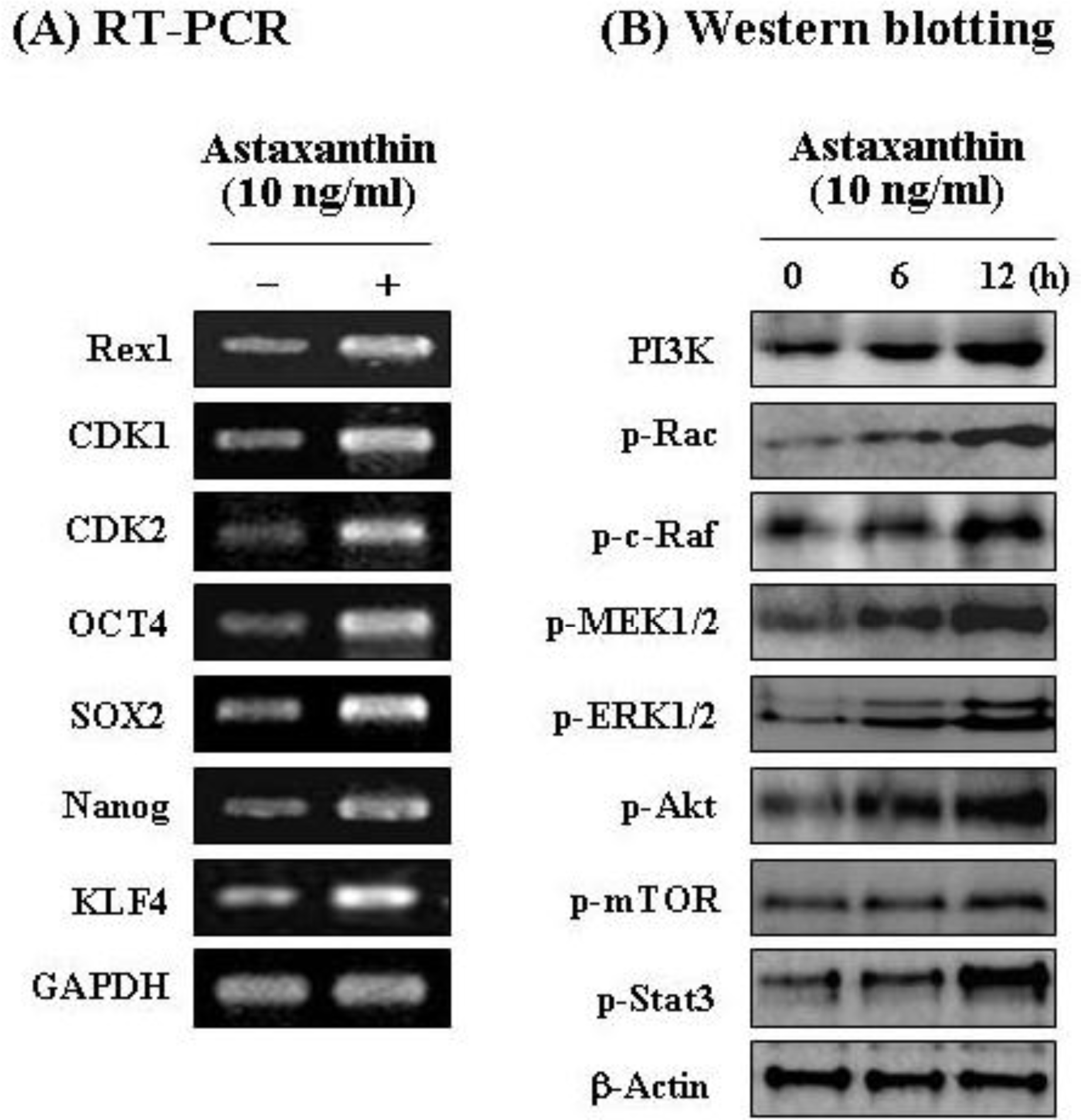
2.1. Astaxanthin Induces Active Cell Proliferation and Improves Stem Cell Potency in NPCs via Stemness Acting Signals
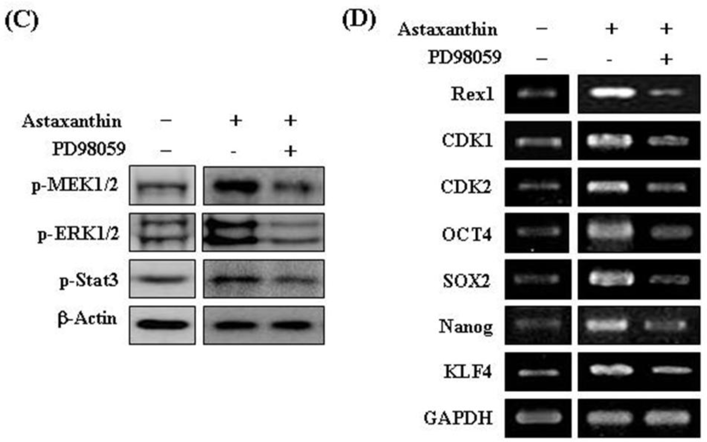
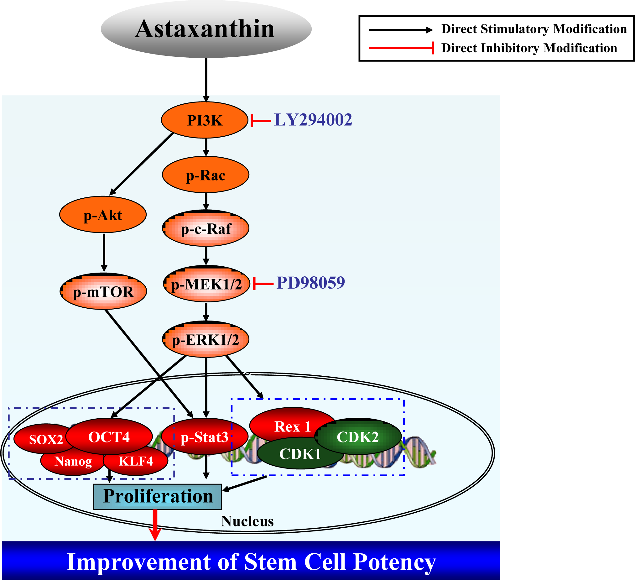
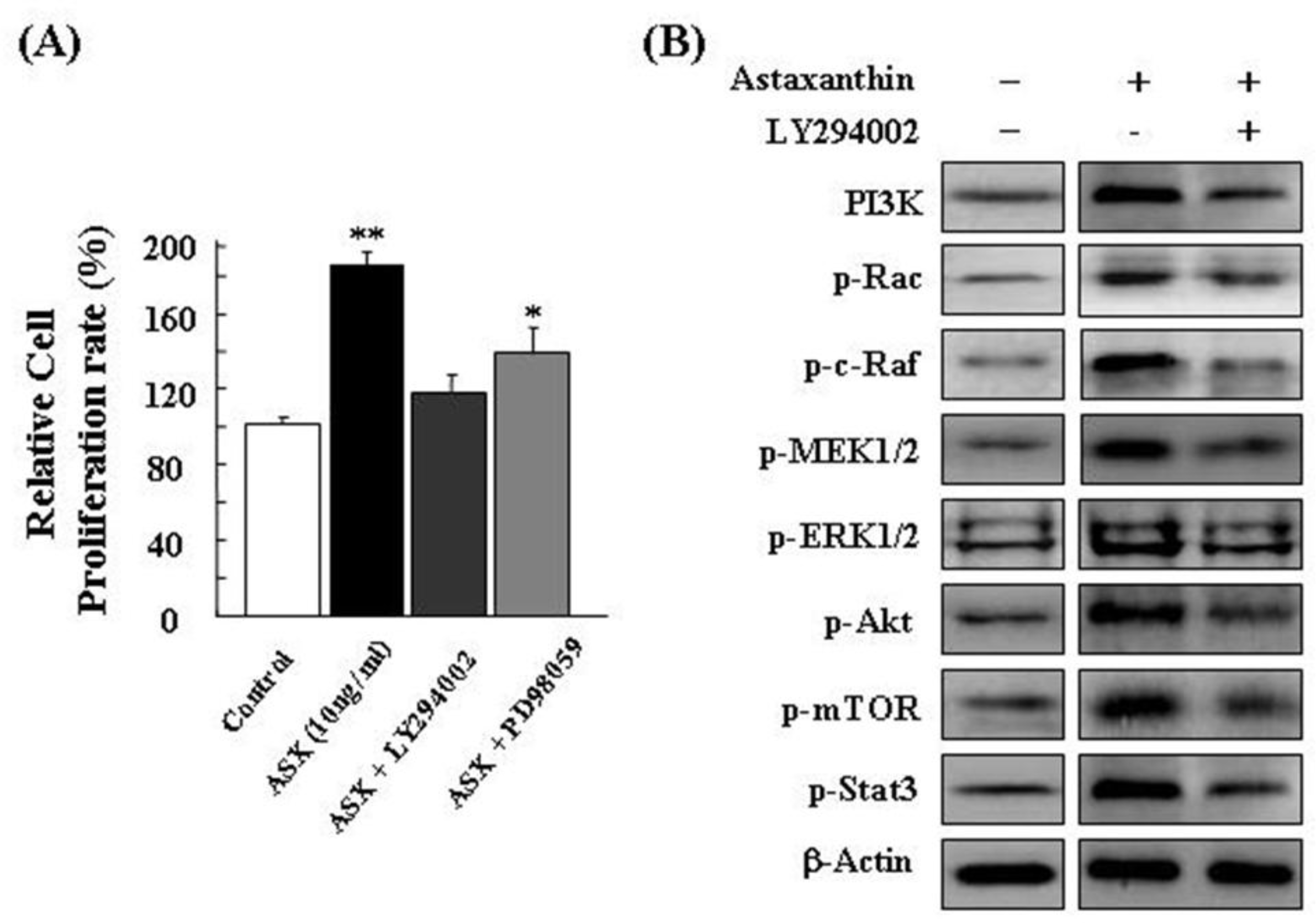
2.2. Astaxanthin Induces Proliferation of NPCs via PI3K and MEK Signaling Pathways
3. Experimental Section
3.1. Astaxanthin Reagent
3.2. Mouse Neural Progenitor Cells (NPCs) Culture
3.3. Selenium Treatment in NPCs and Analysis of Cell Viability
3.4. Colony-Forming Cell (CFU) Assay
3.5. Western Blot Analysis
3.6. Reverse Transcription-Polymerase Chain Reaction (RT-PCR)
3.7. Inhibition Assays
3.8. Statistical Analysis
4. Conclusions
Acknowledgments
References
- Gokhale, PJ; Andrews, PW. Human embryonic stem cells: 10 years on. Lab. Invest 2009, 89, 259–262. [Google Scholar]
- Lui, KO; Waldmann, H; Fairchild, PJ. Embryonic stem cells: overcoming the immunological barriers to cell replacement therapy. Curr. Stem Cell Res. Ther 2009, 4, 70–80. [Google Scholar]
- Singh, SR; Hou, SX. Multipotent stem cells in the Malpighian tubules of adult Drosophila. J. Exp. Biol 2009, 212, 413–423. [Google Scholar]
- Andrew, AG; Phaff, HJ; Starr, MP. Carotenoids of Phaffia rhodozyma, a red pigmented fermenting yeast. Phytochemistry 1976, 15, 1003–1007. [Google Scholar]
- Johnson, EA; An, GH. Astaxanthin from microbial sources. Crit. Rev. Biotechnol 1991, 11, 297–326. [Google Scholar]
- Johnson, EA; Schroeder, WA. Microbial carotenoids. Adv. Biochem. Eng 1995, 53, 119–178. [Google Scholar]
- Johnson, EA; Conklin, DE; Lewis, MJ. The yeast Phaffia rhodozyma as a dietary pigment source for salmonoids and crustaceans. J. Fish. Res. Board Can 1997, 34, 2417–2421. [Google Scholar]
- Johnson, EA; Lewis, MJ. Astaxanthin formation by the yeast Phaffia rhodozyma. J. Gen. Microbiol 1979, 115, 173–183. [Google Scholar]
- Lu, YP; Liu, SY; Sun, H; Wu, XM; Li, JJ; Zhu, L. Neuroprotective effect of astaxanthin on H2O2-induced neurotoxicity in vitro and on focal cerebral ischemia in vivo. Brain Res 2010, 11, 40–48. [Google Scholar]
- Lee, DH; Lee, YJ; Kwon, KH. Neuroprotective effects of astaxanthin in oxygen-glucose deprivation in SH-SY5Y cells and global cerebral ischemia in rat. J. Clin. Biochem. Nutr 2010, 47, 121–129. [Google Scholar]
- Kim, JH; Choi, W; Lee, JH; Jeon, SJ; Choi, YH; Kim, BW; Chang, HI; Nam, SW. Astaxanthin inhibits H2O2-mediated apoptotic cell death in mouse neural progenitor cells via modulation of P38 and MEK signaling pathways. J. Microbiol. Biotechnol 2009, 19, 1355–1363. [Google Scholar]
- Chan, KC; Mong, MC; Yin, MC. Antioxidative and anti-inflammatory neuroprotective effects of astaxanthin and canthaxanthin in nerve growth factor differentiated PC12 cells. J. Food Sci 2009, 74, H225–H231. [Google Scholar]
- Shen, H; Kuo, CC; Chou, J; Delvolve, A; Jackson, SN; Post, J; Woods, AS; Hoffer, BJ; Wang, Y; Harvey, BK. Astaxanthin reduces ischemic brain injury in adult rats. FASEB J 2009, 23, 1958–1968. [Google Scholar]
- Jyonouchi, H; Zhang, L; Gross, M; Tomita, Y. Immunomodulating actions of carotenoids: enhancement of in vivo and in vitro antibody production to T-dependent antigens. Nutr. Cancer 1994, 21, 47–58. [Google Scholar]
- Okai, Y; Higashi-Okai, K. Possible immunomodulating activities of carotenoids in in vitro cell culture experiments. Int. J. Immunopharmacol 1996, 8, 753–758. [Google Scholar]
- Tanaka, T; Morishita, Y; Suzuki, M; Kojima, T; Okumura, A; Mori, H. Chemoprevension of mouse urinary bladder carcinogenesis by the naturally occurring carotenoid astaxanthin. Carcinogenesis 1994, 15, 15–19. [Google Scholar]
- Jyonouchi, H; Sun, S; Lijima, K; Gross, MD. Antitumor activity of astaxanthin and its mode of action. Nutr. Cancer 2000, 36, 59–65. [Google Scholar]
- Kim, JH; Choi, SK; Choi, SY; Kim, HK; Chang, HI. Suppressive effect of astaxanthin isolated from the Xanthophyllomyces dendrorhous mutant on ethanol-induced gastric mucosal injury in rats. Biosci. Biotechnol. Biochem 2005, 69, 1300–1305. [Google Scholar]
- Kim, JH; Kim, YS; Song, GG; Park, JJ; Chang, HI. Protective effect of astaxanthin on naproxen-induced gastric antral ulceration in rats. Eur. J. Pharmacol 2005, 514, 53–59. [Google Scholar]
- Kurashige, M; Okimasu, E; Inoue, M; Utsumi, K. Inhibition of oxidative injury of biological membranes by astaxanthin. Physiol. Chem. Phys. Med. NMR 1990, 22, 27–38. [Google Scholar]
- Naguib, YM. Antioxidant activities of astaxanthin and related carotenoids. J. Agric. Food. Chem 2000, 48, 1150–1154. [Google Scholar]
- Lim, BP; Nagao, A; Terao, J; Tanaka, K; Suzuki, T; Takama, K. Antioxidant activity of xanthophylls on peroxyl radical mediated phospholipid peroxidation. Biochim. Biophys. Acta 1992, 1126, 178–184. [Google Scholar]
- Palozza, P; Krinsky, NI. Astaxanthin and canthaxanthin are potent antioxidants in a membrane model. Arch. Biochem. Biophys 1992, 297, 291–295. [Google Scholar]
- Nakajima, Y; Inokuchi, Y; Shimazawa, M; Otsubo, K; Ishibashi, T; Hara, H. Astaxanthin, a dietary carotenoid, protects retinal cells against oxidative stress in vitro and in mice in vivo. J. Pharm. Pharmacol 2008, 60, 1365–1374. [Google Scholar]
- Forte, A; Schettino, MT; Finicelli, M; Cipollaro, M; Colacurci, N; Cobellis, L; Galderisi, U. Expression pattern of stemness-related genes in human endometrial and endometriotic tissues. Mol. Med 2009, 15, 392–401. [Google Scholar]
- Dhodapkar, MV. Immunity to stemness genes in human cancer. Curr. Opin. Immunol 2010, 22, 245–250. [Google Scholar]
- Kim, JH; Lee, MR; Kim, JH; Jee, MK; Kang, SK. Selenium induces improvement of stem cell behaviors in human adipose-tissue stromal cells via SAPK/JNK and stemness acting signals. Stem Cells 2008, 26, 2724–2734. [Google Scholar]
- Lin, T; Chao, C; Saito, S. p53 induces differentiation of mouse embryonic stem cells by suppressing Nanog expression. Nat. Cell. Biol 2005, 7, 165–171. [Google Scholar]
- Chan, S. Targeting the mammalian target of rapamycin (mTOR): a new approach to treating cancer. Br. J. Cancer 2004, 91, 1420–1424. [Google Scholar]
- Castro-Malaspina, H; Gay, RE; Resnick, G; Kapoor, N; Meyers, P; Chiarieri, D; McKenzie, S; Broxmeyer, HE; Moore, MA. Characterization of human bone marrow fibroblast colony-forming cells (CFU-F) and their progeny. Blood 1980, 56, 289–301. [Google Scholar]
- Jang, KJ; Han, MH; Lee, BH; Kim, BW; Kim, CH; Yoon, HM; Choi, YH. Induction of apoptosis by ethanol extracts of Ganoderma lucidum in human gastric carcinoma cells. J. Acupunct. Meridian Stud 2010, 3, 24–31. [Google Scholar]
- Kim, KO; Park, SY; Han, CW; Chung, HK; Ryu, DH; Han, JS. Effect of sildenafil citrate on interleukin-1-induced nitric oxide synthesis and iNOS expression in SW982 cells. Exp. Mol. Med 2008, 40, 286–293. [Google Scholar]


© 2010 by the authors; licensee MDPI, Basel, Switzerland. This article is an open-access article distributed under the terms and conditions of the Creative Commons Attribution license (http://creativecommons.org/licenses/by/3.0/).
Share and Cite
Kim, J.-H.; Nam, S.-W.; Kim, B.-W.; Choi, W.; Lee, J.-H.; Kim, W.-J.; Choi, Y.-H. Astaxanthin Improves Stem Cell Potency via an Increase in the Proliferation of Neural Progenitor Cells. Int. J. Mol. Sci. 2010, 11, 5109-5119. https://doi.org/10.3390/ijms11125109
Kim J-H, Nam S-W, Kim B-W, Choi W, Lee J-H, Kim W-J, Choi Y-H. Astaxanthin Improves Stem Cell Potency via an Increase in the Proliferation of Neural Progenitor Cells. International Journal of Molecular Sciences. 2010; 11(12):5109-5119. https://doi.org/10.3390/ijms11125109
Chicago/Turabian StyleKim, Jeong-Hwan, Soo-Wan Nam, Byung-Woo Kim, Woobong Choi, Jong-Hwan Lee, Wun-Jae Kim, and Yung-Hyun Choi. 2010. "Astaxanthin Improves Stem Cell Potency via an Increase in the Proliferation of Neural Progenitor Cells" International Journal of Molecular Sciences 11, no. 12: 5109-5119. https://doi.org/10.3390/ijms11125109