Article

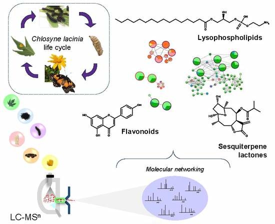
Natural Products Diversity in Plant-Insect Interaction between Tithonia diversifolia (Asteraceae) and Chlosyne lacinia (Nymphalidae)

by
Marília Elias Gallon
1,
Eduardo Afonso Silva-Junior
1,2,
Juliano Geraldo Amaral
3,
Norberto Peporine Lopes
1 and
Leonardo Gobbo-Neto
1,*
1
Núcleo de Pesquisa em Produtos Naturais e Sintéticos, School of Pharmaceutical Sciences of Ribeirão Preto, University of São Paulo, Av. do Café s/n°, Ribeirão Preto, SP 14040-903, Brazil
2
Centro Universitário do Vale do Araguaia, R. Moreira Cabral, Barra do Garças, MT 78600-000, Brazil
3
Instituto Multidisciplinar em Saúde–Campus Anísio Teixeira, Universidade Federal da Bahia, R. Hormindo Barros, 58, Qd 17, Lt 58, Vitória da Conquista, BA 45029-094, Brazil
*
Author to whom correspondence should be addressed.
Molecules 2019, 24(17), 3118; https://doi.org/10.3390/molecules24173118
Submission received: 5 July 2019 / Revised: 17 August 2019 / Accepted: 19 August 2019 / Published: 28 August 2019
(This article belongs to the Section Natural Products Chemistry)

Abstract

:
The chemical ecology of plant-insect interactions has been driving our understanding of ecosystem evolution into a more comprehensive context. Chlosyne lacinia (Lepidoptera: Nymphalidae) is an olygophagous insect herbivore, which mainly uses host plants of Heliantheae tribe (Asteraceae). Herein, plant-insect interaction between Tithonia diversifolia (Heliantheae) and Chlosyne lacinia was investigated by means of untargeted LC-MS/MS based metabolomics and molecular networking, which aims to explore its inherent chemical diversity. C. lacinia larvae that were fed with T. diversifolia leaves developed until fifth instar and completed metamorphosis to the adult phase. Sesquiterpene lactones (STL), flavonoids, and lipid derivatives were putatively annotated in T. diversifolia (leaves and non-consumed abaxial surface) and C. lacinia (feces, larvae, pupae, butterflies, and eggs) samples. We found that several furanoheliangolide-type STL that were detected in T. diversifolia were ingested and excreted in their intact form by C. lacinia larvae. Hence, C. lacinia caterpillars may have, over the years, developed tolerance mechanisms for STL throughout effective barriers in their digestive canal. Flavonoid aglycones were mainly found in T. diversifolia samples, while their glycosides were mostly detected in C. lacinia feces, which indicated that the main mechanism for excreting the consumed flavonoids was through their glycosylation. Moreover, lysophospholipids were predominately found in C. lacinia samples, which suggested that they were essential metabolites during pupal and adult stages. These findings provide insights into the natural products diversity of this plant-insect interaction and contribute to uncovering its ecological roles.

Graphical Abstract

1. Introduction

The integration of ecological and chemical researches has enlightened how plant-insect interactions shape community dynamics and ecosystem evolution [1,2]. Recently, plant phytochemical diversity has been directly and positively correlated with the diversity of plant-associated insects [3]. In this context, the metabolomics approach is a valuable tool for unraveling biological interfaces, including the ecological bases for interactions among metabolites and the interplay between plants and insects [4,5,6,7].
Secondary metabolites (e.g., alkaloids, terpenoids and glucosinolates) have been extensively studied as important plant defensive metabolites, while the role that primary metabolites play in plant-insect interactions remains less explored [8]. Seasonal and spatial effects on plant secondary metabolite contents were associated with variation in the concentrations of chlorogenic acids and flavonoids [9]. Additionally, plants that were collected at the interface between two forest ecosystems exhibited an amplification of defensive compounds, which was related to sesquiterpene lactones (STL) content and then confirmed by the maximal cytotoxic activity [10]. Thus, studies that are based on several analytical platforms for the exploration of primary and secondary metabolites are essential for expanding the coverage of the analysis and to enable a more comprehensive understanding of these interactions [11,12].
Tithonia diversifolia (Hemsl.) A. Gray belongs to Heliantheae tribe (Asteraceae family) and it is chemically characterized by its ability to biosynthesize flavonoids, hydroxycinnamic acid derivatives, and STL, especially heliangolides, furanoheliangolides, and eudesmanolides [13,14,15]. The leaves of T. diversifolia present several ethnobotanical uses and biological activities (e.g., anti-inflammatory, antimalarial, analgesic, and antimicrobial activities), which are associated with their constituents [16,17]. Chlosyne lacinia (Geyer), popularly known as bordered path or “sunflower caterpillar”, is an oligophagous insect that feeds mostly on plants belonging to Heliantheae tribe. Butterfly wings are mainly black with yellow-orange spots of varying sizes [18,19,20]. Its larvae pass through five instars and they are usually black with dorsal orange stripes, however color polymorphism is significant within populations [21,22]. In Brazil, T. diversifolia leaves herbivory by C. lacinia mainly occurs from April to June (autumn) [23].
Herein, we explored the plant-insect interaction between T. diversifolia and C. lacinia by means of insect performance bioassay, untargeted LC-MSn, and molecular networking analyses. We thereby unraveled the natural products diversity in this plant-insect interaction and investigated the influence of the plant metabolites on the insect metabolism.

2. Results and Discussion

2.1. C. lacinia Development

C. lacinia larvae that were fed with T. diversifolia leaves were reared in the laboratory (16/8 h of light/dark; 25 ± 2 °C) and exhibited a survival rate of 100%. Larvae passed through five instars and each took about four days (Table 1), as expected from previous reports [18,20]. Pupation occurred for approximately eight days and all the pupae were able to complete metamorphosis to the adult phase. C. lacinia butterflies lived for almost 12 days. During the adult life stage, butterflies mated and later laid their eggs on T. diversifolia leaves.

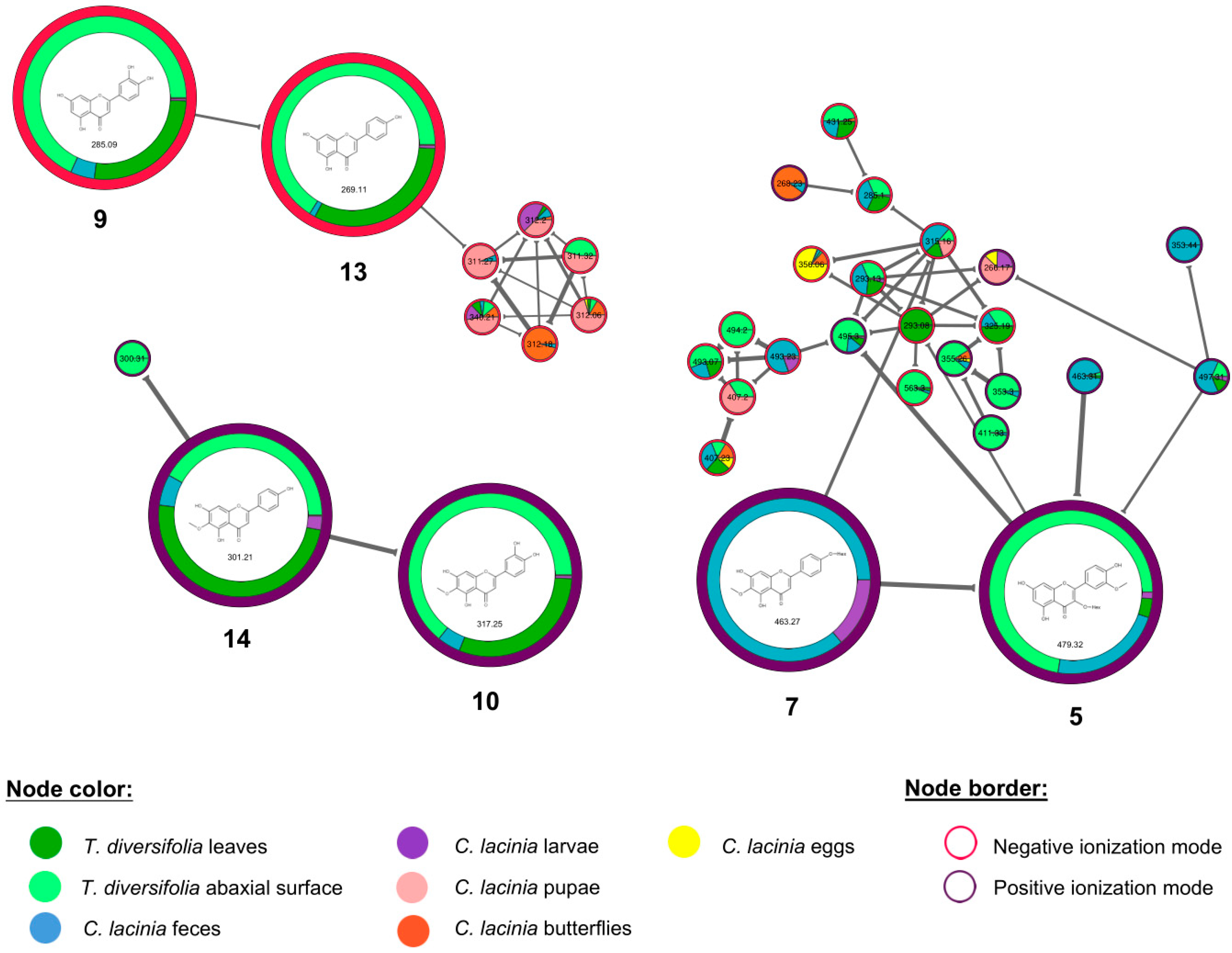
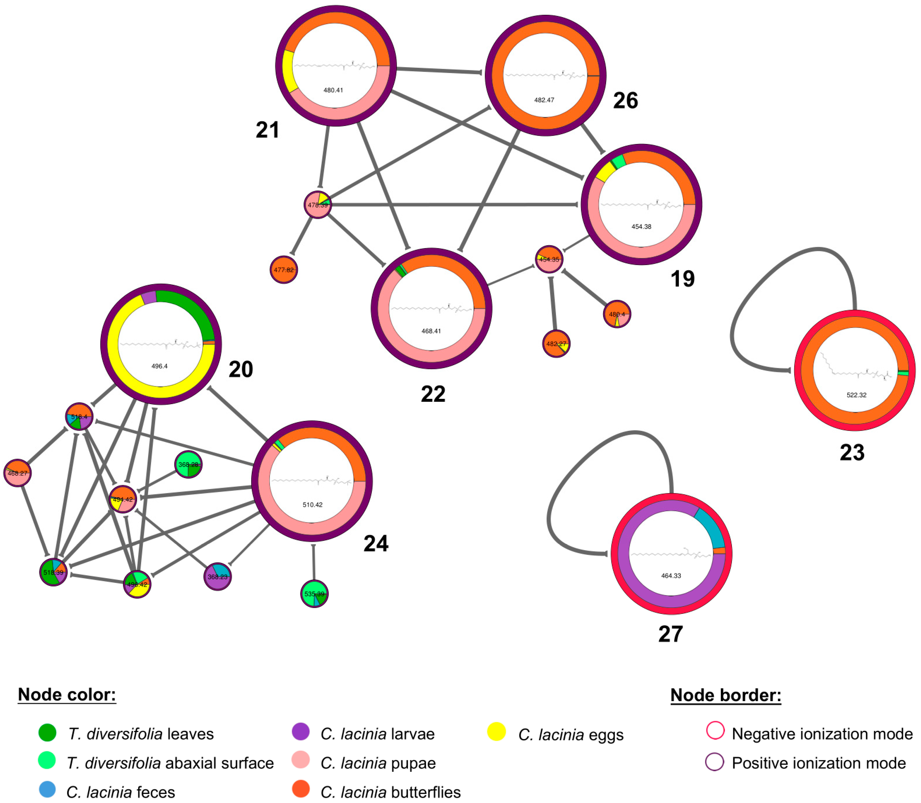
2.2. Natural Products Diversity

The chemical profiles of T. diversifolia samples (leaves and non-consumed abaxial surface) and C. lacinia samples (feces, larvae, pupae, butterflies, and eggs) were clearly distinct (Supplementary Material, Figures S1 and S2). In general, T. diversifolia samples and C. lacinia feces exhibited polar compounds as the major ones, especially STL and flavonoids. C. lacinia pupae, butterflies, and eggs were characterized by the presence of medium polarity compounds, mainly lysophospholipids. Besides, C. lacinia feces and larvae showed medium polarity compounds, as well as low polarity unidentified compounds.
The heatmap that is based on mass features showed that the T. diversifolia samples were clustered apart from C. lacinia, which indicated that there were significant chemical differences between plant and insect (Figure 1). Among C. lacinia samples, feces samples exhibited several unique metabolites and they were clustered apart from other C. lacinia samples (larvae, pupae, butterflies, and eggs), which exhibited some exclusive metabolites but also shared some.
Molecular networking was built with the MS data that were generated in positive and negative ionization modes, aiming to better represent the chemical diversity in this plant-insect interaction and to allow the annotation of as many compounds as possible (Figure 2) [24]. Searching against the GNPS library and previous literature reports, we were able to putatively annotate 28 compounds, according to the minimum standards that are required for level 2 non-novel metabolite identification proposed by Sumner et al. (2007) (Table 2) [25,26]. The spectrometric and spectroscopic information that were used to annotate these compounds were presented at Supplementary Material (Table S1).
Seven furanoheliangolides-type STL, typical of Heliantheae species, were putatively annotated (6, 8, 11, 12, 15, 16, and 17). We were able to identify these STL with a high confidence level when considering the GNPS information with the main fragmentation pathways and diagnostic product ions described in the literature for furanoheliangolides [27]. STL were mainly detected in T. diversifolia leaves and abaxial surface (Figure 3). They were also found in C. lacinia samples, especially C. lacinia feces. Hydrocortisone, which is used as internal standard, was clustered together with the annotated STL. This proximity can be explained by the same biosynthetic origin that these compounds share (mevalonate/methylerythritol phosphate pathway) [28,29].
STL are representative secondary metabolites of Asteraceae family and exhibit particular distribution within its tribes. Several chemotaxonomic studies have applied the differential incidence of their skeleton subtype to deduce taxonomic relationships [30,31,32]. For instance, Vernonieae species are characterized by the presence of glaucolides, hirsutinolides, furanoheliangolides, and eremantholides; the Eupatorieae tribe is characterized by guaianolides, heliangolides, and mikanolides; and, the Heliantheae tribe, which comprises T. diversifolia, is characterized by eudesmanolides, furanoheliangolides, heliangolides, and melampolides [33,34]. More than their chemotaxonomic significance, STL play important roles in plant protection against herbivores and microorganisms, as well as in plant growth inhibition [35].
Field observations have shown that, during their early instars, C. lacinia larvae exclusively fed on the adaxial surface of T. diversifolia leaves [23]. This finding was associated with a behavioral strategy to avoid or reduce the intake of STL-rich glandular trichomes present in the abaxial surface of T. diversifolia, which may be toxic to C. lacinia. We found out that several STL were detected in C. lacinia feces during the third, fourth, and fifth larval instars, which indicated that C. lacinia caterpillars ingested and excreted them. Additionally, the relative abundances of some STL were higher in the whole leaves of T. diversifolia rather than the non-consumed abaxial surface. Thus, our results suggested that most likely C. lacinia larvae exclusively feeding on T. diversifolia adaxial surface during their early instars is not related with avoiding toxic metabolite strategy.
Previous studies have suggested that glaucolide- and hirsutinolide-type STL may be toxic to C. lacinia larvae, while the larvae may have developed tolerance mechanisms to deterrent compounds that are present in T. diversifolia (especially heliangolide- and furanoheliangolide-type STL) [36]. Herein, we only annotated STL typical of Heliantheae tribe (furanoheliangolide-type), which, as expected, were not detrimental to C. lacinia development. According to Ahern & Whitney (2014), the stereochemistry of the lactone ring (cis- or trans-fused) in STL was associated with herbivore resistance and plant fitness. Plants that exhibited xanthanolide-type STL with cis-fused lactone ring suffered higher levels of herbivore damage than plants with trans-fused STL [37]. Thus, the difference between the lactone moiety in glaucolide/hirsutinolide-type STLs (with endocyclic double bond) and heliangolide/furanoheliangolide-type STL (trans-fused γ-lactone ring) might explain the preference of C. lacinia for Heliantheae species. However, further studies, including a wider variety of STL skeletons, are required to better understand if specific subtypes are responsible for toxic and/or deterrent activity of plants and how they influence herbivore-feeding behavior.
Additionally, by correlating the putatively annotated STL with mass differences of unknown metabolite nodes in the molecular networking, we annotated a putative tagitinin derivative STL lacking one OH group (m/z 353), which was predominantly detected in C. lacinia samples (feces, larvae and pupae) (Figure 3). The exact position of the missing OH could not be attributed. Thus, our results suggested that dehydroxylation may have been a potential mechanism for the excretion of STL, since a putative dehydroxylated STL was mostly detected in C. lacinia feces.
STL usually act as plant defensive compounds against insects through an unspecific mechanism of alkylation. The toxic effects of STL on insects were associated with morphological deformities, delayed molting, and impaired metamorphosis [38]. According to Isman (1985), some insects have developed tolerance mechanisms for toxic STL throughout the adaptation of the integument and alimentary canal, which provide an effective physical barrier for the absorption of STL and reduce their bioavailability [39]. Our data agreed with these findings, since the STL were mainly detected in C. lacinia feces in their intact form, which suggested that they were not absorbed from the insect digestive canal.
Six flavonoids, including four flavones (9, 10, 13, and 14) and two flavonoids O-glycosides (5 and 7), were also putatively annotated. Flavones were found especially in T. diversifolia leaves and abaxial surface, while flavonoids O-glycosides were mainly found in C. lacinia feces and larvae (Figure 4). For instance, hispidulin (14) was found at higher relative abundance in T. diversifolia leaves and abaxial surface and at lower abundance in C. lacinia feces and larvae, while its glycosylated flavone (7) was exclusively found in C. lacinia feces and larvae. These results showed that C. lacinia caterpillar excreted a minor part of the consumed flavones in their intact form and a major part was metabolized and excreted as flavone O-glycosides.
Flavonoids are present in plant leaves and they are known by their ability to protect plant tissue against the damaging effects of ultraviolet radiation [40,41,42]. Flavonoids also play an important role in the ecology of plant-insect interactions, especially regarding insect feeding and oviposition behavior. In spite of this, some flavonoid aglycones (e.g., quercetin) were pointed out as being responsible for detrimental effects on the development of insect larvae, because they may inhibit mitochondrial ATPase and cytochrome P450-dependent mixed function oxidases [43]. Moreover, hispidulin has exhibited insect larvicidal properties [44]. This toxicity and inhibitory activity of aglycones might explain why hispidulin was metabolized to hispidulin 4′-O-hexoside before its excretion.
Along with, Martucci et al. (2016) suggested that glucuronyl flavonoids from Vernonanthura phosphorica may be toxic to C. lacinia larvae [36]. We found that flavone O-glycosides were detected mainly in C. lacinia feces, which suggested that glycosylation was the main metabolism mechanism for excretion of the consumed flavonoid aglycones. Besides, these findings corroborated the potentially toxic activity of glucuronyl flavonoids that was previously described, since C. lacinia larvae excreted them.
Host-plant selection by herbivores is widely influenced by plant chemical diversity. While several ordinary plant compounds exist, flavonoid profiles usually vary among families, genera and species. This suggests that flavonoids can modulate host selection; however, there are still few studies in this area [45,46].
Additionally, eight lysophospholipids were putatively annotated, including five phosphatidylethanolamines (19, 21, 22, 26, and 27), two phosphatidylcholines (20 and 24), and one phosphatidylserine (23). In general, these lysophospholipids were found in all samples; however, they were found in higher relative abundance in C. lacinia samples. Interestingly, they were clustered according to their headgroup that was attached to the phosphate group (ethanolamine, choline, or serine), forming separate clusters in the molecular networking (Figure 5).
Phospholipids are widely known by their structural function, i.e., they are the primary building blocks for cell membranes. Phosphatidylcholines and phosphatidylethanolamines are the most abundant phospholipids in insects. The proportion of each one of them is species specific and also depends on the insect physiological state [47,48]. Lysophospholipids are derivatives of phospholipids, in which one of the acyl groups have been removed by hydrolysis (containing only one fatty acid and a phosphate group). Our results suggest that lysophospholipids may play a role in C. lacinia metabolism, because they were predominantly found in insect samples.
Dietary lipid demands vary substantially between insect species. Usually, linoleic or linolenic acids adequately satisfy this nutritional demand. The lack of essential lipids may cause several developmental and reproduction deformations. Many insects are able to biosynthesize lipids; hence, part of the required lipids is obtained from insect diet and the other part from insect lipogenic ability [49,50]. The ability of C. lacinia to synthesize lipids was evidenced herein, since a greater variety of lysophospholipids was detected in C. lacinia samples (especially pupae and butterflies) rather than T. diversifolia leaves. Additionally, lysophospholipids were found at higher abundance in C. lacinia and at minor levels in T. diversifolia samples.
Over the years, lipid metabolism in insects has been extensively studied due to the importance of lipids as substrate for metamorphosis, egg development, and flight. Besides, several insect pheromones and growth hormones exhibit lipophilic character [51,52]. Recently, lysophosphatidylcholine has been associated with antimicrobial activity in adult honeybees [53]. We found out that, in general, the putatively annotated lysophospholipids were detected in higher abundance in C. lacinia pupae and butterflies. Taken together, our findings suggested that lysophospholipids, along with their structural function, may play important roles by protecting the insects against microorganisms, as corroborated by previous reports [53], during pupation and by acting as an energy source for diapause and metamorphosis. Moreover, 1-(9-octadecenoyl)-glycero-3-phosphoserine (23) and 1-stearoyl-2-hydroxy-glycero-3-phosphoethanolamine (26) were predominately found in C. lacinia butterflies, which indicated that these metabolites may be important substrates for flight in the adult life stage, as previously reported [51,52].
The amount of lipids varies considerably during insect life and between different species. Few generalizations can be drawn regarding all the members of this animal class due to several metabolic variations that are brought about by different behavioral characteristics and ecological niches. These facts emphasize the importance of gathering specific plant-insect interactions into a broad background before drawing general conclusions.
Additionally, we putatively annotated two amino acids (2 and 4), two vitamins (1 and 3), and three fatty acid derivatives (18, 25, and 28). Tryptophan (2) and phenylalanine-acetyl (4) were found in T. diversifolia and C. lacinia samples, especially in pupae. Pantothenic acid (1) was detected in all samples; however, it was found at higher abundance in C. lacinia pupae and butterflies. Riboflavin (3) was found in T. diversifolia leaves and mainly in C. lacinia butterflies. These findings suggested that the annotated amino acids and vitamins were essential during all life stages, but mostly during pupation and the adult life stage. Moreover, riboflavin may be the major compound that is responsible for the characteristic yellow-orange color of butterfly wings [54].
12,13-DiHome (18) was detected in all samples and it may be a precursor for other lipid derivatives. Linolenoyl-tyrosine (25) was exclusively found in C. lacinia samples (feces, larvae, and pupae), which indicated that it was produced by C. lacinia during their development. Moreover, it was observed in this study that oleamide (28), which is a fatty acid primary amide, was mostly detected in C. lacinia eggs.
In summary, lipid derivatives were biosynthesized during the insect life cycle and they played essential functions in C. lacinia development. Typical STL and flavonoids from T. diversifolia probably act as defensive plant metabolites, protecting T. diversifolia against external damage (e.g., herbivores, ultraviolet radiation). Whereas, C. lacinia may have, over the years, developed a strategy to reduce the bioavailability of furanoheliangolide-type STL through effective barriers in the insect digestive canal, since STL were excreted in their intact form by C. lacinia larvae. Altogether, our findings enlightened the main metabolism mechanisms of STL, flavonoids, and lipid derivatives in T. diversifolia and C. lacinia plant-insect interaction and provided insights into the ecological roles for interactions among metabolites.

3. Material and Methods

3.1. Experimental Design

The egg masses of C. lacinia were collected in the field (21.0°10.0′9.3″ S; 47.0°50.0′48.8″ W; Medicinal Plant Garden of the School of Pharmaceutical Sciences of Ribeirão Preto, University of São Paulo, Ribeirão Preto, SP, Brazil), on the adaxial surfaces of T. diversifolia leaves. One egg mass was selected and maintained in a rearing box (size 30 × 25 × 20 cm, plastic floor and sides, nylon mesh top) with T. diversifolia fresh leaves. Seven days after the egg eclosion, 20 individuals of C. lacinia larvae in the second instar were randomly selected and maintained in other rearing box at laboratory conditions (16/8 h of light/dark; temperature of 25 ± 2 °C). Fresh leaves of T. diversifolia were collected in the same place that was reported for the egg masses and provided daily until the pupal stage. Butterflies were fed with honey:water solution (1:10, v/v).
C. lacinia larval development was measured by means of survival rate from larvae to pupae and conversion from pupae to butterflies.
C. lacinia that were fed with T. diversifolia leaves were sampled at different stages of development, including third instar larvae (n = 5), pupae (n = 5), butterflies (n = 5), and eggs (one egg mass). Additionally, C. lacinia feces were sampled at third, fourth, and fifth larval instars. For the chemical analysis, three leaves from the same T. diversifolia plant were collected on three different days at the Medicinal Plant Garden of the School of Pharmaceutical Sciences of Ribeirão Preto, University of São Paulo, Ribeirão Preto, SP, Brazil. Additionally, during the larval development experiment, we sampled, on three different days, the surfaces of T. diversifolia leaves that were not consumed by C. lacinia larvae (non-consumed abaxial surfaces). All of the samples were immediately frozen and stored in a −20 °C freezer.

3.2. Sample Preparation

Before extraction, T. diversifolia (leaves and non-consumed abaxial surface) and C. lacinia (feces, larvae, pupae, and butterflies) samples were freeze-dried for 48 h and pulverized using mortar and pestle. Extracts were prepared using 50 mg of each sample and 1 mL of MeOH:H2O solution (7:3, v/v) with 10 µg·mL−1 hydrocortisone as internal standard, followed by vortex agitation and sonication. The extracts of T. diversifolia samples (whole leaves and non-consumed abaxial surface) were prepared in triplicates (n = 3). Extracts of C. lacinia feces were separately prepared for each feces instar sampled (third, fourth, and fifth). C. lacinia egg extract was prepared using a single egg mass (about 100 eggs) and the previously described procedure. After centrifugation, the supernatant was filtered through a 0.2 µm PTFE membrane into HPLC vials.

3.3. LC-MSn Analysis

Chemical profiles of T. diversifolia and C. lacinia samples were obtained on a Shimadzu UFLC system that was coupled to an ion trap mass spectrometer (AmaZon SL, Billerica, MA, USA). Separations were achieved while using a C18 Supelco column (Ascenti® Express C18, 5 µm, 15 cm × 3.0 mm); water with 0.1% of formic acid (A) and acetonitrile with 0.1% of formic acid (B) as mobile phase, with a flow rate of 1.0 mL·min−1. The gradient was 0–2 min, 10% B; 2–40 min, 10–100% B; 40–45 min, 100% B; 45–50 min, 100–10% B; 50–55 min, 10% B. Column oven was set at 40 °C and 20 µL of each sample were injected.
The ion trap mass spectrometer was operated in positive and negative ionization modes, separately. Instrument parameters were as follows: capillary voltage, of 3.5 kV; end plate offset, 500 V; nebulizer, 60 psi, dry gas (N2) flow, 11 L·min−1; dry temperature, 350 °C; auto MS/MS acquiring data between m/z 50 and 1300, average of three spectra; enhanced resolution for scan mode and UltraScan mode for MS/MS; spectra rate acquisition, three spectra/s; exclusion of a particular ion after three spectra and released after 30 s. The mass spectrometer was controlled by Hystar software (Bruker Daltonics Inc., Billerica, MA, USA).

3.4. Data Processing and Analysis

LC-MSn data of the positive and negative ionization modes, previously converted to .mzXML format while using MSconvert software (Proteowizard Software Foundation, Palo Alto, CA, USA), were separately preprocessed using MzMine TM (BMC Bioinformatics, London, UK) and employing the following parameters: mass detection using the centroid algorithm, scan MS level 1 (noise level, 1.0E3), and scan MS level 2 (noise level, 1.0E2); chromatogram builder (minimum time span in minutes, 0.1; minimum height, 2E3; m/z tolerance, 0.3 m/z or 20 ppm); chromatogram deconvolution using the baseline cut-off (minimum peak height, 2E3; peak duration, 0.1 to 1.0; baseline level, 1.0E3); isotopic peak grouper (m/z tolerance, 0.3 m/z or 20 ppm; retention time tolerance in minutes, 0.2; maximum charge, 2; representative isotope, most intense) alignment using the join aligner (m/z tolerance, 0.3 m/z or 20 ppm; weight for m/z, 50; retention time tolerance, 0.3 (abs); weight for retention time, 50 and filtering using peaks list rows filter (keep only peaks with MS2 scan). After processing, the data were exported as .csv table and as .mgf file for GNPS analysis.
The output data from positive and negative ionization modes were combined into a single .mgf file, as well as into a single .csv spreadsheet and then uploaded to the GNPS platform. Molecular networking was built using the “advanced analysis tools for feature networking” and the following parameters: precursor ion mass tolerance, 2.0 Da; fragment ion mass tolerance, 0.5 Da; minutes pairs cos, 0.6; minimum matched fragment ions, 4; maximum shift between precursors, 500 Da; network topK, 10; maximum connected component size, 100; library search minutes matched peaks, 4; score threshold, 0.7; search analogues, don’t search; maximum analog search mass difference, 100 Da; top results to report per query, 1; minimum peak intensity, 0; filter precursor window, filter; filter library, filter library; filter peaks in 500 Da window, filter; normalization per file, no norm; aggregation method for peak abundances per group, sum. Finally, molecular networking was generated and analyzed with Cytoscape version 3.8 (Institute for Systems Biology, Seattle, WA, USA).
All of the LC-MSn data were deposited on the Mass spectrometry Interactive Virtual Environment (MassIVE) at https://massive.ucsd.edu/ with the identifiers MSV000082571 (positive ionization mode) and MSV000082574 (negative ionization mode).
The online platform MetaboAnalyst (https://www.metaboanalyst.ca/faces/home.xhtml) was used to generate the heatmap. The same .csv spreadsheet used in the GNPS platform (comprising mass features in the positive and negative ionization modes) was uploaded to the statistical module, while using peak intensity table as data type and samples in rows (unpaired) as format. None data filtering was applied. Sample normalization was carried out using hydrocortisone as reference feature, followed by data auto scaling. Heatmap was built selecting the following parameters: distance measure, Euclidean; clustering Algorithm, Ward; data source, normalized data; standardization, autoscale features.

3.5. Chemical Analysis and Dereplication

The MS/MS spectra of molecular networking nodes were searched against GNPS spectral library and hit compounds were appointed. The compounds that matched with structurally characterized compounds from the GNPS spectral library were compared with additional information (UV data, fragmentation pattern and previous reports in the species) and then annotated. The stereochemistry of the sugar moieties that was indicated by the GNPS library was not considered in the compound annotation (i.e., sugars were characterized as hexosides, pentosides, deoxyhexosides, etc.).

Supplementary Materials

The following are available online, Figure S1: Base peak chromatograms of T. diversifolia and C. lacinia samples analyzed in the positive ionization mode title, Figure S2: Base peak chromatograms of T. diversifolia and C. lacinia samples analyzed in the negative ionization mode, Table S1: UV and MS spectral data of the annotated compounds in T. diversifolia and C. lacinia samples.

Author Contributions

M.E.G., E.A.S.-J. and L.G.-N. designed the experiments. M.E.G. and E.A.S.-J. performed the experiments. M.E.G., E.A.S.-J., J.G.A., N.P.L. and L.G.-N. analyzed the data and wrote the manuscript. All authors discussed and approved the manuscript.

Funding

The authors thank São Paulo Research Foundation (FAPESP)—grants #2017/17023-4 and #2014/50265-3 and National Council for Scientific and Technological Development (CNPq). This study was financed in part by the Coordenação de Aperfeiçoamento de Pessoal de Nível Superior—Brasil (CAPES)—Finance Code 001.

Acknowledgments

The authors thank SisGen for the authorization to access the samples from the Brazilian Genetic Heritage (A0AD857).

Conflicts of Interest

The authors declare no conflict of interest.

References

  1. Burkepile, D.E.; Parker, J.D. Recent advances in plant-herbivore interactions. F1000Research 2017, 6, 119. [Google Scholar] [CrossRef] [PubMed] [Green Version]
  2. Sedio, B.E. Recent breakthroughs in metabolomics promise to reveal the cryptic chemical traits that mediate plant community composition, character evolution and lineage diversification. New Phytol. 2017, 214, 952–958. [Google Scholar] [CrossRef] [PubMed] [Green Version]
  3. Richards, L.A.; Dyer, L.A.; Forister, M.L.; Smilanich, A.M.; Dodson, C.D.; Leonard, M.D.; Jeffrey, C.S. Phytochemical diversity drives plant–insect community diversity. Proc. Natl. Acad. Sci. USA 2015, 112, 10973–10978. [Google Scholar] [CrossRef] [PubMed]
  4. Maag, D.; Erb, M.; Glauser, G. Metabolomics in plant-herbivore interactions: Challenges and applications. Entomol. Exp. Appl. 2015, 157, 18–29. [Google Scholar] [CrossRef]
  5. Sardans, J.; Peñuelas, J.; Rivas-Ubach, A. Ecological metabolomics: Overview of current developments and future challenges. Chemoecology 2011, 21, 191–225. [Google Scholar] [CrossRef]
  6. Kuhlisch, C.; Pohnert, G. Metabolomics in chemical ecology. Nat. Prod. Rep. 2015, 32, 937–955. [Google Scholar] [CrossRef] [PubMed] [Green Version]
  7. Brunetti, A.E.; Neto, F.C.; Vera, M.C.; Taboada, C.; Pavarini, D.P.; Bauermeister, A.; Lopes, N.P. An integrative omics perspective for the analysis of chemical signals in ecological interactions. Chem. Soc. Rev. 2018, 47, 1574–1591. [Google Scholar] [CrossRef]
  8. Zhou, S.; Lou, Y.-R.; Tzin, V.; Jander, G. Alteration of plant primary metabolism in response to insect herbivory. Plant Physiol. 2015, 169, 1488–1498. [Google Scholar] [CrossRef] [Green Version]
  9. Gobbo-Neto, L.; Bauermeister, A.; Sakamoto, H.; Gouvea, D.; Lopes, J.; Lopes, N. Spatial and temporal variations in secondary metabolites content of the Brazilian arnica leaves (Lychnophora ericoides Mart., Asteraceae). J. Braz. Chem. Soc. 2017, 28, 2382–2390. [Google Scholar] [CrossRef]
  10. Gobbo-Neto, L.; Guaratini, T.; Pessoa, C.; De Moraes, M.O.; Costa-Lotufo, L.V.; Vieira, R.F.; Colepicolo, P.; Lopes, N.P. Differential metabolic and biological profiles of Lychnophora ericoides mart. (Asteraceae) from different localities in the Brazilian “campos rupestres”. J. Braz. Chem. Soc. 2010, 21, 750–759. [Google Scholar] [CrossRef]
  11. Barah, P.; Bones, A.M. Multidimensional approaches for studying plant defence against insects: From ecology to omics and synthetic biology. J. Exp. Bot. 2015, 66, 479–493. [Google Scholar] [CrossRef]
  12. Dyer, L.A.; Philbin, C.S.; Ochsenrider, K.M.; Richards, L.A.; Massad, T.J.; Smilanich, A.M.; Forister, M.L.; Parchman, T.L.; Galland, L.M.; Hurtado, P.J.; et al. Modern approaches to study plant–insect interactions in chemical ecology. Nat. Rev. Chem. 2018, 2, 50–64. [Google Scholar] [CrossRef]
  13. Baruah, N.C.; Sharma, R.P.; Madhusudanan, K.P.; Thyagarajan, G.; Herz, W.; Murari, R. Sesquiterpene lactones of Tithonia diversifolia. Stereochemistry of the tagitinins and related compounds. J. Org. Chem. 1979, 44, 1831–1835. [Google Scholar] [CrossRef]
  14. Zhao, G.-J.; Xi, Z.-X.; Chen, W.-S.; Li, X.; Sun, L.; Sun, L.-N. Chemical constituents from Tithonia diversifolia and their chemotaxonomic significance. Biochem. Syst. Ecol. 2012, 44, 250–254. [Google Scholar] [CrossRef]
  15. Sampaio, B.L.; Edrada-Ebel, R.; Da Costa, F.B. Effect of the environment on the secondary metabolic profile of Tithonia diversifolia: A model for environmental metabolomics of plants. Sci. Rep. 2016, 6, 29265. [Google Scholar] [CrossRef] [PubMed]
  16. Chagas-Paula, D.A.; Oliveira, R.B.; Rocha, B.A.; Da Costa, F.B.; Chagas-Paula, D.A. Ethnobotany, chemistry, and biological activities of the genus Tithonia (Asteraceae). Chem. Biodivers. 2012, 9, 210–235. [Google Scholar] [CrossRef] [PubMed]
  17. Ajao, A.A.; Moteetee, A.N. Tithonia diversifolia (Hemsl) A. Gray. (Asteraceae: Heliantheae), an invasive plant of significant ethnopharmacological importance: A review. S. Afr. J. Bot. 2017, 113, 396–403. [Google Scholar] [CrossRef]
  18. Drummond, B.A., III; Bush, G.L.; Emmel, T.C. The biology and laboratory culture of Chlosyne lacinia Geyer (Nymphalidae). J. Lepid. Soc. 1970, 24, 135–142. [Google Scholar]
  19. Justus, C.M.; Pasini, A.; De Oliveira, É.D. Biologia e preferência da lagarta do girassol, Chlosyne lacinia saundersii (Lepidoptera: Nymphalidae) na planta daninha losna branca, Parthenium hysterophorus (Asteraceae). Neotrop. Entomol. 2003, 32, 163–166. [Google Scholar] [CrossRef]
  20. Neck, R.W. Foodplant ecology of the butterfly Chlosyne lacinia (Geyer) (Nymphalidae). I. Larval foodplants. J. Lepid. Soc. 1973, 27, 22–33. [Google Scholar]
  21. Lopes-Da-Silva, M.; Casagrande, M.M. Color polymorphism and allele frequency in a Brazilian population of the sunflower caterpillar Chlosyne lacinia saundersi (Doubleday) (Lepidoptera: Nymphalidae). Neotrop. Entomol. 2003, 32, 159–161. [Google Scholar] [CrossRef]
  22. Neck, R.W. Larval morph variation in Chlosyne lacinia (Nymphalidae). J. Lepid. Soc. 1976, 30, 91–94. [Google Scholar]
  23. Ambrósio, S.R.; Oki, Y.; Heleno, V.C.G.; Chaves, J.S.; Nascimento, P.G.B.D.; Lichston, J.E.; Constantino, M.G.; Varanda, E.M.; Da Costa, F.B. Constituents of glandular trichomes of Tithonia diversifolia: Relationships to herbivory and antifeedant activity. Phytochemistry 2008, 69, 2052–2060. [Google Scholar] [CrossRef] [PubMed]
  24. Aksenov, A.A.; Da Silva, R.; Knight, R.; Lopes, N.P.; Dorrestein, P.C. Global chemical analysis of biology by mass spectrometry. Nat. Rev. Chem. 2017, 1, 54. [Google Scholar] [CrossRef]
  25. Sumner, L.W.; Amberg, A.; Barrett, D.; Beger, R.; Beale, M.H.; Daykin, C.A.; Fan, T.W.M.; Fiehn, O.; Goodacre, R.; Griffin, J.L.; et al. Proposed minimum reporting standards for chemical analysis: Chemical Analysis Working Group (CAWG) Metabolomics Standards Initiative (MSI). Metabolomics 2007, 3, 211–221. [Google Scholar] [CrossRef] [PubMed]
  26. Kind, T.; Fiehn, O. Strategies for dereplication of natural compounds using high-resolution tandem mass spectrometry. Phytochem. Lett. 2017, 21, 313–319. [Google Scholar] [CrossRef]
  27. Crotti, A.E.M.; Lopes, J.L.C.; Lopes, N.P. Triple quadrupole tandem mass spectrometry of sesquiterpene lactones: A study of goyazensolide and its congeners. J. Mass Spectrom. 2005, 40, 1030–1034. [Google Scholar] [CrossRef] [PubMed]
  28. Herz, W. Biogenetic aspects of sesquiterpene lactone chemistry. Isr. J. Chem. 1977, 16, 32–44. [Google Scholar] [CrossRef]
  29. Dewick, P.M. The mevalonate and methylerythritol phosphate pathways: Terpenoids and steroids. In Medicinal Natural Products; Wiley: Hoboken, NJ, USA, 2009; pp. 187–310. [Google Scholar]
  30. Seaman, F.C. Sesquiterpene lactones as taxonomic characters in the Asteraceae. Bot. Rev. 1982, 48, 121–594. [Google Scholar] [CrossRef]
  31. Emerenciano, V.D.P.; Ferreira, Z.S.; Kaplan, M.A.C.; Gottlieb, O.R. A chemosystematic analysis of tribes of Asteraceae involving sesquiterpene lactones and flavonoids. Phytochemistry 1987, 26, 3103–3115. [Google Scholar] [CrossRef]
  32. Zdero, C.; Bohlmann, F. Systematics and evolution within the Compositae, seen with the eyes of a chemist. Plant Syst. Evol. 1990, 171, 1–14. [Google Scholar] [CrossRef]
  33. Da Costa, F.; Terfloth, L.; Gasteiger, J. Sesquiterpene lactone-based classification of three Asteraceae tribes: A study based on self-organizing neural networks applied to chemosystematics. Phytochemistry 2005, 66, 345–353. [Google Scholar] [CrossRef] [PubMed]
  34. Gallon, M.E.; Monge, M.; Casoti, R.; Da Costa, F.B.; Semir, J.; Gobbo-Neto, L. Metabolomic analysis applied to chemosystematics and evolution of megadiverse Brazilian Vernonieae (Asteraceae). Phytochemistry 2018, 150, 93–105. [Google Scholar] [CrossRef] [PubMed]
  35. Padilla-Gonzalez, G.F.; Dos Santos, F.A.; Da Costa, F.B. Sesquiterpene lactones: More than protective plant compounds with High Toxicity. Crit. Rev. Plant Sci. 2016, 35, 1–20. [Google Scholar] [CrossRef]
  36. Martucci, M.E.P.; Gobbo-Neto, L. Differential secondary metabolite accumulation and performance of Chlosyne lacinia fed with Tithonia diversifolia or Vernonia polyanthes. Biochem. Syst. Ecol. 2016, 68, 156–162. [Google Scholar] [CrossRef]
  37. Ahern, J.R.; Whitney, K.D. Sesquiterpene lactone stereochemistry influences herbivore resistance and plant fitness in the field. Ann. Bot. 2014, 113, 731–740. [Google Scholar] [CrossRef] [PubMed]
  38. Amorim, M.H.R.; Gil Da Costa, R.M.; Lopes, C.; Bastos, M.M.S.M. Sesquiterpene lactones: Adverse health effects and toxicity mechanisms. Crit. Rev. Toxicol. 2013, 43, 559–579. [Google Scholar] [CrossRef]
  39. Isman, M.B. Toxicity and tolerance of sesquiterpene lactones in the migratory grasshopper, Melanoplus sanguinipes (Acrididae). Pestic. Biochem. Physiol. 1985, 24, 348–354. [Google Scholar] [CrossRef]
  40. Dewick, P.M. The shikimate pathway: Aromatic amino acids and phenylpropanoids. In Medicinal Natural Products; Wiley: Hoboken, NJ, USA, 2009; pp. 137–186. [Google Scholar]
  41. Bone, K.; Mills, S. Principles of herbal pharmacology. In Principles and Practice of Phytotherapy: Modern Herbal Medicine; Churchill Livingstone: New York, NY, USA, 2013; p. 1056. ISBN 9780443069925. [Google Scholar]
  42. Silva, D.B.; Turatti, I.C.C.; Gouveia, D.R.; Ernst, M.; Teixeira, S.P.; Lopes, N.P. Mass spectrometry of flavonoid vicenin-2, based sunlight barriers in Lychnophora species. Sci. Rep. 2014, 4, 4309. [Google Scholar] [CrossRef]
  43. Simmonds, M.S. Importance of flavonoids in insect-plant interactions: Feeding and oviposition. Phytochemistry 2001, 56, 245–252. [Google Scholar] [CrossRef]
  44. Patel, K.; Patel, D.K. Medicinal importance, pharmacological activities, and analytical aspects of hispidulin: A concise report. J. Tradit. Complement. Med. 2017, 7, 360–366. [Google Scholar] [CrossRef] [PubMed]
  45. Simmonds, M.S. Flavonoid–insect interactions: Recent advances in our knowledge. Phytochemistry 2003, 64, 21–30. [Google Scholar] [CrossRef]
  46. Preiß, S.; Degenhardt, J.; Gershenzon, J. Plant-animal dialogues. In Ecological Biochemistry: Enviromental and Interspecies Interactions; Wiley-VCH: Weinheim, Germany, 2014; pp. 312–330. ISBN 9783527322909. [Google Scholar]
  47. Downer, R.G.H.; Matthews, J.R. Patterns of lipid distribution and utilization in insects. Am. Zool. 1976, 16, 733–745. [Google Scholar] [CrossRef]
  48. Fast, P.G. A comparative study of the phospholipids and fatty acids of some insects. Lipids 1966, 1, 209–215. [Google Scholar] [CrossRef] [PubMed]
  49. Visser, B.; Willett, D.S.; Harvey, J.A.; Alborn, H.T. Concurrence in the ability for lipid synthesis between life stages in insects. R. Soc. Open Sci. 2017, 4, 160815. [Google Scholar] [CrossRef] [PubMed] [Green Version]
  50. Canavoso, L.E.; Jouni, Z.E.; Karnas, K.J.; Pennington, J.E.; Well, M.A. Fat metabolism in insects. Annu. Rev. Nutr. 2001, 21, 23–46. [Google Scholar] [CrossRef] [PubMed]
  51. Gilbert, L.I. Lipid Metabolism and Function in Insects. Adv. Insect Physiol. 1967, 4, 69–211. [Google Scholar]
  52. Arrese, E.L.; Soulages, J.L. Insect fat body: Energy, metabolism, and regulation. Annu. Rev. Entomol. 2010, 55, 207–225. [Google Scholar] [CrossRef]
  53. Riessberger-Gallé, U.; Hernández-López, J.; Rechberger, G.; Crailsheim, K.; Schuehly, W. Lysophosphatidylcholine acts in the constitutive immune defence against American foulbrood in adult honeybees. Sci. Rep. 2016, 6, 30699. [Google Scholar] [CrossRef] [Green Version]
  54. Pintea, A.M. Other natural pigments. In Food Colorants: Chemical and Functional Properties; Socaciu, C., Ed.; Taylor & Francis Group: Boca Raton, FL, USA, 2008; pp. 101–124. [Google Scholar]
Sample Availability: Samples are not available from the authors.
Figure 1. Heatmap based on mass features obtained by LC-MSn (IT) in positive and negative ionization modes. Td, T. diversifolia (green); Cl, C. lacinia (orange); Fe, feces (dark blue); AbSurf., abaxial surface. Numbers in parentheses represent the replicates.
Molecules 24 03118 g001
Figure 2. Molecular networking of T. diversifolia and C. lacinia samples analyzed by LC-MSn (IT) in the positive and negative ionization modes. Nodes represent detected compounds and are colored according to the type of sample. Borders of the nodes are colored according to the ionization mode in which the mass feature was obtained. Edges between nodes represent molecular structural similarity between compounds. Nodes without GNPS spectral library matching with other nodes are represented as self-loops (bottom), i.e., edges from a vertex to itself.
Molecules 24 03118 g002
Figure 3. Molecular networking highlighting the cluster of annotated sesquiterpene lactones. Nodes are colored according to the type of sample. Borders of the nodes are colored according to the ionization mode in which the mass feature was obtained. IS, internal standard.
Molecules 24 03118 g003
Figure 4. Molecular networking highlighting the cluster of annotated flavonoids. Nodes are colored according to the type of sample. Borders of the nodes are colored according to the ionization mode in which the mass feature was obtained.
Molecules 24 03118 g004
Figure 5. Molecular networking highlighting the clusters of annotated lysophospholipids. Nodes are colored according to the type of sample. Borders of the nodes are colored according to the ionization mode in which the mass feature was obtained.
Molecules 24 03118 g005
Table 1. Developmental stages of C. lacinia reared on T. diversifolia leaves in the laboratory and their respective duration.
Stage1st Instar2nd Instar3rd Instar4th Instar5th InstarPupationAdult StageTotal Development
Mean 15.253.703.253.453.557.6511.3538.20
SD 21.120.650.440.830.690.671.040.85
1 mean duration of insect instars in days; 2 standard deviation in days.
Table 2. Putatively annotated compounds in T. diversifolia and C. lacinia samples.
RtUsual Name
(InChI)
Compound ClassSamples
1.2pantothenic acid (1)
(1/C9H17NO5/c1-9(2,5-11)7(14)8(15)10-4-3-6(12)13/h7,11,14H,3-5H2,1-2H3,(H,10,15)(H,12,13)/t7-/s2)
vitaminT. diversifolia leaves and abaxial surface; C. lacinia feces, larvae, pupae, butterflies and eggs
1.4tryptophan (2)
(1/C11H12N2O2/c12-9(11(14)15)5-7-6-13-10-4-2-1-3-8(7)10/h1-4,6,9,13H,5,12H2,(H,14,15)/t9-/s2)
amino acidT. diversifolia leaves and abaxial surface; C. lacinia larvae, pupae and butterflies
3.2riboflavin (3)
(1/C17H22N4O6/c1-7-3-9-10(4-8(7)2)21(5-11(23)14(25)12(24)6-22)15-13(18-9)16(26)20-17(27)19-15/h3-4,11-12,14-15,22-25H,5-6H2,1-2H3,(H2,19,20,26,27)/t11-,12+,14-,15?/s2)
vitaminT. diversifolia leaves and abaxial surface; C. lacinia butterflies
4.8phenylalanine-acetyl (4)
(1/C11H13NO3/c1-8(13)12-10(11(14)15)7-9-5-3-2-4-6-9/h2-6,10H,7H2,1H3,(H,12,13)(H,14,15)/t10-/s2)
amino acidT. diversifolia leaves; C. lacinia larvae, pupae and butterflies
7.2isorhamnetin 3-O-hexoside (5)
(1/C22H22O12/c1-31-12-4-8(2-3-10(12)25)20-21(17(28)15-11(26)5-9(24)6-13(15)32-20)34-22-19(30)18(29)16(27)14(7-23)33-22/h2-6,14,16,18-19,22-27,29-30H,7H2,1H3/t14-,16+,18+,19-,22+/s2)
flavonoidT. diversifolia leaves and abaxial surface; C. lacinia feces and larvae
7.6orizabin (6)
(1/C19H26O7/c1-9(2)16(21)25-13-7-18(5)14(20)8-19(23,26-18)10(3)6-12-15(13)11(4)17(22)24-12/h6,9,12-15,20,23H,4,7-8H2,1-3,5H3/b10-6-/t12-,13-,14+,15+,18-,19-/s2)
STLT. diversifolia leaves and abaxial surface; C. lacinia feces and pupae
7.7hispidulin 4′-O-hexoside (7)
(1/C22H22O11/c1-30-21-12(25)7-14-16(18(21)27)11(24)6-13(32-14)9-2-4-10(5-3-9)31-22-20(29)19(28)17(26)15(8-23)33-22/h2-7,15,17,19-20,22-23,25-29H,8H2,1H3/t15-,17-,19+,20-,22-/s2)
flavonoidC. lacinia feces and larvae
8.71-hydroxy-3-O-methyltirotundin (8)
(1/C20H30O7/c1-10(2)17(22)26-14-8-19(5)15(21)9-20(24-6,27-19)11(3)7-13-16(14)12(4)18(23)25-13/h10-11,13-16,21H,4,7-9H2,1-3,5-6H3/t11-,13+,14+,15-,16-,19+,20+/s2)
STLT. diversifolia leaves and abaxial surface; C. lacinia butterflies
9.4luteolin (9)
(1S/C15H10O6/c16-8-4-11(19)15-12(20)6-13(21-14(15)5-8)7-1-2-9(17)10(18)3-7/h1-6,16-19H)
flavonoidT. diversifolia leaves and abaxial surface, C. lacinia feces
9.6nepetin (10)
(1S/C16H12O7/c1-22-16-11(20)6-13-14(15(16)21)10(19)5-12(23-13)7-2-3-8(17)9(18)4-7/h2-6,17-18,20-21H,1H3)
flavonoidT. diversifolia leaves and abaxial surface, C. lacinia feces
9.9tagitinin A (11)
(1/C19H28O7/c1-9(2)16(21)25-13-7-18(5)14(20)8-19(23,26-18)10(3)6-12-15(13)11(4)17(22)24-12/h9-10,12-15,20,23H,4,6-8H2,1-3,5H3/t10-,12+,13+,14-,15-,18+,19+/s2)
STLT. diversifolia leaves and abaxial surface; C. lacinia feces
10.3tagitinin B (12)
(1/C19H26O7/c1-9(2)16(21)25-13-7-18(5)8-14(20)19(23,26-18)10(3)6-12-15(13)11(4)17(22)24-12/h6,9,12-15,20,23H,4,7-8H2,1-3,5H3/b10-6-/t12-,13-,14+,15+,18-,19+/s2)
STLT. diversifolia leaves and abaxial surface; C. lacinia feces
10.9apigenin (13)
(1S/C15H10O5/c16-9-3-1-8(2-4-9)13-7-12(19)15-11(18)5-10(17)6-14(15)20-13/h1-7,16-18H)
flavonoidT. diversifolia leaves and abaxial surface, C. lacinia feces
11.2hispidulin (14)
(1S/C16H12O6/c1-21-16-11(19)7-13-14(15(16)20)10(18)6-12(22-13)8-2-4-9(17)5-3-8/h2-7,17,19-20H,1H3)
flavonoidT. diversifolia leaves and abaxial surface, C. lacinia feces and larvae
11.42-hydroxytirotundin (15)
(1/C19H28O7/c1-9(2)16(21)25-13-7-18(5)8-14(20)19(23,26-18)10(3)6-12-15(13)11(4)17(22)24-12/h9-10,12-15,20,23H,4,6-8H2,1-3,5H3/t10-,12+,13+,14+,15-,18+,19-/s2)
STLT. diversifolia leaves and abaxial surface; C. lacinia feces and butterflies
11.7tagitinin C (16)
(1/C19H24O6/c1-10(2)17(21)25-15-9-19(5,23)7-6-13(20)11(3)8-14-16(15)12(4)18(22)24-14/h6-8,10,14-16,23H,4,9H2,1-3,5H3/b7-6+,11-8-/t14-,15+,16-,19-/s2)
STLT. diversifolia leaves and abaxial surface; C. lacinia feces
14.82-O-methyltagitinin B (17)
(1/C20H28O7/c1-10(2)17(21)26-14-8-19(5)9-15(24-6)20(23,27-19)11(3)7-13-16(14)12(4)18(22)25-13/h7,10,13-16,23H,4,8-9H2,1-3,5-6H3/b11-7-/t13-,14-,15+,16+,19-,20+/s2)
STLT. diversifolia leaves and abaxial surface; C. lacinia feces
18.212,13-DiHOME (18)
(1/C18H34O4/c1-2-3-10-13-16(19)17(20)14-11-8-6-4-5-7-9-12-15-18(21)22/h8,11,16-17,19-20H,2-7,9-10,12-15H2,1H3,(H,21,22)/b11-8-)
fatty acid derivativeT. diversifolia leaves and abaxial surface; C. lacinia feces, larvae, pupae, butterflies and eggs
22.11-hexadecanoyl-glycero-3-phosphoethanolamine (19)
(1/C21H44NO7P/c1-2-3-4-5-6-7-8-9-10-11-12-13-14-15-21(24)27-18-20(23)19-29-30(25,26)28-17-16-22/h20,23H,2-19,22H2,1H3,(H,25,26)/t20-/s2)
lysoPLT. diversifolia leaves and abaxial surface; C. lacinia pupae, butterflies and eggs
22.51-palmitoyl-glycerol-3-phosphorylcholine (20)
(1/C24H50NO7P/c1-5-6-7-8-9-10-11-12-13-14-15-16-17-18-24(27)30-21-23(26)22-32-33(28,29)31-20-19-25(2,3)4/h23,26H,5-22H2,1-4H3/p+1/t23-/s2)
lysoPLT. diversifolia leaves; C. lacinia larvae, butterflies and eggs
22.91-(9-octadecenoyl)-glycero-3-phosphoethanolamine (21)
(1/C23H46NO7P/c1-2-3-4-5-6-7-8-9-10-11-12-13-14-15-16-17-23(26)29-20-22(25)21-31-32(27,28)30-19-18-24/h9-10,22,25H,2-8,11-21,24H2,1H3,(H,27,28)/b10-9-/t22-/s2)
lysoPLC. lacinia pupae, butterflies, eggs
23.61-heptadecanoyl-glycero-3-phosphoethanolamine (22)
(1/C22H46NO7P/c1-2-3-4-5-6-7-8-9-10-11-12-13-14-15-16-22(25)28-19-21(24)20-30-31(26,27)29-18-17-23/h21,24H,2-20,23H2,1H3,(H,26,27)/t21-/s2)
lysoPLT. diversifolia leaves; C. lacinia pupae and butterflies
23.81-(9-octadecenoyl)-glycero-3-phosphoserine (23)
(1/C24H46NO9P/c1-2-3-4-5-6-7-8-9-10-11-12-13-14-15-16-17-23(27)32-18-21(26)19-33-35(30,31)34-20-22(25)24(28)29/h9-10,21-22,26H,2-8,11-20,25H2,1H3,(H,28,29)(H,30,31)/b10-9-/t21-,22+/s2)
lysoPLT. diversifolia leaves and abaxial surface; C. lacinia butterflies
24.21-heptadecanoyl-glycero-3-phosphocholine (24)
(1/C25H52NO7P/c1-5-6-7-8-9-10-11-12-13-14-15-16-17-18-19-25(28)31-22-24(27)23-33-34(29,30)32-21-20-26(2,3)4/h24,27H,5-23H2,1-4H3/p+1/t24-/s2)
lysoPLT. diversifolia leaves and abaxial surface; C. lacinia pupae, butterflies and eggs
24.4linolenoyl-tyrosine (25)
(1/C27H39NO4/c1-2-3-4-5-6-7-8-9-10-11-12-13-14-15-16-17-26(30)28-25(27(31)32)22-23-18-20-24(29)21-19-23/h3-4,6-7,9-10,18-21,25,29H,2,5,8,11-17,22H2,1H3,(H,28,30)(H,31,32)/b4-3-,7-6-,10-9-/t25-/s2)
fatty acid derivativeC. lacinia feces, larvae and pupae
25.31-stearoyl-2-hydroxy-glycero-3-phosphoethanolamine (26)
(1/C23H48NO7P/c1-2-3-4-5-6-7-8-9-10-11-12-13-14-15-16-17-23(26)29-20-22(25)21-31-32(27,28)30-19-18-24/h22,25H,2-21,24H2,1H3,(H,27,28)/t22-/s2)
lysoPLC. lacinia butterflies
26.3phosphatidylethanolamine lyso alkenyl 18:0 (27)
(1/C23H48NO6P/c1-2-3-4-5-6-7-8-9-10-11-12-13-14-15-16-17-19-28-23(21-25)22-30-31(26,27)29-20-18-24/h17,19,23,25H,2-16,18,20-22,24H2,1H3,(H,26,27)/b19-17+)
lysoPLC. lacinia feces, larvae and butterflies
29.3oleamide (28)
(1S/C18H35NO/c1-2-3-4-5-6-7-8-9-10-11-12-13-14-15-16-17-18(19)20/h9-10H,2-8,11-17H2,1H3,(H2,19,20)/b10-9-)
fatty acid derivativeT. diversifolia leaves and abaxial surface; C. lacinia butterflies and eggs
Rt, retention time in minutes; STL, sesquiterpene lactone; lysoPL, lysophospholipids. Compound structures were presented in Figure 3, Figure 4 and Figure 5.

