Article

Berberine Inhibits Human Melanoma A375.S2 Cell Migration and Invasion via Affecting the FAK, uPA, and NF-κB Signaling Pathways and Inhibits PLX4032 Resistant A375.S2 Cell Migration In Vitro

1
Department of Biological Science and Technology, China Medical University, Taichung 40402, Taiwan
2
Department of Medical Laboratory Science and Biotechnology, College of Medicine and Life Science, Chung Hwa University of Medical Technology, Tainan 71703, Taiwan
3
Department of Surgery, China Medical University Beigang Hospital, Beigang, Yunlin 65152, Taiwan
4
Department of Medical Research, China Medical University Hospital, Taichung 40402, Taiwan
5
Biomedical Sciences Program, Graduate School, Khon Kaen University, Khon Kaen 40002, Thailand
6
Department of Physiology, College of Medicine, China Medical University, Taichung 40402, Taiwan
7
Department of Nursing, St. Mary’s Junior College of Medicine, Nursing and Management, Yilan 26644, Taiwan
8
Department of Food Nutrition and Health Biotechnology, Asia University, Taichung 41354, Taiwan
9
Department of Biotechnology, Asia University, Taichung 41354, Taiwan
*
Authors to whom correspondence should be addressed.
Molecules 2018, 23(8), 2019; https://doi.org/10.3390/molecules23082019
Submission received: 25 June 2018 / Revised: 10 August 2018 / Accepted: 10 August 2018 / Published: 13 August 2018

Abstract

:
Many studies have demonstrated that berberine inhibited the cell migration and invasion in human cancer cell lines. However, the exact molecular mechanism of berberine inhibiting the cell migration and invasion of human melanoma A375.S2 and A375.S2/PLX (PLX4032 induced resistant A375.S2) skin cancer cells remains unknown. In this study, we investigated the anti-metastasis mechanisms of berberine in human melanoma cancer A375.S2 cells and A375.S2/PLX resistant cells in vitro. Berberine at low concentrations (0, 1, 1.5 and 2 μM) induced cell morphological changes and reduced the viable cell number and inhibited the mobility, migration, and invasion of A375.S2 cells that were assayed by wound healing and transwell filter. The gelatin zymography assay showed that berberine slightly inhibited MMP-9 activity in A375.S2 cells. Results from western blotting indicated that berberine inhibited the expression of MMP-1, MMP-13, E-cadherin, N-cadherin, RhoA, ROCK1, SOS-1, GRB2, Ras, p-ERK1/2, p-c-Jun, p-FAK, p-AKT, NF-κB, and uPA after 24 h of treatment, but increased the PKC and PI3K in A375.S2 cells. PLX4032 is an inhibitor of the BRAFV600E mutation and used for the treatment of cancer cells harboring activated BRAF mutations. Berberine decrease cell number and inhibited the cell mobility in the resistant A375.S2 (A375.S2/PLX, PLX4032 generated resistant A375.S2 cells). Based on these observations, we suggest that the potential of berberine as an anti-metastatic agent in melanoma that deserves to be investigated in more detail, including in vivo studies in future.

1. Introduction

Skin cancer, including melanoma and non-melanoma skin cancer, is one of the most common types of malignancy in the white population and the incidence rate is increasing worldwide [1]. Melanoma, the most lethal skin cancer [2], is much more common in the white population than in other ethnic groups [3] and more frequent in males than in females; after age 75, the incidence of melanoma in males was almost three times that in females [4,5]. In the USA, melanoma is the fifth and seventh most common cancer among men and women, respectively [6]. Moreover, the metastatic melanoma has been recognized to be a highly aggressive malignancy and its morbidity has increased in the past years [7]. Current therapies for metastatic melanoma include chemotherapy and a variety of immunotherapeutic choices [7], however, these are still unsatisfactory. Therefore, numerous studies have focused on finding and identify new compounds from natural products for the treatment of melanoma.
Metastasis, the most important characteristic of malignant tumors, has been playing a critical role in the treatment efficacy and quality of life of patients with metastatic tumors [8]. Numerous pieces of evidence have shown that tumor cells can undergo migration, adhesion, and invasion via the lymphatic system and/or the bloodstream and they can undergo infiltration through the extracellular matrix (ECM) in order to form new tumors in other sites of the human body [9,10]. Tumor metastasis is a critical cause of cancer-related deaths [11] and is difficult to treat. ECM acts as a mechanical barrier to cell movement, thus, the degradation of the ECM is a vital step in the metastatic process [12]. Numerous pieces of evidence have shown that the matrix metalloproteinases (MMPs) degrade the ECM when facilitating a tumor invasion [13,14]. Inhibition of tumor metastasis will significantly increase the survival rate of cancer patients. Reports have shown that elevated levels of MMPs in melanoma were involved with the rapid progression of metastatic melanoma [15,16]. Thus, one of the treatments for metastasis melanoma could be blocking cancer cell migration and invasion.
Berberine, an isoquinoline alkaloid, can be isolated from the roots and bark of plants from the Berberis genus (Berberidaceae family) and other medical plants [17]. Berberines have biological activities such as anti-microbial [18], anti-inflammatory [19], antioxidant [20,21], and anti-cancer activities [22,23]. Numerous studies have shown that berberine decreased the cell number of many human cancer cell lines through the induction of the cell cycle arrest and apoptotic cell death [22,23,24,25]. Berberine inhibited the migration and invasion of human chondrosarcoma cells via the downregulation of the αvβ3 integrin through the protein kinase C (PKC δ), c-Src, and AP-1 [26]. Berberine suppressed the migration and invasion of prostate cancer cells through the suppression of epithelial–mesenchymal transition (EMT)-related genes [27] and inhibited the invasion and metastasis of colorectal cancer cells via the down-regulation of the COX-2/PGE2- JAK2/STAT3 signaling pathway [28]. Recently, it was reported that berberine induced the apoptosis of cells and inhibited the migration of skin squamous cell carcinoma A431 cells [29]. Based on these findings, it can be asserted that berberine suppressed the migration and invasion of cancer cells through multiple mechanisms on different tumor cell types. Thus, we investigated the molecular mechanism involved in cell metastasis in human melanoma cells in vitro and the results indicated that berberine suppressed the migration and invasion of A375.S2 cells in vitro through the FAK, uPA and NF-κB signaling pathways.

2. Results

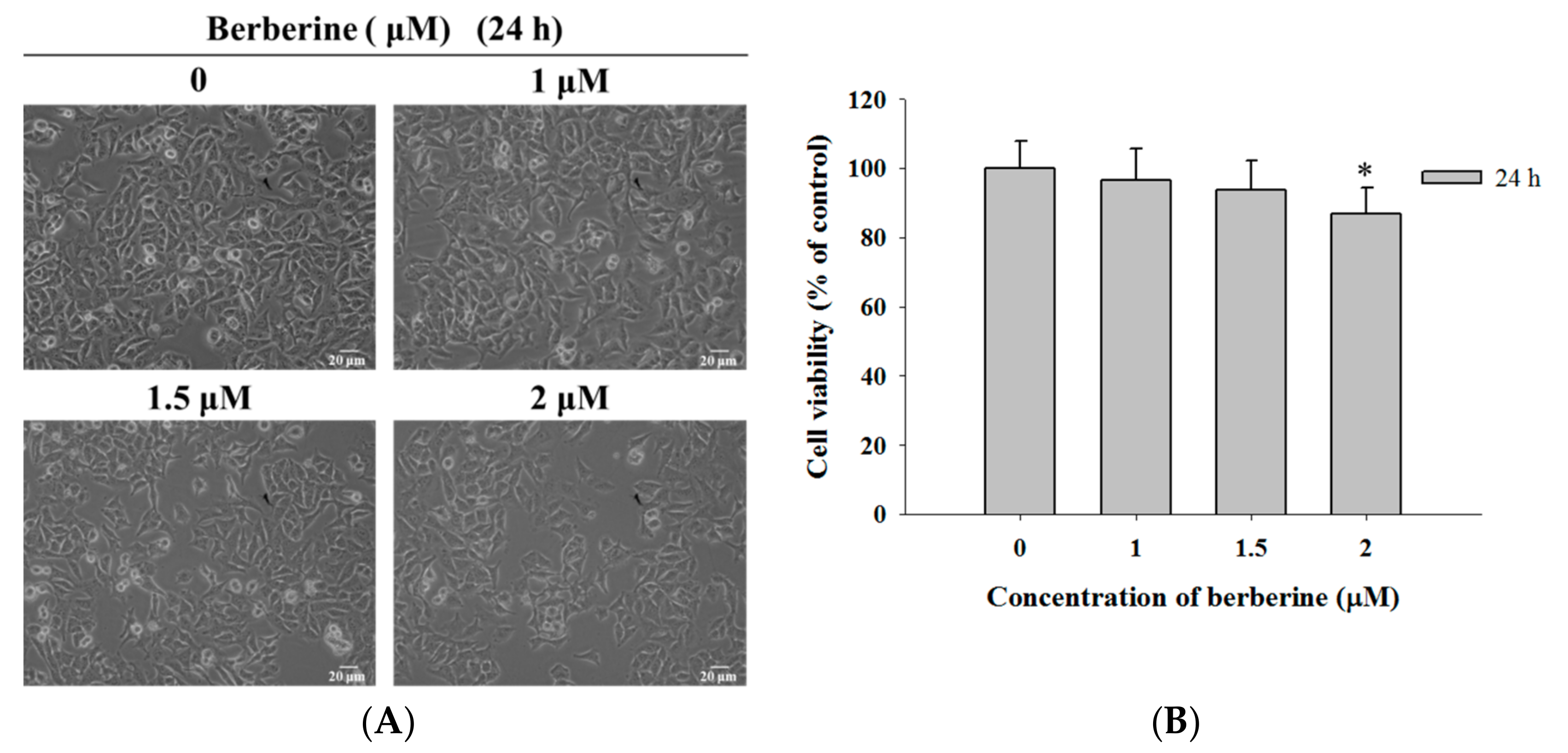
2.1. Berberine Induces Cell Morphological Changes and Decreases the Total Viability of A375.S2 Cells

As indicated in Figure 1A,B, berberine, at a 1–1.5 μM treatment dosage, did not show significant cytotoxic activity for morphology changes and reduced the total viable cell number in A375.S2 cells at 24 h (0 μM: 100% ± 7.99; 1 μM: 96.45% ± 9.07; 1.5 μM: 93.91% ± 8.22; 2 μM: 86.74% ± 7.58). However, berberine at 2 μM showed slightly induced cell morphological changes and reduced the cell number (reduced the total viable cells by 13.26%).

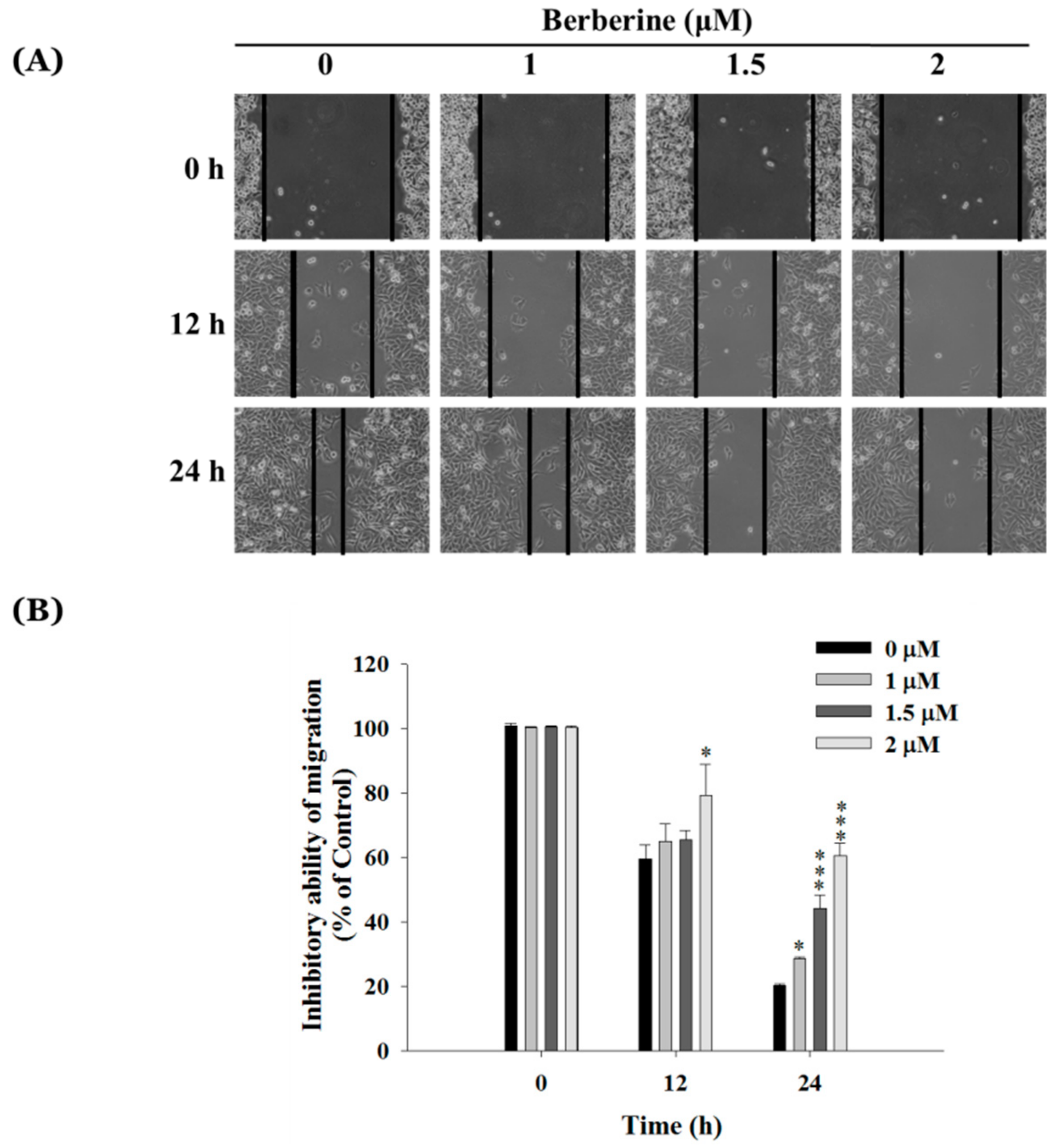
2.2. Berberine Inhibits Cell Mobility in A375.S2 Cells

The results from the wound healing assay that were presented in Figure 2A,B showed that berberine treatment at 1–2 μM inhibited the closure rate of the scratch in A375.S2 cells. The berberine treated cells remained creviced on the scratched plate but the control (untreated cells) wounds healed after 24 h of treatment. The edge distance was significantly higher in the high dosage (2 μM) group after 24 h, compared to that observed at a low dose (1 μM) (Figure 2B).

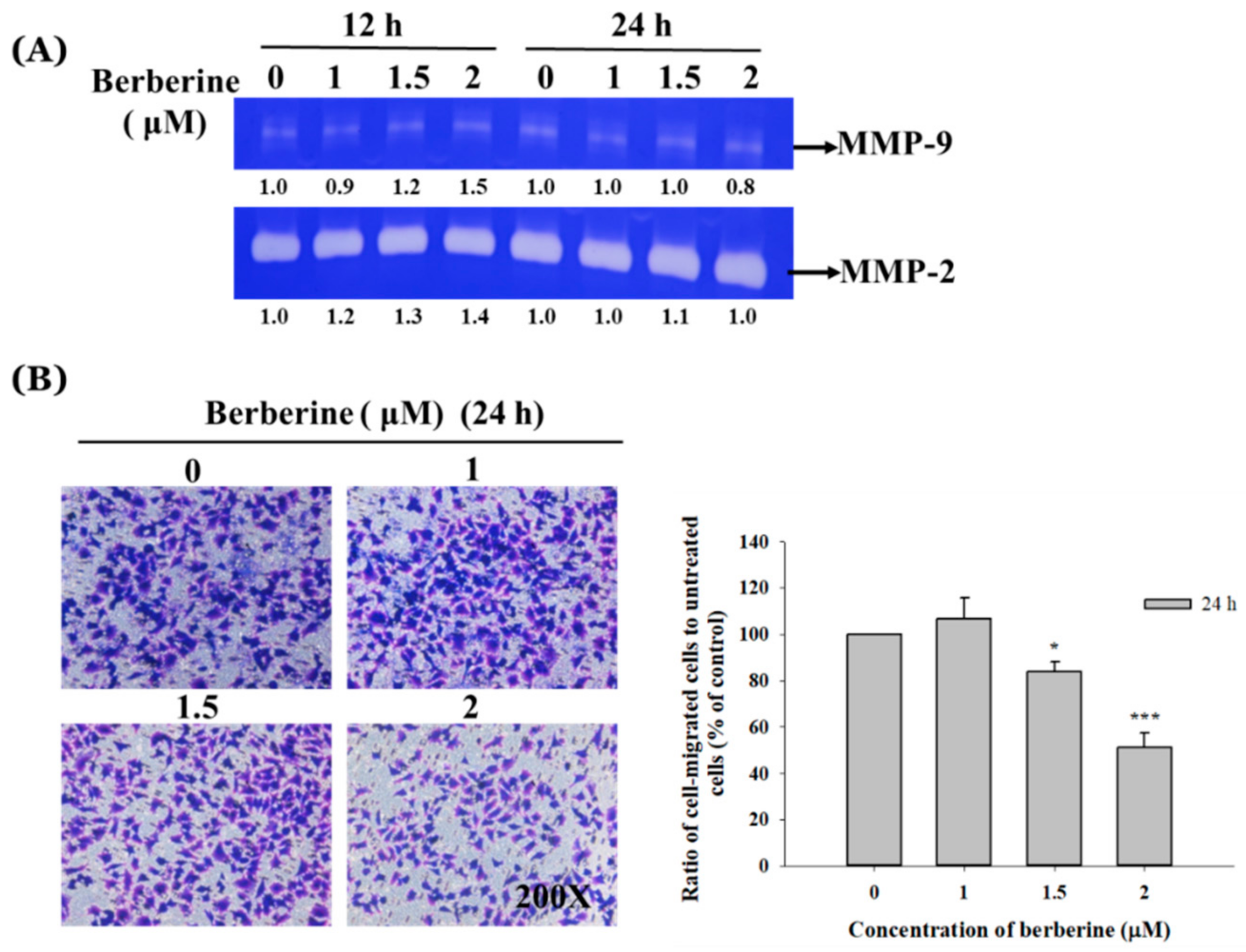
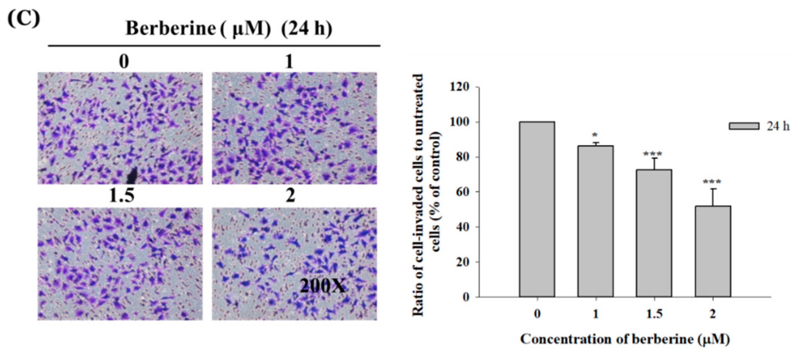
2.3. Berberine Affects the Matrix Metalloproteinase Activity and Cell Migration and Invasion in A375.S2 Cells

After the A375.S2 cells were treated with berberine (1–2 μM) for 12 and 24 h, conditioned media were collected for determining the MMP-2 or MMP-9 activity by using gelatin zymography and the results are shown in Figure 3A. The results indicated that the berberine treatment at 1 μM concentration for 12 h and 2 μM for 24 h slightly inhibited the MMP-9 activity. The transwell chambers were coated with collagen for cell migration examination and coated with Matrigel for cell invasion examinations. The results are shown in Figure 3B,C. Figure 3B indicates that berberine (1.5–2 μM) significantly inhibited the migration of A375.S2 cells and Figure 3C indicates that berberine (1–2 μM) significantly inhibited the invasion of A375.S2 cells and that these effects are dose-dependent (Figure 3C).

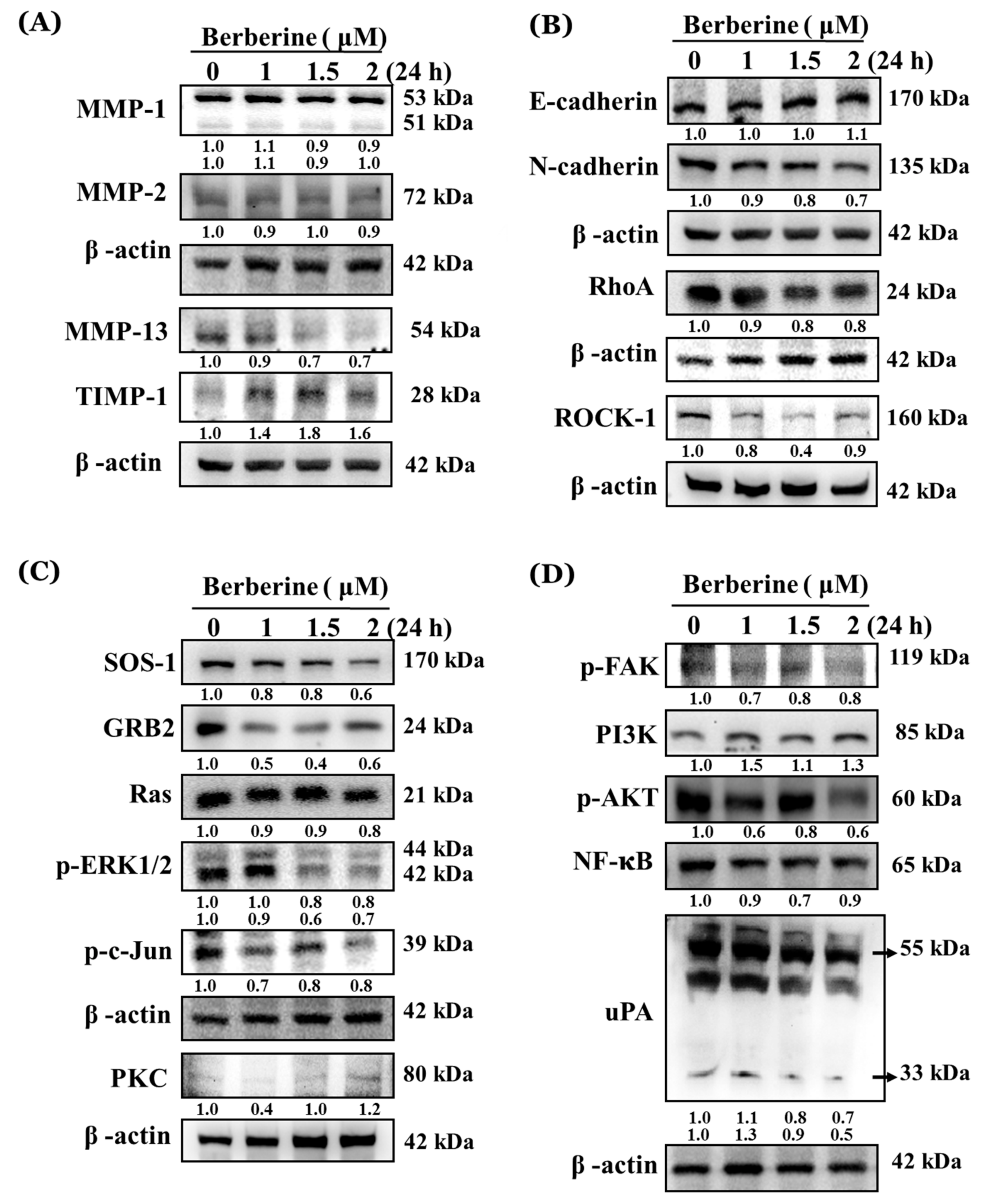
2.4. Berberine Affects Key Metastasis-Related Proteins in A375.S2 Cells

As indicated in Figure 4A–D, berberine (1–2 μM) significantly decreased MMP-13 (Figure 4A), N-cadherin, RhoA and ROCK-1 (Figure 4B), SOS-1, GRB2, Ras, p-ERK1/2 and p-c-Jun (Figure 4C), p-FAK, p-AKT, NF-κB, and uPA (Figure 4D). However, it increased TIMP-1 (Figure 4A), E-cadherin, PKC (Figure 4C), and PI3K (Figure 4D) in A375.S2 cells after 24 h of treatment, but did not significantly affect MMP-1 and MMP-2 (Figure 4A). Based on these findings, berberine may have suppressed the cell metastasis of A375.S2 cells through multiple signaling pathways.

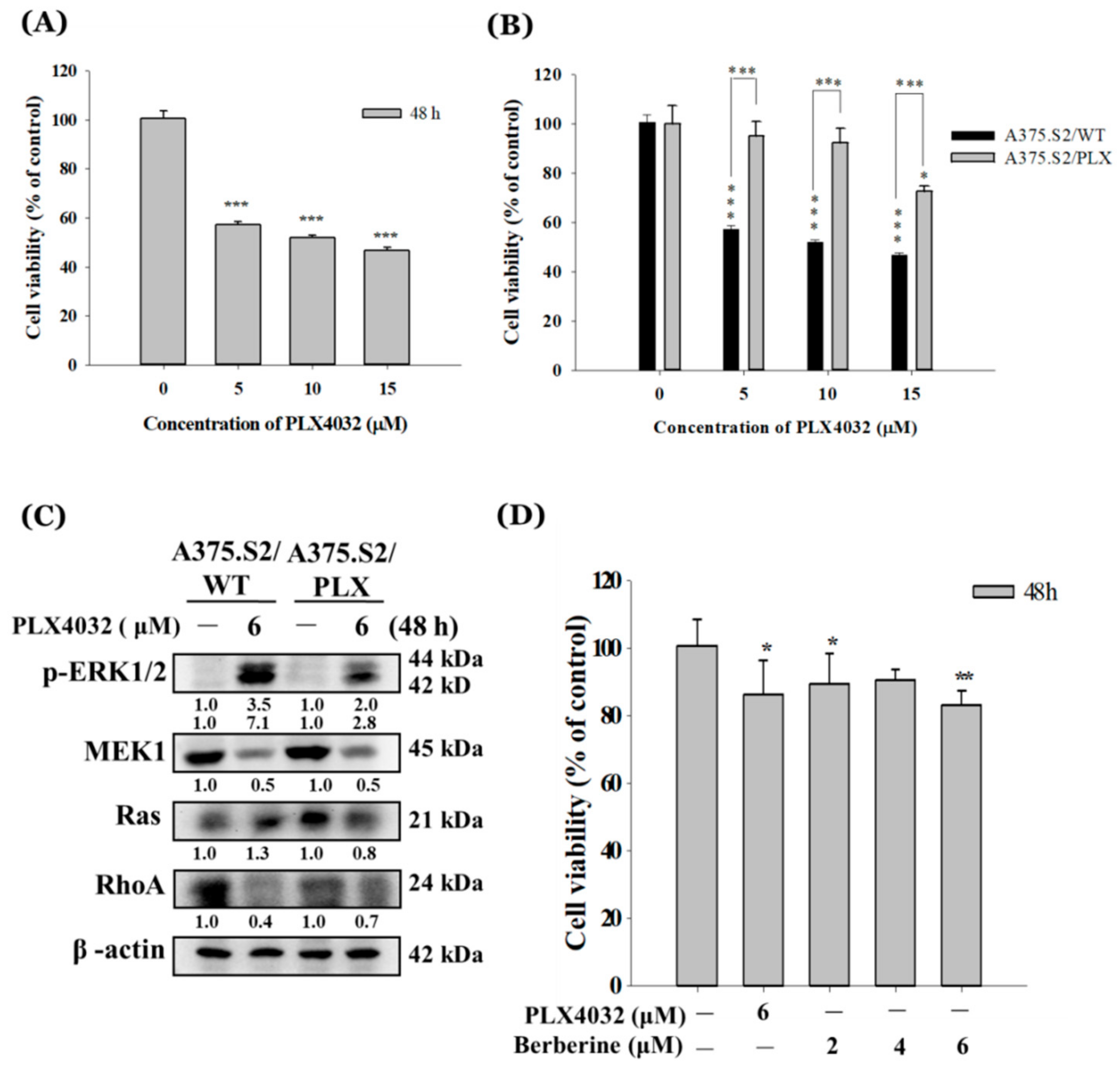
2.5. Berberine Decreases Cell Viability of A375.S2/PLX Resistant Cells

The A375.S2 cells were treated with 0, 5, 10, and 15 μM of PLX4032 (an inhibitor of the BRAFV600E mutation) for 48 h and the cells were collected for measuring the total viable cell numbers. The results are given in Figure 5A. The results indicated that PXL4032 decreased the total viable cell number at 5–15 μM after 48 h of treatment. The IC50 was calculated and the value is 6 μM of berberine. The 6 μM concentration of PLX4302 was used to generate the PLX4032 resistant A375.S2 cells (A375.S2/PLX cells). The A375.S2/PLX and A375.S2 (A375.S2/WT) cells were treated with various concentrations of PLX4032 and the results were shown in Figure 5B. The original A375.S2 cells decreased the total viable cells at 5–15 μM of PLX4032 but the A375.S2/PLX cells only reduced the total viable cells with a treatment of 15 μM, but treatments of 5–10 μM did not significantly affect the total viable cells. In order to confirm the PLX4032 resistance of the A375.S2/PLX cells and whether they were involved in the ERK pathway, the western blotting of the associated protein expressions were examined and the results are shown in Figure 5C, which indicates that PLX4032 has a higher inhibition of p-ERK1/2 in A375.S2/PLX cells than in A375.S2/WT cells. PLX4032 inhibited a MEK1 expression in A375.S2/WT and A375.S2/PLX cells. However, PLX4032 inhibited the expression of Ras in A375.S2/PLX cells but increased it in A375.S2/WT cells. Nevertheless, PLX4032 decreased the expression of RhoA in both cells. The A375.S2/PLX cells were treated with PLX4032 (6 μM) and berberine at 2–6 μM for 48 h and the results are shown in Figure 5D, which indicate that berberine reduced the total viable A375.S2/PLX cells at 2–6 μM.

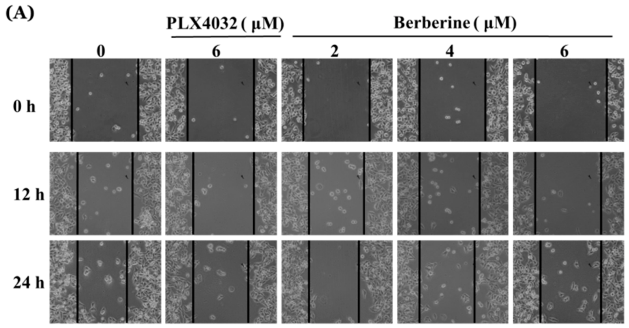
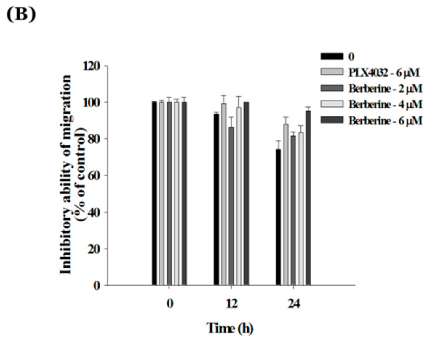
2.6. Berberine Suppresses Cell Mobility in A375.S2/PLX Resistant Cells

As indicated in Figure 6A,B, berberine treatment of 2–6 μM for cell mobility inhibited the scratch area of the wound healing assay and the results indicated that berberine significantly suppressed the cell mobility in a dose-dependent manner. These effects indicated that berberine has a higher inhibition of cell mobility than that of PLX4032 at 6 μM (Figure 6A,B).

3. Discussion

It is well documented that chemotherapy drugs contain anti-tumor activities involved in the inhibition of the proliferation, induced cell apoptosis, or suppressed cell metastasis of tumor cells [30,31,32,33]. Numerous pieces of evidence have shown that tumor metastasis may lead to multiple organ failures and dyscrasia, which are the leading causes of death in patients with malignant tumors. Therefore, one of the best strategies against cancer cells is to block the signaling pathway of the cancer cell metastasis [33,34,35]. Many clinical drugs are used for patients with cancer metastasis and some of these drugs are obtained from natural products. Berberine, a compound obtained naturally from plants, has shown to induce cancer cell death in many human cancer cell lines. Moreover, berberine suppresses the migration and invasion of B16F10 murine melanoma cells and A375 human melanoma cells through a reduction in the activity of the ERK signaling pathway and the COX-2 protein levels [36]. However, there is no available information to shows that berberine affects the migration and invasion of A375.S2 and PLX4032-resistant A375.S2 cells. PLX4302 is an inhibitor of the BRAFV600E mutation for the treatment of cancers harboring activated BRAF mutations [37]. Thus, herein, we investigated the effects of berberine cell migration and invasion in A375.S2 cells and PLX4032-resistant A375.S2 cells (A375.S2/PLX) in vitro.
The results indicated that berberine slightly induced cell morphological changes and decreased the viable number of A375.S2 cells at 1–2 μM after 24 h of treatment (Figure 1A,B). Berberine has significantly suppressed the migration of A375.S2 cells in a dose-dependent manner (Figure 2A), however, only at 2 μM after 12 h of treatment does cell mobility get inhibited (Figure 2B). This is in agreement with other reports that showed that berberine suppresses cell mobility in murine melanoma B16 cells [38] and in hepatocellular carcinoma cells [39]. Berberine inhibits the invasion of human skin squamous cell carcinoma A431 cells [29].
For further investigation, the results of the cell migration and invasion assay indicated that berberine suppressed the cell migration at 1.5–2 μM after 24 h of treatment (Figure 3B) and inhibited the cell invasion at 1–2 μM after 24 h of treatment (Figure 3C) in A375.S2 cells. These findings are in agreement with other reports that indicated that berberine suppressed the migration and invasion of murine melanoma B16 cells [38], human colorectal cancer SW620 and LoVo cells [28], and human prostate cancer cells in vitro [27]. Gelatin zymography was used to measure the MMP-2 and MMP-9 gelatin activities and the results show that at 2 μM of berberine, MMP-9 activity is significantly reduced after 24 h of treatment. Meanwhile, other investigators have shown that the MMPs activities were markedly suppressed by berberine in a dose-dependent manner in murine melanoma B16 cells [38]. Furthermore, the agent-inhibited MMP-2 could lead to the suppression of tumor metastasis [40,41].
The proteins involved in migration and invasion were investigated by western blotting and the results are shown in Figure 4A–D. As shown in Figure 4B, berberine increased E-cadherin and decreased N-cadherin in A375.S2 cells. It is well known that cancer cell migration and invasion will result in decreased E-cadherin and increased N-cadherin rates [42,43], which play an important role in cancer cell migration and invasion [44,45]. Thus, berberine increased E-cadherin rate and berberine decreased N-cadherin rate (Figure 4B) may be involved in the inhibition of the migration and invasion of A375.S2 cells. Berberine decreased the RhoA (Figure 4B), p-FAK, and p-AKT (Figure 4D) protein expressions at 1–2 μM after berberine treatment for 24 h in A375.S2 cells. The activated PI3K/AKT signaling has shown to involve tumor cell invasion and oncogenesis [45,46], including melanoma cells [47]. Numerous pieces of evidence have shown that RhoA plays a critical role in cell metastasis [48,49,50]. Akt and focal adhesion kinase (FAK) play important roles in glioma and prostate cell migration and invasion [51,52]. The results also showed that berberine (1–2 μM) significantly inhibited the protein expressions, such as SOS-1, GRB2, Ras, p-ERK1/2, p-c-Jun (Figure 4C), NF-κB, and uPA (Figure 4D) in A375.S2 cells. GRB2, SOS-1, Ras, p-ERK1/2, and p-c-Jun have been shown also to involve cell metastasis [53,54]. However, berberine increased the expression of PKC (Figure 4C) and PI3K (Figure 4D) in A375.S2 cells. Thus, further investigations are needed in the future.
More interesting is that the inhibition of the PI3k/Akt pathway led to the decrease in the invasion of melanoma cells [48,55]. the results from Figure 4D also showed that berberine suppressed the expression of p-AKT in A375.S2 cells. The PI3K/Akt pathway plays a role in the MMPs for uPA gene regulation, cell survival, and cell invasion [56,57]. The AKT activation induced the invasion and metastasis of cancer cells by stimulating secretions of MMPs [48,53]. Figure 4D also shows that berberine inhibited the uPA and NF-κB protein expressions in A375.S2 cells. It was reported that the down-regulation of uPA by berberine decreased the HCC cell invasion and migration [39]. MMPs were up-regulation by uPA and tPA and down-regulated by TIMPs and PAI-1 [39,58]. NF-κB was linked with tumor cell proliferation, survival, invasion, and metastasis [59]. Based on these results, we suggest that the berberine-inhibited cell migration, and invasion are involved with NF-κB in A375.S2 cells in vitro.
PLX4032, also known as vemurafenib, is an inhibitor that binds to the ATP-binding site of mutated BRAF kinase, inhibiting ERK signaling only in tumor cells expressing BRAFV600E mutations [60]. The cells develop a resistance to PLX4032 usually within one-year of therapy [61]. Therefore, we generated PLX4032 (inhibitor of the BRAFV600E mutation) resistant A375.S2 cells (A375.S2/PLX cells), which are shown in Figure 5A,B. The A375.S2/PLX cells were treated with berberine at 2, 4, and 6 μM, leading to the significantly reduced total cell number. Furthermore, we also investigated the cell mobility by using the wound healing assay and the results showed that berberine significantly suppressed the cell mobility of the A375.S2/PLX cells (Figure 6A,B). Based on these results, berberine suppressed the cell migration in A375.S2/PLX cells.
In conclusion, our results indicated that berberine significantly suppressed the mobility, migration, and invasion of A375.S2 cells involved in the inhibition of metastasis-associated proteins such as FAK, RhoA, ROCK1, or p-AKT, NF-κB, and uPA, which lead to the inhibition of MMP-1 and MMP-13 in vitro. Overall, the possible signal pathway for berberine-suppressed cell mobility, migration, and invasion of A375.S2 cells and by the FAK, uPA and NF-κB signaling pathways. Furthermore, we also found that berberine suppressed the mobility of PLX4032 resistant A375.S2 cells (A375.S2/PLX cells). Berberine can be considered to have potential as a chemotherapeutic agent in melanoma, which will have to be proven in further in-depth studies, including investigations on in vivo efficacy.

4. Materials and Methods

4.1. Test Chemicals, Reagents and Culture Medium

Berberine, dimethyl sulfoxide (DMSO), Tris-HCl, trypan blue, trypsin, propidium iodide (PI), gelatin, Coomassie blue R-250, and PLX4032 were purchased from Sigma Chemical Co. (St. Louis, MO, USA). Fetal bovine serum (FBS), Minimum Essential Medium (MEM) culture medium, and penicillin-streptomycin were purchased from Invitrogen (Carlsbad, CA, USA). Primary antibody anti-MMP-1, -MMP-2, and -MMP-13 were obtained from Santa Cruz Biotechnology (Santa Cruz, CA, USA) and anti-TIMP-1, -E-cadherin, -N-cadherin, -RhoA, -ROCK-1, -SOS-1, -GRB2, -Ras, -p-ERK1/2, -p-c-Jun, -PKC, -p-FAK, -PI3K, -p-AKT, NF-κB, -uPA, and the peroxidase conjugated secondary antibodies were purchased from Cell Signaling Technology, Inc. (Beverly, MA, USA). Berberine was dissolved in DMSO as a carrier solvent and control cultures were 0.5% DMSO. Berberine was further diluted in a culture medium to the appropriate final concentrations prior to use.

4.2. Cell Line and Culture

The human melanoma A375.S2 cell line with a BRAFV600E mutation was obtained from the Food Industry Research and Development Institute (Hsinchu, Taiwan). The A375.S2 cells were cultured in the MEM medium supplemented with 10% FBS, 2 mM L-glutamine, 10 g/L non-essential amino acid, 100 μg/mL streptomycin, and 100 units/mL penicillin in a humidified atmosphere of 5% CO2 at 37 °C and at 70% confluence. The cells displayed a normal morphology as previously described [62,63].

4.3. Cell Morphological Examination and Viability Assay

The A375.S2 cells (1 × 10⁵ cells/well) were seeded onto 12-well plates overnight with a MEM culture medium and they were incubated with berberine at final concentrations (0, 1, 1.5, and 2 μM) in triplicate for 24 h. After treatment, the cells were examined and photographed under contrast-phase microscopy at 200×. The cells were harvested, washed with PBS, and were stained with PI (5 μg/mL) for measuring the total percentage of cell viability by using flow cytometry (Becton-Dickinson, San Jose, CA, USA) as described previously [62,63].
The A375.S2 cells (1 × 10⁵ cells/well) were treated with various concentrations (5, 10, or 15 μM) of PLX4032, an inhibitor of the BRAFV600E mutation, for 48 h and the cells were harvested to measure the total viable cell number as described previously [62,63]. To calculate the IC50 of the PLX4032, 6 μM of A375.S2 cells were used to generate the PLX4032-resistant A375.S2 cells, as described previously with modifications [64]. The resistant A375.S2 cells were incubated with various concentrations (0, 5, 10, or 15 μM) of PLX4032 for 48 h to measure the total viable cell number. The resistant A375.S2 cells were treated with various concentrations (2, 4, and 6 μM) of berberine for 48 h and were harvested for measuring the total viable cell number as described previously [62,63].

4.4. In-Vitro Scratch Wound Healing Assay

Cell mobility characteristics were analyzed by a wound healing assay, as described previously [62,63]. Briefly, A375.S2 or A375.S2/PLX cells (2 × 10⁵ cells/well) were placed in 12-well plates for 24 h and grown until reaching a confluent monolayer. The culture media were replaced with serum-free MEM to wash the cell monolayers. Cell monolayers were scratched (wound) using a sterile 200 μL-pipette tip and to remove the cell debris by PBS washing. A375.S2 or A375.S2/PLX were incubated with various concentrations of berberine (0, 1, 1.5, and 2 μM, or 0, 2, 4, 6 μM, respectively) for different time-periods (0, 12, and 24 h). The migrating cells in the denuded zone were monitored and photographed under phase contrast microscopy. The scratch experiments were done 3 times. We quantitated the relative wound size by using the Image J version 1.49o software. The inhibitory ability of migration is as follows: (% of control) = ((wound areaberberine 12 h or 24 h/wound areaberberine 0 h)/(wound areacontrol 12 h or 24 h/wound areacontrol 0 h)) × 100%, as described previously [65,66].

4.5. Gelatin Zymography Assay

The A375.S2 cells (2 × 10⁵ cells/well) were maintained in 12-well plates for 24 h and a serum-free MEM medium containing berberine (0, 1, 1.5, and 2 μM) was individually added to each plate and cultured for 12 and 24 h. The conditioned medium was collected and the total proteins from each treatment were quantitated to load them onto 10% polyacrylamide gels. They were then copolymerized with 0.2% gelatin and then the gel was incubated in a zymogen developing buffer (Sigma-Aldrich, St. Louis, Missouri, USA) containing 50 mM Tris (pH 7.5), 200 mM NaCl, 5 mM CaCl2, 1 μM ZnCl2, and 0.02% Brij-35. The gel was kept overnight at 37 °C and soaked twice in 2.5% Triton X-100 in dH2O at 25 °C for 30 min. The bands of MMP-2 and -9 corresponded to the activity, which was stained with 0.2% Coomassie blue in 10% acetic acid and 50% methanol. After staining, the gel was photographed and the band of gelatinolytic activity was determined using the NIH Image J software, version 1.47 (National Institutes of Health, Bethesda, MA, USA), as described previously [65,67].

4.6. Transwell Assay for Cell Migration and Invasion Examinations

Collagens and the Matrigel cell migration and invasion assay system were used to measure the migration and invasion of cell in vitro, as described previously [63,65]. For the cell migration assay, A375.S2 cells (5 × 10⁴ cells/well) in serum-free MEM with berberine (0, 1, 1.5 and 2 μM) were placed in the upper chamber (8 μm pore size; Millipore, Temecula, CA, USA) and coated with 50 μL collagen in each transwell insert overnight, and 800 μL of MEM with 10% FBS was placed in the lower chamber and incubated for 24 h. After incubation, the cells adhering to the upper surface of the membrane were removed and all the migrated cells which adhered to the lower surface were fixed with 4% formaldehyde in PBS, treated with methanol, and stained with 2% crystal violet. After staining, all the samples were examined and photographed under light microscopy to count the total cells and calculate the percentage of inhibition based on the cells on each picture, as described previously [63,65]. The invasion assay was performed in almost the same way with a cell migration assay, except matrigel (matrigel:serum-free medium 1:10) was on the transwell membrane, as described previously [63,65].

4.7. Western Blotting Analysis for Cell Metastasis-Associated Protein Expressions

The A375.S2 cells were placed in 10-cm culture dishes at a density of 1 × 10⁶ cells/dish and they were incubated with berberine (0, 1, 1.5, and 2 μM) for 24 h. After treatment, the cells were harvested and re-suspended in a lysis buffer of 50 mM, Tris-HCl pH 7.5, 400 mM NaCl, 2 mM EGTA, 1 mM EDTA, 1 mM DTT, and a protease inhibitor cocktail (Roche, Mannheim, Germany). The cell lysates were centrifuged at 10,000× g at 4 °C for 10 min. We quantitated the total protein concentration of the supernatants by using a Bradford protein assay kit. A total of 30 μg of total proteins were separated by 12% SDS-polyacrylamide gel electrophoresis. They were then transferred onto a PVDF membrane (Millipore, Bedford, MA, USA). The membrane was blocked with 5% non-fat milk which was in the TBS-T buffer (10 mM Tris–HCl, 150 mM NaCl, and 0.05% Tween-20, pH 7.8) for 1 h at room temperature. The membranes were washed with the TBS-T buffer and were incubated with monoclonal antibodies such as anti-MMP-1, -MMP-2, -MMP-13, -TIMP-1, -E-cadherin, -N-cadherin, -RhoA, -ROCK1, -SOS-1, -GRB2, -Ras, -p-ERK1/2, p-c-Jung, -PKC, -p-FAK, -PI3K, -p-AKT, -NF-κB, and -uPA. After washing, the membranes were incubated with diluted corresponding HRP-conjugated secondary antibodies (diluted 1:5000; Santa Cruz Biotechnology), developed with ECL (Amersham, Piscataway, NJ), and they were detected directly with a Biospectrum Imaging System (UVP, Inc., Upland, CA, USA) [65,68].
After the PLX4032 treatment, the A375.S2 and PLX4032-resistant A375.S2 cells were harvested for examining the cell migration and invasion expression associated proteins such as p-ERK1/2, MEK1, Ras, and RhoA, as described in the western blotting section.

4.8. Statistical Analysis

The results are presented as mean ± SD. The data were statistically analyzed with one-way ANOVA analysis of variance. * p < 0.05, ** p < 0.01, *** p < 0.001 are determined as significant between the control and berberine treated groups.

Author Contributions

J.-F.L., K.C.L., W.-W.H. and J.-G.C. conceived and designed the experiments; J.-F.L., K.C.L., S.-F.P. and P.M. performed the experiments; Y.-P.H., A.-C.H. and F.-S.C. analyzed the data; K.C.L. and W.-W.H. and J.-G.C. contributed reagents, materials, and analysis tools; and W.-W.H. and J.-G.C. wrote the paper.

Funding

This work supported by the grant CMUBH R106-002 from China Medical University Beigang Hospital, Yunlin, Taiwan. This work was supported by the Ministry of Science and Technology, Taipei, Taiwan (grant numbers: 105-2815-C-039-082-B).

Acknowledgments

Experiments and data analysis performed in part through the using of the Medical Research Core Facilities Center, Office of Research & Development at China Medical University, Taichung, Taiwan.

Conflicts of Interest

The authors have declared that no competing interests exist.

References

  1. Leiter, U.; Eigentler, T.; Garbe, C. Epidemiology of skin cancer. Adv. Exp. Med. Biol. 2014, 810, 120–140. [Google Scholar] [PubMed]
  2. Linos, E.; Swetter, S.M.; Cockburn, M.G.; Colditz, G.A.; Clarke, C.A. Increasing burden of melanoma in the United States. J. Investig. Dermatol. 2009, 129, 1666–1674. [Google Scholar] [CrossRef] [PubMed]
  3. American Cancer Society. Available online: http://www.cancer.org/cancer/skincancer-melanoma/detailedguide/melanoma-skin-cancer-key-statistics (accessed on 3 June 2016).
  4. Rastrelli, M.; Tropea, S.; Rossi, C.R.; Alaibac, M. Melanoma: Epidemiology, risk factors, pathogenesis, diagnosis and classification. In Vivo 2014, 28, 1005–1011. [Google Scholar] [PubMed]
  5. Chen, S.T.; Geller, A.C.; Tsao, H. Update on the Epidemiology of Melanoma. Curr. Dermatol. Rep. 2013, 2, 24–34. [Google Scholar] [CrossRef] [PubMed] [Green Version]
  6. Siegel, R.L.; Miller, K.D.; Jemal, A. Cancer statistics, 2015. CA A Cancer J. Clin. 2015, 65, 5–29. [Google Scholar] [CrossRef] [PubMed]
  7. Hamm, C.; Verma, S.; Petrella, T.; Bak, K.; Charette, M. Biochemotherapy for the treatment of metastatic malignant melanoma: A systematic review. Cancer Treat. Rev. 2008, 34, 145–156. [Google Scholar] [CrossRef] [PubMed]
  8. Martin, T.A.; Ye, L.; Sanders, A.J.; Lane, J.; Jiang, W.G. Cancer invasion and metastasis: Molecular and cellular perspective. In Metastatic Cancer Clinical Biological Perspectives; Landes Bioscience: Austin, TX, USA, 2013; pp. 135–168. [Google Scholar]
  9. Liu, P.L.; Tsai, J.R.; Charles, A.L.; Hwang, J.J.; Chou, S.H.; Ping, Y.H.; Lin, F.Y.; Chen, Y.L.; Hung, C.Y.; Chen, W.C.; et al. Resveratrol inhibits human lung adenocarcinoma cell metastasis by suppressing heme oxygenase 1-mediated nuclear factor-kappaB pathway and subsequently downregulating expression of matrix metalloproteinases. Mol. Nutr. Food Res. 2010, 54 (Suppl. 2), S196–S204. [Google Scholar] [CrossRef] [PubMed]
  10. Tsuchiya, Y.; Sawada, S.; Yoshioka, I.; Ohashi, Y.; Matsuo, M.; Harimaya, Y.; Tsukada, K.; Saiki, I. Increased surgical stress promotes tumor metastasis. Surgery 2003, 133, 547–555. [Google Scholar] [CrossRef] [PubMed]
  11. Hainaut, P.; Plymoth, A. Targeting the hallmarks of cancer: Towards a rational approach to next-generation cancer therapy. Curr. Opin. Oncol. 2013, 25, 50–51. [Google Scholar] [CrossRef] [PubMed]
  12. Yilmaz, M.; Christofori, G.; Lehembre, F. Distinct mechanisms of tumor invasion and metastasis. Trends Mol. Med. 2007, 13, 535–541. [Google Scholar] [CrossRef] [PubMed]
  13. Folgueras, A.R.; Pendas, A.M.; Sanchez, L.M.; Lopez-Otin, C. Matrix metalloproteinases in cancer: From new functions to improved inhibition strategies. Int. J. Dev. Biol. 2004, 48, 411–424. [Google Scholar] [CrossRef] [PubMed]
  14. Kessenbrock, K.; Plaks, V.; Werb, Z. Matrix metalloproteinases: Regulators of the tumor microenvironment. Cell 2010, 141, 52–67. [Google Scholar] [CrossRef] [PubMed]
  15. Nikkola, J.; Vihinen, P.; Vuoristo, M.S.; Kellokumpu-Lehtinen, P.; Kahari, V.M.; Pyrhonen, S. High serum levels of matrix metalloproteinase-9 and matrix metalloproteinase-1 are associated with rapid progression in patients with metastatic melanoma. Clin. Cancer Res. 2005, 11, 5158–5166. [Google Scholar] [CrossRef] [PubMed]
  16. Malaponte, G.; Zacchia, A.; Bevelacqua, Y.; Marconi, A.; Perrotta, R.; Mazzarino, M.C.; Cardile, V.; Stivala, F. Co-regulated expression of matrix metalloproteinase-2 and transforming growth factor-beta in melanoma development and progression. Oncol. Rep. 2010, 24, 81–87. [Google Scholar] [CrossRef] [PubMed]
  17. Bhadra, K.; Kumar, G.S. Therapeutic potential of nucleic acid-binding isoquinoline alkaloids: Binding aspects and implications for drug design. Med. Res. Rev. 2011, 31, 821–862. [Google Scholar] [CrossRef] [PubMed]
  18. Hwang, B.Y.; Roberts, S.K.; Chadwick, L.R.; Wu, C.D.; Kinghorn, A.D. Antimicrobial constituents from goldenseal (the Rhizomes of Hydrastis canadensis) against selected oral pathogens. Planta Med. 2003, 69, 623–627. [Google Scholar] [PubMed]
  19. Kuo, C.L.; Chi, C.W.; Liu, T.Y. The anti-inflammatory potential of berberine in vitro and in vivo. Cancer Lett. 2004, 203, 127–137. [Google Scholar] [CrossRef] [PubMed]
  20. Tomosaka, H.; Chin, Y.W.; Salim, A.A.; Keller, W.J.; Chai, H.; Kinghorn, A.D. Antioxidant and cytoprotective compounds from Berberis vulgaris (barberry). Phytother. Res. PTR 2008, 22, 979–981. [Google Scholar] [CrossRef] [PubMed]
  21. Abd El-Wahab, A.E.; Ghareeb, D.A.; Sarhan, E.E.; Abu-Serie, M.M.; El Demellawy, M.A. In vitro biological assessment of Berberis vulgaris and its active constituent, berberine: Antioxidants, anti-acetylcholinesterase, anti-diabetic and anticancer effects. BMC Complement. Altern. Med. 2013, 13, 218. [Google Scholar] [CrossRef] [PubMed]
  22. Tillhon, M.; Guaman Ortiz, L.M.; Lombardi, P.; Scovassi, A.I. Berberine: New perspectives for old remedies. Biochem. Pharmacol. 2012, 84, 1260–1267. [Google Scholar] [CrossRef] [PubMed]
  23. Zou, K.; Li, Z.; Zhang, Y.; Zhang, H.Y.; Li, B.; Zhu, W.L.; Shi, J.Y.; Jia, Q.; Li, Y.M. Advances in the study of berberine and its derivatives: A focus on anti-inflammatory and anti-tumor effects in the digestive system. Acta Pharmacol. Sin. 2017, 38, 157–167. [Google Scholar] [CrossRef] [PubMed]
  24. Eo, S.H.; Kim, J.H.; Kim, S.J. Induction of G(2)/M Arrest by Berberine via Activation of PI3K/Akt and p38 in Human Chondrosarcoma Cell Line. Oncol. Res. 2014, 22, 147–157. [Google Scholar] [CrossRef] [PubMed]
  25. Yip, N.K.; Ho, W.S. Berberine induces apoptosis via the mitochondrial pathway in liver cancer cells. Oncol. Rep. 2013, 30, 1107–1112. [Google Scholar] [CrossRef] [PubMed]
  26. Wu, C.M.; Li, T.M.; Tan, T.W.; Fong, Y.C.; Tang, C.H. Berberine Reduces the Metastasis of Chondrosarcoma by Modulating the alpha v beta 3 Integrin and the PKC delta, c-Src, and AP-1 Signaling Pathways. Evid.-Based Complement. Altern. Med. ECAM 2013, 2013, 423164. [Google Scholar]
  27. Liu, C.H.; Tang, W.C.; Sia, P.; Huang, C.C.; Yang, P.M.; Wu, M.H.; Lai, I.L.; Lee, K.H. Berberine inhibits the metastatic ability of prostate cancer cells by suppressing epithelial-to-mesenchymal transition (EMT)-associated genes with predictive and prognostic relevance. Int. J. Med. Sci. 2015, 12, 63–71. [Google Scholar] [CrossRef] [PubMed]
  28. Liu, X.; Ji, Q.; Ye, N.; Sui, H.; Zhou, L.; Zhu, H.; Fan, Z.; Cai, J.; Li, Q. Berberine Inhibits Invasion and Metastasis of Colorectal Cancer Cells via COX-2/PGE2 Mediated JAK2/STAT3 Signaling Pathway. PLoS ONE 2015, 10, e0123478. [Google Scholar] [CrossRef] [PubMed]
  29. Li, D.X.; Zhang, J.; Zhang, Y.; Zhao, P.W.; Yang, L.M. Inhibitory effect of berberine on human skin squamous cell carcinoma A431 cells. Genet. Mol. Res. 2015, 14, 10553–10568. [Google Scholar] [CrossRef] [PubMed]
  30. Ricci, M.S.; Zong, W.X. Chemotherapeutic approaches for targeting cell death pathways. Oncologist 2006, 11, 342–357. [Google Scholar] [CrossRef] [PubMed]
  31. Zhu, W.; Liang, Q.; Yang, X.; Yu, Y.; Shen, X.; Sun, G. Combination of sorafenib and Valproic acid synergistically induces cell apoptosis and inhibits hepatocellular carcinoma growth via down-regulating Notch3 and pAkt. Am. J. Cancer Res. 2017, 7, 2503–2514. [Google Scholar] [PubMed]
  32. Zanotto-Filho, A.; Rajamanickam, S.; Loranc, E.; Masamsetti, P.; Gorthi, A.; Romero, J.C.; Tonapi, S.; Goncalves, R.M.; Reddick, R.L.; Benavides, R.; et al. Sorafenib improves alkylating therapy by blocking induced inflammation, invasion and angiogenesis in breast cancer cells. Cancer Lett. 2018. [Google Scholar] [CrossRef] [PubMed]
  33. Yin, P.; Song, G.; Jiang, Z. Cisplatin suppresses proliferation, migration and invasion of nasopharyngeal carcinoma cells in vitro by repressing the Wnt/beta-catenin/Endothelin-1 axis via activating B cell translocation gene 1. Cancer Chemother. Pharmacol. 2018, 81, 863–872. [Google Scholar] [CrossRef] [PubMed]
  34. Shi, C.; Zhang, N.; Feng, Y.; Cao, J.; Chen, X.; Liu, B. Aspirin Inhibits IKK-beta-mediated Prostate Cancer Cell Invasion by Targeting Matrix Metalloproteinase-9 and Urokinase-Type Plasminogen Activator. Cell. Physiol. Biochem. 2017, 41, 1313–1324. [Google Scholar] [CrossRef] [PubMed]
  35. Lai, K.C.; Hsiao, Y.T.; Yang, J.L.; Ma, Y.S.; Huang, Y.P.; Chiang, T.A.; Chung, J.G. Benzyl isothiocyanate and phenethyl isothiocyanate inhibit murine melanoma B16F10 cell migration and invasion in vitro. Int. J. Oncol. 2017, 51, 832–840. [Google Scholar] [CrossRef] [PubMed]
  36. Kim, H.S.; Kim, M.J.; Kim, E.J.; Yang, Y.; Lee, M.S.; Lim, J.S. Berberine-induced AMPK activation inhibits the metastatic potential of melanoma cells via reduction of ERK activity and COX-2 protein expression. Biochem. Pharmacol. 2012, 83, 385–394. [Google Scholar] [CrossRef] [PubMed]
  37. Smalley, K.S. PLX-4032, a small-molecule B-Raf inhibitor for the potential treatment of malignant melanoma. Curr. Opin. Investig. Drugs 2010, 11, 699–706. [Google Scholar] [PubMed]
  38. Kou, Y.; Li, L.; Li, H.; Tan, Y.; Li, B.; Wang, K.; Du, B. Berberine suppressed epithelial mesenchymal transition through cross-talk regulation of PI3K/AKT and RARalpha/RARbeta in melanoma cells. Biochem. Biophys. Res. Commun. 2016, 479, 290–296. [Google Scholar] [CrossRef] [PubMed]
  39. Wang, X.; Wang, N.; Li, H.; Liu, M.; Cao, F.; Yu, X.; Zhang, J.; Tan, Y.; Xiang, L.; Feng, Y. Up-Regulation of PAI-1 and Down-Regulation of uPA Are Involved in Suppression of Invasiveness and Motility of Hepatocellular Carcinoma Cells by a Natural Compound Berberine. Int. J. Mol. Sci. 2016, 17, 577. [Google Scholar] [CrossRef] [PubMed]
  40. Wang, Z.; You, D.; Lu, M.; He, Y.; Yan, S. Inhibitory effect of norcantharidin on melanoma tumor growth and vasculogenic mimicry by suppressing MMP-2 expression. Oncol. Lett. 2017, 13, 1660–1664. [Google Scholar] [CrossRef] [PubMed] [Green Version]
  41. Fan, S.; Liao, Y.; Liu, C.; Huang, Q.; Liang, H.; Ai, B.; Fu, S.; Zhou, S. Estrogen promotes tumor metastasis via estrogen receptor beta-mediated regulation of matrix-metalloproteinase-2 in non-small cell lung cancer. Oncotarget 2017, 8, 56443–56459. [Google Scholar] [CrossRef] [PubMed] [Green Version]
  42. Chen, H.F.; Ma, R.R.; He, J.Y.; Zhang, H.; Liu, X.L.; Guo, X.Y.; Gao, P. Protocadherin 7 inhibits cell migration and invasion through E-cadherin in gastric cancer. Tumour Biol. 2017, 39. [Google Scholar] [CrossRef] [PubMed] [Green Version]
  43. Canel, M.; Serrels, A.; Frame, M.C.; Brunton, V.G. E-cadherin-integrin crosstalk in cancer invasion and metastasis. J. Cell Sci. 2013, 126 Pt 2, 393–401. [Google Scholar] [CrossRef]
  44. Labernadie, A.; Kato, T.; Brugues, A.; Serra-Picamal, X.; Derzsi, S.; Arwert, E.; Weston, A.; Gonzalez-Tarrago, V.; Elosegui-Artola, A.; Albertazzi, L.; et al. A mechanically active heterotypic E-cadherin/N-cadherin adhesion enables fibroblasts to drive cancer cell invasion. Nat. Cell Biol. 2017, 19, 224–237. [Google Scholar] [CrossRef] [PubMed] [Green Version]
  45. Kim, D.; Kim, S.; Koh, H.; Yoon, S.O.; Chung, A.S.; Cho, K.S.; Chung, J. Akt/PKB promotes cancer cell invasion via increased motility and metalloproteinase production. FASEB J. 2001, 15, 1953–1962. [Google Scholar] [CrossRef] [PubMed]
  46. Shukla, S.; Maclennan, G.T.; Hartman, D.J.; Fu, P.; Resnick, M.I.; Gupta, S. Activation of PI3K-Akt signaling pathway promotes prostate cancer cell invasion. Int. J. Cancer 2007, 121, 1424–1432. [Google Scholar] [CrossRef] [PubMed] [Green Version]
  47. Thang, N.D.; Yajima, I.; Kumasaka, M.Y.; Iida, M.; Suzuki, T.; Kato, M. Deltex-3-like (DTX3L) stimulates metastasis of melanoma through FAK/PI3K/AKT but not MEK/ERK pathway. Oncotarget 2015, 6, 14290–14299. [Google Scholar] [CrossRef] [PubMed] [Green Version]
  48. Veit, C.; Genze, F.; Menke, A.; Hoeffert, S.; Gress, T.M.; Gierschik, P.; Giehl, K. Activation of phosphatidylinositol 3-kinase and extracellular signal-regulated kinase is required for glial cell line-derived neurotrophic factor-induced migration and invasion of pancreatic carcinoma cells. Cancer Res. 2004, 64, 5291–5300. [Google Scholar] [CrossRef] [PubMed]
  49. Gunther, W.; Skaftnesmo, K.O.; Arnold, H.; Terzis, A.J. Molecular approaches to brain tumour invasion. Acta Neurochir. 2003, 145, 1029–1036. [Google Scholar] [CrossRef] [PubMed]
  50. Senapati, S.; Rachagani, S.; Chaudhary, K.; Johansson, S.L.; Singh, R.K.; Batra, S.K. Overexpression of macrophage inhibitory cytokine-1 induces metastasis of human prostate cancer cells through the FAK-RhoA signaling pathway. Oncogene 2010, 29, 1293–1302. [Google Scholar] [CrossRef] [PubMed]
  51. Zhong, D.; Ran, J.H.; Tang, W.Y.; Zhang, X.D.; Tan, Y.; Chen, G.J.; Li, X.S.; Yan, Y. Mda-9/syntenin promotes human brain glioma migration through focal adhesion kinase (FAK)-JNK and FAK-AKT signaling. Asian Pac. J. Cancer Prev. 2012, 13, 2897–2901. [Google Scholar] [CrossRef] [PubMed]
  52. Kwiatkowska, A.; Kijewska, M.; Lipko, M.; Hibner, U.; Kaminska, B. Downregulation of Akt and FAK phosphorylation reduces invasion of glioblastoma cells by impairment of MT1-MMP shuttling to lamellipodia and downregulates MMPs expression. Biochim. Biophys. Acta 2011, 1813, 655–667. [Google Scholar] [CrossRef] [PubMed] [Green Version]
  53. Shih, Y.L.; Chou, H.M.; Chou, H.C.; Lu, H.F.; Chu, Y.L.; Shang, H.S.; Chung, J.G. Casticin impairs cell migration and invasion of mouse melanoma B16F10 cells via PI3K/AKT and NF-kappaB signaling pathways. Environ. Toxicol. 2017, 32, 2097–2112. [Google Scholar] [CrossRef] [PubMed]
  54. Rosenberg, L.; Yoon, C.H.; Sharma, G.; Bertagnolli, M.M.; Cho, N.L. Sorafenib inhibits proliferation and invasion in desmoid-derived cells by targeting Ras/MEK/ERK and PI3K/Akt/mTOR pathways. Carcinogenesis 2018, 39, 681–688. [Google Scholar] [CrossRef] [PubMed]
  55. Li, C.Y.; Wang, Q.; Shen, S.; Wei, X.L.; Li, G.X. Oridonin inhibits migration, invasion, adhesion and TGF-beta1-induced epithelial-mesenchymal transition of melanoma cells by inhibiting the activity of PI3K/Akt/GSK-3beta signaling pathway. Oncol. Lett. 2018, 15, 1362–1372. [Google Scholar] [PubMed]
  56. Chou, Y.C.; Chang, M.Y.; Wang, M.J.; Yu, F.S.; Liu, H.C.; Harnod, T.; Hung, C.H.; Lee, H.T.; Chung, J.G. PEITC inhibits human brain glioblastoma GBM 8401 cell migration and invasion through the inhibition of uPA, Rho A, and Ras with inhibition of MMP-2, -7 and -9 gene expression. Oncol. Rep. 2015, 34, 2489–2496. [Google Scholar] [CrossRef] [PubMed] [Green Version]
  57. Yodkeeree, S.; Chaiwangyen, W.; Garbisa, S.; Limtrakul, P. Curcumin, demethoxycurcumin and bisdemethoxycurcumin differentially inhibit cancer cell invasion through the down-regulation of MMPs and uPA. J. Nutr. Biochem. 2009, 20, 87–95. [Google Scholar] [CrossRef] [PubMed]
  58. Hsu, H.H.; Hu, W.S.; Lin, Y.M.; Kuo, W.W.; Chen, L.M.; Chen, W.K.; Hwang, J.M.; Tsai, F.J.; Liu, C.J.; Huang, C.Y. JNK suppression is essential for 17beta-Estradiol inhibits prostaglandin E2-Induced uPA and MMP-9 expressions and cell migration in human LoVo colon cancer cells. J. Biomed. Sci. 2011, 18, 61. [Google Scholar] [CrossRef] [PubMed]
  59. Perkins, N.D. The diverse and complex roles of NF-kappaB subunits in cancer. Nat. Rev. Cancer 2012, 12, 121–132. [Google Scholar] [CrossRef] [PubMed]
  60. Joseph, E.W.; Pratilas, C.A.; Poulikakos, P.I.; Tadi, M.; Wang, W.; Taylor, B.S.; Halilovic, E.; Persaud, Y.; Xing, F.; Viale, A.; et al. The RAF inhibitor PLX4032 inhibits ERK signaling and tumor cell proliferation in a V600E BRAF-selective manner. Proc. Natl. Acad. Sci. USA 2010, 107, 14903–14908. [Google Scholar] [CrossRef] [PubMed] [Green Version]
  61. Vultur, A.; Villanueva, J.; Herlyn, M. Targeting BRAF in advanced melanoma: A first step toward manageable disease. Clin. Cancer Res. 2011, 17, 1658–1663. [Google Scholar] [CrossRef] [PubMed]
  62. Wu, Z.Y.; Lien, J.C.; Huang, Y.P.; Liao, C.L.; Lin, J.J.; Fan, M.J.; Ko, Y.C.; Hsiao, Y.P.; Lu, H.F.; Chung, J.G. Casticin Inhibits A375.S2 Human Melanoma Cell Migration/Invasion through Downregulating NF-kappaB and Matrix Metalloproteinase-2 and -1. Molecules 2016, 21, 384. [Google Scholar] [CrossRef] [PubMed]
  63. Shiue, Y.W.; Lu, C.C.; Hsiao, Y.P.; Liao, C.L.; Lin, J.P.; Lai, K.C.; Yu, C.C.; Huang, Y.P.; Ho, H.C.; Chung, J.G. Casticin Induced Apoptosis in A375.S2 Human Melanoma Cells through the Inhibition of NF-[Formula: See text]B and Mitochondria-Dependent Pathways In Vitro and Inhibited Human Melanoma Xenografts in a Mouse Model In Vivo. Am. J. Chin. Med. 2016, 44, 637–661. [Google Scholar] [CrossRef] [PubMed]
  64. Huang, A.C.; Yang, M.D.; Hsiao, Y.T.; Lin, T.S.; Ma, Y.S.; Peng, S.F.; Hsia, T.C.; Cheng, Y.D.; Kuo, C.L.; Chung, J.G. Bufalin inhibits gefitinib resistant NCI-H460 human lung cancer cell migration and invasion in vitro. J. Ethnopharmacol. 2016, 194, 1043–1050. [Google Scholar] [CrossRef] [PubMed]
  65. Ma, Y.S.; Hsiao, Y.T.; Lin, J.J.; Liao, C.L.; Lin, C.C.; Chung, J.G. Phenethyl Isothiocyanate (PEITC) and Benzyl Isothiocyanate (BITC) Inhibit Human Melanoma A375.S2 Cell Migration and Invasion by Affecting MAPK Signaling Pathway In Vitro. Anticancer Res. 2017, 37, 6223–6234. [Google Scholar] [PubMed]
  66. Huang, Y.P.; Chang, N.W. PPARalpha modulates gene expression profiles of mitochondrial energy metabolism in oral tumorigenesis. BioMedicine 2016, 6, 3. [Google Scholar] [CrossRef] [PubMed]
  67. Chan, C.Y.; Lien, C.H.; Lee, M.F.; Huang, C.Y. Quercetin suppresses cellular migration and invasion in human head and neck squamous cell carcinoma (HNSCC). BioMedicine 2016, 6, 15. [Google Scholar] [CrossRef] [PubMed]
  68. Chang, Y.-M.; Velmurugan, B.K.; Kuo, W.-W.; Chen, Y.-S.; Ho, T.-J.; Tsai, C.-T.; Ye, C.-X.; Tsai, C.-H.; Tsai, F.-J.; Huang, C.-Y. Inhibitory effect of alpinate Oxyphyllae fructus extracts on Ang II-induced cardiac pathological remodeling-related pathways in H9c2 cardiomyoblast cells. BioMedicine 2013, 3, 148–152. [Google Scholar] [CrossRef]
Sample Availability: Samples of the compounds berberine and PLX4032 are commercially available.
Figure 1. The berberine induced cell morphological changes and decreased the cell viability of A375.S2 cells. The cells (1 × 10⁵ cells/well) were incubated with berberine at different concentrations (0, 1, 1.5, and 2 μM) for 24 h. The cells were examined and photographed for morphological changes (A) or they were collected for the total percentage of the total viable cells (B) as described in Materials and Methods. * p < 0.05, significant difference between berberine-treated groups and the control as analyzed by one-way ANOVA analysis of variance.
Molecules 23 02019 g001
Figure 2. The berberine-affected in vitro wound closure of A375.S2 cells. The cells (2 × 10⁵ cells/well) were kept in 12-well plates for 24 h, scratched (wounded), and then incubated with different berberine concentrations (0, 1, 1.5, and 2 μM) for 12 and 24 h. The relative wound closures were photographed using phase contrast microscopy (A) and the percentage of the inhibitory ability of migration was calculated (B) as described in Materials and Methods. * p < 0.05, *** p < 0.001, significant difference between berberine-treated groups and the control as analyzed by one-way ANOVA analysis of variance.
Molecules 23 02019 g002
Figure 3. The berberine inhibited the matrix metalloproteinase (MMP) activity and suppressed the migration and invasion of A375.S2 cells in vitro. The cells (1 × 10⁵ cells/well) were incubated in 12-well plates and treated with different berberine concentrations (0, 1, 1.5, and 2 μM) for 12 and 24 h. Then the conditioned mediums were harvested for gelatin zymography assay (A) as described in Materials and Methods. The cells (5 × 10⁴ cells/well) were placed on transwell inserts coated with collagen for migration or with Matrigel for invasion and were treated with different berberine concentrations (0, 1, 1.5, and 2 μM) for 24 h. The A375.S2 cells penetrated to the lower surface of the transwell membrane for migration (B) or invasion (C) stained with crystal violet and photographed under a light microscope at 200×. The penetrated cells were counted as described in Materials and Method. The results were obtained from the three independent experiments. * p < 0.05, *** p < 0.001, significant difference between berberine-treated groups and the control as analyzed by one-way ANOVA analysis of variance.
Molecules 23 02019 g003aMolecules 23 02019 g003b
Figure 4. Berberine affected the levels of the associated proteins in the migration and invasion of A375.S2 cells. The cells (1 × 10⁶ cells/dish) were treated with berberine (0, 1, 1.5, and 2 μM) for 24 h. The cells were collected and the total protein was determined for sodium dodecyl sulfate polyacrylamide gel electrophoresis (SDS page) as described in the Materials and Methods. The levels of MMP-1, MMP-2, MMP-13, and TIMP-1 (A); E-cadherin, N-cadherin, RhoA, and ROCK1 (B); SOS-1, GRB2, Ras, p-ERK1/2, p-c-Jun, and PKC (C); p-FAK, PI3K, p-AKT, NF-κB, and uPA (D) expressions were estimated by western blotting as described in the Materials and Methods.
Molecules 23 02019 g004
Figure 5. PLX4032 and berberine decreased the viable cell number in A375.S2 cells and PLX4032 resistant A375.S2 cells. The A375.S2 cells were treated with 0, 5, 10, and 15 μM of PLX4032 and were harvested for cell viability (A). PLX4032 (6 μM) was used to generate the resistant A375.S2 cells and wild-type A375.S2 cells (A375.S2/WT cells) and the PLX403 resistant A375.S2 cells (A375.S2/PLX cells) were treated with PLX4032 at 0, 5, 10, and 15 μM and were measured for the total viable cell number (B). Alternatively, cells were treated with PLX4032 (6 μM) and were harvested for western blotting and protein expression of p-ERK1/2, MEK1, Ras, and RhoA were examined (C) as described in the Materials and Methods. PLX4032 resistant A375.S2 cells treated with berberine (0, 2, 4 and 6 μM) for 48 h and were collected for measuring the total viable cell number (D) as described in the Materials and Methods. The results were obtained from three independent experiments. * p < 0.05, ** p < 0.01, *** p < 0.001, significant difference between berberine-treated groups and the control as analyzed by one-way ANOVA analysis of variance.
Molecules 23 02019 g005
Figure 6. Berberine affected the in vitro wound closure of A375.S2/PLX resistant cells. The cells (2 × 10⁵ cells/well) were kept in 12-well plates for 24 h, were scratched, and were incubated with PLX4032 or berberine (0, 2, 4, and 6 μM) for 48 h. The relative wound closures were photographed using phase contrast microscopy (A) and the percentage of inhibitory abilities was calculated (B) as described in the Materials and Methods. * p < 0.05, significant difference between berberine-treated groups and the control, as analyzed by one-way ANOVA analysis of variance.
Molecules 23 02019 g006aMolecules 23 02019 g006b

