Rapid Investigation and Screening of Bioactive Components in Simo Decoction via LC-Q-TOF-MS and UF-HPLC-MD Methods

 , ,
, ,
Abstract
:1. Introduction
2. Materials and Methods
2.1. Chemicals and Reagents
2.2. HPLC Conditions
2.3. Q-TOF-MS Apparatus
2.4. Sample Preparations
2.5. UF-HPLC-Based Binding Assay
2.6. Molecular Docking Study
3. Results and Discussion
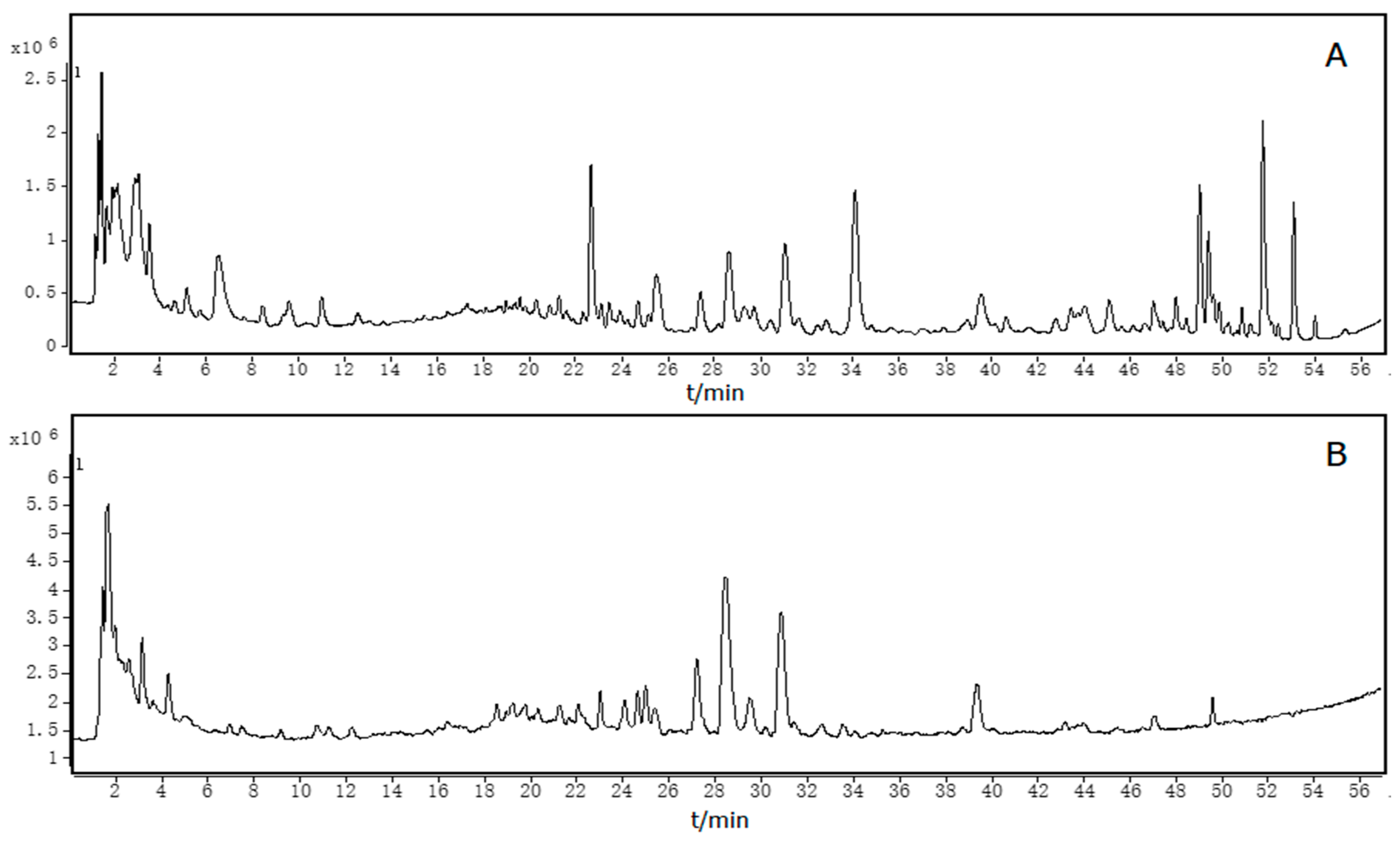
3.1. Optimization of HPLC Conditions
3.2. Identification of Constituents in SMD
3.3. Optimization of Screening Conditions
3.4. Screening Bioactive HSA Ligands from SMD
3.5. Repeatability of Ultrafiltration
3.6. Analysis of Molecular Docking
4. Conclusions
Author Contributions
Funding
Conflicts of Interest
References
- Chen, S.P.; Wang, X.P. Effect of Simotang oral liquid on anal exhaust in patients after abdominal gynecological operation. Chin. J. Integr. Med. 2006, 12, 221–223. [Google Scholar] [PubMed]
- Chong, T.; Jin, H.B.; Zhang, J.D.; Tan, Y. Clinical effects of the treatment of gastrointestinal dysfunction after stable thoracolumbar fractures with Simo decoction oral liquid. Zhongguo Gu Shang 2010, 23, 595–597. [Google Scholar] [PubMed]
- You, X.M.; Mo, X.S.; Ma, L.; Zhong, J.H.; Qin, H.G.; Lu, Z.; Xiang, B.D.; Wu, F.X.; Zhao, X.H.; Tang, J.; et al. Randomized Clinical Trial Comparing Efficacy of Simo Decoction and Acupuncture or Chewing Gum Alone on Postoperative Ileus in Patients With Hepatocellular Carcinoma After Hepatectomy. Medicine 2015, 94, e1968. [Google Scholar] [CrossRef] [PubMed]
- Yang, Y.; Zuo, H.Q.; Li, Z.; Qin, Y.Z.; Mo, X.W.; Huang, M.W.; Lai, H.; Wu, L.C.; Chen, J.S. Comparison of efficacy of simo decoction and acupuncture or chewing gum alone on postoperative ileus in colorectal cancer resection: A randomized trial. Sci. Rep. 2017, 7, 37826. [Google Scholar] [CrossRef] [PubMed]
- Cai, G.X.; Liu, B.Y.; Yi, J.; Chen, X.M.; Liu, F.L. Simotang enhances gastrointestinal motility, motilin and cholecystokinin expression in chronically stressed mice. World J. Gastroenterol. 2011, 17, 1594–1599. [Google Scholar] [CrossRef] [PubMed]
- Dai, C.; Liu, N.; Chen, W.; Qian, W.; Hou, X. Simo decoction promotes contraction of antral circular smooth muscle mainly via muscarinic M3 receptor. J. Ethnopharmacol. 2012, 144, 270–276. [Google Scholar] [CrossRef] [PubMed]
- Yi, Y.N.; Cheng, X.M.; Liu, L.A.; Hu, G.Y.; Wang, Z.T.; Deng, Y.D.; Huang, K.L.; Cai, G.X.; Wang, C.H. Simultaneous determination of synephrine, arecoline, and norisoboldine in Chinese patent medicine Si-Mo-Tang oral liquid preparation by strong cation exchange high performance liquid chromatography. Pharm. Biol. 2012, 50, 832–838. [Google Scholar] [CrossRef] [PubMed] [Green Version]
- Yi, Y.N.; Cheng, X.M.; Liu, L.A.; Hu, G.Y.; Cai, G.X.; Deng, Y.D.; Huang, K.L.; Wang, C.H. HPLC Fingerprint with Multi-components Analysis for Quality Consistency Evaluation of Traditional Chinese Medicine Si-Mo-Tang Oral Liquid Preparation. Chem. Res. Chin. Univ. 2011, 27, 756–763. [Google Scholar]
- Zhang, X.; Han, L.; Liu, J.; Xu, Q.; Guo, Y.; Zheng, W.; Wang, J.; Huang, X.; Ren, P. Pharmacokinetic Study of 7 Compounds Following Oral Administration of Fructus Aurantii to Depressive Rats. Front. Pharmacol. 2018, 9, 131. [Google Scholar] [CrossRef] [PubMed]
- Berezhkovskiy, L.M. On the calculation of the concentration dependence of drug binding to plasma proteins with multiple binding sites of different affinities: Determination of the possible variation of the unbound drug fraction and calculation of the number of binding sites of the protein. J. Pharm. Sci. 2007, 96, 249–257. [Google Scholar] [PubMed]
- Zhu, J.; Yi, X.; Huang, P.; Chen, S.; Wu, Y. Drug-protein binding of Danhong injection and the potential influence of drug combination with aspirin: Insight by ultrafiltration LC-MS and molecular modeling. J. Pharm. Biomed. Anal. 2017, 134, 100–107. [Google Scholar] [CrossRef] [PubMed]
- He, X.M.; Carter, D.C. Atomic structure and chemistry of human serum albumin. Nature 1992, 358, 209–215. [Google Scholar] [CrossRef] [PubMed] [Green Version]
- Gokara, M.; Sudhamalla, B.; Amooru, D.G.; Subramanyam, R. Molecular interaction studies of trimethoxy flavone with human serum albumin. PLoS ONE 2010, 5, e8834. [Google Scholar] [CrossRef] [PubMed]
- Vuignier, K.; Schappler, J.; Veuthey, J.L.; Carrupt, P.A.; Martel, S. Drug-protein binding: A critical review of analytical tools. Anal. Bioanal. Chem. 2010, 398, 53–66. [Google Scholar] [CrossRef] [PubMed]
- Liu, Y.; Liu, S.; Liu, Z. Screening and determination of potential xanthine oxidase inhibitors from Radix Salviae Miltiorrhizae using ultrafiltration liquid chromatography-mass spectrometry. J. Chromatogr. B Anal. Technol. Biomed. Life Sci. 2013, 923–924, 48–53. [Google Scholar] [CrossRef] [PubMed]
- Li, S.; Li, S.; Tang, Y.; Liu, C.; Chen, L.; Zhang, Y. Ultrafiltration-LC-MS combined with semi-preparative HPLC for the simultaneous screening and isolation of lactate dehydrogenase inhibitors from Belamcanda chinensis. J. Sep. Sci. 2016, 39, 4533–4543. [Google Scholar] [CrossRef] [PubMed]
- Zhou, H.; Xing, J.; Liu, S.; Song, F.; Cai, Z.; Pi, Z.; Liu, Z.; Liu, S. Screening and determination for potential alpha-glucosidase inhibitors from leaves of Acanthopanax senticosus harms by using UF-LC/MS and ESI-MS(n). Phytochem. Anal. 2012, 23, 315–323. [Google Scholar] [CrossRef] [PubMed]
- Chen, G.L.; Tian, Y.Q.; Wu, J.L.; Li, N.; Guo, M.Q. Antiproliferative activities of Amaryllidaceae alkaloids from Lycoris radiata targeting DNA topoisomerase I. Sci. Rep. 2016, 6, 38284. [Google Scholar] [CrossRef] [PubMed]
- Qin, S.; Ren, Y.; Fu, X.; Shen, J.; Chen, X.; Wang, Q.; Bi, X.; Liu, W.; Li, L.; Liang, G.; et al. Multiple ligand detection and affinity measurement by ultrafiltration and mass spectrometry analysis applied to fragment mixture screening. Anal. Chim. Acta 2015, 886, 98–106. [Google Scholar] [CrossRef] [PubMed]
- Ferreira, L.G.; Dos Santos, R.N.; Oliva, G.; Andricopulo, A.D. Molecular docking and structure-based drug design strategies. Molecules 2015, 20, 13384–13421. [Google Scholar] [CrossRef] [PubMed]
- Ma, X.H.; Shi, Z.; Tan, C.; Jiang, Y.; Go, M.L.; Low, B.C.; Chen, Y.Z. In-silico approaches to multi-target drug discovery: Computer aided multi-target drug design, multi-target virtual screening. Pharm. Res. 2010, 27, 739–749. [Google Scholar] [CrossRef] [PubMed]
- Scotti, L.; Mendonca Junior, F.J.; Ishiki, H.M.; Ribeiro, F.F.; Singla, R.K.; Barbosa Filho, J.M.; Da Silva, M.S.; Scotti, M.T. Docking Studies for Multi-Target Drugs. Curr. Drug Targets 2017, 18, 592–604. [Google Scholar] [CrossRef] [PubMed]
- Zhang, Y.; Peng, M.; Liu, L.; Shi, S.; Peng, S. Screening, identification, and potential interaction of active compounds from Eucommia ulmodies leaves binding with bovine serum albumin. J. Agric. Food Chem. 2012, 60, 3119–3125. [Google Scholar] [CrossRef] [PubMed]
- Trott, O.; Olson, A.J. AutoDock Vina: Improving the speed and accuracy of docking with a new scoring function, efficient optimization, and multithreading. J. Comput. Chem. 2010, 31, 455–461. [Google Scholar] [CrossRef] [PubMed]
- Ma, X.; He, J.; Yan, J.; Wang, Q.; Li, H. Comparative analysis the binding affinity of mycophenolic sodium and meprednisone with human serum albumin: Insight by NMR relaxation data and docking simulation. Chem. Biol. Interact. 2016, 248, 52–59. [Google Scholar] [CrossRef] [PubMed]
- Qing, Z.X.; Zhao, H.; Tang, Q.; Mo, C.M.; Huang, P.; Cheng, P.; Yang, P.; Yang, X.Y.; Liu, X.B.; Zheng, Y.J.; et al. Systematic identification of flavonols, flavonol glycosides, triterpene and siraitic acid glycosides from Siraitia grosvenorii using high-performance liquid chromatography/quadrupole-time-of-flight mass spectrometry combined with a screening strategy. J. Pharm. Biomed. Anal. 2017, 138, 240–248. [Google Scholar] [CrossRef] [PubMed]
- Srimany, A.; George, C.; Naik, H.R.; Pinto, D.G.; Chandrakumar, N.; Pradeep, T. Developmental patterning and segregation of alkaloids in areca nut (seed of Areca catechu) revealed by magnetic resonance and mass spectrometry imaging. Phytochemistry 2016, 125, 35–42. [Google Scholar] [CrossRef] [PubMed]
- Lee, H.H.; Chen, L.Y.; Wang, H.L.; Chen, B.H. Quantification of Salivary Arecoline, Arecaidine and N-Methylnipecotic Acid Levels in Volunteers by Liquid Chromatography-Tandem Mass Spectrometry. J. Anal. Toxicol. 2015, 39, 714–719. [Google Scholar] [CrossRef] [PubMed]
- Deng, G.M.; Xiang, B.; Xiao, X.Q.; Ge, J.W.; Chen, Z.; Yang, L.P.; Wei, F. Study on Chemical Constituents of Lindera aggregate by GC-MS and UPLC-ESI-MS/MS. Zhong Yao Cai 2016, 39, 2229–2236. [Google Scholar]
- Wu, Y.; Zheng, Y.; Liu, X.; Han, Z.; Ren, Y.; Gan, L.; Zhou, C.; Luan, L. Separation and quantitative determination of sesquiterpene lactones in Lindera aggregata (Wu-yao) by ultra-performance LC-MS/MS. J. Sep. Sci. 2010, 33, 1072–1078. [Google Scholar] [PubMed]
- Wu, Y.J.; Zheng, Y.L.; Luan, L.J.; Liu, X.S.; Han, Z.; Ren, Y.P.; Gan, L.S.; Zhou, C.X. Development of the fingerprint for the quality of Radix Linderae through ultra-pressure liquid chromatography-photodiode array detection/electrospray ionization mass spectrometry. J. Sep. Sci. 2010, 33, 2734–2742. [Google Scholar] [CrossRef] [PubMed]
- Qiang, Y.; Yang, Z.D.; Yang, J.L.; Gao, K. Sesquiterpenoids from the root tubers of Lindera aggregata. Planta Med. 2011, 77, 1610–1616. [Google Scholar] [CrossRef] [PubMed]
- Zhang, J.; Hu, X.; Gao, W.; Qu, Z.; Guo, H.; Liu, Z.; Liu, C. Pharmacokinetic study on costunolide and dehydrocostuslactone after oral administration of traditional medicine Aucklandia lappa Decne. by LC/MS/MS. J. Ethnopharmacol. 2014, 151, 191–197. [Google Scholar] [CrossRef] [PubMed]
- Zhang, J.; Gao, W.; Liu, Z.; Zhang, Z. Identification and Simultaneous Determination of Twelve Active Components in the Methanol Extract of Traditional Medicine Weichang’an Pill by HPLC-DAD-ESI-MS/MS. Iran. J. Pharm. Res. 2013, 12, 15–24. [Google Scholar] [PubMed]
- Ye, X.; Cao, D.; Zhao, X.; Song, F.; Huang, Q.; Fan, G.; Wu, F. Chemical fingerprint and metabolic profile analysis of Citrus reticulate ‘Chachi’ decoction by HPLC-PDA-IT-MS(n) and HPLC-Quadrupole-Orbitrap-MS method. J. Chromatogr. B Anal. Technol. Biomed. Life Sci. 2014, 970, 108–120. [Google Scholar] [CrossRef] [PubMed]
- Lin, Z.; Wang, H.; Xu, Y.; Dong, J.; Hashi, Y.; Chen, S. Identification of antioxidants in Fructus aurantii and its quality evaluation using a new on-line combination of analytical techniques. Food Chem. 2012, 134, 1181–1191. [Google Scholar] [CrossRef] [PubMed]
- Wang, M.; Li, Y.; Huang, Y.; Tian, Y.; Xu, F.; Zhang, Z. Chemomic and chemometric approach based on ultra-fast liquid chromatography with ion trap time-of-flight mass spectrometry to reveal the difference in the chemical composition between Da-Cheng-Qi decoction and its three constitutional herbal medicines. J. Sep. Sci. 2014, 37, 1148–1154. [Google Scholar] [CrossRef] [PubMed]
- Wang, S.; Tu, H.; Wan, J.; Chen, W.; Liu, X.; Luo, J.; Xu, J.; Zhang, H. Spatio-temporal distribution and natural variation of metabolites in citrus fruits. Food Chem. 2016, 199, 8–17. [Google Scholar] [CrossRef] [PubMed]
- Tian, Q.; Schwartz, S.J. Mass spectrometry and tandem mass spectrometry of citrus limonoids. Anal. Chem. 2003, 75, 5451–5460. [Google Scholar] [CrossRef] [PubMed]
- Zhou, W.J.; Song, J.Z.; Fu, W.W.; Tan, H.S.; Bian, Z.X.; Xu, H.X. Chemical comparison of two dosage forms of Hemp Seed Pills by UHPLC-Q-ToF-MS/MS and multivariate statistical techniques. J. Pharm. Biomed. Anal. 2013, 84, 59–68. [Google Scholar] [CrossRef] [PubMed]
- Liu, X.; Gu, Z.; Guo, Y.; Liu, J.; Ma, M.; Chen, B.; Wang, L. Rapid analysis of Aurantii Fructus Immaturus (Zhishi) using paper spray ionization mass spectrometry. J. Pharm. Biomed. Anal. 2017, 137, 204–212. [Google Scholar] [CrossRef] [PubMed]
- Chen, H.F.; Zhang, W.G.; Yuan, J.B.; Li, Y.G.; Yang, S.L.; Yang, W.L. Simultaneous quantification of polymethoxylated flavones and coumarins in Fructus aurantii and Fructus aurantii immaturus using HPLC-ESI-MS/MS. J. Pharm. Biomed. Anal. 2012, 59, 90–95. [Google Scholar] [CrossRef] [PubMed]
- Lee, J.; Chan, B.L.; Mitchell, A.E. Identification/quantification of free and bound phenolic acids in peel and pulp of apples (Malus domestica) using high resolution mass spectrometry (HRMS). Food Chem. 2017, 215, 301–310. [Google Scholar] [CrossRef] [PubMed]
- Zhang, W.M.; Huang, W.Y.; Chen, W.X.; Han, L.; Zhang, H.D. Optimization of extraction conditions of areca seed polyphenols and evaluation of their antioxidant activities. Molecules 2014, 19, 16416–16427. [Google Scholar] [CrossRef] [PubMed]
- Zhang, T.; Wang, H.; Du, G.; Chen, R. Study on chemical constituents from roots of Saussurea lappa. Zhongguo Zhong Yao Za Zhi 2009, 34, 1223–1224. [Google Scholar] [PubMed]
- Wei, H.; He, C.; Peng, Y.; Ma, G.; Xiao, P. Chemical constituents of Dolomiaea souliei. Chin. J. Chin. Mater. Med. 2012, 37, 1249–1253. [Google Scholar] [CrossRef]
- Brito, A.; Ramirez, J.E.; Areche, C.; Sepulveda, B.; Simirgiotis, M.J. HPLC-UV-MS profiles of phenolic compounds and antioxidant activity of fruits from three citrus species consumed in Northern Chile. Molecules 2014, 19, 17400–17421. [Google Scholar] [CrossRef] [PubMed]
- Durand-Hulak, M.; Dugrand, A.; Duval, T.; Bidel, L.P.; Jay-Allemand, C.; Froelicher, Y.; Bourgaud, F.; Fanciullino, A.L. Mapping the genetic and tissular diversity of 64 phenolic compounds in Citrus species using a UPLC-MS approach. Ann. Bot. 2015, 115, 861–877. [Google Scholar] [CrossRef] [PubMed]
- Raman, G.; Cho, M.; Brodbelt, J.S.; Patil, B.S. Isolation and purification of closely related Citrus limonoid glucosides by flash chromatography. Phytochem. Anal. 2005, 16, 155–160. [Google Scholar] [CrossRef] [PubMed]
- Mencherini, T.; Campone, L.; Piccinelli, A.L.; Mesa, M.G.; Sanchez, D.M.; Aquino, R.P.; Rastrelli, L. HPLC-PDA-MS and NMR characterization of a hydroalcoholic extract of Citrus aurantium L. var. amara peel with antiedematogenic activity. J. Agric. Food Chem. 2013, 61, 1686–1693. [Google Scholar] [PubMed]
- Kuroyanagi, M.; Ishii, H.; Kawahara, N.; Sugimoto, H.; Yamada, H.; Okihara, K.; Shirota, O. Flavonoid glycosides and limonoids from Citrus molasses. J. Nat. Med. 2008, 62, 107–111. [Google Scholar] [CrossRef] [PubMed]
- Matsubara, Y.; Yusa, T.; Sawabe, A.; Iizuka, Y.; Takekuma, S.; Yoshida, Y. Structures of new cyclic peptides in young unshiu (Citrus unshiu Marcov.), orange (Citrus sinensis Osbeck.) and amanatsu (Citrus natsudaidai) peelings. Agric. Biol. Chem. 1991, 55, 2923–2929. [Google Scholar] [PubMed]
- Di Donna, L.; De Luca, G.; Mazzotti, F.; Napoli, A.; Salerno, R.; Taverna, D.; Sindona, G. Statin-like principles of bergamot fruit (Citrus bergamia): Isolation of 3-hydroxymethylglutaryl flavonoid glycosides. J. Nat. Prod. 2009, 72, 1352–1354. [Google Scholar] [CrossRef] [PubMed]
- Xing, T.T.; Zhao, X.J.; Zhang, Y.D.; Li, Y.F. Fast Separation and Sensitive Quantitation of Polymethoxylated Flavonoids in the Peels of Citrus Using UPLC-Q.-TOF-MS. J. Agric. Food Chem. 2017, 65, 2615–2627. [Google Scholar] [CrossRef] [PubMed]
- Dugrand, A.; Olry, A.; Duval, T.; Hehn, A.; Froelicher, Y.; Bourgaud, F. Coumarin and furanocoumarin quantitation in citrus peel via ultraperformance liquid chromatography coupled with mass spectrometry (UPLC-MS). J. Agric. Food Chem. 2013, 61, 10677–10684. [Google Scholar] [CrossRef] [PubMed]
- Wang, Z.; Kwon, S.H.; Hwang, S.H.; Kang, Y.H.; Lee, J.Y.; Lim, S.S. Competitive binding experiments can reduce the false positive results of affinity-based ultrafiltration-HPLC: A case study for identification of potent xanthine oxidase inhibitors from Perilla frutescens extract. J. Chromatogr. B Anal. Technol. Biomed. Life Sci. 2017, 1048, 30–37. [Google Scholar] [CrossRef] [PubMed]
- Anguizola, J.; Debolt, E.; Suresh, D.; Hage, D.S. Chromatographic analysis of the effects of fatty acids and glycation on binding by probes for Sudlow sites I and II to human serum albumin. J. Chromatogr. B Anal. Technol. Biomed. Life Sci. 2016, 1021, 175–181. [Google Scholar] [CrossRef] [PubMed] [Green Version]
- Fu, L.; Sun, Y.; Ding, L.; Wang, Y.; Gao, Z.; Wu, Z.; Wang, S.; Li, W.; Bi, Y. Mechanism evaluation of the interactions between flavonoids and bovine serum albumin based on multi-spectroscopy, molecular docking and Q-TOF HR-MS analyses. Food Chem. 2016, 203, 150–157. [Google Scholar] [CrossRef] [PubMed]
- Deng, S.; Xia, L.; Xiao, H. Screening of alpha-glucosidase inhibitors from green tea extracts using immobilized enzymes affinity capture combined with UHPLC-QTOF MS analysis. Chem. Commun. 2014, 50, 2582–2584. [Google Scholar] [CrossRef] [PubMed]
Sample Availability: Samples of the compounds are available from the authors. |


| No. | TR (min) | ESI+ (m/z) | ESI− (m/z) | Fragment Ions (Positive/Negative) | MW (Mea.) | MW (MFG) | Formula | Compound | Ref. | Error (ppm) b |
|---|---|---|---|---|---|---|---|---|---|---|
| Areca catechu | ||||||||||
| 01 | 1.899 | 128.0704 | 109.0289 | 127.0631 | 127.0633 | C6H9NO2 | Guvacine | [27] | 1.61 | |
| 02 | 2.113 | 142.0860 | / | 141.0787 | 141.0790 | C7H11NO2 | Guvacoline | [27] | 1.96 | |
| 03 | 3.001 | 144.1021 | / | 143.0948 | 143.0946 | C7H13NO2 | N-Methylnipecotic Acid | [28] | −1.06 | |
| 04 | 5.163 | 142.0864 | 124.0252, 109.0289 | 141.0791 | 141.0790 | C7H11NO2 | Arecaidine a | [27,28] | −0.80 | |
| 05 | 7.021 | 156.1018 | 127.0410 | 155.0946 | 155.0946 | C8H13NO2 | Arecoline a | [27,28] | 0.24 | |
| 06 | 16.247 | 137.0240 | 138.0312 | 138.0317 | C7H6O3 | 4-Hydroxybenzoic acid | [43] | 3.23 | ||
| 07 | 17.508 | 199.0597 | 198.0525 | 198.0528 | C9H10O5 | Syringic acid | [44] | 1.62 | ||
| 08 | 19.379 | 291.0866 | 290.0793 | 290.0790 | C15H14O6 | Epicatechin | [44] | −0.93 | ||
| 09 | 25.337 | 193.0500 | 194.0572 | 194.0579 | C10H10O4 | Ferulic acid | [43] | 3.4 | ||
| Radix linderae | ||||||||||
| 10 | 5.722 | 165.0545 | 164.0472 | 164.0473 | C9H8O3 | p-Coumaric acid | [29] | 0.6 | ||
| 11 | 20.782 | 328.1546 | 327.1474 | 327.1471 | C19H21NO4 | Boldine | [29,31] | −0.91 | ||
| 12 | 21.558 | 314.1388 | 297.1125, 265.0839, 237.0743 | 313.1315 | 313.1314 | C18H19NO4 | Norboldine | [29,31] | −0.26 | |
| 13 | 22.677 | 314.1387 | 297.1141, 265.0787, 237.0619 | 313.1314 | 313.1314 | C18H19NO4 | Norisoboldine a | [29] | −0.12 | |
| 14 | 23.466 | 328.1543 | 297.1110, 265.0859, 237.0627 | 327.1470 | 327.1471 | C19H21NO4 | Isoboldine | / | 0.03 | |
| 15 | 24.093 | 261.1116 | 243.1018, 173.0132 | 260.1043 | 260.1049 | C15H16O4 | Linderane a | [30,31,32] | 2.07 | |
| 16 | 24.698 | 330.1698 | 330.1691, 299.1472, 192.0682 | 329.1625 | 329.1627 | C19H23NO4 | Reticuline | [29,31] | 0.57 | |
| 17 | 25.234 | 263.1270 | 262.1197 | 262.1205 | C15H18O4 | Linderagalactone D | [30,31] | 3.13 | ||
| 18 | 35.746 | 277.1068 | 276.0995 | 276.0998 | C15H16O5 | Linderanlide A | [32] | 1.01 | ||
| 19 | 40.231 | 245.1169 | 244.1096 | 244.1099 | C15H16O3 | Neolinderalactone | [30,31] | 1.50 | ||
| 20 | 40.590 | 291.1223 | 290.1150 | 290.1154 | C16H18O5 | Linderanlide D | [32] | 1.51 | ||
| 21 | 44.323 | 305.1376 | 304.1303 | 304.1311 | C17H20O5 | Linderanlide F | [32] | 2.54 | ||
| 22 | 46.623 | 263.1279 | 262.1207 | 262.1205 | C15H18O4 | Linderagalactone C | [30] | −0.57 | ||
| 23 | 47.462 | 247.1326 | 246.1254 | 246.1256 | C15H18O3 | Hydroylindestenolide isomer | [29] | 0.92 | ||
| 24 | 49.032 | 261.1117 | 260.1044 | 260.1049 | C15H16O4 | Linderane isomer | / | 1.64 | ||
| 25 | 50.118 | 247.1325 | 246.1252 | 246.1256 | C15H18O3 | Hydroylindestenolide | [30,31] | 1.53 | ||
| 26 | 50.126 | 247.1329 | 246.1256 | 246.1256 | C15H18O3 | Lindenenol E | [29] | 0.08 | ||
| 27 | 54.009 | 231.1380 | 230.1307 | 230.1307 | C15H18O2 | Lindenenol | [31] | −0.01 | ||
| Radix aucklandiae | ||||||||||
| 28 | 11.014 | 127.0388 | / | 126.0315 | 126.0317 | C6H6O3 | 5-HydroxymethylFurfual | [45] | 1.24 | |
| 29 | 25.283 | 193.0490 | 192.0417 | 192.0423 | C10H8O4 | 5,7-dihydroxy-2-methylchromone | [45] | 2.89 | ||
| 30 | 44.022 | 233.1534 | 232.1461 | 232.1463 | C15H20O2 | Mokko lactone | [46] | 0.91 | ||
| 31 | 46.101 | 233.1532 | 232.1459 | 232.1463 | C15H20O2 | Costunolide isomer | / | 1.84 | ||
| 32 | 47.095 | 233.1536 | 232.1464 | 232.1463 | C15H20O2 | Cyclocostunolide | [46] | −0.16 | ||
| 33 | 49.408 | 233.1530 | 187.1475, 121.0516 | 232.1457 | 232.1463 | C15H20O2 | Costunolide a | [33,34] | 2.75 | |
| 34 | 49.895 | 235.1691 | 234.1618 | 234.1620 | C15H22O2 | Costus acid | [46] | 0.71 | ||
| 35 | 51.221 | 239.2003 | 238.1930 | 238.1933 | C15H26O2 | 4-α-hydroxy-4-β-methyldihydrocostol | [46] | 1.2 | ||
| 36 | 54.001 | 231.1373 | 230.1301 | 230.1307 | C15H18O2 | Dehydrocostus lactone a | [33,34] | 2.69 | ||
| Aurantii fructus | ||||||||||
| 37 | 3.201 | 191.0189 | 192.0262 | 192.0270 | C6H8O7 | Citric acid | [37,47] | 4.06 | ||
| 38 | 3.802 | 168.1017 | 167.0944 | 167.0946 | C9H13NO2 | Synephrine a | [35] | 1.45 | ||
| 39 | 9.369 | 268.1035 | 267.0962 | 267.0968 | C10H13N5O4 | Adenosine | [37] | 2.03 | ||
| 40 | 17.108 | 196.0967 | 195.0894 | 195.0895 | C10H13NO3 | N-Acetylnorsynephrine | / | 0.8 | ||
| 41 | 20.439 | 611.1590 | 465.0874, 303.0511 | 610.1518 | 610.1534 | C27H30O16 | Quercetin-3-O-rutinoside (Rutin) a | [36] | 2.53 | |
| 42 | 21.337 | 595.1659 | 593.1500 | 594.1587 | 594.1585 | C27H30O15 | Isovitexin-7-O-glucoside (Saponarin) | [48] | −0.31 | |
| 43 | 21.903 | 625.1766 | 623.1615 | 301.0723 | 624.1693 | 624.1690 | C28H32O16 | Diosmetin-6,8-di-C-glucoside | [47] | −0.46 |
| 44 | 22.234 | 741.2245 | 579.1833, 417.1323, 271.0756 | 742.2318 | 742.2320 | C33H42O19 | Naringenin-7-O-triglycoside | [36,40] | 0.36 | |
| 45 | 22.377 | 625.1761 | 624.1688 | 624.1690 | C28H32O16 | Diosmetin 6,8-di-C-glucoside (isomer) | [47] | 0.41 | ||
| 46 | 23.947 | 471.2007 | 470.1935 | 470.1941 | C26H30O8 | Limonin a | [37,38,39] | 1.28 | ||
| 47 | 24.235 | 597.1813 | 595.1663 | 435.1278, 417.1185, 331.1826, 289.0702 | 596.1740 | 596.1741 | C27H32O15 | Eriodictyol-7-O-rutinoside (Eriocitrin) a | [36] | 0.12 |
| 48 | 24.460 | 481.1683 | 480.1610 | 480.1632 | C23H28O11 | Paeoniflorin, Albiflorin | [40] | 4.44 | ||
| 49 | 24.603 | 649.2501 | 650.2573 | 650.2575 | C32H42O14 | Limonin-17-β-d-glucoside | [39,49] | 0.16 | ||
| 50 | 25.124 | 597.1807 | 595.1656 | 451.1287, 289.0699 | 596.1735 | 596.1741 | C27H32O15 | Eriodictyol-7-O-neohesperidoside (Neoeriocitrin) | [36,37] | 1.06 |
| 51 | 26.946 | 461.1067 | 462.1140 | 462.1162 | C22H22O11 | Diosmetin-7-O-glucoside | [35] | 4.85 | ||
| 52 | 26.986 | 595.1653 | 463.1303, 287.0559 | 594.1579 | 594.1585 | C27H30O15 | Luteolin-7-O-rutinoside (Veronicastroside) | / | 0.9 | |
| 53 | 27.421 | 581.1853 | 579.1705 | 435.1274, 273.0757 | 580.1781 | 580.1792 | C27H32O14 | Naringenin-7-O-rutinoside (Narirutin) a | [36,41] | 1.97 |
| 54 | 27.989 | 625.2107 | 643.1461, 267.1224 | 624.2034 | 624.2054 | C29H36O15 | Magnoloside A | [37,47] | 3.31 | |
| 55 | 28.694 | 581.1857 | 579.1687 | 435.1278, 419.1330, 273.0754, 153.0186 | 580.1785 | 580.1792 | C27H32O14 | naringenin-7-O-neohesperidoside (Naringin) a | [36,41] | 1.26 |
| 56 | 29.032 | 435.1274 | 273.0757 | 434.1201 | 434.1213 | C21H20O10 | Naringenin-7-O-glucoside | 2.71 | ||
| 57 | 29.692 | 611.1965 | 609.1803 | 465.1432, 303.0858, 273.0757 | 610.1891 | 610.1898 | C28H34O15 | Hesperetin-7-O-rutinoside (Hesperidin) a | [36,41] | 1.13 |
| 58 | 30.385 | 579.1708 | 577.1549 | 433.1323, 271.0596 | 578.1636 | 578.1636 | C27H30O14 | Apigenin-7-O-rutinoside (Isorhoifolin) | [40] | −0.01 |
| 59 | 31.051 | 611.1962 | 609.1811 | 465.1434, 303.0862, 153.0188 | 610.1889 | 610.1898 | C28H34O15 | Hesperetin-7-O-neohesperidoside (Neohesperidin) a | [36,41] | 1.37 |
| 60 | 31.121 | 465.1395 | 331.1881, 303.0861, 155.0372, 121.0216 | 464.1322 | 464.1319 | C22H24O11 | Hesperitin-7-O-glucoside | [37] | −0.73 | |
| 61 | 31.638 | 609.1819 | 463.1409, 301.0723 | 608.1747 | 608.1741 | C28H32O15 | Diosmetin-7-O-rutinoside (Diosmin) | −0.88 | ||
| 62 | 32.515 | 609.1806 | 463.1411, 301.0723 | 608.1734 | 608.1741 | C28H32O15 | Diosmetin-7-O-neohesperidoside (Neodiosmin) | [47] | 1.12 | |
| 63 | 32.531 | 693.2756 | 694.2829 | 694.2837 | C34H46O15 | Nominin-17-β-d-glucoside | [39,49] | 1.12 | ||
| 64 | 32.787 | 651.1541 | 652.1614 | 652.1639 | C29H32O17 | Obacunoic acid-17-β-d-glucoside | [39] | 3.92 | ||
| 65 | 33.509 | 711.2850 | 712.2923 | 712.2942 | C34H48O16 | Nomilinic acid 17-O-β-d-glucoside | [39,49] | 2.74 | ||
| 66 | 34.361 | 261.1120 | 260.1047 | 260.1049 | C15H16O4 | Meranzin hydrate | [50] | 0.67 | ||
| 67 | 39.559 | 595.2016 | 593.1875 | 449.1505, 287.0917 | 594.1944 | 594.1949 | C28H34O14 | Isosakuranetin-7-O-neohesperidoside, (Poncirin) a | [36,41] | 0.77 |
| 68 | 39.546 | 287.0913 | 286.0840 | 286.0841 | C16H14O5 | Oxypeucedanin | [48] | 0.48 | ||
| 69 | 41.776 | 697.1975 | 696.1901 | 696.1902 | C31H36O18 | Isovitexin-7-O-xylocoside 2″-O-arabinoside | [40] | 0.05 | ||
| 70 | 42.803 | 728.3970 | 727.3896 | 727.3905 | C36H53N7O9 | Citrusin III | [35,51,52] | 1.15 | ||
| 71 | 43.149 | 271.0609 | 272.0682 | 272.0685 | C15H12O5 | Naringenin a | [36] | 1.18 | ||
| 72 | 44.264 | 725.2283 | 724.2210 | 724.2215 | C33H40O18 | Melitidin | [53] | 0.65 | ||
| 73 | 45.375 | 301.0714 | 302.0787 | 302.0790 | C16H14O6 | Hesperetin a | [36] | 1.27 | ||
| 74 | 47.003 | 704.3968 | 703.3895 | 703.3905 | C34H53N7O9 | Citrusin I | [52] | 1.40 | ||
| 75 | 47.187 | 329.1023 | 314.0762, 299.0543 | 328.0950 | 328.0947 | C18H16O6 | Monohydroxytrimethoxyflavone | [54] | −1.08 | |
| 76 | 47.976 | 355.1533 | 354.1460 | 354.1467 | C21H22O5 | Epoxybergamottin or Cnidicin | [55] | 2.01 | ||
| 77 | 48.440 | 359.1119 | 344.0877, 326.0771 | 358.1046 | 358.1053 | C19H18O7 | 5-Hydroxy-6,7,3′,4′-tetramethoxy-flavone | [54] | 1.74 | |
| 78 | 49.028 | 261.1117 | 260.1044 | 260.1049 | C15H16O4 | Meranzin, IsoMeranzin | [50] | 1.64 | ||
| 79 | 49.634 | 471.2005 | 470.1932 | 470.1941 | C26H30O8 | Limonin isomer | [37,38] | 1.76 | ||
| 80 | 50.227 | 373.1276 | 358.1024, 343.0811 | 372.1204 | 372.1209 | C20H20O7 | 5,7,8,3′,4′-Pentamethoxyflavone (Isosinensetin) | [36,54] | 1.44 | |
| 81 | 50.853 | 373.1278 | 358.1036, 343.0812 | 372.1205 | 372.1209 | C20H20O7 | 5,6,7,3′,4′-Pentamethoxyflavone (Sinensetin) | [54] | 1.04 | |
| 82 | 51.721 | 403.1385 | 388.1025, 373.1253 | 402.1312 | 402.1315 | C21H22O8 | 5,6,7,8,3′,4′-Hexamethoxyflavone (Nobiletin) a | [36,42] | 0.74 | |
| 83 | 51.847 | 433.1485 | 403.1021, 388.0773 | 432.1413 | 432.1420 | C22H24O9 | 3′,4′,3,5,6,7,8-Heptamethox-yflavone | [36] | 1.79 | |
| 84 | 52.030 | 343.1174 | 328.0927, 285.0749 | 342.1101 | 342.1103 | C19H18O6 | 5,6,8,4′-Tetramethoxyflavone | [54] | 0.63 | |
| 85 | 52.381 | 343.1175 | 328.0919, 313.0705 | 342.1102 | 342.1103 | C19H18O6 | 4′,5,7,8-Tetramethoxyflavone | [54] | 0.36 | |
| 86 | 53.099 | 373.1281 | 358.1007, 343.1182 | 372.1208 | 372.1209 | C20H20O7 | 5,6,7,8,4′-Pentamethoxyflavone, (Tangeretin) a | [36,42] | 0.25 | |
| Common Compounds | ||||||||||
| 87 | 1.711 | 116.0705 | 115.0632 | 115.0633 | C5H9NO2 | Proline | [38] | 0.83 | ||
| 88 | 2.849 | 118.0865 | 117.0792 | 117.0790 | C5H11NO2 | Valine | [38] | −1.83 | ||
| 89 | 4.328 | 132.1016 | 131.0943 | 131.0946 | C6H13NO2 | Isoleucine | [38] | 2.32 | ||
| 90 | 4.678 | 132.1019 | 131.0947 | 131.0946 | C6H13NO2 | Leucine | [38] | −0.32 | ||
| 91 | 5.813 | 182.0810 | 181.0737 | 181.0739 | C9H11NO3 | tyrosine | [38] | 0.83 | ||
| 92 | 9.561 | 166.0859 | 165.0786 | 165.0790 | C9H11NO2 | Phenylalanine | [38] | 2.36 | ||
| 93 | 17.316 | 205.0969 | 204.0896 | 204.0899 | C11H12N2O2 | Tryptophan | [38] | 1.34 | ||
| 94 | 25.552 | 113.0597 | 112.0524 | 112.0524 | C6H8O2 | Sorbic acid a | / | 0.25 | ||
| No. | Ligand | Binding Affinity | Docking Score | |
|---|---|---|---|---|
| Site I | Site II | |||
| A | Norisoboldine | 26.1 | −34.7 | −36.1 |
| B | Eriocitrin | 14.2 | −39.7 | −30.5 |
| C | Neoeriocitrin | 15.3 | −38.9 | −31.8 |
| D | Narirutin | 15.5 | −40.6 | −31.4 |
| E | Hesperidin | 11.6 | −39.3 | −30.1 |
| F | Naringin | 13.9 | −39.7 | −33.9 |
| G | Neohesperidin | 12.8 | −39.3 | −30.1 |
| H | Hesperitin-7-O-glucoside | 9.8 | −36.4 | −35.5 |
| I | Linderane | 22.5 | −34.3 | −36.0 |
| J | Poncirin | 16.7 | −38.9 | −31.0 |
| K | Costunolide | 19.6 | −33.5 | −35.9 |
| L | Nobiletin | 14.7 | −32.2 | −33.5 |
| M | Tangeretin | 12.9 | −31.4 | −34.7 |
| Drugs a | Warfarin | − | −33.5 | − |
| Ibuprofen | − | − | −32.2 | |
© 2018 by the authors. Licensee MDPI, Basel, Switzerland. This article is an open access article distributed under the terms and conditions of the Creative Commons Attribution (CC BY) license (http://creativecommons.org/licenses/by/4.0/).
Share and Cite
He, Y.; Cheng, P.; Wang, W.; Yan, S.; Tang, Q.; Liu, D.; Xie, H. Rapid Investigation and Screening of Bioactive Components in Simo Decoction via LC-Q-TOF-MS and UF-HPLC-MD Methods. Molecules 2018, 23, 1792. https://doi.org/10.3390/molecules23071792
He Y, Cheng P, Wang W, Yan S, Tang Q, Liu D, Xie H. Rapid Investigation and Screening of Bioactive Components in Simo Decoction via LC-Q-TOF-MS and UF-HPLC-MD Methods. Molecules. 2018; 23(7):1792. https://doi.org/10.3390/molecules23071792
Chicago/Turabian StyleHe, Yingjie, Pi Cheng, Wei Wang, Sien Yan, Qi Tang, Dongbo Liu, and Hongqi Xie. 2018. "Rapid Investigation and Screening of Bioactive Components in Simo Decoction via LC-Q-TOF-MS and UF-HPLC-MD Methods" Molecules 23, no. 7: 1792. https://doi.org/10.3390/molecules23071792