Review

Carbonic Anhydrase Inhibitors as Novel Drugs against Mycobacterial β-Carbonic Anhydrases: An Update on In Vitro and In Vivo Studies

1
Faculty of Medicine and Health Technology, University of Tampere, 33014 Tampere, Finland
2
Institut des Biomolécules Max Mousseron (IBMM) UMR 5247 CNRS, ENSCM, Université de Montpellier, 34296 Montpellier CEDEX 05, France
3
Neurofarba Department, Sezione di Chimica Farmaceutica e Nutraceutica, Università degli Studi di Firenze, 50019 Sesto Fiorentino (Firenze), Italy
4
Oral and Maxillofacial Unit, Tampere University Hospital, 33521 Tampere, Finland
5
Fimlab Ltd. and Tampere University Hospital, 33520 Tampere, Finland
*
Author to whom correspondence should be addressed.
Molecules 2018, 23(11), 2911; https://doi.org/10.3390/molecules23112911
Submission received: 23 October 2018 / Revised: 5 November 2018 / Accepted: 6 November 2018 / Published: 8 November 2018
(This article belongs to the Special Issue Metalloenzyme Inhibitors and Activators)

Abstract

:
Mycobacteria cause a variety of diseases, such as tuberculosis, leprosy, and opportunistic diseases in immunocompromised people. The treatment of these diseases is problematic, necessitating the development of novel treatment strategies. Recently, β-carbonic anhydrases (β-CAs) have emerged as potential drug targets in mycobacteria. The genomes of mycobacteria encode for three β-CAs that have been cloned and characterized from Mycobacterium tuberculosis (Mtb) and the crystal structures of two of the enzymes have been determined. Different classes of inhibitor molecules against Mtb β-CAs have subsequently been designed and have been shown to inhibit these mycobacterial enzymes in vitro. The inhibition of these centrally important mycobacterial enzymes leads to reduced growth of mycobacteria, lower virulence, and impaired biofilm formation. Thus, the inhibition of β-CAs could be a novel approach for developing drugs against the severe diseases caused by pathogenic mycobacteria. In the present article, we review the data related to in vitro and in vivo inhibition studies in the field.

1. Introduction

Mycobacteria are rod-shaped, non-motile, and acid-fast bacteria that contain a high amount of G + C in their genome [1,2]. The genus mycobacterium includes a variety of clinically relevant human pathogens, including Mycobacterium tuberculosis (Mtb), the main causative agent of human tuberculosis (TB). A group of closely related bacteria referred to as the Mycobacterium tuberculosis complex (MTC) composes of a variety of pathogens causing TB in humans and other mammals. These include M. tuberculosis, M. bovis, M. caprae, M. africanum, M. canettii, M. microti, M. orygis, and M. pinnipedii [3]. Mtb is typically transmitted through air by a droplet contact. Mtb can affect many organs in humans, but the main target organ is the lung, causing pulmonary TB in 80% of the patients. In 2017, 10 million new cases were diagnosed [4]. In addition to the diagnosed active Mtb infections, the World Health Organization (WHO) estimates that 23% of the world’s population has developed a latent TB [4], which is asymptomatic but can become reactivated and cause a difficult-to-treat and potentially lethal disease. Mtb is currently one of the deadliest bacteria killing 1.3 million people every year. Multi-drug-resistant strains of Mtb are on the rise making TB increasingly difficult to treat. This development poses an enormous global threat necessitating immediate action to find new ways to treat this devastating disease [4].
Leprosy is another example of a clinically relevant mycobacterial disease. Leprosy is caused by M. leprae, which is transmitted through droplets due to close and frequent contact with untreated patients. As the multiplication rate of M. leprae is very slow, the incubation period of disease ranges between 1 and 20 years. The disease mainly affects the skin, peripheral nerves, mucosa of the upper respiratory tract, and eye. If left untreated, the disease usually causes permanent tissue damage. In many developing countries, leprosy is still a serious health problem and the people suffering from the disease often face social problems that go hand in hand with the disease progression. The latest WHO report shows that there were 216,108 new leprosy cases in 145 countries from the 6 WHO regions [5].
The nontuberculous mycobacteria (NTM) group includes all mycobacteria other than MTC and M. leprae, and about 40 species of them are pathogenic [6]. NTM are ubiquitously found in a wide variety of environmental reservoirs [7,8]. Although they are mostly nonpathogenic, they are important opportunistic pathogens of humans [9]. The species of NTM associated with human disease are: M. avium, M. intracellulare, M. kansasii, M. fortuitum, M. chelonae, M. szulgai, M. paratuberculosis, M. scrofulaceum [10] as well as bacteria belonging to the M. abscessus complex [11]. NTM can cause pulmonary disease resembling tuberculosis, lymphadenitis, and skin disease. The pulmonary disease represents about 80% of infections caused by NTM [12]. Recent reports suggest that the NTM pulmonary disease is increasing in several parts of the world [13,14]. However, standardized diagnostics and effective treatment protocols for NTM infections are lacking [15].
Genomes of many mycobacterial species from both MTC and NTM categories have been sequenced [16,17,18,19,20]. Bioinformatic and molecular analysis of mycobacterial genomes revealed that they code for several novel proteins that are essential for the alternative pathways and critical for the life cycle of these pathogens [21,22,23]. Recent progress in the structural and functional analyses of genomes and proteomes has opened new avenues for the design of mechanism-based drugs targeting proteins crucial for pathogenesis of mycobacteria [24]. Among many such proteins, β-carbonic anhydrases (β-CAs) of mycobacteria could be possible targets for developing novel antimycobacterial agents with the potential to treat even infections caused by drug-resistant mycobacteria.
M. tuberculosis genome codes for three β-CA genes Rv1284 (β-CA1), Rv3588c (β-CA2) and Rv3273 (β-CA3) as shown in Table 1 [25]. Database searches and our bioinformatic analyses showed the presence of all the three β-CAs in both NTM and MTC bacteria [26,27,28,29,30]. β-CAs catalyze the reversible hydration of CO2 to HCO3 and H+, thus generating a buffering weak base (bicarbonate) and a strong acid (H+) [31,32]. Mycobacterial β-CAs are zinc-containing metalloenzymes with characteristics similar to many other bacterial β-CAs. All conserved amino acid residues typical of β-CAs and involved in the catalytic cycle, i.e., the four zinc-binding residues, Cys42, Asp44, His97 and Cys101 are shown in Figure 1.
The mycobacterial β-CAs are essential during starvation for the growth and survival of the bacteria [22,23,33,34]. Recent studies showed that the bicarbonate ion, which is a product of reversible hydration of CO2, is essential for the transport of extracellular DNA (eDNA) and the formation of biofilm in NTM bacteria in vitro [35]. Inhibition of β-CAs using ethoxzolamide (EZA), a CA inhibitor, reduced the transport of eDNA and the formation of biofilm [35]. EZA also inhibited the PhoPR regulon, a two-component regulatory system in Mtb, as well as Esx-1 protein secretion system centrally important for the virulence of Mtb bacterium, and showed efficacy in infected macrophages and mice [36], suggesting that β-CAs perform very important roles in mycobacterial infections. Using M. marinum, an NTM model bacterium, we were the first to show that dithiocarbamate Fc14-584b, a β-CA inhibitor impairs mycobacterial growth in zebrafish larvae in vivo [26]. These essential enzymes are thus potential drug targets and are currently under investigation by several groups, including ours [26,27,28,29,35,36,37,38,39]. Similarly, several in vitro studies have shown that all the Mtb β-CAs could be efficiently (KI in nanomolar ranges) inhibited by sulfonamides/sulfamates (Table 1). In the present review, we update the data on in vitro and in vivo studies using CA inhibitors on mycobacterial β-CAs.

2. In Vitro Inhibition Studies of M. tuberculosis β-CAs

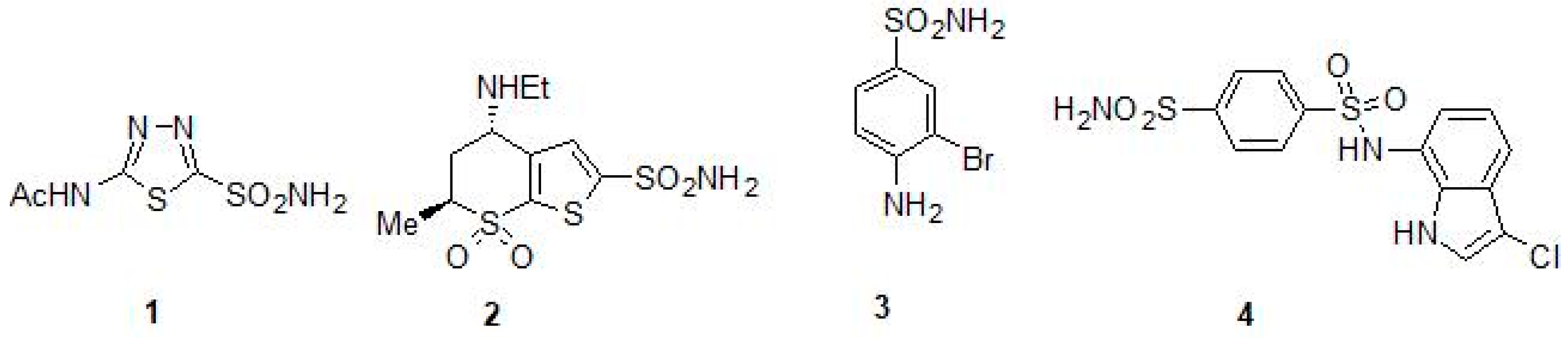
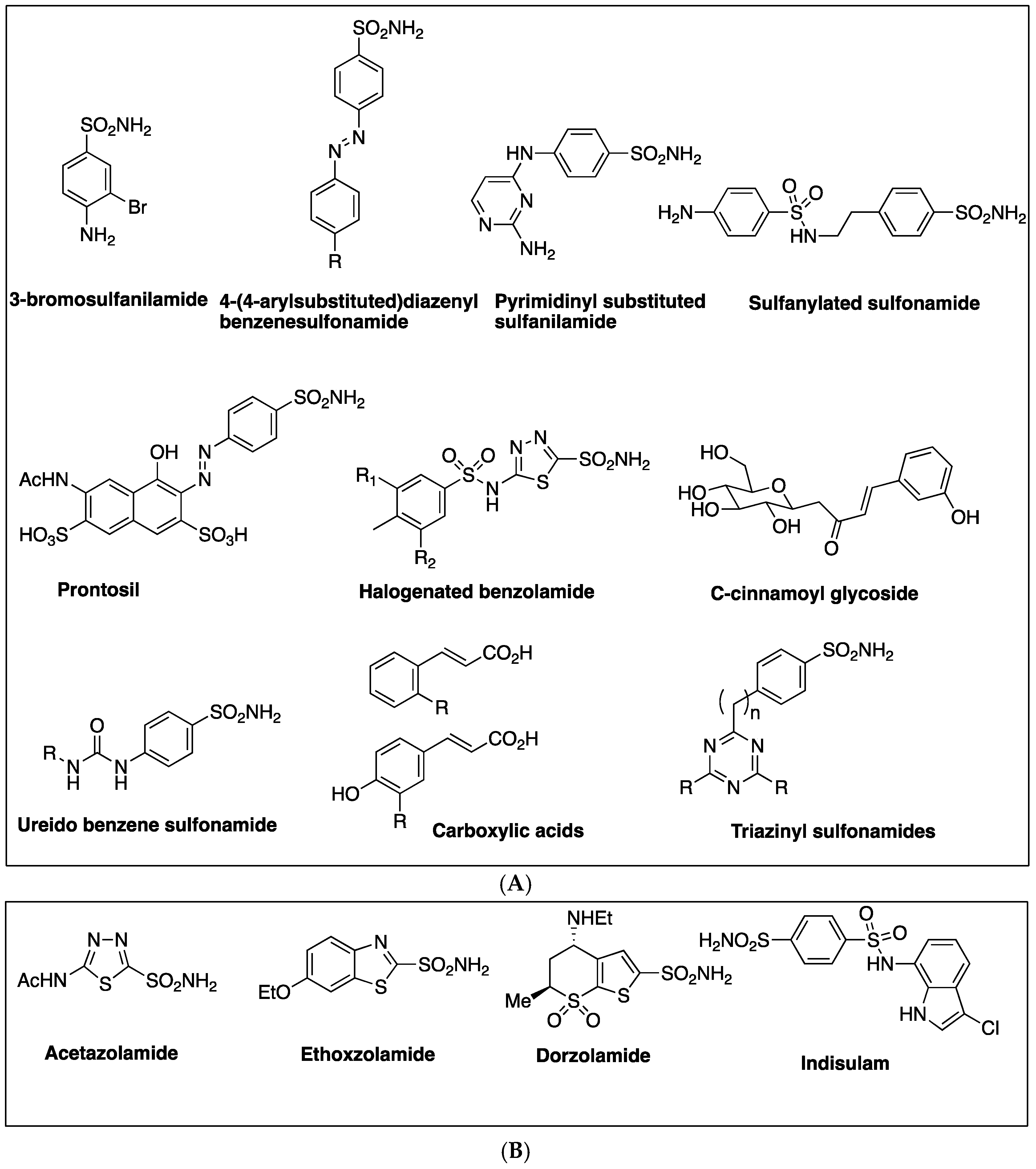
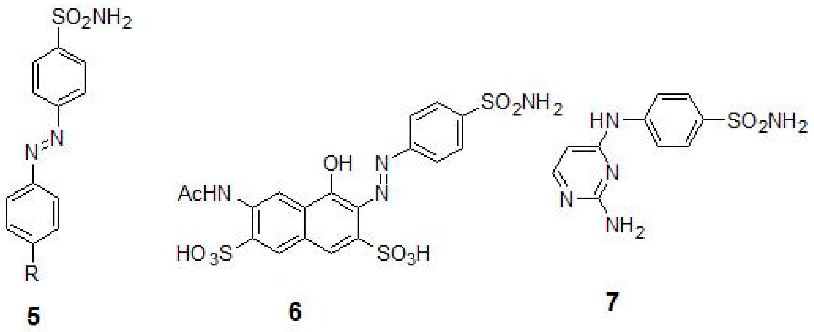
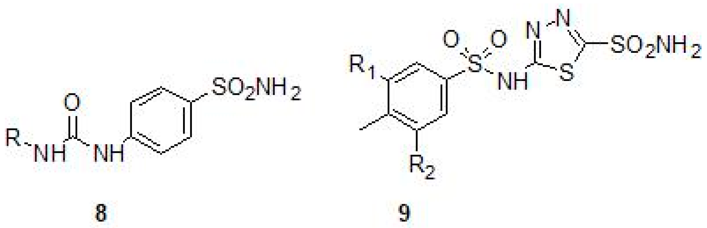
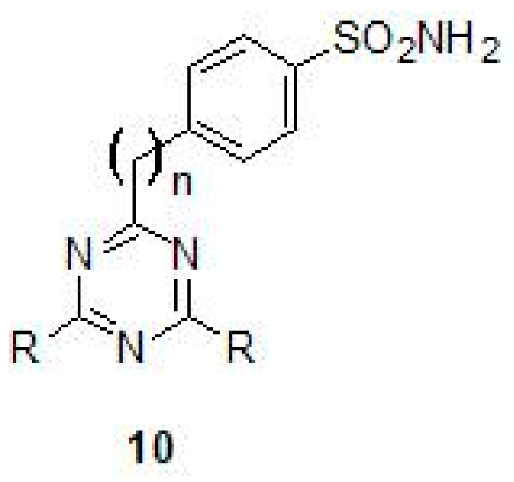
2.1. Sulfonamides as Inhibitors of M. tuberculosis β-CAs

The cloning and characterization of the M. tuberculosis β-CAs were done in the 2000s and these enzymes were identified as novel drug targets for developing anti-TB agents [27,28,29]. β-CA1 was the first Mtb β-CA cloned and characterized and in the same study, the first in vitro inhibition studies were performed using a panel of sulfonamides, sulfamates and their derivatives [27]. For in vitro inhibition studies, the CO2 hydration activity of β-CA1 was measured by applying Applied Photophysics stopped-flow instrument using phenol red as an indicator [27]. Among the tested sulfonamides, most of them inhibited the activity of β-CA1 in the range of 1–10 µM. Many of the derivatives, including sulfanilyl-sulfonamides acetazolamide (ATZ) (1), methazolamide, dichlorophenamide, dorzolamide (DZA) (2), brinzolamide, benzolamide, and the sulfamate topiramate, exhibited sub-micromolar inhibition (KI values of 0.481–0.905 µM) [27] (Table 2). Among the tested sulfonamides 3-bromosulfanilamide (3) and indisulam (4) inhibited the activity of β-CAs most efficiently (KI values of 97–186 nM) (Table 2 and Figure 2). This was the first study to show that Mtb β-CA1 is a potential target for developing anti-TB drugs that have a different mechanism of action [27]. Several studies in vitro inhibition studies were performed using these inhibitor molecules on Mtb β-CA1 and human CA II that showed similar inhibition profiles suggesting reliability of the method used for the studies [29,39,40,41,42,43,44,45].
Inhibition of Mtb β-CA2 was investigated using a series of diazenylbenzenesulfonamides (5) that were derived from sulfanilamide or metanilamide (Table 2) [40]. To increase the inhibitory properties, new molecules were synthesized by diazotization of aminosulfonamide and by coupling with phenols or amines [29]. The molecules were subsequently incorporated with various R moieties in the molecule such as hydroxy, amino, methylamino and dimethylamino and sulfonate that may induce water solubility to these compounds as sodium salts. The aminomethylene sodium sulfonate derivatives and their corresponding N-methylated analogue showed the best inhibition constants (KIs of 45–59 nM) [29]. In these compounds, the para position had bulky substituent with respect to the sulfonyl moiety, suggesting that this strategy may be good in obtaining low nanomolar range inhibitors that are selective against Mtb β-CA2 [29]. In addition to Mtb β-CA1 and β-CA2, the diazenylbenzenesulfonamides (5) were also tested for the inhibition of the Mtb β-CA3, and the prontosil (6) (Table 2 and Figure 3) was found to be the best inhibitor with inhibition constants in the range of (KIs) of 126–148 nM [39]. In another study, several compounds were studied for their inhibitory properties against Mtb β-CA3 and among them 2-amino-pyrimidin-4-yl-sulfanilamide (7) (KI 90 nM) and sulfonylated sulfonamide (KI of 170 nM) showed that Mtb CA3 can be successfully targeted using CAIs with a potential for developing agents targeting mycobacteria (Table 2) [28].
Inhibition studies on Mtb β-CA1 and β-CA3 using sulfonamides prepared by reaction of sulfanilamide with aryl/alkyl isocyanates (Ureido-sulfonamides) (8) have been carried out and the KIs were found to be in the range of 4.8–6500 nM and of 6.4–6850 nM, respectively (Table 2) [41]. Similarly, inhibition studies on all the three β-CAs of Mtb were performed using a number of halogenated sulfanilamides and halogenated benzolamide (9) derivatives that showed the efficacies of inhibition in the sub-micromolar to micromolar range (Table 2 and Figure 4). The inhibition range was dependent on the substitution pattern at the sulfanilamide moiety/fragment of the molecule. Best inhibitors were the halogenated benzolamides (KIs in the range of 0.12–0.45 μM), whereas the halogenated sulfanilamides were slightly less inhibitory (KIs in the range of 0.41–4.74 μM) [42].
A new series of fluorine containing sulfonamides (Triazinyl sulfonamides) (10) that were incorporated with amino, amino alcohol and amino acid moieties were used for the inhibition of all the three β-CAs of Mtb [43] (Figure 5). Among the compounds tested, some of them inhibited β-CA2 efficiently with KI values in the nanomolar range and also with very good potency (KIs in sub-micromolar range) against β-CA1 and β-CA3 [43] (Table 2). In a recent study, novel sulfonamides were obtained from sulfanilamide, which was N4-alkylated with ethyl bromoacetate, followed by reaction with hydrazine hydrate and further reacted with various aromatic aldehydes [44]. The inhibition studies using these sulfonamides showed KIs in the range of 127 nM–2.12 μM for Mtb β-CA3 [44].

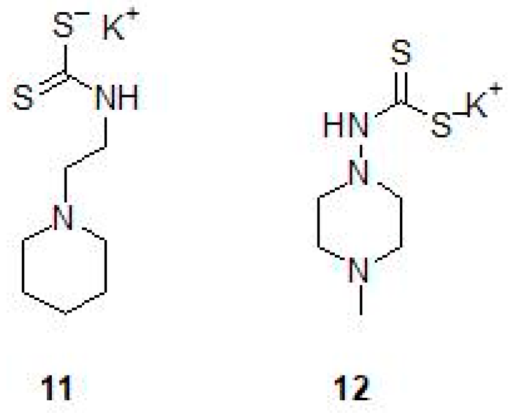
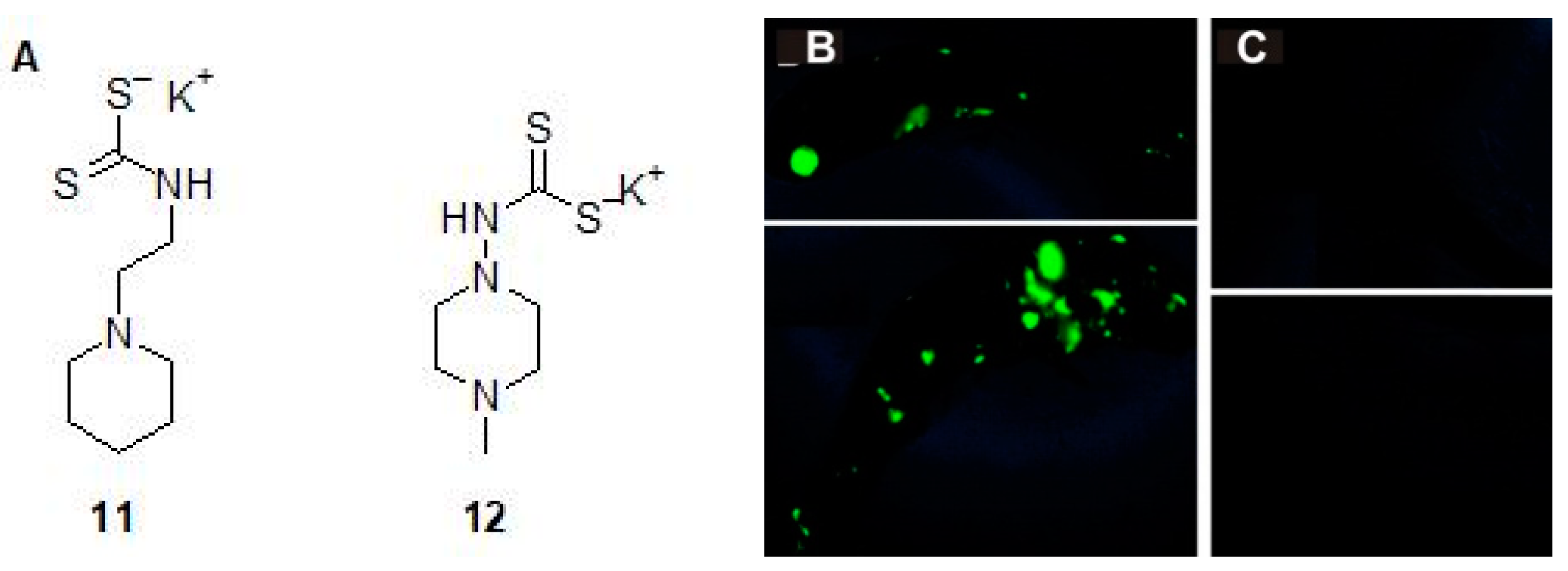
2.2. Mono and Dithiocarbamates

A series of N-mono- and N,N-disubstituted dithiocarbamates (DTCs) (11,12) have been tested for inhibition of β-CA1 and β-CA3 from Mtb (Table 2 and Figure 6) [45]. Both enzymes could be inhibited with sub-nanomolar to micromolar efficacies, depending on the substitution pattern at the nitrogen atom from the dithiocarbamate zinc-binding group. Aryl, arylalkyl-, heterocyclic as well as aliphatic and amino acyl moieties led to potent Mtb β-CA1 and β-CA3 inhibitors in both the N-mono- and N,N-disubstituted dithiocarbamate series [45].

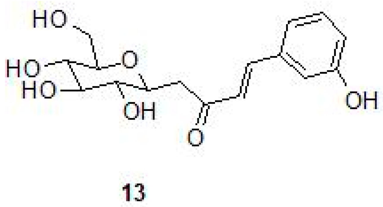
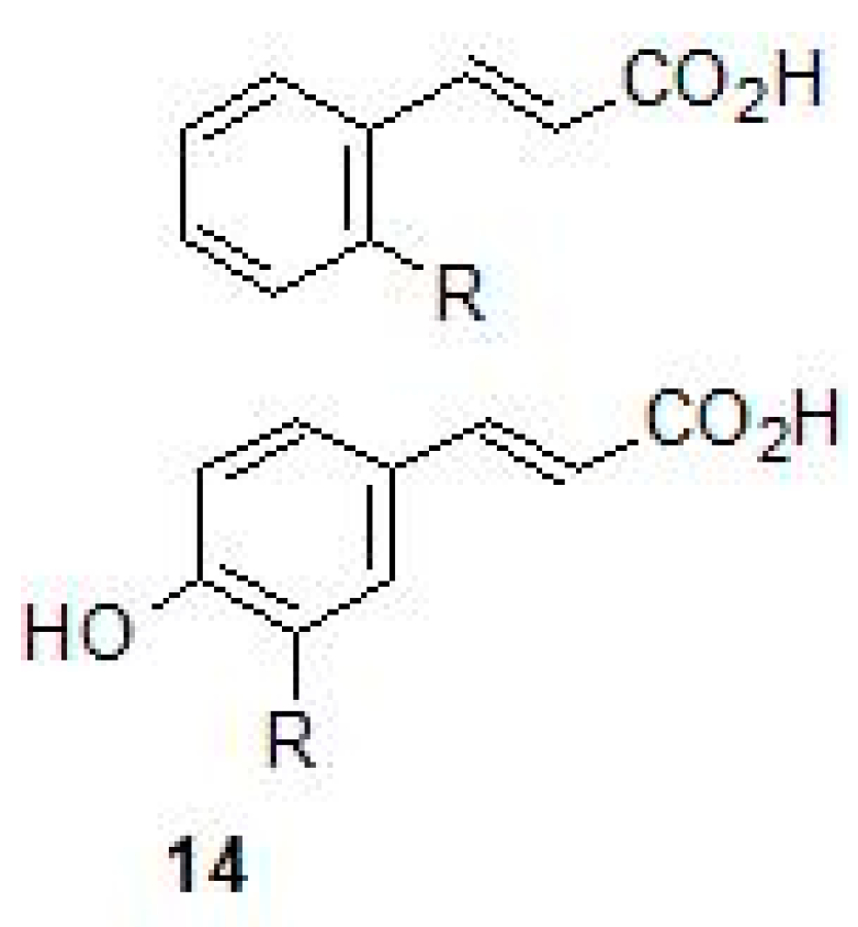
2.3. Phenolic Natural Products and Phenolic Acids

Several studies have shown that sulfonamides inhibit the Mtb β-CAs efficiently as discussed above. Similarly, in an effort to discover novel inhibitors that could selectively inhibit β-CAs through novel mechanism of action, Supuran’s group screened a series of phenolic-based natural products (NPs) against the Mtb β-CAs [46]. Enzyme inhibition properties of 21 NP compounds were investigated against β-CAs of Mtb as well as against human α-CAs I and II for comparison. Sulfonamides that are used clinically inhibited human CAs efficiently (at nM range), whereas β-CAs required micromolar concentrations. In contrast, 8 and 7 of the 21 phenolic compounds had sub-micromolar affinity for β-CA1 and β-CA3, respectively. The selectivity of some compounds was significantly higher against β-CAs than human α-CAs (the inhibition range being 8 μM to 430 μM) [46]. These NPs are the first nonclassical CA inhibitors that are more potent against mycobacteria β-CAs compared to host CA enzymes, suggesting usefulness of NPs for targeting β-CA of Mtb [46]. In addition to natural phenolic products, a series of phenolic acids and their esters, derivatives of caffeic, ferulic, and p-coumaric acids were tested against all the β-CAs of Mtb [47]. Among the screened compounds, esters 6–9 showed good inhibitory activity against all the Mtb β-CAs (KIs 1.87 Μm–7.05 μM), whereas they showed no inhibitory activity against human CAI and CAII, suggesting that they could be potentially developed as anti-mycobacterial compounds [47]. Computational analysis of binding mode of the compounds suggested that the inhibitors anchor to the zinc-coordinated water molecule from the CA active site interfering with the nucleophilic attack of the zinc hydroxide on the substrate CO2 [47]. These results provided insights into mechanism of inhibition of β-CAs, which may be valuable for developing new mycobacterial agents with a novel mechanism of action [47].
In another study, inhibition profiles of series of C-cinnamoyl glycosides (13) containing the phenol moiety were investigated against the three β-CAs of Mtb [48] (Table 2 and Figure 7). Among the compounds investigated, most of them (compounds 13 and 57) inhibited Mtb β-CA2 at nanomolar concentrations (KI 130-640 nM), and for Mtb β-CA1 (compounds 5 and 6) the KI range was between 140–930 nM and showed preference for β-CA1 over human CAII. Only one compound inhibited Mtb β-CA1 at nanomolar quantities (KI 140 nM) over human CAII [48].

2.4. Carboxylic Acids

Weak acids are known to inhibit the growth of mycobacterium but the mechanism of action of these compounds is not known. Carboxylic acids (14) that contain scaffolds such as benzoic acids, nipecotic acid, ortho and para coumaric acid and ferulic acid were investigated for the inhibition of all the three β-CAs of Mtb (Figure 8). These compounds inhibited all the three β-CA enzymes of Mtb at sub-micromolar to micromolar concentration range (KIs in the range of 0.11–0.97 µM). The KIs for the inhibition of β-CA2 was in the range of 0.59–8.10 µM, whereas against β-CA1, the carboxylic acids showed inhibition constants in the range of 2.25–7.13 µM [49]. This class of relatively underexplored β-CA inhibitors warrant further in vivo studies, as they may have the potential for developing antimycobacterial agents.

3. In Vitro Inhibition of Mycobacterial strains Using CA Inhibitors

A new class of compounds prepared by reaction of 6-mercaptopurine with sulfony/sulfenyl halides known as 9-sulfonylated/sulfenylated-6-mercaptopurines inhibit growth of Mtb H37Rv, a wild type bacilli in the range of 0.39–3.39 μg/mL [50] (Table 3). In addition, one of the derivatives showed an appreciable (minimal inhibitory concentration (MIC) under 1 μg/mL) inhibitory activity against several drug resistant strains of Mtb [50]. The compounds that exhibit MIC of less than 1 μg/mL are considered as excellent leads and were the first CAIs with anti-tubercular activity. Thus, these compounds may indeed constitute interesting leads for discovering more efficient antimycobacterial drugs. Similarly, C-cinnamoyl glycosides containing the phenol moiety that inhibit Mtb β-CA1 and β-CA2 in nanomolar quantities were tested for inhibition of the Mtb H37Rv strain, leading to the identification of compounds having anti-tubercular activity (Table 3) [48]. The MIC of the C-cinnamoyl glycosides was 100 μg/mL; though high, the compounds inhibited the growth of the bacterium completely. Interestingly, one of the C-cinnamoyl glycosides, (E)-1-(2,3,4,6-tetra-O-acetyl-β-d-glucopyranosyl)-4-(3-hydroxyphenyl) but-3-en-2-one inhibited the growth of the bacterium efficiently (3.125–6.25 μg/mL) on a solid medium (Table 3) [48].
Inhibition studies on M. marinum, an NTM and a close relative of Mtb, were carried out in liquid cultures using DTCs Fc14-584b and Fc14-594a. These drugs were prepared by reaction of corresponding amine with carbon disulfide in the presence of a base and shown to be specific inhibitors of Mtb β-CA1 and β-CA3 [45]. In vitro inhibition studies showed that the concentration required for the inhibition of the M. marinum was 17–18 μg/mL for both compounds after six days of exposure to the inhibitors. Further studies to find if the compounds were bacteriostatic or bactericidal showed that there was no growth resumption of M. marinum with inhibitor concentration below MIC after inhibitor dilution by 1:4, suggesting that these compounds were bactericidal [26] (Table 3).
Similar to other bacteria that contain extracellular DNA (eDNA) in the matrix of the bacterial biofilms, NTM bacteria also contain significant amounts of eDNA in their biofilms and are responsible for phenotypic resistance of the bacteria to antibiotics, in addition to other biological functions [51]. A recent study showed that bicarbonate ion positively influences eDNA export in NTM and it is well established that bicarbonate is generated by the hydration of carbon dioxide via CA [31,35]. Screening of a mutant library for eDNA export in NTM bacteria M. avium identified mutants that were inactivated for CA gene and these mutants when complemented with the CA gene restored the transport of eDNA, suggesting that CAs play important roles in the transport of eDNA and formation of biofilms in NTM [35]. The surface exposed proteome of M. avium in eDNA containing biofilms showed presence of abundant CAs and inhibition studies exposing these bacteria to 6-ethoxy-1,3-benzathiazole-2-sulfonamide/ethoxzolamide (15) (EZA) showed reduction in eDNA transport significantly (Table 4 and Figure 9) [35]. Thus, in addition to having an effect on mycobacterial growth, CA-inhibition may also be a potential strategy to inhibit biofilm formation of mycobacteria.

4. CA Inhibitors and In Vivo Inhibition of Mycobacteria

The first in vivo study to show the effect of CA inhibitor on Mtb was published in 2015 by Johnson et al. [36]. The authors showed that EZA (Figure 10) inhibits the signaling of PhoPR in Mtb [36,52]. EZA is a sulfonamide compound (Figure 10) that is a general inhibitor of CA enzyme activity and is an FDA approved drug used in the treatment of glaucoma, epilepsy and duodenal ulcers and is a diuretic.
The study showed that Mtb treated with EZA induces phenotypes similar to the mutants of the PhoPR, downregulating PhoPR regulon, reducing the production of virulence-associated lipids, and inhibiting Esx-1 protein secretion (Table 4) [36]. In addition, quantitative single cell imaging of a PhoPR dependent fluorescent reporter strain showed that EZA inhibits PhoPR regulated genes in infected macrophages and mouse lungs [36]. Similarly, the efficacy assessment in Mtb-infected mice, orally treated with EZA, showed a significant reduction in bacterial growth in the lungs compared to the mock-treated control group [36].
Dithiocarbamates, another class of compounds that strongly inhibit β-CAs of Mtb in vitro have been recently used by our group to show in vivo inhibition of M. marinum [26,45]. Among the two DTCs 11 and 12 that were first evaluated for toxicity in the zebrafish larval model, 12 was found to be less toxic and was taken further to study the inhibition of M. marinum in vivo. The zebrafish larvae infected with green fluorescent M. marinum strain with an infection dose of 471 ± 143 bacteria treated with 300 μM concentration of 12 showed significant reduction (p > 0.0096) in bacterial load compared to the larvae not treated with the inhibitor (Figure 11C). The study suggested that the inhibitors of CAs could be useful as a new class of antimycobacterial compounds that can potentially treat MDR-TB.

5. Future Prospects

In this review, we discussed the progress made on the discovery and development of antimycobacterial agents that target mycobacterial β-CAs. The chemical inhibitors that selectively bind to the mycobacterial β-CAs could be developed as antimycobacterial agents for treating not only drug-resistant tuberculosis, but also other diseases caused by pathogenic mycobacteria that are resistant to clinically used drugs. The M. tuberculosis contains three β-CAs, among them, β-CA1 and β-CA 2 are cytoplasmic, and β-CA3 is membrane associated. Many of the inhibitors reported so far have been shown to inhibit β-CA1 and β-CA2 in vitro efficiently, however it is not known if these inhibitors are permeable through the mycobacterial membrane. Similarly, many of the inhibitors that have been shown to inhibit mycobacterial β-CAs efficiently have also been shown to inhibit human α-CAs though in higher concentrations.
The current strategy of developing inhibitors against the mycobacterial β-CAs for treating mycobacterial diseases can be more successful in the future by designing inhibitors that bind Mtb β-CAs selectively and specifically. For designing such inhibitors, information regarding the active site residues of the enzymes that interact with inhibitor molecules need to be obtained. Resolving crystal structures in complex with potential inhibitors of β-CAs is one way of getting insights into such residues that will help in the design and synthesis of β-CA specific inhibitors.
Studies have shown that these enzymes can be inhibited in vivo using CAIs exhibiting antimycobacterial effect showing proof-of-concept. However, only very few studies have shown antimycobacterial effects of the inhibitors possibly through the inhibition of β-CA3 of the bacterium. In future, to achieve more success for in vivo inhibition of these enzymes, there is a need to design and synthesize inhibitors that are not only selective for β-CAs but also permeable through the membrane. It will also be useful to design inhibitor molecules with a tag that will help in tracking the fate of the molecule once it is inside the bacterium.
Zebrafish represents an excellent vertebrate model for tuberculosis research, because it is a natural host for M. marinum that causes a TB-like disease in the fish. Safety and toxicity of the potential β-CA inhibitors can be first evaluated using zebrafish larvae. Subsequently, preclinical in vivo inhibition studies can be done in a zebrafish larval model after causing an active TB by M. marinum infection. The recent developments of the in vivo zebrafish models that mimic the human TB disease, coupled with new imaging technologies, provide much better predictive preclinical models to produce new combinations of treatments/drugs that are more effective against the hard-to-treat mycobacterial diseases. Unlike the drugs that are in clinical use, the β-CA inhibitors have a different mechanism of action. These drugs will probably have minimal off-target effects due to the absence of β-CAs in humans and other vertebrates in whom mycobacteria cause infections.

Author Contributions

A.A. is responsible for conceiving the idea and for preparing the manuscript. A.A., J.-Y.W., F.C., C.T.S., M.H., M.P., and S.P. contributed to the writing of the article and have read and approved the final version of the manuscript.

Funding

This research was funded by grants from Sigrid Jusélius Foundation (S.P., M.P.), Finnish Cultural Foundation (A.A., M.H.), Academy of Finland (S.P., M.P.), Jane and Aatos Erkko Foundation (M.P. and S.P.), Orion-Farmos Foundation (M.H.), and Tampere Tuberculosis Foundation (S.P., M.H., and M.P.).

Conflicts of Interest

The authors declare no conflict of interest.

Abbreviations

CAcarbonic anhydrase
MtbMycobacterium tuberculosis
MICminimal inhibitory concentration
CAIcarbonic anhydrase inhibitor
TBtuberculosis
MTCMycobacterium tuberculosis complex
NTMnontuberculous mycobacteria
EZAethoxzolamide
DTCsdithiocarbamates
ATZacetazolamide
DZAdorzolamide
eDNAextracellular DNA

References

  1. Stahl, D.A.; Urbance, J.W. The division between fast- and slow-growing species corresponds to natural relationships among the mycobacteria. J. Bacteriol. 1990, 172, 116–124. [Google Scholar] [CrossRef] [PubMed]
  2. Kim, C.J.; Kim, N.H.; Song, K.H.; Choe, P.G.; Kim, E.S.; Park, S.W.; Kim, H.B.; Kim, N.J.; Kim, E.C.; Park, W.B.; et al. Differentiating rapid- and slow-growing mycobacteria by difference in time to growth detection in liquid media. Diagn. Microbiol. Infect. Dis. 2013, 75, 73–76. [Google Scholar] [CrossRef] [PubMed]
  3. Sinha, P.; Gupta, A.; Prakash, P.; Anupurba, S.; Tripathi, R.; Srivastava, G.N. Differentiation of Mycobacterium tuberculosis complex from non-tubercular mycobacteria by nested multiplex PCR targeting IS6110, MTP40 and 32kD alpha antigen encoding gene fragments. BMC Infect. Dis. 2016, 16, 123. [Google Scholar] [CrossRef] [PubMed]
  4. WHO. Global Tuberculosis Report 2018; World Health Organization: Geneva, Switzerland, 2018; pp. 1–277. ISBN 978-92-4-156564-6. [Google Scholar]
  5. WHO. World Health Organization Lepracy: Fact Sheet on Leprosy; World Health Organization: Geneva, Switzerland, 2017; pp. 1–5. [Google Scholar]
  6. Daley, C.L.; Griffith, D.E. Pulmonary non-tuberculous mycobacterial infections. Int. J. Tuberc. Lung Dis. 2010, 14, 665–671. [Google Scholar] [PubMed]
  7. Adrados, B.; Julián, E.; Codony, F.; Torrents, E.; Luquin, M.; Morató, J. Prevalence and concentration of non-tuberculous mycobacteria in cooling towers by means of quantitative PCR: A prospective study. Curr. Microbiol. 2011, 62, 313–319. [Google Scholar] [CrossRef] [PubMed] [Green Version]
  8. King, H.C.; Khera-Butler, T.; James, P.; Oakley, B.B.; Erenso, G.; Aseffa, A.; Knight, R.; Wellington, E.M.; Courtenay, O. Environmental reservoirs of pathogenic mycobacteria across the Ethiopian biogeographical landscape. PLoS ONE 2017, 12, e0173811. [Google Scholar] [CrossRef] [PubMed]
  9. Falkinham, J.O. Ecology of nontuberculous mycobacteria--where do human infections come from? Semin. Respir. Crit. Care Med. 2013, 34, 95–102. [Google Scholar] [CrossRef] [PubMed]
  10. Gopinath, K.; Singh, S. Non-tuberculous mycobacteria in TB-endemic countries: Are we neglecting the danger? PLoS Negl. Trop. Dis. 2010, 4, e615. [Google Scholar] [CrossRef] [PubMed]
  11. Johnson, M.M.; Odell, J.A. Nontuberculous mycobacterial pulmonary infections. J. Thorac. Dis. 2014, 6, 210–220. [Google Scholar] [CrossRef] [PubMed]
  12. Faria, S.; Joao, I.; Jordao, L. General Overview on Nontuberculous Mycobacteria, Biofilms, and Human Infection. J. Pathog. 2015, 809014. [Google Scholar] [CrossRef] [PubMed]
  13. Misch, E.A.; Saddler, C.; Davis, J.M. Skin and Soft Tissue Infections Due to Nontuberculous Mycobacteria. Curr. Infect. Dis. Rep. 2018, 20, 6. [Google Scholar] [CrossRef] [PubMed]
  14. Al-Ghafli, H.; Al-Hajoj, S. Nontuberculous Mycobacteria in Saudi Arabia and Gulf Countries: A Review. Can. Respir. J. 2017, 5035932. [Google Scholar] [CrossRef] [PubMed]
  15. Nishiuchi, Y.; Iwamoto, T.; Maruyama, F. Infection Sources of a Common Non-tuberculous Mycobacterial Pathogen, Mycobacterium avium Complex. Front. Med. 2017, 27. [Google Scholar] [CrossRef] [PubMed]
  16. Fedrizzi, T.; Meehan, C.J.; Grottola, A.; Giacobazzi, E.; Fregni Serpini, G.; Tagliazucchi, S.; Fabio, A.; Bettua, C.; Bertorelli, R.; De Sanctis, V.; et al. Genomic characterization of Nontuberculous Mycobacteria. Sci. Rep. 2017, 7, 45258. [Google Scholar] [CrossRef] [PubMed]
  17. Stinear, T.P.; Seemann, T.; Harrison, P.F.; Jenkin, G.A.; Davies, J.K.; Johnson, P.D.; Abdellah, Z.; Arrowsmith, C.; Chillingworth, T.; Churcher, C.; et al. Insights from the complete genome sequence of Mycobacterium marinum on the evolution of Mycobacterium tuberculosis. Genome Res. 2008, 18, 729–741. [Google Scholar] [CrossRef] [PubMed]
  18. Cole, S.T.; Brosch, R.; Parkhill, J.; Garnier, T.; Churcher, C.; Harris, D.; Gordon, S.V.; Eiglmeier, K.; Gas, S.; Barry, C.E.; et al. Deciphering the biology of Mycobacterium tuberculosis from the complete genome sequence. Nature 1998, 393, 537–544. [Google Scholar] [CrossRef] [PubMed]
  19. Vissa, V.D.; Brennan, P.J. The genome of Mycobacterium leprae: A minimal mycobacterial gene set. Genome Biol. 2001, 2, Reviews1023. [Google Scholar] [CrossRef] [PubMed]
  20. Cole, S.T.; Eiglmeier, K.; Parkhill, J.; James, K.D.; Thomson, N.R.; Wheeler, P.R.; Honoré, N.; Garnier, T.; Churcher, C.; Harris, D.; et al. Massive gene decay in the leprosy bacillus. Nature 2001, 409, 1007–1011. [Google Scholar] [CrossRef] [PubMed]
  21. Showalter, H.D.; Denny, W.A. A roadmap for drug discovery and its translation to small molecule agents in clinical development for tuberculosis treatment. Tuberculosis 2008, 88, S3–S17. [Google Scholar] [CrossRef]
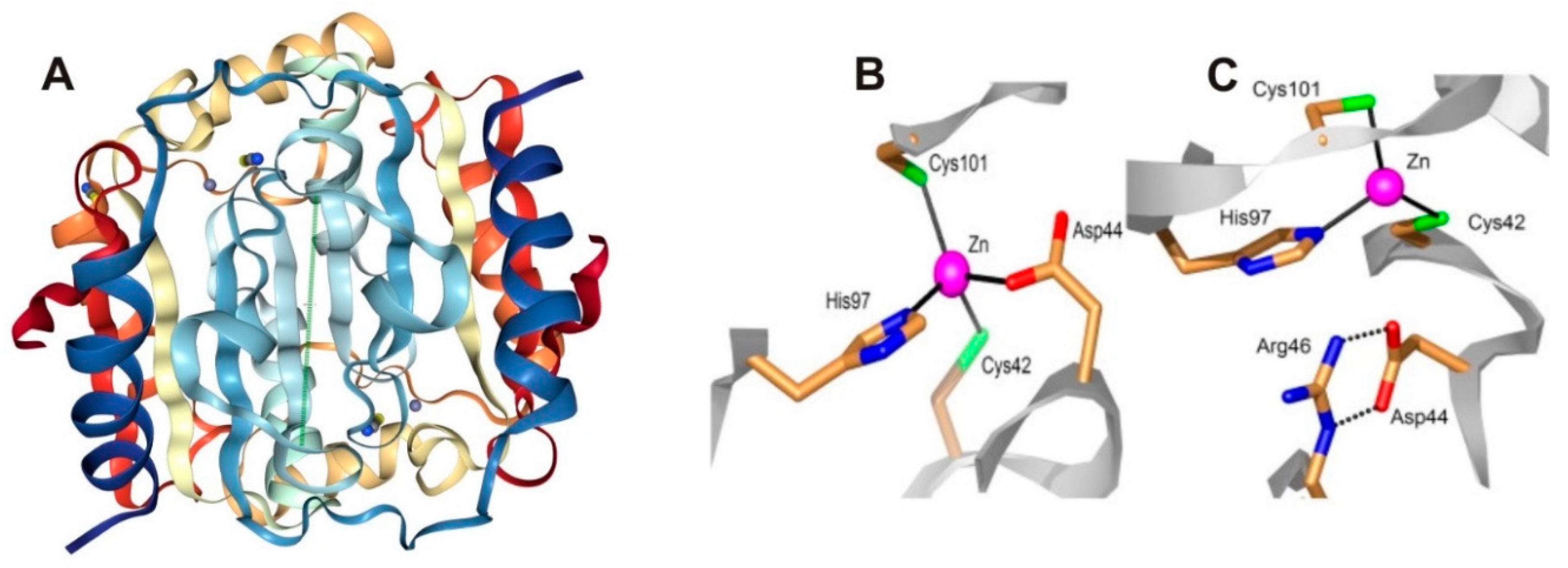
  22. Covarrubias, A.S.; Larsson, A.M.; Högbom, M.; Lindberg, J.; Bergfors, T.; Björkelid, C.; Mowbray, S.L.; Unge, T.; Jones, T.A. Structure and function of carbonic anhydrases from Mycobacterium tuberculosis. J. Biol. Chem. 2005, 280, 18782–18789. [Google Scholar] [CrossRef] [PubMed]
  23. Covarrubias, A.S.; Bergfors, T.; Jones, T.A.; Högbom, M. Structural mechanics of the pH-dependent activity of beta-carbonic anhydrase from Mycobacterium tuberculosis. J. Biol. Chem. 2006, 281, 4993–4999. [Google Scholar] [CrossRef] [PubMed]
  24. Winum, J.-Y.; Stephan, K.; Scozzafava, A.; Montero, J.-L.; Supuran, C.T. Targeting bacterial metalloenzymes: A new strategy for the development of anti-infective agents. Anti-Infect. Agents Med. Chem. 2008, 7, 169–179. [Google Scholar] [CrossRef]
  25. Kapopoulou, A.; Lew, J.M.; Cole, S.T. The MycoBrowser portal: A comprehensive and manually annotated resource for mycobacterial genomes. Tuberculosis 2011, 91, 8–13. [Google Scholar] [CrossRef] [PubMed]
  26. Aspatwar, A.; Hammarén, M.; Koskinen, S.; Luukinen, B.; Barker, H.; Carta, F.; Supuran, C.T.; Parikka, M.; Parkkila, S. beta-CA-specific inhibitor dithiocarbamate Fc14-584B: A novel antimycobacterial agent with potential to treat drug-resistant tuberculosis. J. Enzyme Inhib. Med. Chem. 2017, 32, 832–840. [Google Scholar] [CrossRef] [PubMed]
  27. Minakuchi, T.; Nishimori, I.; Vullo, D.; Scozzafava, A.; Supuran, C.T. Molecular cloning, characterization, and inhibition studies of the Rv1284 beta-carbonic anhydrase from Mycobacterium tuberculosis with sulfonamides and a sulfamate. J. Med. Chem. 2009, 52, 2226–2232. [Google Scholar] [CrossRef] [PubMed]
  28. Nishimori, I.; Minakuchi, T.; Vullo, D.; Scozzafava, A.; Innocenti, A.; Supuran, C.T. Carbonic anhydrase inhibitors. Cloning, characterization, and inhibition studies of a new beta-carbonic anhydrase from Mycobacterium tuberculosis. J. Med. Chem. 2009, 52, 3116–3120. [Google Scholar] [CrossRef] [PubMed]
  29. Carta, F.; Maresca, A.; Covarrubias, A.S.; Mowbray, S.L.; Jones, T.A.; Supuran, C.T. Carbonic anhydrase inhibitors. Characterization and inhibition studies of the most active beta-carbonic anhydrase from Mycobacterium tuberculosis, Rv3588c. Bioorg. Med. Chem. Lett. 2009, 19, 6649–6654. [Google Scholar] [CrossRef] [PubMed]
  30. Nishimori, I.; Minakuchi, T.; Morimoto, K.; Sano, S.; Onishi, S.; Takeuchi, H.; Vullo, D.; Scozzafava, A.; Supuran, C.T. Carbonic anhydrase inhibitors: DNA cloning and inhibition studies of the alpha-carbonic anhydrase from Helicobacter pylori, a new target for developing sulfonamide and sulfamate gastric drugs. J. Med. Chem. 2006, 49, 2117–2126. [Google Scholar] [CrossRef] [PubMed]
  31. Supuran, C.T. Carbonic anhydrases: Novel therapeutic applications for inhibitors and activators. Nat. Rev. Drug Discov. 2008, 168–181. [Google Scholar] [CrossRef] [PubMed]
  32. Supuran, C.T. Structure and function of carbonic anhydrases. Biochem. J. 2016, 473, 2023–2032. [Google Scholar] [CrossRef] [PubMed]
  33. Sassetti, C.M.; Boyd, D.H.; Rubin, E.J. Genes required for mycobacterial growth defined by high density mutagenesis. Mol. Microbiol. 2003, 48, 77–84. [Google Scholar] [CrossRef] [PubMed] [Green Version]
  34. Sassetti, C.M.; Rubin, E.J. Genetic requirements for mycobacterial survival during infection. Proc. Natl. Acad. Sci. USA 2003, 100, 12989–12994. [Google Scholar] [CrossRef] [PubMed] [Green Version]
  35. Rose, S.J.; Bermudez, L.E. Identification of Bicarbonate as a Trigger and Genes Involved with Extracellular DNA Export in Mycobacterial Biofilms. MBio 2016, 7, e01597-16. [Google Scholar] [CrossRef] [PubMed]
  36. Johnson, B.K.; Colvin, C.J.; Needle, D.B.; Mba Medie, F.; Champion, P.A.; Abramovitch, R.B. The Carbonic Anhydrase Inhibitor Ethoxzolamide Inhibits the Mycobacterium tuberculosis PhoPR Regulon and Esx-1 Secretion and Attenuates Virulence. Antimicrob Agents Chemother. 2015, 59, 4436–4445. [Google Scholar] [CrossRef] [PubMed] [Green Version]
  37. Nishimori, I.; Minakuchi, T.; Maresca, A.; Carta, F.; Scozzafava, A.; Supuran, C.T. The beta-carbonic anhydrases from Mycobacterium tuberculosis as drug targets. Curr. Pharm. Des. 2010, 16, 3300–3309. [Google Scholar] [CrossRef] [PubMed]
  38. Güzel, O.; Maresca, A.; Scozzafava, A.; Salman, A.; Balaban, A.T.; Supuran, C.T. Discovery of low nanomolar and subnanomolar inhibitors of the mycobacterial beta-carbonic anhydrases Rv1284 and Rv3273. J. Med. Chem. 2009, 52, 4063–4067. [Google Scholar] [CrossRef] [PubMed]
  39. Maresca, A.; Carta, F.; Vullo, D.; Scozzafava, A.; Supuran, C.T. Carbonic anhydrase inhibitors. Inhibition of the Rv1284 and Rv3273 beta-carbonic anhydrases from Mycobacterium tuberculosis with diazenylbenzenesulfonamides. Bioorg. Med. Chem. Lett. 2009, 19, 4929–4932. [Google Scholar] [CrossRef] [PubMed]
  40. Carta, F.; Maresca, A.; Scozzafava, A.; Vullo, D.; Supuran, C.T. Carbonic anhydrase inhibitors. Diazenylbenzenesulfonamides are potent and selective inhibitors of the tumor-associated isozymes IX and XII over the cytosolic isoforms I and II. Bioorg. Med. Chem. 2009, 17, 7093–7099. [Google Scholar] [CrossRef] [PubMed]
  41. Pacchiano, F.; Carta, F.; Vullo, D.; Scozzafava, A.; Supuran, C.T. Inhibition of beta-carbonic anhydrases with ureido-substituted benzenesulfonamides. Bioorg. Med. Chem. Lett. 2011, 21, 102–105. [Google Scholar] [CrossRef] [PubMed]
  42. Maresca, A.; Scozzafava, A.; Vullo, D.; Supuran, C.T. Dihalogenated sulfanilamides and benzolamides are effective inhibitors of the three beta-class carbonic anhydrases from Mycobacterium tuberculosis. J. Enzym. Inhib. Med. Chem. 2013, 28, 384–387. [Google Scholar] [CrossRef] [PubMed]
  43. Ceruso, M.; Vullo, D.; Scozzafava, A.; Supuran, C.T. Sulfonamides incorporating fluorine and 1,3,5-triazine moieties are effective inhibitors of three beta-class carbonic anhydrases from Mycobacterium tuberculosis. J. Enzym. Inhib. Med. Chem. 2014, 29, 686–689. [Google Scholar] [CrossRef] [PubMed]
  44. Wani, T.V.; Bua, S.; Khude, P.S.; Chowdhary, A.H.; Supuran, C.T.; Toraskar, M.P. Evaluation of sulphonamide derivatives acting as inhibitors of human carbonic anhydrase isoforms I, II and Mycobacterium tuberculosis beta-class enzyme Rv3273. J. Enzym. Inhib. Med. Chem. 2018, 33, 962–971. [Google Scholar] [CrossRef] [PubMed]
  45. Maresca, A.; Carta, F.; Vullo, D.; Supuran, C.T. Dithiocarbamates strongly inhibit the beta-class carbonic anhydrases from Mycobacterium tuberculosis. J. Enzym. Inhib. Med. Chem. 2013, 28, 407–411. [Google Scholar] [CrossRef] [PubMed]
  46. Davis, R.A.; Hofmann, A.; Osman, A.; Hall, R.A.; Mühlschlegel, F.A.; Vullo, D.; Innocenti, A.; Supuran, C.T.; Poulsen, S.A. Natural product-based phenols as novel probes for mycobacterial and fungal carbonic anhydrases. J. Med. Chem. 2011, 54, 1682–1692. [Google Scholar] [CrossRef] [PubMed]
  47. Cau, Y.; Mori, M.; Supuran, C.T.; Botta, M. Mycobacterial carbonic anhydrase inhibition with phenolic acids and esters: Kinetic and computational investigations. Org. Biomol. Chem. 2016, 14, 8322–8330. [Google Scholar] [CrossRef] [PubMed]
  48. Buchieri, M.V.; Riafrecha, L.E.; Rodríguez, O.M.; Vullo, D.; Morbidoni, H.R.; Supuran, C.T.; Colinas, P.A. Inhibition of the beta-carbonic anhydrases from Mycobacterium tuberculosis with C-cinnamoyl glycosides: Identification of the first inhibitor with anti-mycobacterial activity. Bioorg. Med. Chem. Lett. 2013, 23, 740–743. [Google Scholar] [CrossRef] [PubMed]
  49. Maresca, A.; Vullo, D.; Scozzafava, A.; Manole, G.; Supuran, C.T. Inhibition of the beta-class carbonic anhydrases from Mycobacterium tuberculosis with carboxylic acids. J. Enzym. Inhib. Med. Chem. 2013, 28, 392–396. [Google Scholar] [CrossRef] [PubMed]
  50. Scozzafava, A.; Mastrolorenzo, A.; Supuran, C.T. Antimycobacterial activity of 9-sulfonylated/sulfenylated-6-mercaptopurine derivatives. Bioorg. Med. Chem. Lett. 2001, 11, 1675–1678. [Google Scholar] [CrossRef]
  51. Rose, S.J.; Babrak, L.M.; Bermudez, L.E. Mycobacterium avium Possesses Extracellular DNA that Contributes to Biofilm Formation, Structural Integrity, and Tolerance to Antibiotics. PLoS ONE 2015, 10, e0128772. [Google Scholar] [CrossRef] [PubMed]
  52. Johnson, B.K.; Abramovitch, R.B. Small Molecules That Sabotage Bacterial Virulence. Trends Pharmacol. Sci. 2017, 38, 339–362. [Google Scholar] [CrossRef] [PubMed] [Green Version]
Figure 1. Crystal Structure of Rv1284 (β-CA1) from M. tuberculosis. (A) Structure of β-CA1 (1YLK) [22]. Coordination of the Zn(II) ion in the β-CA1 of Mtb. (B) Closed active site, with the Zn(II) ion (violet sphere) coordinated by a histidine, two cysteines and one aspartate residue. (C) Open active site, with three protein ligands coordinated to Zn(II); the aspartate makes a salt bridge with a conserved arginine residue in all β-CAs [22,29]. The images adapted from Covarrubias et al. https://www.rcsb.org/structure/1YLK [22]. Licensed under CC BY 4.0.
Molecules 23 02911 g001
Figure 2. Chemical structures of acetazolamide (1) (ATZ), dorzolamide (2) (DZA), 3-bromosulfanilamide (3) and indisulam (4).
Molecules 23 02911 g002
Figure 3. Chemical structures of diazenylbenzenesulfonamides (5), prontosil (6) and 2-amino-pyrimidin-4-yl-sulfanilamide (7).
Molecules 23 02911 g003
Figure 4. General chemical structures of ureido containing sulfonamides (8) and halogenated benzene sulfonamides (9).
Molecules 23 02911 g004
Figure 5. General chemical structure of triazinyl sulfonamides (10).
Molecules 23 02911 g005
Figure 6. Chemical structures of dithiocarbamates (11) and (12).
Molecules 23 02911 g006
Figure 7. Chemical structure of C-cinnamoyl glycoside (13).
Molecules 23 02911 g007
Figure 8. General chemical structures ferulic and coumaric acids (14).
Molecules 23 02911 g008
Figure 9. Chemical structure of ethoxzolamide (15).
Molecules 23 02911 g009
Figure 10. Novel compounds that inhibit M. tuberculosis β-CAs at nanomolar quantities. (Upper panel; A): structures of the compounds with a potential to be developed as anti-mycobacterial agents for treating the mycobacterial diseases caused by MTC and NTM bacteria that are resistant to clinically used drugs. (Lower panel; B): compounds that efficiently inhibit the M. tuberculosis β-CAs in addition to human CAII and are in clinical use or testing to treat human diseases.
Molecules 23 02911 g010
Figure 11. The in vivo inhibition of M. marinum in zebrafish larval model. (A) The β-CA inhibitors dithiocarbamate 11 and 12 that were evaluated for toxicity and safety for further use to inhibit growth of M. marinum in zebrafish larvae. (B) The zebrafish larvae injected with M. marinum wasabi strain, which expresses a green fluorescent protein. The green fluorescence showing the infection in zebrafish larvae after 6-day post infection not treated with any inhibitor. (C) Zebrafish larvae injected with M. marinum and treated with 300 μM concentration of 12 [26].
Molecules 23 02911 g011
Table 1. Activity and inhibition properties of Mtb β-CAs compared to human CA II.
CAsGene IDProteinKcat (s−1)Kcat/Km (M−1 s−1)ActivityKI (nM) aReference
Mtb β-CA1Rv1284163 aa3.9 × 1053.7 × 107Moderate480[27]
Mtb β-CA2Rv3588c207 aa9.8 × 1059.3 × 107High9.8[29]
Mtb β-CA3Rv3273b 215 aa4.3 × 1054.0 × 107Moderate104[28]
hCAIICA2260 aa1.4 × 1061.5 × 108Very high12[29]
a Inhibition using acetazolamide. b CA domain of β-CA3.
Table 2. Compounds that inhibit M. tuberculosis β-CAs at nM concentrations compared to inhibition of human CAII enzyme in vitro.
CAIshCAIIβ-CA1β-CA2β-CA3Reference
3-bromosulfanilamide40186NSNS[27]
Diazenylbenzenesulfonamides105-45–955-[29]
2-amino-pyrimidin-4-yl-sulfanilamide33-NA91[28]
Sulfonylated sulfonamideNS--170[28]
prontosilNS126 148[39]
Halogenated benzolamidesNS120–580410–450170–340[42]
N-mono- and N,N-dithiocarbamates0.7–3250.9–481NA0.91–431[45]
Ureido-sulfonamides2.1–2265–560NA6.4–533[41]
Cinnamoyl glycosidesNS140130–640-[48]
Triazinyl Sulfonamides4.9–5.542–5808.1–102.1–210[43]
a Acetazolamide (ATZ)124819104[29]
a Ethoxzolamide (EZA)8-59427[29]
a Dorzolamide (DZA)974499137[29]
a Indisulam1597NSNS[27]
a Clinically, the most relevant or promising compounds that inhibit human CAs efficiently. NA—not assayed, NS–non-significant.
Table 3. Studies on minimal inhibitory concentrations (MICs) of the CA inhibitors in mycobacterial cultures.
InhibitorBacilliConcentrationReference
(E)-1-(2,3,4,6-tetra-O-acetyl-β-d-glucopyranosyl)-4-(3-hydroxyphenyl) but-3-en-2-oneMtb H37Rvb 3.125–6.25 μg/mL[48]
100 μg/mL[48]
DTCs (Fc14-584b and Fc14-594a)M. marinum17–18 μg/mL[26]
9-sulfonylated/sulfenylated-6-mercaptopurinesMtb H37Rv0.39–3.39 μg/mL[50]
9-sulfonylated/sulfenylated-6-mercaptopurinesa Mtb1 μg/mL[50]
a Drug resistant Mtb strains. b Antimycobacterial activity on solid medium.
Table 4. Details of in vivo inhibition studies on different mycobacterial species.
InhibitorBacteriumInhibitory EffectReferences
EZAM. tuberculosisaAttenuates virulence, inhibits PhoPR[36]
EZAM. aviumbTransport of eDNA and biofilm formation[35]
Dithiocarbamate 12M. marinumbc Impairs grow of bacterium in the larvae[26]
a Mycobacterium tuberculosis complex. b Nontuberculous mycobacteria. c In zebrafish larval model.

