Article

Novel 5′-Norcarbocyclic Derivatives of Bicyclic Pyrrolo- and Furano[2,3-d]Pyrimidine Nucleosides

by
Anna A. Klimenko
1,
Elena S. Matyugina
1,
Evgeniya B. Logashenko
2,
Pavel N. Solyev
1,
Marina A. Zenkova
2,
Sergey N. Kochetkov
1 and
Anastasia L. Khandazhinskaya
1,*
1
Engelhardt Institute of Molecular Biology, Russian Academy of Sciences, 32 Vavilov St., Moscow 119991, Russia
2
Institute of Chemical Biology and Fundamental Medicine, Siberian Branch of Russian Academy of Sciences, 8 Lavrentiev Ave., Novosibirsk 630090, Russia
*
Author to whom correspondence should be addressed.
Molecules 2018, 23(10), 2654; https://doi.org/10.3390/molecules23102654
Submission received: 3 September 2018 / Revised: 28 September 2018 / Accepted: 12 October 2018 / Published: 16 October 2018

Abstract

:
Here we report the synthesis and biological activity of new 5′-norcarbocyclic derivatives of bicyclic pyrrolo- and furano[2,3-d]pyrimidines with different substituents in the heterocyclic ring. Lead compound 3i, containing 6-pentylphenyl substituent, displays inhibitory activity with respect to a number of tumor cells with a moderate selectivity index value. Compound 3i induces cell death by the apoptosis pathway with the dissipation of mitochondrial potential.

Graphical Abstract

1. Introduction

Nucleic acid components are involved in many vitally important metabolic processes (DNA and RNA synthesis, cell signaling, enzyme regulation and metabolism); this is why their synthetic analogues are convenient tools for studying and influencing these processes. Nucleoside and nucleotide analogues can interact with and inhibit essential enzymes such as human and viral polymerases (DNA-dependent DNA polymerases, RNA-dependent DNA polymerases or RNA-dependent RNA polymerases), kinases, ribonucleotide reductase, DNA methyltransferases, purine and pyrimidine nucleoside phosphorylase and thymidylate synthase [1]. As a result, nucleoside analogues have been in clinical use for almost 50 years and have become cornerstones of treatment for patients with cancer or viral infections [1]. However, the clinical use of these compounds is limited by important side-effects and primary or acquired drug resistance [2]. Thus, the development of new antiviral and anticancer agents is of crucial importance.
Bicyclic furano[2,3-d]pyrimidine nucleosides were first developed by McGuigan et al. as herpes virus family inhibitors [3]. The compounds bearing the 2′-deoxyribose residue are non-toxic and are highly effective inhibitors of the Varicella–Zoster virus [4], and analogues containing the 2′,3′-dideoxyribose or acyclic fragments suppress human cytomegalovirus [5]. The corresponding carbocyclic analogue was also synthesized by the same group but turned out to be less active [6]. It was shown that in order to display antiviral activity bicyclic furano[2,3-d]pyrimidine nucleosides have to be phosphorylated by viral deoxythymidine kinase, but the complete mechanism of their inhibitory effect has not yet been elucidated [5]. At the same time, significant anticancer activity was found for several small molecules which include a furo[2,3-d]pyrimidine scaffold due to their inhibitory effect against different protein kinases [7,8]. Recent data have shown that some pyrrolo- and furano[2,3-d]pyrimidine nucleosides are able not only to suppress the growth of various lines of tumor cells, but also to induce apoptosis [9,10,11]. The first 5′-norcarbocyclic derivatives of bicyclic furano[2,3-d]pyrimidines with various alkyl substituents at the 6-position of the heterocyclic base have shown antitumor activity against different cell lines [12]. Here we synthesized new representatives of bicyclic furano[2,3-d]pyrimidine nucleosides and novel bicyclic pyrrolo[2,3-d]pyrimidine nucleosides to obtain structure–activity relationship data for this family of compounds and to get additional information on the mechanisms of action and potential cellular targets for these bicyclic nucleosides.

2. Results and Discussion
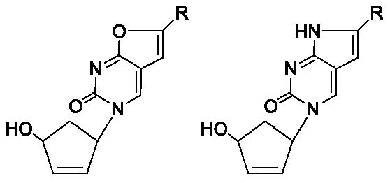
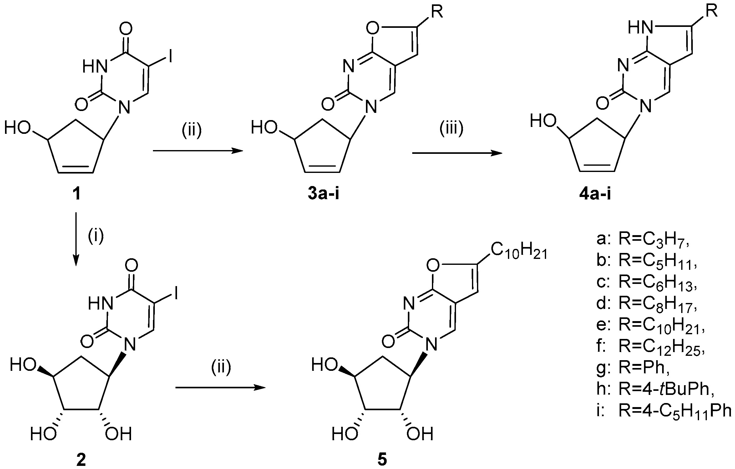
2.1. Chemistry

All the 5′-norcarbocyclic analogs of bicyclic furano- and pyrrolo[2,3d]pyrimidine nucleosides were synthesized starting from the general precursor racemic 1-(4′-hydroxy-2′-cyclopentene-1′-yl)-5-ioduracil 1 (Figure 1) which was obtained as described earlier [12,13]. 1-(2′,3′,4′-Trihydroxycyclopent-1′-yl)-5-iodouracil 2 was synthesized by oxidation of compound 1 using osmium tetroxide in the presence of N-methylmorpholine-N-oxide (NMMO) [14]. This procedure allows the cis-2′,3′-diol to be obtained selectively [15,16]. To prepare furano[2,3d]pyrimidine nucleosides we used Cu/Pd-catalyzed cyclisation of 1 (for 3ai) or its oxidized derivative 2 (for 5) with corresponding alkynes in refluxing CH3CN. This afforded target furano[2,3d]pyrimidine nucleosides in good yields (36–82%). Such a deviation in yields was due both to the difference in alkyne structures and to the fact that isolation and purification of some products turned to be laborious. Subsequent treatment of compounds 3ai with 32% ammonia in methanol resulted in corresponding pyrrolo[2,3-d]pyrimidine analogs 4ai (Scheme 1). The reactions at 40 °C were rather slow, but such mild conditions gave us an opportunity to obtain products 4ai with good yields (57–89%) without using a bomb. It is worth remarking that preparative liquid chromatography on silica gel plates turned to be more effective for the isolation of pyrrolo[2,3-d]pyrimidine analogues 4ai than the column chromatography on silica gel, which was our choice in the case of furano[2,3-d]pyrimidine derivatives 3ai and 5. All the compounds were synthesized as racemic mixtures.
As a result, a set of novel bicyclic furano[2,3-d]pyrimidine nucleosides 3ai, early unknown bicyclic pyrrolo[2,3-d]pyrimidine nucleosides 4ai and a new 1-(2′,3′,4′-trihydroxycyclopent1′-yl)6-decyl-3H-furano[2,3-d]pyrimidine-2-one 5 were obtained. The last one was synthesized as a first representative of trihydroxycyclopentyl derivatives of furano[2,3-d]pyrimidine-2-one in order to estimate the potential of this modification for antitumor activity and to gain a better structure activity relationship (SAR) understanding.

2.2. Biological Evaluation

2.2.1. Cell Viability Assay

The target compounds were tested on different lines of tumor cells, HuTu-80 (human duodenal cancer), B16 (mouse melanoma), A549 (human lung adenocarcinoma), KB-3-1 (human squamous cell carcinoma), HeLa (human squamous cell carcinoma of the cervix), as well as on human noncancer cells hFF3.
Compounds 3b and 4a had no toxic effect on either normal untransfected hFF3 cells or on tumor cells in concentrations up to 100 μM. Compounds 3df,h and 4f,i were almost equally toxic for cancer and noncancer cells (Table 1). The pyrrole-containing compounds 4e and 4h were less toxic than the corresponding furan analogues 3e and 3h, but also reduced the viability of all tested cell lines with IC50 in the range from 11 μM (HeLa) to 63 μM (hFF3) for 4e and from 15 μM (KB-3-1) to 70 μM (hFF3) for 4h. First 2′,3′-dihydroxy derivative 5, proved to be less toxic than the corresponding 2′,3′-didehydro-2′,3′-dideoxy analogue 3e. Compounds 3a, 3c, 3g, 3i, 4bd, 4g and 5 inhibited the growth of some tested tumor cells, mainly KB-3-1 and HeLa (Table 1), while they were not toxic for normal cells in concentrations up to 100 μM. Melanoma cells B16 were the most resistant towards action of these new nucleoside analogues. Only compounds 3f, 3i, 4e, 4h and 4i had the selective toxic effect on this line with IC50 4.5, 21, 25, 35 and 13.4 μM, respectively.
The nucleoside analogue 3i was among most active compounds (Table 1) and had the most selective antiproliferative antitumor effect, especially against the HuTu80, KB-3-1 and HeLa cell lines (Table 2). Therefore, we used it as a lead compound to study the mechanism of induced cells death.

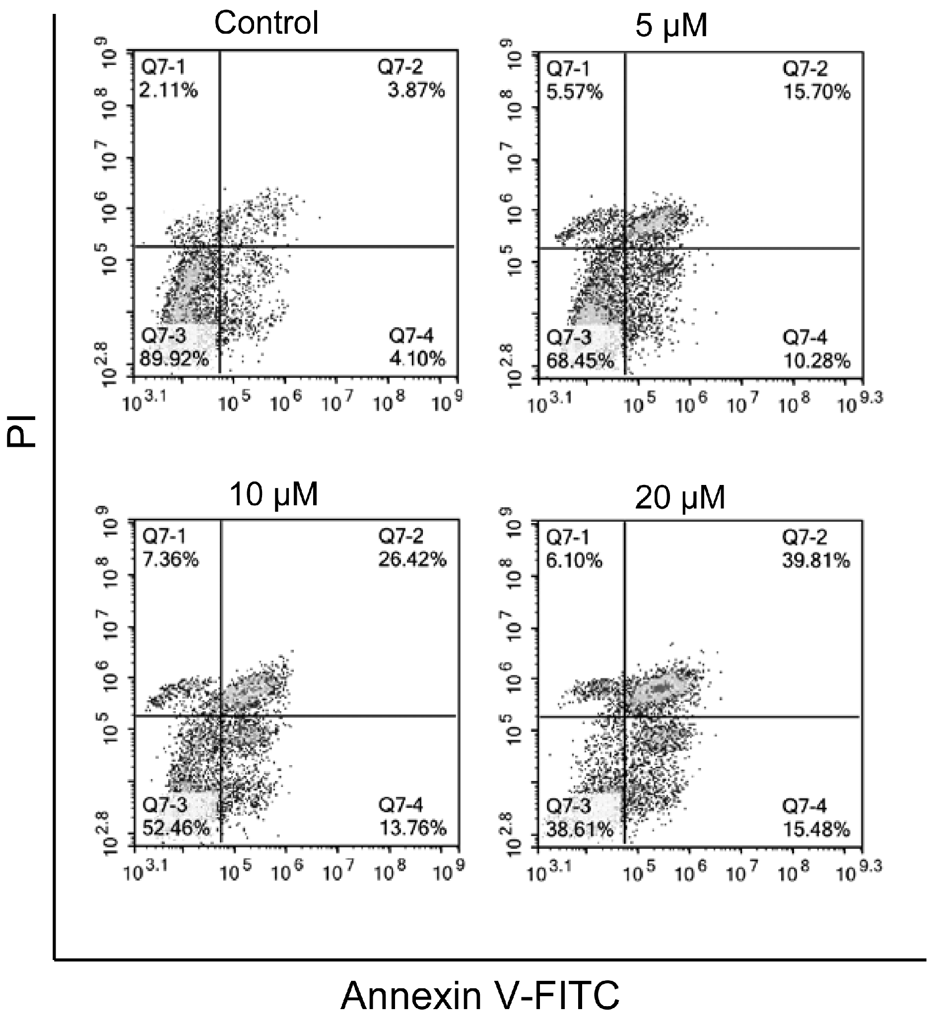
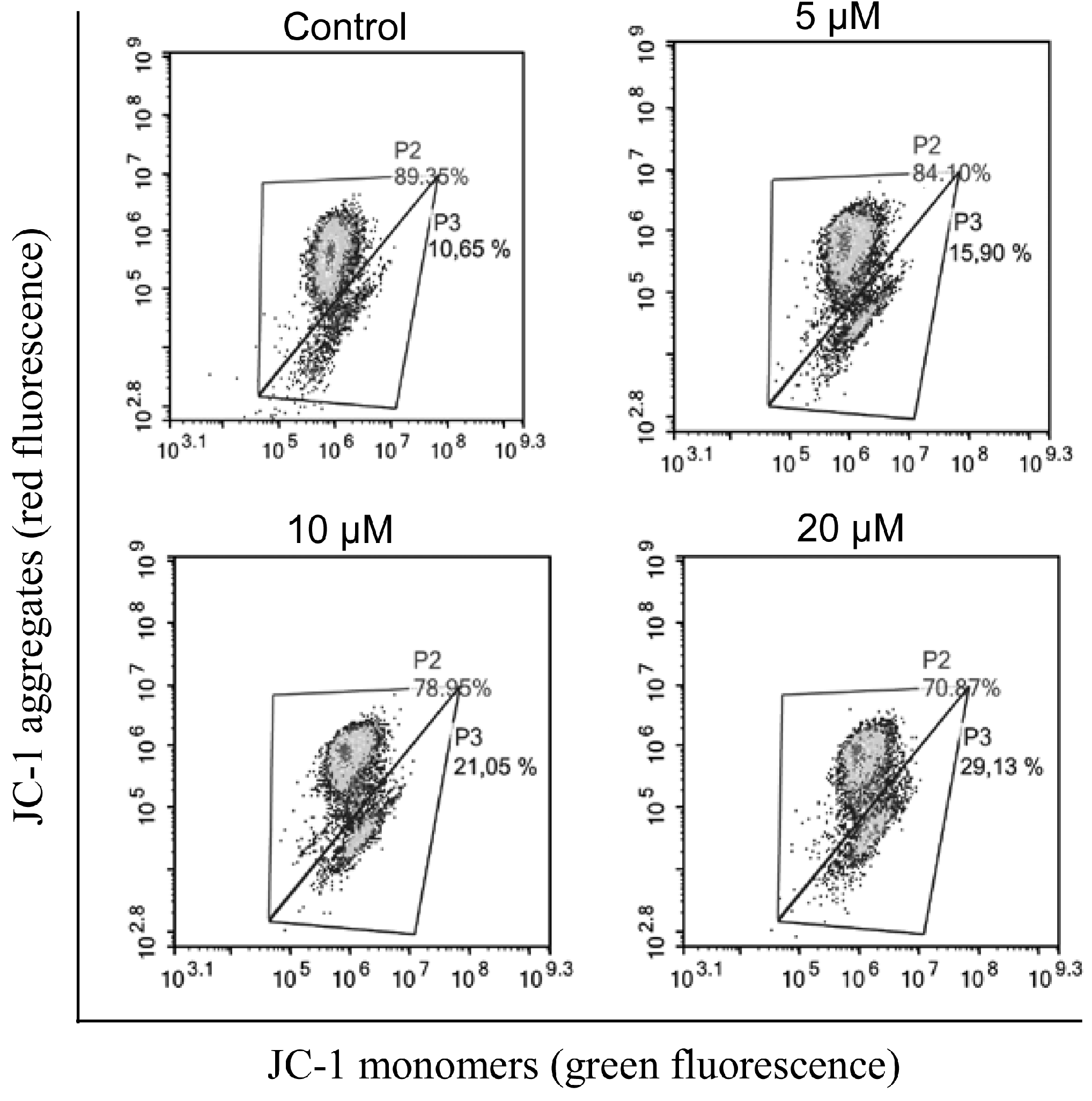
2.2.2. Induction of Apoptosis

To examine whether the tested 5′-norcarbocyclic derivatives induce cell death via apoptosis Annexin V and propidium iodide analysis were used (Figure 1). KB-3-1 cells were exposed to 3i, the most active among tested compounds, for 48 h and then flow cytometric analysis was undertaken. Annexin V binds phosphatidylserine residues, which are asymmetrically distributed toward the inner plasma membrane, and migrate to the outer plasma membrane during apoptosis [17]. The data shows that 3i induces apoptotic cell death in 26% of KB-3-1 cells at concentrations of 5 µM. The increasing of 3i concentration to 20 µM resulted in 55.3% apoptotic cells after 48 h of incubation of KB-3-1 cells with the analogue. Hence, the 5′-norcarbocyclic derivative 3i induced dose-dependent apoptotic cell death.
We next investigated whether 3i utilizes the mitochondrial ‘intrinsic’ pathway in the apoptotic death of KB-3-1 cells, since the pivotal role of mitochondria in triggering apoptosis is well established. We evaluated the mitochondrial transmembrane potential (∆Ψm) in KB-3-1 cells exposed to 3i using cytofluorometric analysis. Cells were stained with the specific mitochondrial cationic dye JC-1 (5,5′,6,6′-tetrachloro-1,1′,3,3′-tetraethyl benzimidazole carbocyanine iodide) that accumulates in the transmembrane space of polarized mitochondria and forms the so–called «J–aggregates», emitting red fluorescence. A decrease in ∆Ψm results in disappearance of J–aggregates and formation of JC-1 monomers, which emit in a green fluorescence. The cytometric analysis of KB-3-1 cells stained with JC-1 is shown in Figure 2.
In the control cells incubated in the presence of 0.1% DMSO the majority of cells shows a high emission of fluorescence in both channels due to the equilibrium between J-aggregates and monomers (Figure 2). The exposure of KB-3-1 cells to 20 µM of compound 3i leads to a decrease of the red fluorescence value as compared to the control (0.1% DMSO).

3. Materials and Methods

3.1. Chemistry

N-Methylmorpholine-N-oxide (NMMO), peracetic acid, Pd(PPh3)4, CuI, 10% Pd/C, triethylamine, 3-(4,5-dimethylthiazol-2-yl)-2,5-diphenyltetrazolium bromide, propidium iodide and organic solvents were obtained from “Acros” (Belgium) or “Aldrich” (USA) and were used without further purification. 1-(4′-Hydroxy-2′-cyclopenten-1′-yl)-5-iodouracil (1) was synthesized according an earlier published protocol [12]; 1-(4′-hydroxy-2′-cyclopenten-1′-yl)-6-octyl-3Hfurano[2,3-d]-pyrimidine-2-one (3d), 1-(4′-hydroxy-2′-cyclopenten-1′-yl)-6-decyl-3Hfurano[2,3-d]-pyrimidine-2-one (3e) and 1-(4′-hydroxy-2′-cyclopenten-1′-yl)-6-dodecyl-3Hfurano[2,3-d]-pyrimidine-2-one (3f) were prepared as described earlier [12,13]. Annexin-FITC apoptosis staining/detection kit was from “Abcam” (Eugene, CA USA); 5,5′,6,6′-tetrachloro-1,1′,3,3′tetraethylbenzimidazolylcarbocyanine iodide (JC-1) was from “Invitrogen” (San Diego, CA, USA).
Column chromatography was performed on Silica Gel 60 0.040–0.063 mm (Merck, Germany), and systems for elution are indicated in the text. Thin layer chromatography (TLC) was performed on TLC Silica gel 60 F254 plates (Merck, Germany) in chloroform–methanol, 9:1 (A), or chloroform–methanol, 4:1 (B) systems. Preparative layer chromatography (PLC) was performed on PLC Silica gel 60 F254 plates (Merck, Germany), systems for elution are indicated in the text.
1H and 13C nuclear magnetic resonance (NMR) spectra were registered on a Bruker Avance 400 spectrometer (Bruker, Newark, Germany) using tetramethylsilane (TMS) in CDCl3, CD3OD, CDCl3/CD3OD mixture, or DMSO-d6 as internal standard. Chemical shifts are given in ppm, and the letter “J” indicates normal 3JHH couplings and all J values are given in Hz.
High-resolution mass spectra (HRMS) were registered on a Bruker Daltonics micrOTOF-Q II instrument using electrospray ionization (ESI). The measurements were acquired in a negative ion mode with the following parameters: interface capillary voltage—3700 V; mass range from m/z 50 to 3000; external calibration (Electrospray Calibrant Solution, Fluka); nebulizer pressure—0.3 Bar; flow rate—3 µL/min; dry gas nitrogen (4.0 L/min); interface temperature was set at 180 or 190 °C. A syringe injection was used.
The absorbance (MTT assay) was measured on a plate reader Multiscan RC (Thermo LabSystems, Vantaa, Finland) at 570 nm. Mitochondrial transmembrane potential and the amount of apoptotic cells in samples were analyzed by flow cytometer «FC500» (Beckman Coulter, Indianapolis, IN, USA).

3.1.1. General Method for the Synthesis of 1-(4′-hydroxy-2′-cyclopenten-1′-yl)-6-alkyl-3H-furano[2,3-d]pyrimidine-2-ones and 1-(4′-hydroxy-2′-cyclopenten-1′-yl)-6-aryl-3H-furano[2,3-d]-pyrimidine-2-ones (3ai)

To the solution of 1-(4′-hydroxy-2′-cyclopenten-1′-yl)-5-iodouracil (100 mg, 0.31 mmol) in acethonitrile (5 mL) CuI (19 mg, 0.1 mmol), 10% Pd/C (15 mg) and appropriate 1-alkyne (0.38 mmol) were added and the reaction mixture was refluxed for 4 h. The progress of the reaction was monitored by TLC. Reaction mixtures were evaporated to dryness in vacuo, the residues were dissolved in the appropriate solvent mixture and 3ai were isolated and purified using column chromatography on silica gel.
1-(4′-Hydroxy-2′-cyclopenten-1′-yl)-6-propyl-3H-furano[2,3-d]-pyrimidine-2-one (3a) was purified on a silica gel column using chloroform: methanol (97:3) as an eluent with 41% yield. Rf 0.35 (system A). 1H-NMR (CD3OD): 8.29 (1H, s, H4), 6.39 (1H, m, H5), 6.34-6.31 (1H, m, H2′), 6.00–5.97 (1H, m, H3′), 5.77–5.75 (1H, m, H1′), 4.87–4.82 (1H, m, H4′), 3.07–3.02 (1H, m, H5′a), 2.71–2.66 (2H, m, CH2CH2CH3), 1.81–1.69 (2H, m, CH2CH2CH3), 1.59–1.52 (1H, m, H5′b), 1.03 (3H, t, J = 8 Hz, CH2CH2CH3). 13C-NMR (CD3OD): 172.4, 160.9, 157.2, 141.1, 139.2, 131.8, 109.7, 100.2, 75.1, 63.2, 41.9, 30.5, 20.9, 13.4. HRMS (ESI, m/z) of C14H16N2O3: calcd. for [M + Na]+ 283.1053, found 283.1061, see Supplementary Materials.
1-(4′-Hydroxy-2′-cyclopenten-1′-yl)-6-pentyl-3H-furano[2,3-d]-pyrimidine-2-one (3b) was purified on silica gel column using chloroform: methanol (98:2 to 95:5) as an eluent with 69% yield. Rf 0.36 (system A). 1H-NMR (CD3OD): 8.29 (1H, s, H4), 6.38 (1H, m, H5), 6.35–6.31 (1H, m, H2′), 6.00–5.97 (1H, m, H3′), 5.81–5.75 (1H, m, H1′), 4.87–4.82 (1H, m, H4′), 3.05–3.00 (1H, m, H5′a), 2.71 (2H, t, J = 8 Hz, CH2CH2(CH2)2CH3), 1.75–1.70 (2H, m, CH2CH2(CH2)2CH3), 1.59–1.52 (1H, m, H5′b), 1.43–1.37 (4H, m, CH2CH2(CH2)2CH3), 0.94 (3H, t, J = 8 Hz, CH2CH2(CH2)2CH3). 13C-NMR (CD3OD): 171.4, 160.1, 156.3, 140.1, 138.2, 130.8, 108.7, 99.1, 74.1, 62.2, 40.9, 30.9, 27.5, 26.3, 21.9, 12.8. HRMS (ESI, m/z) of C16H20N2O3: calcd. for [M + H]+ 289.1547, found 289.1545.
1-(4′-Hydroxy-2′-cyclopenten-1′-yl)-6-hexyl-3H-furano[2,3-d]-pyrimidine-2-one (3c) was purified using two-step chromatography on silicа gel. The first column was eluted with chloroform: methanol (98:2 to 95:5) and the second one with hexane: ethyl acetate (1:4) to ethyl acetate: methanol (97:3) as an eluent to give (3c) with 52% yield. Rf 0.36 (system A). 1H-NMR (CD3OD): 8.27 (1H, s, H4), 6.36 (1H, s, H5), 6.32–6.30 (1H, m, H2′), 5.98–5.96 (1H, m, H3′), 5.76–5.75 (1H, m, H1′), 4.85–4.83 (1H, m, H4′), 3.05–3.01 (1H, m, H5′a), 2.69 (2H, t, J = 8Hz, CH2CH2(CH2)3CH3), 1.76–1.68 (2H, m, CH2CH2(CH2)3CH3), 1.56–1.51 (1H, m, H5′b), 1.35–1.29 (6H, m, CH2CH2(CH2)3CH3), 0.92 (3H, t, J = 8Hz, CH2CH2(CH2)3CH3). 13C-NMR (CD3OD): 172.8, 161.5, 157.7, 141.5, 139.6, 132.2, 110.1, 100.5, 75.5, 63.6, 42.3, 32.6, 29.7, 29.0, 28.0, 23.6, 14.3. HRMS (ESI, m/z) of C17H22N2O3: calcd. for [M + H]+ 303.1703, found 303.1698.
1-(4′-Hydroxy-2′-cyclopenten-1′-yl)-6-phenyl-3H-furano[2,3-d]-pyrimidine-2-one (3g) was purified using column chromatography with chloroform: methanol (97:3) as an eluent and then on a PLC in ethyl acetate with 37% yield. Rf 0.34 (system A). 1H-NMR (CDCl3-CD3OD): 8.17 (1H, s, H4), 7.71–7.69 (2H, m, Ph), 7.41–7.32 (3H, m, Ph), 6.69 (1H, s, H5), 6.27–6.26 (1H, m, H2′), 5.87–5.86 (1H, m, H3′), 5.81–5.80 (1H, m, H1′), 4.85–4.83 (1H, m, H4′), 3.01–2.94 (1H, m, H5′a), 1.60–1.56 (1H, m, H5′b). 13C-NMR (CDCl3-CD3OD): 171.3, 156.0, 140.2, 138.4, 131.7, 129.80, 129.0 × 2, 128.3, 125.0 × 2, 109.1, 97.7, 74.4, 62.4, 41.1, 29.7. HRMS (ESI, m/z) of C17H14N2O3: calcd. for [M + H]+ 295.1077, found 295.1077.
1-(4′-Hydroxy-2′-cyclopenten-1′-yl)-6-tertbutylphenyl-3H-furano[2,3-d]-pyrimidine-2-one (3h) was purified on a silica gel column using chloroform: methanol (98:2) as an eluent with 36% yield. Rf 0.36 (system A). 1H-NMR (CDCl3-CD3OD): 8.14 (1H, s, H4), 7.63 (2H, d, J = 8 Hz, Ph), 7.41 (2H, d, J = 8 Hz, Ph), 6.63 (1H, s, H5), 6.26 (1H, m, H2′), 5.86 (1H, m, H3′), 5.80 (1H, m, H1′), 4.84 (1H, m, H4′), 3.01–2.93 (1H, m, H5′a), 1.61–1.57 (1H, m, H5′b), 1.28 (9H, s, tBu). 13C-NMR (CDCl3-CD3OD): 171.3, 156.2, 155.9, 153.4, 140.1, 137.9, 131.7, 125.9 × 2, 125.5, 124.8 × 2, 109.2, 96.9, 74.4, 62.3, 41.2, 34.9, 31.1 × 3. HRMS (ESI, m/z) of C21H22N2O3: calcd. for [M + H]+ 351.1703, found 351.1698.
1-(4′-Hydroxy-2′-cyclopenten-1′-yl)-6-(4-pentylphenyl)-3H-furano[2,3-d]-pyrimidine-2-one (3i) was purified using two-step chromatography on silicа gel. The first column was eluted with chloroform: methanol (98:2 to 97:3) and the second one with hexane: ethyl acetate (1:4) to ethyl acetate: methanol (97:3) as an eluent to give (3i) with 82% yield. Rf 0.36 (system A). 1H-NMR (CDCl3-CD3OD): 8.33 (1H, s, H4), 7.68 (2H, d, J = 8.5 Hz, Ph), 7.27 (2H, d, J = 8.5 Hz, Ph), 6.84 (1H, s, H5), 6.35–6.32 (1H, m, H2′), 5.95–5.93 (1H, m, H3′), 5.85–5.82 (1H, m, H1′), 4.88–4.86 (1H, m, H4′), 3.07–3.00 (1H, m, H5′a), 2.65 (2H, t, J = 9Hz, CH2CH2(CH2)2CH3), 1.66–1.57 (3H, m, H5′b and CH2CH2(CH2)2CH3), 1.35–1.32 (4H, m, CH2CH2(CH2)2CH3), 0.89 (3H, t, J = 9Hz, CH2CH2(CH2)2CH3). 13C-NMR (CDCl3-CD3OD): 171.1, 156.3, 156.0, 145.0, 140.0, 138.4, 130.09, 128.8 × 2, 125.5, 124.6 × 2, 109.5, 96.8, 73.9, 62.1, 41.9, 35.4, 31.1, 30.6, 22.1, 13.3. HRMS (ESI, m/z) of C22H24N2O3: calcd. for [M + H]+ 365.1861, found 365.1860.
1-(2′,3′,4′-Trihydroxycyclopent1’-yl)-5-iodouracil (2) was synthesized using the common procedure [14] 1H-NMR(DMSO-d6): 11.58 (1H, s, NH), 8.12 (1H, s, H6), 5.14 (1H, d, J = 4 Hz, OH), 4.95 (1H, d, J = 6 Hz, OH), 4.77 (1H, d, J = 4 Hz, OH), 4.73–4.68 (1H, m, H2′), 4.17–4.12 (1H, m, H1′), 3.82–3.79 (1H, m, H3′), 3.69–3.66 (1H, m, H4′), 2.43–2.40 (1H, m, H5′a), 1.42–1.37 (1H, m, H5′b).
1-(2′,3′,4′-Trihydroxycyclopent-1′-yl)-6-decyl-3H-furano[2,3-d]-pyrimidine-2-one (5) was synthesized as described for compounds 3ai, starting from 1-(2′,3′,4′-trihydroxycyclopenten-1′-yl)-5-iodouracil 2. The product was purified using chloroform: methanol (4:1) as an eluent with 54% yield. Rf 0.25 (system B). 1H-NMR(CD3OD): 8.44 (1H, s, H4), 6.36 (1H, s, H5), 5.05–4.97 (1H, m, H2′), 4.55–4.50 (1H, m, H1′), 4.06–4.03 (1H, m, H3′), 3.94–3.92 (1H, m, H4′), 2.76–2.71 (1H, m, H5′a), 2.69–2.64 (2H, m, CH2(CH2)8CH3), 1.73–1.66 (3H, m, H5′b + CH2CH2(CH2)7CH3), 1.39–1.26 (14H, m, CH2CH2(CH2)7CH3), 0.68 (3H, t, J = 8 Hz, CH2(CH2)8CH3). 13C-NMR(CDCl3-CD3OD): 171.35, 160.15, 156.56, 139.75, 108.87, 99.08, 76.68, 75.61, 73.77, 65.02, 35.54, 31.63, 29.25, 29.19, 29.00, 28.94, 28.66, 27.56, 26.57, 22.29, 13.01. HRMS: found m/z 393.2384, calculated for C21H32N2O5 [M + H]+ 393.2388.

3.1.2. General Method for the Synthesis of 1-(4′-hydroxy-2′-cyclopenten-1′-yl)-6-alkyl-3H-pyrrolo[2,3-d]pyrimidine-2-ones and 1-(4′-hydroxy-2′-cyclopenten-1′-yl)-6-aryl-3H-pyrrolo[2,3-d]-pyrimidine-2-ones (4ai).

To the corresponding 1-(4′-hydroxy-2′-cyclopenten-1′-yl)-6-alkyl-3H-furano[2,3-d]pyrimidine-2-one or 1-(4′-hydroxy-2′-cyclopenten-1′-yl)-6-aryl-3H-furano[2,3-d]-pyrimidine-2-one (50 mg) a solution of 32% NH3 in MeOH (15 mL) was added. The reaction mixture was kept at 40 °C for 48 h. Solvent then was evaporated in vacuo and a new portion of 32% NH3 in MeOH was added (15 mL). The procedure was repeated three times controlling the progress of the reaction by TLC.
1-(4′-Hydroxy-2′-cyclopenten-1′-yl)-6-propyl-3H-pyrrolo[2,3-d]-pyrimidine-2-one (4a) was purified using PLC with chloroform: methanol (9:1) as an eluent with 70% yield. Rf 0.33 (system A). 1H-NMR (DMSO-d6): 11.05 (1H, s, NH), 8.03 (1H, s, H4), 6.18–6.16 (1H, m, H2′), 5.91 (1H, s, H5), 5.87–5.86 (1H, m, H3′), 5.69–5.66 (1H, m, H1′), 5.21 (1H, m, OH), 4.68 (1H, m, H4′), 2.87–2.83 (1H, m, H5′a), 2.50 (2H, t, J = 8 Hz, CH2CH2CH3), 1.64–1.59 (2H, m, CH2CH2CH3), 1.38–1.34 (1H, m, H5′b), 0.89 (3H, t, J = 8 Hz, CH2CH2CH3). 13C-NMR (DMSO-d6): 158.9, 154.4, 142.1, 139.8, 135.9, 131.4, 109.2, 96.2, 73.5, 60.4, 41.2, 29.4, 20.9, 13.4. HRMS (ESI, m/z) of C14H17N3O2: calcd. for [M + Na]+ 282.1213, found 282.1211.
1-(4′-Hydroxy-2′-cyclopenten-1′-yl)-6-pentyl-3H-pyrrolo[2,3-d]-pyrimidine-2-one (4b). The product was purified using PLC with chloroform: methanol (9:1) as an eluent with 57% yield. Rf 0.33 (system A). 1H-NMR (CD3OD): 8.18 (1H, s, H4), 6.31–6.28 (1H, m, H2′), 6.01 (1H, m, H5), 5.99–5.96 (1H, m, H3′), 5.85–5.81 (1H, m, H1′), 4.89 (1H, m, H4′), 3.08–3.03 (1H, m, H5′a), 2.64 (2H, t, J = 8 Hz, CH2CH2(CH2)2CH3), 1.75–1.68 (2H, m, CH2CH2(CH2)2CH3), 1.57–1.49 (1H, m, H5′b), 1.40–1.35 (4H, m, CH2CH2(CH2)2CH3), 0.93 (3H, t, J = 8 Hz, CH2CH2(CH2)2CH3). 13C-NMR (CD3OD): 158.6, 156.3, 143.8, 139.5, 136.0, 131.3, 111.2, 96.4, 74.2, 61.7, 41.2, 31.1, 27.6, 27.5, 22.0, 12.9. HRMS (ESI, m/z) of C16H21N3O2: calcd. for [M + H]+ 288.1706, found 288.1710.
1-(4′-Hydroxy-2′-cyclopenten-1′-yl)-6-hexyl-3H-pyrrolo[2,3-d]-pyrimidine-2-one (4c). The product was purified using PLC with chloroform: methanol (9:1) as an eluent with 64% yield. Rf 0.33 (system A). 1H-NMR (CD3OD): 8.16 (1H, s, H4), 6.28–6.27 (1H, m, H2′), 6.00 (1H, m, H5), 5.96–5.95 (1H, m, H3′), 5.81 (1H, m, H1′), 4.83–4.80 (1H, m, H4′), 3.08–3.00 (1H, m, H5′a), 2.62 (2H, t, J = 8 Hz, CH2CH2(CH2)3CH3), 1.70–1.66 (2H, m, CH2CH2(CH2)3CH3), 1.54–1.48 (1H, m, H5′b), 1.39–1.33 (6H, m, CH2CH2(CH2)3CH3), 0.90 (3H, t, J = 8 Hz,CH2CH2(CH2)3CH3). 13C-NMR (CD3OD): 160.0, 157.8, 145.2, 140.9, 137.4, 132.8, 112.6, 97.8, 75.6, 63.1, 42.6, 32.7, 29.9, 29.3, 29.0, 23.6, 14.3. HRMS (ESI, m/z) of C17H23N3O2: calcd. for [M + H]+ 302.1863, found 302.1868.
1-(4′-Hydroxy-2′-cyclopenten-1′-yl)-6-octyl-3H-pyrrolo[2,3-d]-pyrimidine-2-one (4d). The product was purified using PLC with chloroform: methanol (9:1) as an eluent with 89% yield. Rf 0.33 (system A). 1H-NMR (CD3OD): 8.18 (1H, s, H4), 6.31–6.29 (1H, m, H2′), 6.02 (1H, s, H5), 5.98–5.96 (1H, m, H3′), 5.83 (1H, m, H1′), 4.85–4.82 (1H, m, H4′), 3.08–3.00 (1H, m, H5′a), 2.64 (2H, t, J = 8 Hz, CH2CH2(CH2)5CH3), 1.72–1.67 (2H, m, CH2CH2(CH2)5CH3), 1.56–1.48 (1H, m, H5′b), 1.35–1.27 (10H, m, CH2CH2(CH2)5CH3), 0.91 (3H, t, J = 8 Hz, CH2CH2(CH2)5CH3). 13C-NMR (DMSO-d6): 160.0, 157.8, 145.2, 140.9, 137.4, 132.8, 112.6, 97.9, 75.6, 63.1, 42.6, 33.0, 30.4, 30.3, 30.2, 29.3, 29.0, 23.7, 14.4. HRMS (ESI, m/z) of C19H27N3O2: calcd. for [M + Na]+ 352.2176, found 352.2177.
1-(4′-Hydroxy-2′-cyclopenten-1′-yl)-6-decyl-3H-pyrrolo[2,3-d]-pyrimidine-2-one (4e) was purified using PLC with chloroform: methanol (9:1) as an eluent with 67% yield. Rf 0.33 (system A). 1H-NMR (CD3OD): 8.18 (1H, s, H4), 6.31–6.28 (1H, m, H2′), 6.02 (1H, s, H5), 5.99–5.96 (1H, m, H3′), 5.85–5.81 (1H, m, H1′), 4.85–4.82 (1H, m, H4′), 3.08–3.01 (1H, m, H5′a), 2.64 (2H, t, J = 8 Hz, CH2CH2(CH2)7CH3), 1.72–1.67 (2H, m, CH2CH2(CH2)7CH3), 1.57–1.49 (1H, m, H5′b), 1.35–1.30 (14H, m, CH2CH2(CH2)7CH3), 0.91 (3H, t, J = 8 Hz, CH2CH2(CH2)7CH3). 13C-NMR (DMSO-d6): 160.0, 157.7, 145.2, 140.9, 137.4, 132.8, 112.6, 97.9, 75.6, 63.1, 42.6, 33.0, 30.6, 30.6, 30.4 × 2, 30.2, 29.3, 29.0, 23.7, 14.4. HRMS (ESI, m/z) of C21H31N3O2: calcd. for [M + Na]+ 380.2308, found 380.2309.
1-(4′-Hydroxy-2′-cyclopenten-1′-yl)-6-dodecyl-3H-pyrrolo[2,3-d]-pyrimidine-2-one (4f) was purified using PLC with chloroform: methanol (9:1) as an eluent with 66% yield. Rf 0.38 (system A). 1H-NMR (CDCl3): 8.28 (1H, s, H4), 6.37–6.35 (1H, m, H2′), 5.92 (1H, s, H5), 5.88–5.87 (1H, m, H3′), 5.80–5.77 (1H, m, H1′), 4.95–4.92 (1H, m, H4′), 3.04–2.094 (1H, m, H5′a), 2.70 (2H, t, J = 8 Hz, CH2CH2(CH2)9CH3), 1.82–1.77 (2H, m, CH2CH2(CH2)9CH3), 1.71–1.66 (1H, m, H5′b), 1.36–1.26 (18H, m, CH2CH2(CH2)9CH3), 0.89 (3H, t, J = 8 Hz, CH2CH2(CH2)9CH3). 13C-NMR (CDCl3): 156.9, 153.8, 144.6, 140.4, 138.2, 131.6, 111.2, 96.9, 74.9, 62.5, 41.3, 32.0, 29.7 × 4, 29.5, 29.4, 29.3, 28.1 × 2, 22.7, 14.2. HRMS (ESI, m/z) of C23H35N3O2: calcd. for [M + H]+ 386.2802, found 386.2804.
1-(4′-Hydroxy-2′-cyclopenten-1′-yl)-6-phenyl-3H-pyrrolo[2,3-d]-pyrimidine-2-one (4g) was purified using PLC with chloroform: methanol (9:1) as an eluent with 71% yield. Rf 0.33 (system A). 1H-NMR (CD3OD): 8.33 (1H, s, H4), 7.75–7.73 (2H, m, Ph), 7.45–7.42 (2H, m, Ph), 7.38–7.36 (1H, m, Ph), 6.66 (1H, s, H5), 6.32–6.30 (1H, m, H2′), 6.00–5.98 (1H, m, H3′), 5.82 (1H, m, H1′), 4.86–4.82 (1H, m, H4′), 3.10–3.02 (1H, m, H5′a), 1.59–1.53 (1H, m, H5′b). 13C-NMR (CD3OD): 160.7, 157.8, 142.4, 141.1, 139.1, 132.6, 132.0, 130.1 × 2, 129.8, 126.4 × 2, 112.8, 97.9, 75.6, 63.4, 42.7. HRMS (ESI, m/z) of C17H15N3O2: calcd. for [M + H]+ 294.1237, found 294.1233.
1-(4′-Hydroxy-2′-cyclopenten-1′-yl)-6-(4-tertbutylphenyl)-3H-pyrrolo[2,3-d]-pyrimidine-2-one (4h) was purified using PLC with chloroform: methanol (9:1) as an eluent with 61% yield. Rf 0.40 (system A). 1H-NMR (CDCl3): 8.12 (1H, s, H4), 7.58 (2H, d, J = 8 Hz, Ph), 7.43 (2H, d, J = 8 Hz, Ph), 6.39 (1H, s, H5), 6.29–6.27 (1H, m, H2′), 5.88–5.86 (1H, m, H3′), 5.82 (1H, m, H1′), 4.87 (1H, m, H4′), 3.06–3.00 (1H, m, H5′a), 1.66–1.62 (1H, m, H5′b), 1.32 (9H, s, tBu). 13C-NMR (CDCl3): 159.0, 155.8, 152.2, 140.9, 139.7, 137.2, 131.9, 127.5, 126.0 × 2, 124.9 × 2, 111.5, 95.6, 74.6, 62.3, 41.3, 34.7, 31.1 × 3. HRMS (ESI, m/z) of C21H23N3O2: calcd. for [M + H]+ 350.1863, found 350.1864.
1-(4′-Hydroxy-2′-cyclopenten-1′-yl)-6-(4-pentylphenyl)-3H-pyrrolo[2,3-d]-pyrimidine-2-one (4i). The product was purified using two-step chromatography. The first column was eluted with chloroform: methanol (98:2 to 97:3) and the second one with hexane: ethyl acetate (1:4) to ethyl acetate: methanol (97:3) as an eluent to give (4i) with 72% yield. Rf 0.36 (system A). 1H-NMR (CDCl3-CD3OD): 8.27 (1H, s, H4), 7.62 (2H, d, J = 8.5 Hz, Ph), 7.23 (2H, d, J = 8.5 Hz, Ph), 6.58 (1H, s, H5), 6.29–6.27 (1H, m, H2′), 5.97–5.95 (1H, m, H3′), 5.81–5.79 (1H, m, H1′), 4.83–4.80 (1H, m, H4′), 3.09–2.97 (1H, m, H5′a), 2.62 (2H, t, J = 9 Hz, CH2CH2(CH2)2CH3), 1.64–1.55 (3H, m, H5′b and CH2CH2(CH2)2CH3), 1.34–1.29 (4H, m, CH2CH2(CH2)2CH3), 0.88 (3H, t, J = 9 Hz, CH2CH2(CH2)2CH3). 13C-NMR (CDCl3-CD3OD): 159.2, 156.4, 143.8, 141.2, 139.67, 137.2, 131.3, 128.7 × 2, 125.7, 125.0 × 2, 111.6, 95.8, 78.0, 74.2, 61.9, 41.3, 35.2, 31.2, 30.8, 22.2, 13.0. HRMS (ESI, m/z) of C22H25N3O2: calcd. for [M + H]+ 364.2020, found 364.2020.

3.2. Biological Assay

3.2.1. Cell Cultures

Human KB-3-1 epidermoid carcinoma cell line, human HeLa cervical epithelioid carcinoma cell line, human HuTu-80 duodenal cancer cells, human A549 lung carcinoma epithelial cells, mouse B16 melanoma cell line and human hFF3 fibroblast cells were obtained from the Russian Cell Culture Collection (St. Petersburg, Russia) and were cultured in DMEM (hFF3 cells in IMDM) supplemented with 10% (v/v) heat-inactivated fetal bovine serum, penicillin (100 U mL−1), streptomycin (100 µg mL−1) and amphotericin (250 µg mL−1). Cells were maintained in a humidified atmosphere of 5% CO2 at 37 °C.
All compounds were dissolved in dimethylsulfoxide (DMSO) and stock solutions (10 mmol L−1) were stored at −20 °C.
After treatments, both floating and adherent scrapped cells were collected by centrifugation and used for further analysis.

3.2.2. Cell Viability Analysis by MTT Assay

Cells growing in the logarithmic phase were seeded in triplicate in 96-well plates at a density of 5 × 10³ cells per well for HeLa and HuTu-80 cells, 7 × 10³ cells per well for KB-3-1 and hFF3, 10 × 103 for A549 and 20 × 103 for B16. The plates were incubated at 37 °C in a humidified 5% CO2 atmosphere. Cells were allowed to adhere to the surface for 24 h and then tested compounds were added at different concentrations and incubation was continued for 48 h. Then [3-(4,5-dimethylthiazol-2-yl)-2,5-diphenyltetrazolium bromide] (MTT) solution (10 µL, 5 mg mL−1) was added to each well and the incubation was continued for an additional 3 h. The dark blue formazan crystals formed within the healthy cells were solubilized with DMSO and the absorbance was measured using a Multiscan RC plate reader at 570 nm. The IC50 was determined as the compound concentration required to decrease the A570 to 50% compared to the control (no tested compounds, DMSO) and was determined by interpolation from dose-response curves.

3.2.3. Apoptosis Detection by Annexin V Staining

Exponentially growing KB-3-1 cells in 6–well plates (5 × 10⁵ cells per well) were treated with 3i (5, 10 and 20 µM) or with 0.1% (v/v) DMSO as a control for 48 h. The cells were stained with Annexin V-FITC and propidium iodide by the Annexin-FITC apoptosis staining/detection kit (Abcam) according to the instruction of the manufacturer. Briefly, cells were collected by scrapping, washed twice with cold PBS, and centrifuged (400 g, 5 min). Cells were resuspended in binding buffer (500 µL) and Annexin V–FITC (5 µL) and PI (5 µL) were added. Cells were incubated for 5 min at 20 °C in the dark. Finally, binding buffer (300 µL) was added to each tube, and the amount of apoptotic cells in samples were analyzed by flow cytometry. For each sample, 10,000 ungated events were acquired. Annexin V + PI − cells represent the early apoptotic populations. Annexin V + PI + cells represent either late apoptotic or secondary necrotic populations.

3.2.4. Mitochondria Depolarization Analysis

Mitochondria involvement in apoptosis was measured by mitochondrial depolarization occurring early during onset of apoptosis. KB-3-1 cells were incubated with 3i (5, 10 and 20 µM) or 0.1% (v/v) DMSO as a control for 48 h. Then, cells were collected and incubated in complete media in the dark with mitochondrial potential sensor JC-1 (5 µg mL−1) at 37 °C for 30 min, washed with cold PBS and resuspended in PBS (400 µL). Fluorescences of J–aggregate and J–monomer were recorded in the fluorescence channels 2 (FL2) and 1 (FL1), respectively, with flow cytometer «FC500». Necrotic fragments were electronically gated out, on the basis of morphological characteristics on the forward light scatter versus side light scatter dot plot.

4. Conclusions

The comparative evaluation of the effects of synthesized nucleoside analogues on the growth and viability of tumor cell cultures from various origins and on normal cells has revealed that cytotoxicity depends on both the type of bicyclic system (pyrrolo- or furano[2,3-d]pyrimidine) and the structure of a substituent in the 6th position of the heterocyclic base. Furano[2,3-d]pyrimidine 3i, bearing pentylphenyl substituent, is the most promising among synthesized 5′-norcarbocyclic derivatives of 6-substituted bicyclic pyrrolo- and furano[2,3-d]pyrimidines. This demonstrated inhibitory activities with respect to tumor cells with the selectivity index value about 15–20 depending on the nature and origin of tumor cells. In an attempt to understand the mechanism of the action, we showed that 3i induces cell death by apoptosis pathway with the dissipation of mitochondrial potential.

Supplementary Materials

Copies of the NMR spectra are available online.

Author Contributions

A.A.K., E.S.M. and A.L.K. conceived, designed and performed the chemical synthesis; E.B.L. and M.A.Z. designed and performed cell assays and evaluated biological properties of the compounds; P.N.S. performed HRMS analysis and analyzed the relevant data. A.A.K., E.S.M., E.B.L., M.A.Z., P.N.S., S.N.K. and A.L.K. analyzed the data; S.N.K. contributed reagents/materials/analysis tools; A.A.K., E.S.M., E.B.L., P.N.S. and A.L.K. wrote the paper.

Funding

This research was supported by the Russian Foundation for Basic Research (RFBR, grant No. 16-04-01022) and by the Program of fundamental research for state academies for 2013–2020 years (No. 01201363818).

Conflicts of Interest

The authors declare no conflict of interest.

Abbreviations

MTT3-(4,5-Dimethylthiazol-2-yl)-2,5-diphenyltetrazolium bromide
NMMON-methylmorpholine N-oxide
JC-15,5′,6,6′-tetrachloro-1,1′,3,3′-tetraethylbenzimidazolylcarbocyanine iodide
PIpropidium iodide
PLCpreparative layer chromatography
IC50the compound concentration that results in 50% cell survival as measured by the MTT assay

References

  1. Jordheim, L.P.; Durantel, D.; Zoulim, F.; Dumontet, C. Advances in the development of nucleoside and nucleotide analogues for cancer and viraldiseases. Nat. Rev. Drug Discov. 2013, 12, 447–464. [Google Scholar] [CrossRef] [PubMed]
  2. Galmarini, C.; Popowycz, F.; Joseph, B. Cytotoxic Nucleoside Analogues: Different Strategies to Improve their Clinical Efficacy. Curr. Med. Chem. 2008, 15, 1072–1082. [Google Scholar] [CrossRef] [PubMed]
  3. McGuigan, C.; Yarnold, C.J.; Jones, G.; Velázquez, S.; Barucki, H.; Brancale, A.; Andrei, G.; Snoeck, R.; De Clercq, E.; Balzarini, J. Potent and selective inhibition of varicella-zoster virus (VZV) by nucleoside analogues with an unusual bicyclic base. J. Med. Chem. 1999, 42, 4479–4484. [Google Scholar] [CrossRef] [PubMed]
  4. Balzarini, J.; McGuigan, C. Bicyclic pyrimidine nucleoside analogues (BCNAs) as highly selective and potent inhibitors of varicella-zoster virus replication. J. Antimicrob. Chemother. 2002, 50, 5–9. [Google Scholar] [CrossRef] [PubMed] [Green Version]
  5. Jahnz-Wechmann, Z.; Framski, G.; Januszczyk, P.; Boryski, J. Bioactive fused heterocycles: Nucleoside analogues with an additional ring. Eur. J. Med. Chem. 2015, 97, 388–396. [Google Scholar] [CrossRef] [PubMed]
  6. Migliore, M.D.; Zonta, N.; McGuigan, C.; Henson, G.; Andrei, G.; Snoeck, R.; Balzarini, J. Synthesis and Antiviral Activity of the Carbocyclic Analogue of the Highly Potent and Selective Anti-VZV Bicyclo Furano Pyrimidines. J. Med. Chem. 2007, 50, 6485–6492. [Google Scholar] [CrossRef] [PubMed]
  7. Dincer, S.; Cetin, K.T.; Onay-Besikci, A.; Olgen, S. Synthesis, biological evaluation and docking studies of new pyrrolo[2,3-d] pyrimidine derivatives as Src family-selective tyrosine kinase inhibitors. J. Enzyme Inhib. Med. Chem. 2013, 28, 1080–1087. [Google Scholar] [CrossRef] [PubMed]
  8. Aziz, M.A.; Serya, R.A.T.; Lasheen, D.S.; Abouzid, K.A.M. Furo[2,3-d]pyrimidine based derivatives as kinase inhibitors and anticancer agents. Future J. Pharm. Sci. 2016, 2, 1–8. [Google Scholar] [CrossRef]
  9. Romeo, R.; Giofre, S.V.; Garozzo, A.; Bisignano, B.; Corsaro, A.; Chiacchio, M.A. Synthesis and biological evaluation of furopyrimidine N,O-nucleosides. Bioorg. Med. Chem. 2013, 21, 5688–5693. [Google Scholar] [CrossRef] [PubMed]
  10. Framski, G.; Wawrzyniak, D.; Jahnz-Wechmann, Z.; Szymanska-Michalak, A.; Barciszewski, J.; Boryski, J.; Kraszewski, A.; Stawinski, J. New Applications of 6-Alkyl-2,3dihydrofurano[2,3-d]pyrimidin-2(1H)-one and 6-Alkyl-2,3-dihydropyrrolo[2,3-d]pyrimidin-2(3H,7H)-one Nucleosides: Anticancer Properties. In Proceedings of the XXI Round Table on Nucleosides, Nucleotides and Nucleic Acids “Chemical Biology of Nucleic Acids”, Poznań, Poland, 24–28 August 2014. Electronic Abstract Book, Poster 13. IS3NA, 2014. [Google Scholar]
  11. Framski, G.; Wawrzyniak, D.; Jahnz-Wechmann, Z.; Szymanska-Michalak, A.; Kraszewski, A.; Barciszewski, J.; Boryski, J.; Stawinski, J. Searching for anti-glioma activity. Ribonucleoside analogues with modifications in nucleobase and sugar moieties. Acta Biochim. Pol. 2016, 63, 765–771. [Google Scholar] [CrossRef] [PubMed]
  12. Matyugina, E.; Logashenko, E.; Zenkova, M.; Kochetkov, S.; Khandazhinskaya, A. 5′-Norcarbocyclic analogues of furano[2,3-d] pyrimidine nucleosides. Heterocycl. Commun. 2015, 21, 259–262. [Google Scholar] [CrossRef]
  13. Matyugina, E.S.; Khandazhinskaya, A.L.; Chernousova, L.N.; Andreevskaya, S.N.; Smirnova, T.G.; Chizhov, A.O.; Karpenko, I.L.; Kochetkov, S.N.; Alexandrova, L.A. The Synthesis and Antituberculosis Activity of 5’-Nor Carbocyclic Uracil Derivatives. Bioorg. Med. Chem. 2012, 20, 6680–6686. [Google Scholar] [CrossRef] [PubMed]
  14. Khandazhinskaya, A.L.; Shirokova, E.A.; Shipitsin, A.V.; Karpenko, I.L.; Belanov, E.F.; Kukhanova, M.K.; Yasko, M.V. Adenosine N1-oxide analogues as inhibitors of orthopox virus replication. Collect. Czech. Chem. Commun. 2006, 71, 1107–1121. [Google Scholar] [CrossRef]
  15. Matyugina, E.S.; Khandazhinskaya, A.L.; Kochetkov, S.N. Carbocyclic nucleoside analogues: Classification, target enzymes, mechanisms of action and synthesis. Russ. Chem. Rev. 2012, 81, 729–746. [Google Scholar] [CrossRef]
  16. Ainai, T.; Wang, Y.-G.; Tokoro, Y.; Kobayashi, Y. Highly Stereoselective Synthesis of Aristeromycin through Dihydroxylation of 4-Aryl-1-azido-2-cyclopentenes. J. Org. Chem. 2004, 69, 655–659. [Google Scholar] [CrossRef] [PubMed]
  17. Fuertes, M.A.; Castilla, J.; Alonso, C.; Pérez, J.M. Novel concepts in the development of platinum antitumor drugs. Curr. Med. Chem. Anticancer Agents 2002, 2, 539–551. [Google Scholar] [CrossRef] [PubMed]
Sample Availability: Samples of the compounds are not available from the authors.
Scheme 1. Synthesis of the compounds. Reaction conditions: (i) OsO4, NMMO; (ii) CuI, 10%Pd/C, RC≡CH, NEt3, CH3CN; (iii) NH3/MeOH.
Molecules 23 02654 sch001
Figure 1. Quantification of apoptosis with annexin V binding to KB-3-1 cells. Cells were incubated in the presence of 3i (5, 10 or 20 µM), or in the presence of DMSO (0.1% v/v) for 48 h and then Annexin V/PI staining was analyzed by flow cytometry. Etoposide was used as a standard apoptosis inducer to confirm the correct work of the system (data not shown). The results of one of three independent experiments are represented.
Molecules 23 02654 g001
Figure 2. Analysis of mitochondrial transmembrane potential of KB-3-1 cells treated with the compound 3i by flow cytometry after JC-1 staining. Cells were incubated with 3i (5, 10 or 20 µM) or DMSO (0.1% v/v) for 48 h. In normal cells, the dye is aggregated in mitochondria, and fluoresces red. In cells with altered mitochondrial potential, the dye fails to accumulate in the mitochondria, remained as monomers in the cytoplasm, and fluoresces green.
Molecules 23 02654 g002
Table 1. Antiproliferative activity of the compounds, IC50 (µM).
Molecules 23 02654 i001
CompoundRHuTu80B16A549KB-3-1HeLahHFF3
3aC3H7>100>100>10040.1 ± 5.263.4 ± 6.8>100
4a>100>100>100>100>100>100
3bC5H11>100>100>100>100>100>100
4b80.4 ± 0.9>10045.3 ± 3.925.2 ± 3.450.1 ± 4.7>100
3cC6H13100>10050.3 ± 6.145.3 ± 5.1>100>100
4c20.4 ± 3.9>10035.2 ± 4.125.6 ± 2.950.7 ± 6.2>100
3dC8H1746.3 ± 5.746.2 ± 3.454.1 ± 6.147.3 ± 4.240.6 ± 3.948.2 ± 5.1
4d30.2 ± 2.9>10010025.4 ± 3.120.1 ± 1.9>100
3eC10H217.3 ± 2.421.3 ± 3.136.2 ± 11.311.2 ± 5.34.5 ± 0.911.6 ± 2.1
4e23.4 ± 7.825.1 ± 2.436.2 ± 6.218.4 ± 7.111 ± 2.162.5 ± 8.3
3fC12H257.1 ± 0.84.5 ± 0.310.5 ± 1.910.2 ± 4.12.5 ± 0.310.2 ± 1.8
4f3.1 ± 0.43.1 ± 0.34.5 ± 0.24.5 ± 0.93.2 ± 0.43.4 ± 0.7
3gPh>100>100>10030.1 ± 4.145.3 ± 5.1>100
4g>100>10050.2 ± 6.745.4 ± 5.390.2 ± 9.6>100
3h4-tBuPh9.9 ± 1.710.1 ± 2.37.2 ± 0.81.7 ± 0.22.3 ± 0.35.1 ± 0.7
4h25.4 ± 3.135.6 ± 4.225.3 ± 2.915.3 ± 1.125.8 ± 4.670.9 ± 9.1
3i4-C5H11Ph5.1 ± 0.621.3 ± 2.947.5 ± 5.88.2 ± 1.36.5 ± 0.9>100
4i8.5 ± 1.113.4 ± 3.215.6 ± 4.19.8 ± 0.711.1 ± 0.921.9 ± 4.8
5C10H21>100>100>10050.3 ± 9.870.2 ± 8.9>100
Table 2. Selectivity index of the tested compounds.
CompoundRHuTu80B16A549KB-3-1HeLa
3aC3H7111>2.5>1.6
4a11111
3bC5H1111111
4b>1.21>2.2>3.9>1.9
3cC6H1311>1.9>2.21
4c>4.91>2.8>3.9>1.9
3dC8H17110.911.2
4d>3.311>3.9>4.9
3eC10H211.60.50.312.6
4e2.62.51.73.45.7
3fC12H251.42.3114.1
4f1.11.10.80.81
3gPh111>3.3>2.2
4g11>2>2.2>1.1
3h4-tBuPh0.50.50.732.2
4h2.81.92.84.62.7
3i4-C5H11Ph>19.6>4.7>2.1>12.2>15.3
4i2.61.61.42.21.9
5C10H21111>2>1.4
The selectivity index (SI) was the ratio of IChFF3 (cytotoxicity on normal hFF3 cells) to IC50 of cancer cells.

