Synthesis and Structural Identification of a Biaryl Ether-Linked Zearalenone Dimer


1
Department Analytical Chemistry, Reference Materials, Bundesanstalt für Materialforschung und -prüfung (BAM), Richard-Willstätter-Straße 11, 12489 Berlin, Germany
2
Department of Food Chemistry and Toxicology, Technische Universität Berlin, Gustav-Meyer-Allee 25, 13355 Berlin, Germany
*
Author to whom correspondence should be addressed.
Molecules 2018, 23(10), 2624; https://doi.org/10.3390/molecules23102624
Submission received: 14 September 2018
/
Revised: 8 October 2018
/
Accepted: 11 October 2018
/
Published: 12 October 2018
(This article belongs to the Collection Advances in Food Analysis)
Abstract
:A new dimer of the food-relevant mycotoxin zearalenone was isolated after electrochemical and chemical oxidation. The structure was determined as a 16-O-15′-biaryl ether-linked dimer based on spectroscopic analyses (1H- and 13C-NMR, COSY, HMBC, and HSQCAD) and high-resolution mass spectrometry analysis (Q-TOF).
1. Introduction
The fungal secondary metabolite zearalenone (ZEN) is found worldwide and is primarily produced by Fusarium species [1,2,3]. Often found in common crops like corn, wheat, rice, soybeans, sorghum, spices, or walnuts, it poses a health risk to human and animals [4,5,6]. As mycoestrogen, it causes swelling of the uterus and vulva, infertility, and atrophy of ovaries reported in swine and cattle [7,8]. Several metabolites derived from plants, fungi, and mammalian metabolism are part of ongoing research due to unknown toxic effects and occurrence [9,10,11,12].
Oxidative reactions of ZEN lead to hydroxylated species obtained from in vitro assays with liver microsomes of rodent and non-rodent liver cells [13,14]. A recent study proposed the production of numerous hydroxylated as well as new dimeric species of ZEN by using electrochemistry coupled to mass spectrometry [15]. The production, isolation, and structural elucidation of the predominant dimeric species is now achieved.
2. Results and Discussion
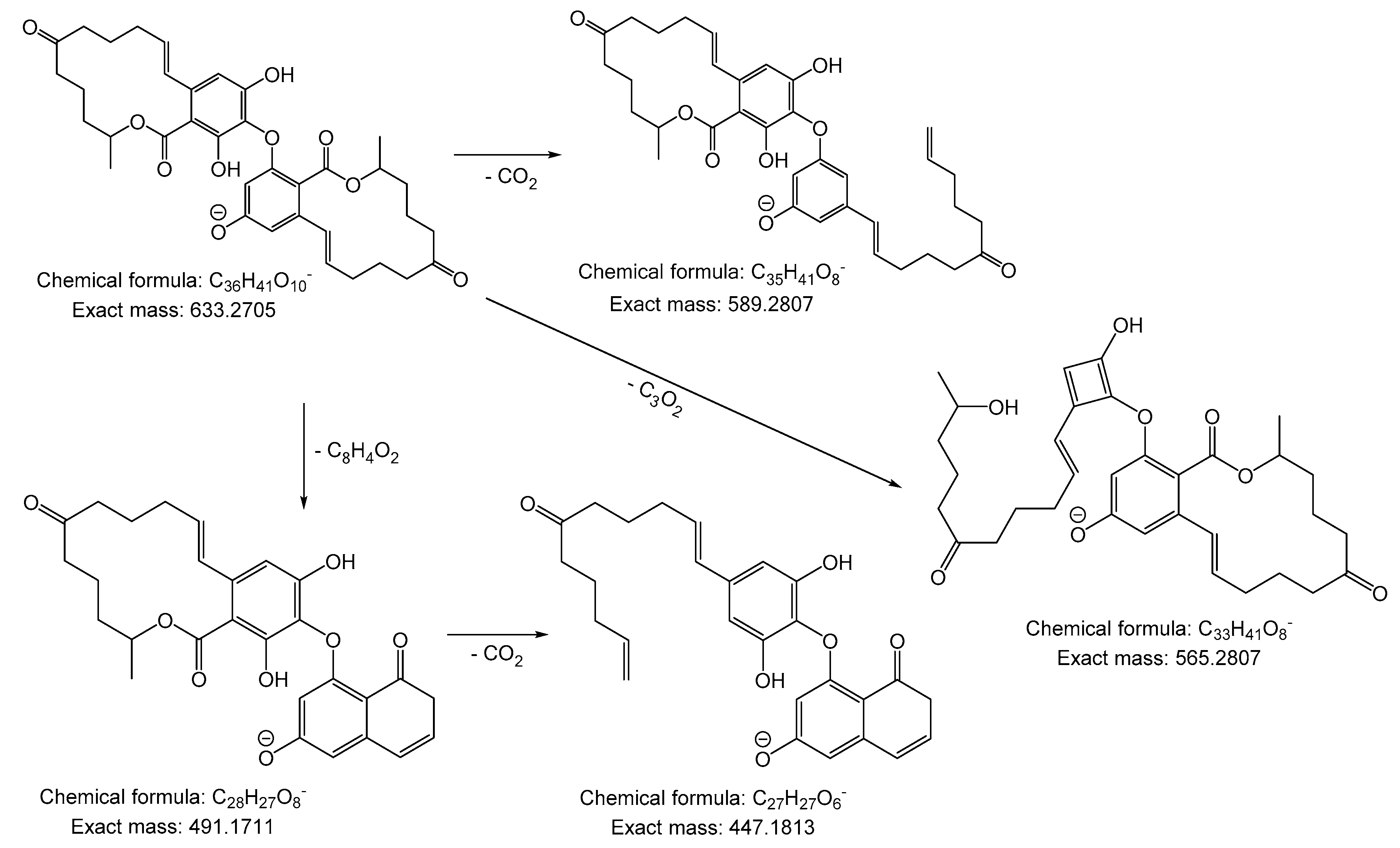
Compound 1 was obtained as a pale orange solid substance after electrochemical and chemical oxidation of ZEN with a molecular formula of C36H42O10, which was measured by high-resolution mass spectrometry with m/z 633.2658 [M − H]− in an ESI negative ionization mode (theoretical exact mass m/z 633.2702), as previously described [15]. The MS/MS measurements of compound 1 revealed a fragment with m/z 589.2756, which is due to the loss of CO2, and a fragment with m/z 565.2746 because of a loss of C3O2. The signal with m/z 491.1658, corresponds to a loss of C8H4O2, which led to the fragment m/z 447.1763 after the loss of CO2 (Supplement Figure S1). The assumed fragments and their chemical formulas with theoretical exact masses are shown in Figure 1.
The 1H-NMR spectrum of compound 1 measured at 400 MHz in MeOH-d4 gave the following information with 1.00–2.85 (m, H, J = 6.8 Hz), 1.34 (d, 3H, J = 6.2 Hz), 1.39 (d, 3H, J = 6.4 Hz), 5.06–5.16 (m, 1H), 5.20–5.36 (m, 1H), 5.71 (ddd, 1H, J = 4.0, 9.8, 15.6 Hz), 5.77 (d, 1H, J = 2.1 Hz), 6.03 (d, 1H, J = 4.4, 9.6, 14.6 Hz), 6.34–6.41 (m, 2H), 6.42 (s, 1H), and 6.58 (d, 1H, J = 2.1 Hz). The 13C-NMR measurements with 100 MHz revealed the following chemical shifts with 20.2, 21.0, 22.1, 22.6, 23.4, 32.4, 35.9, 37.6, 38.3, 43.9, 44.7, 73.7, 74.3, 100.8, 104.3, 106.7, 126.4, 130.2, 134.4, 137.2, 139.4, 158.0, 161.0, 172.1, 213.8, and 214.1 ppm (Supplement Figures S2 and S3). The assignments of the carbons and protons are summarized in Table 1.
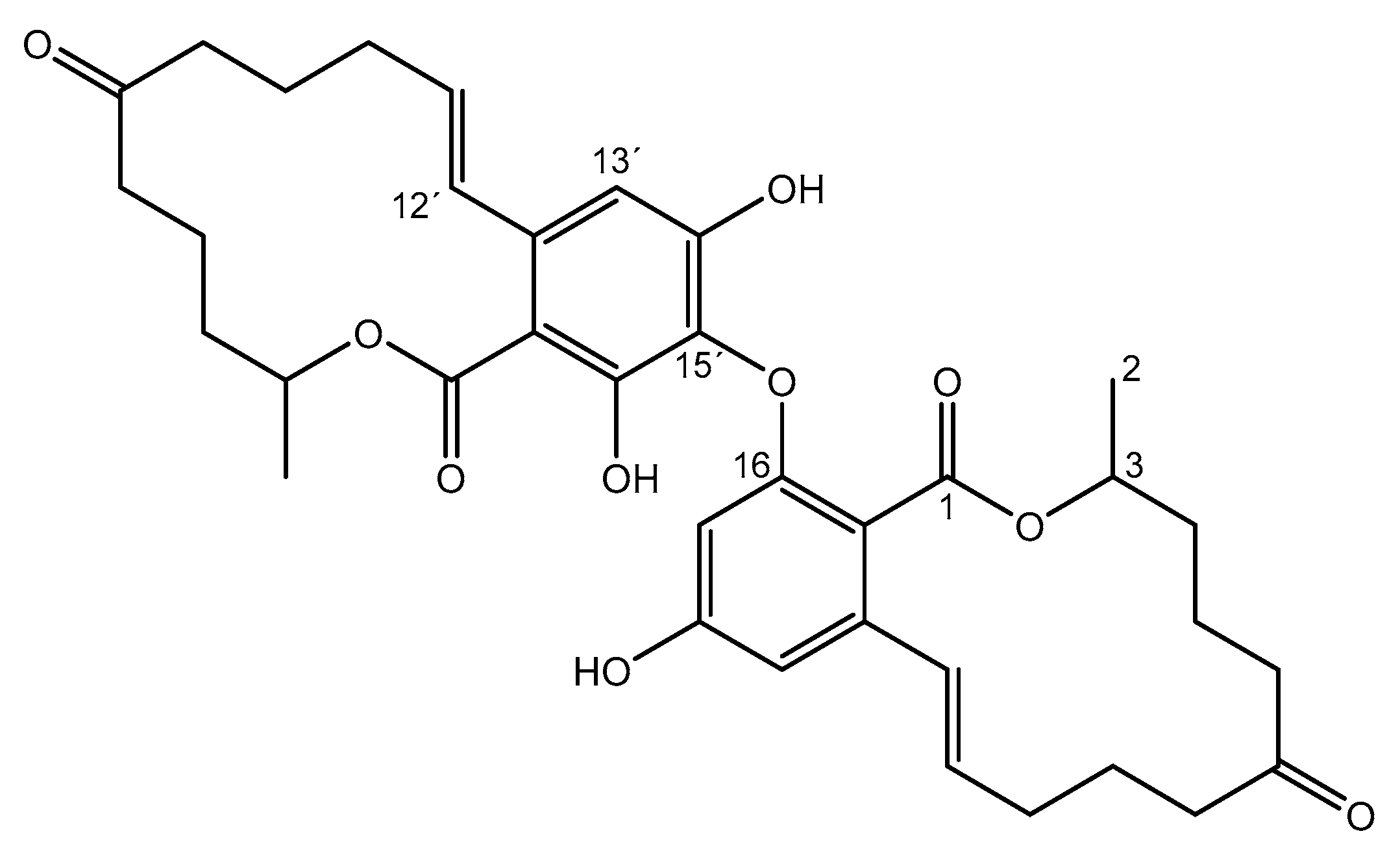
The 1H-NMR and COSY spectra revealed two aromatic systems (Supplement Figures S2 and S4). One of these aromatic systems contained two protons (d 6.58 ppm, 1H, J = 2.1 Hz, and d 5.77 ppm, 1H, J = 2.1 Hz) while the second aromatic system had only one proton (s 6.42 ppm, 1H). From this observation, the structures connected over the C-C linkages 15, 15′, 13, 13′, and 13, 15′ can be excluded since these dimers would have two aromatic systems with only one proton. Thus, only dimers having an C-O-C ether bridge between the monomers are possible. The COSY spectrum showed nearly identical chemical structures for the spectral part of the molecule (Supplement Figure S4). As a result, the dimerization of two ZEN molecules is only likely over an ether-link between 14-O-13′, 14-O-15′, 16-O-13′, or 16-O-15′. The HMBC spectrum (8 Hz) indicates a common coupling partner of the single aromatic proton and 12′-H, which is located in the aromatic-olefinic region (Supplement Figure S5). Due to the spatial proximity, it should be at the singlet (6.42 ppm) and, as a result, act around the 13′-H position. Consequently, an O-15′-linkage is conceivable.
The observed significant difference of the chemical shifts of 3-H and 3′-H indicated a different chemical environment. For a C-14 link, the closer chemical environment of 3-H and 3′-H would be very similar. A C-16 link, on the other hand, would be a greater steric influence and inductive effects would occur. Thus, the compound 16-O-15′-biaryl ether bond is the most likely structure, which is shown in Figure 2. Whether this dimer can be found naturally in food or feed remains to be analyzed in detail. Especially in plants, lichen, bacteria, and fungi regio-selective and stereo-selective biaryl C-C and biaryl ether C-O-C linkages are often found and it is conceivable that dimers of ZEN might not be uncommon in nature [16].
3. Materials and Methods
3.1. Chemicals and General Experimental Procedures
Zearalenone with a purity over 98% was obtained from Fermentek (Jerusalem, Israel) and Cerium(IV)sulfate was purchased from Sigma-Aldrich (Steinheim, Germany). Ultrapure water was generated by a Seralpur PRO 90 CN system (Ransbach-Baumbach, Germany). All standard chemicals were of p.a. grade and all solvents were of an HPLC grade. Electrochemical oxidation was achieved by using the Roxy® system synthesis cell (Antec, Zoeterwoude, The Netherlands) equipped with a platinum working electrode. The HPLC system used for fractionation consisted of an Agilent 1200 series autosampler, a 1260 series pump, a 1200 series diode array detector, and a column oven. A Macherey-Nagel Nucleosil C18 100-5 150 × 4.6 mm column (Düren, Germany) was used. The TripleTOF® 6600 Quadrupole Time-Of-Flight (QTOF) mass analyzer (Sciex, Darmstadt, Germany) was operated in negative ionization mode and 10 µM of the dimer sample were dissolved in methanol with 0.1% of formic acid. The used parameters were as follows:
Gas temperature 350 °C, ion source gas 1 (nitrogen) 20 L/min, ion source gas 2 (nitrogen) 15 L/min, curtain gas (nitrogen) 20 L/min, and ion spray voltage floating −4500 V. The MS/MS-spectrum was recorded in the targeted MS/MS mode with the following parameters: De-clustering potential (−15 V), collision energy (−40 V), and TOF Masses 100–640 Da. Confirmation of the 16-O-15′-biaryl ether-linked dimer structure was conducted by nuclear magnetic resonance spectroscopy (NMR). The NMR spectra were recorded in methanol-d4 on an Agilent 400-MR NMR spectrometer (Agilent Technologies, Waldbronn, Germany) at 30 °C. For the measurements, the ATB 5mm-probe head was operated at 399.8 MHz for 1H and 100.5 MHz for 13C data.
3.2. Electrochemical and Chemical Production of ZEN Dimer
Electrochemical: The optimal potential was tested by taking aliquots after different time points and using potentials of 0, 1.0, 1.4, and 1.8 V vs. Pd/H2. For the electrochemical oxidation, 80 mL of 250 µM ZEN in acetonitrile/water (50/50, v/v) was stirred for 48 h using 1.4 V vs. Pd/H2. The solution was subsequently evaporated to dryness by using a rotary evaporator and dissolved in water/acetonitrile (65/35, v/v) for HPLC fractionation.
Chemical: For the oxidative production of ZEN dimers, Ce(IV)sulfate was used. About 100 mg of ZEN and 350 mg of Ce(IV)sulfate were dissolved in 200 mL of acetonitrile/water (50/50, v/v) and stirred for two hours at 70 °C. Subsequently, the sample was stirred for 24 h at room temperature and a white precipitate was formed. The yellow solvent mixture was extracted three times with 20 mL of ethyl acetate. After the extraction, the ethyl acetate was colored yellow and the acetonitrile water mixture was colorless. After evaporation, a deep orange and highly viscous fluid was obtained. After freeze-drying, a pale orange solid was formed with a yield of 10%.
3.3. Purification of ZEN Dimer
For the separation of ZEN dimers, an already described HPLC method was adapted [15] by using a flowrate of 1.2 mL/min and an isocratic eluent consisting of water/acetonitrile, 65/35, v/v without modifiers. The ZEN dimer was isolated by collecting the fraction between 15.4 min and 16.2 min of the retention time by using a Foxy® R1 fraction collector (Teledyne ISCO, Nebraska, NE, USA). The purity of the dimer was determined to be 92% based on DAD spectra by using a wavelength at λ = 254 nm (Supplement Figure S7).
4. Conclusions
A new dimeric species of the food-relevant mycotoxin zearalenone was synthesized electrochemically and chemically with Ce(IV)sulfate and structurally identified. Among other possible dimers, the occurrence of the 16-O-15′-biaryl ether-linked dimer in food and feed is conceivable because the dimerization of phenolic compounds is often observed in plants, fungi, or lichen.
Supplementary Materials
The following are available online at https://www.mdpi.com/1420-3049/23/10/2624/s1, Figure S1. MS/MS fragmentation spectrum of the ZEN dimer obtained with Q-TOF in negative ionization mode. Figure S2. 1H-NMR spectrum of the 16-O-15′-biaryl ether-linked zearalenone dimer. Figure S3. 13C-NMR spectrum of the 16-O-15′-biaryl ether-linked zearalenone dimer. Figure S4. COSY spectrum of the 16-O-15′-biaryl ether-linked zearalenone dimer. Figure S5. HMBC spectrum of the 16-O-15′-biaryl ether-linked zearalenone dimer. Figure S6. HSQC spectrum of the 16-O-15′-biaryl ether-linked zearalenone dimer. Figure S7. HPLC-DAD chromatograms (λ = 254 nm) of the zearalenone dimer reaction mixture before fractionation (top) and after fractionation (bottom).
Author Contributions
Conceptualization, J.K. and L.H. Methodology, J.K., L.H., and M.K. Investigation, L.H. Data Curation, J.K. and M.K. Writing-Original Draft Preparation, J.K. Writing-Review & Editing, H.H. and M.K. Supervision, M.K and H.H.
Funding
This research received no external funding.
Acknowledgments
The authors would like to thank René Kudick (ASCA GmbH, Berlin, Germany) for the NMR measurements and analytical advice.
Conflicts of Interest
The authors declare no conflict of interest.
References
- Caldwell, R.W.; Tuite, J.; Stob, M.; Baldwin, R. Zearalenone production by fusarium species. Appl. Microbiol. 1970, 20, 31–34. [Google Scholar] [PubMed]
- Fraeyman, S.; Croubels, S.; Devreese, M.; Antonissen, G. Emerging fusarium and alternaria mycotoxins: Occurrence, toxicity and toxicokinetics. Toxins 2017, 9, 228. [Google Scholar] [CrossRef] [PubMed]
- Placinta, C.M.; D’Mello, J.P.F.; Macdonald, A.M.C. A review of worldwide contamination of cereal grains and animal feed with fusarium mycotoxins. Anim. Feed Sci. Technol. 1999, 78, 21–37. [Google Scholar] [CrossRef]
- Alexander, J.; Benford, D.; Boobis, A.; Ceccatelli, S.; Cottrill, B.; Cravedi, J.P.; Di Domenico, A.; Doerge, D.; Dogliotti, E.; Edler, L.; et al. Scientific opinion on the risks for public health related to the presence of zearalenone in food efsa panel on contaminants in the food chain. EFSA J. 2011, 9, 2197. [Google Scholar]
- Hetmanski, M.T.; Scudamore, K.A. Detection of zearalenone in cereal extracts using high-performance liquid chromatography with post-column derivatization. J. Chromatogr. 1991, 588, 47–52. [Google Scholar] [CrossRef]
- Koppen, R.; Riedel, J.; Proske, M.; Drzymala, S.; Rasenko, T.; Durmaz, V.; Weber, M.; Koch, M. Photochemical trans-/cis-isomerization and quantitation of zearalenone in edible oils. J. Agric. Food Chem. 2012, 60, 11733–11740. [Google Scholar] [CrossRef] [PubMed]
- Drzymala, S.S.; Binder, J.; Brodehl, A.; Penkert, M.; Rosowski, M.; Garbe, L.-A.; Koch, M. Estrogenicity of novel phase i and phase ii metabolites of zearalenone and cis-zearalenone. Toxicon 2015, 105, 10–12. [Google Scholar] [CrossRef] [PubMed]
- Pfohlleszkowicz, A.; Chekirghedira, L.; Bacha, H. Genotoxicity of zearalenone, an estrogenic mycotoxin—DNA adduct formation in female mouse-tissues. Carcinogenesis 1995, 16, 2315–2320. [Google Scholar] [CrossRef]
- Binder, S.B.; Schwartz-Zimmermann, H.E.; Varga, E.; Bichl, G.; Michlmayr, H.; Adam, G.; Berthiller, F. Metabolism of zearalenone and its major modified forms in pigs. Toxins 2017, 9, 56. [Google Scholar] [CrossRef] [PubMed]
- Borzekowski, A.; Drewitz, T.; Keller, J.; Pfeifer, D.; Kunte, H.-J.; Koch, M.; Rohn, S.; Maul, R. Biosynthesis and characterization of zearalenone-14-sulfate, zearalenone-14-glucoside and zearalenone-16-glucoside using common fungal strains. Toxins 2018, 10, 104. [Google Scholar] [CrossRef] [PubMed]
- Brodehl, A.; Moller, A.; Kunte, H.J.; Koch, M.; Maul, R. Biotransformation of the mycotoxin zearalenone by fungi of the genera rhizopus and aspergillus. FEMS Microbiol. Lett. 2014, 359, 124–130. [Google Scholar] [CrossRef] [PubMed]
- Kiessling, K.H.; Pettersson, H. Metabolism of zearalenone in rat liver. Acta Pharmacol. Toxicol. 1978, 43, 285–290. [Google Scholar] [CrossRef]
- Bravin, F.; Duca, R.C.; Balaguer, P.; Delaforge, M. In vitro cytochrome p450 formation of a mono-hydroxylated metabolite of zearalenone exhibiting estrogenic activities: Possible occurrence of this metabolite in vivo. Int. J. Mol. Sci. 2009, 10, 1824–1837. [Google Scholar] [CrossRef] [PubMed]
- Hildebrand, A.A.; Pfeiffer, E.; Rapp, A.; Metzler, M. Hydroxylation of the mycotoxin zearalenone at aliphatic positions: Novel mammalian metabolites. Mycotoxin Res. 2012, 28, 1–8. [Google Scholar] [CrossRef] [PubMed]
- Keller, J.; Haase, H.; Koch, M. Hydroxylation and dimerization of zearalenone: Comparison of chemical, enzymatic and electrochemical oxidation methods. World Mycotoxin J. 2017, 10, 297–307. [Google Scholar] [CrossRef]
- Wezeman, T.; Bräse, S.; Masters, K.-S. Xanthone dimers: A compound family which is both common and privileged. Nat. Prod. Rep. 2015, 32, 6–28. [Google Scholar] [CrossRef] [PubMed]
Figure 1.
Structure of compound 1.

Figure 2.
Postulated fragments of the ZEN-dimer, according to the MS/MS measurements with a molecular structure and formula along with the theoretical exact masses.
Figure 2.
Postulated fragments of the ZEN-dimer, according to the MS/MS measurements with a molecular structure and formula along with the theoretical exact masses.


Table 1.
1H-NMR and 13C-NMR data of 1 (400/100 MHz, Methanol-d4).
| Position | 1H (ppm) | 13C (ppm) |
|---|---|---|
| 1, 1′ | - | 213.8, 214.1 |
| 2 | 1.39 (d, J = 6.4 Hz, 3H) | 35.9 |
| 3 | 5.20–5.36 (m, 1H) | 73.7 |
| 10, 10′ | - | 32.4, 32.4 |
| 11 | 6.03 (ddd, J = 4.4, 9.6, 14.6 Hz, 1H) | 134.4 |
| 12 | 6.38–6.41 (m, 1H) | 130.2 |
| 13 | 6.58 (d, J = 2.1 Hz, 1H) | 106.7 |
| 14 | - | 161.0 |
| 15 | 5.77 (d, J = 2.1 Hz, 1H) | 100.8 |
| 2′ | 1.34 (d, J = 6.2 Hz, 3H) | 35.9 |
| 3′ | 5.06–5.16 (m, 1H) | 74.3 |
| 11′ | 5.71 (ddd, J = 4.0, 9.8, 15.6 Hz, 1H) | 137.2 |
| 12′ | 6.34–6.37 (m, 1H) | 126.4 |
| 13′ | 6.42 (s, 1H) | 104.3 |
© 2018 by the authors. Licensee MDPI, Basel, Switzerland. This article is an open access article distributed under the terms and conditions of the Creative Commons Attribution (CC BY) license (http://creativecommons.org/licenses/by/4.0/).
Share and Cite
MDPI and ACS Style
Keller, J.; Hantschke, L.; Haase, H.; Koch, M. Synthesis and Structural Identification of a Biaryl Ether-Linked Zearalenone Dimer. Molecules 2018, 23, 2624. https://doi.org/10.3390/molecules23102624
AMA Style
Keller J, Hantschke L, Haase H, Koch M. Synthesis and Structural Identification of a Biaryl Ether-Linked Zearalenone Dimer. Molecules. 2018; 23(10):2624. https://doi.org/10.3390/molecules23102624
Chicago/Turabian StyleKeller, Julia, Luisa Hantschke, Hajo Haase, and Matthias Koch. 2018. "Synthesis and Structural Identification of a Biaryl Ether-Linked Zearalenone Dimer" Molecules 23, no. 10: 2624. https://doi.org/10.3390/molecules23102624