Abstract
Sleeping sickness or human African trypanosomiasis (HAT) is a neglected tropical disease (NTD) threatening millions of peoples’ lives with thousands infected. The disease is endemic in poorly developed regions of sub-Saharan Africa and is caused by the kinetoplastid “protozoan” parasite Trypanosoma brucei. The parasites are transmitted to humans through bites of infected tsetse flies of the genus Glossina. The few available drugs for treatment of this disease are highly toxic, difficult to administer, costly and unavailable to poor rural communities bearing the major burden of this infection. Therefore, the search for new efficacious, safe and affordable drugs is of high importance. Vernonia lasiopus O. Hoffm., an indigenous African plant of the Asteraceae family, has been extensively reported to be used ethno-medicinally as a treatment for malaria. Its crude extracts obtained with solvents of different polarity were screened in vitro for anti-protozoal activity and the dichloromethane extract was found to be particularly active against Trypanosoma brucei rhodesiense (IC50 = 0.17 µg/mL). Bioassay-guided chromatographic fractionation of the dichloromethane extract led to the isolation and identification of six elemanolide type sesquiterpene lactones: 8-desacylvernolide, vernolepin, vernomenin, vernodalol, vernodalin and 11,13-dihydrovernodalin. All these elemanolide sesquiterpene lactones showed in vitro anti-trypanosomal activity. They were also tested for cytotoxicity against mammalian cells (L6 cell line). Vernolepin, the main component in the extract, was also the most potent with an IC50 value of 0.05 µg/mL against T.b. rhodesiense trypomastigotes. This compound showed a selectivity index of 14.5, which makes it an interesting candidate for in vivo tests and determination of its mechanism of action.
1. Introduction
Human African trypanosomiasis (HAT), also commonly known as sleeping sickness, is one of the neglected tropical diseases (NTDs) according to the World Health Organization (WHO) []. NTDs are ailments that afflict the poorest of the world’s communities which represent a non-lucrative sector for investment in drug development [,]. NTDs, besides being life-threatening infections, promote and advance poverty by disabling people, reducing their productivity in the case of adults and affecting learning, cognition and development in children, thus condemning these populations to more poverty [].
HAT is caused by two trypanosomatid parasites, Trypanosoma brucei rhodesiense and T. b. gambiense in East and West Africa, respectively []. These “protozoans” are transmitted to humans by the bites of infected tsetse flies of the genus Glossina occurring in 36 sub-Saharan African countries. The disease mainly burdens poor rural communities who lack adequate access to health facilities. Through sustained efforts, the reported cases have been on the decline with only 3796 cases in 2014 []. However, it is believed that many cases go undiagnosed and unreported with an estimated population of about 70 million people at risk of infection []. For the treatment of HAT, only few drugs are available [,]. These drugs are very costly, often unavailable, highly toxic and difficult to administer, particularly in the second stage of infection. Furthermore, an increasing number of relapses are reported after treatment [,,]. It is therefore clear that the search for leads towards new, affordable and available, safe and efficacious drugs is of high importance [,,].
Natural products from plants, microorganisms and marine organism have a long and successful tradition as drugs or as leads in drug development [,]. In continuation of our ongoing search for new anti-trypanosomal agents in plants [,,,], we set out to investigate the activity of Vernonia lasiopus O. Hoffm. (Asteraceae) and its secondary metabolites against protozoan parasites. V. lasiopus is an African indigenous shrub that reaches up to 3 m in height []. It belongs to the Vernonieae tribe in the Asteraceae family []. The plant is used ethnomedicinally to cure indigestion, stomach-ache, gastrointestinal problems, worms, malaria, scabies, venereal diseases, sores and also as a purgative [,,,,]. Organic crude extracts from this plant have been reported to possess antiplasmodial potency and cytotoxic activity in vitro [,,,]. No previous reports exist in the literature with respect to an anti-trypanosomal activity of V. lasiopus or its chemical constituents.
2. Results and Discussion
2.1. Antiprotozoal Activity of V. lasiopus Crude Extracts
Crude extracts of the aerial parts of V. lasiopus obtained with solvents of increasing polarity (n-hexane (Hex), dichloromethane (DCM), ethyl acetate (EtOAc), n-butanol (BuOH), methanol (MeOH) and water (H2O)) were tested for in vitro activity against T. b. rhodesiense (Tbr), T. cruzi (Tcr), Leishmania donovani (Ldon) and Plasmodium falciparum (Pf). For these tests, Tbr (STIB 900 strain) trypomastigotes, Tcr (Tulahuen C4 strain) amastigotes, Ldon (MHOM-ET-67/L82 strain) amastigotes, Pf (NF54 strain) intra-erythrocytic forms, and for cytotoxicity L6 rat-skeletal myoblasts were used. The results are summarized in Table 1. It becomes evident that the DCM extract showed the highest activity against Tbr with an IC50 value of 0.17 µg/mL. It also displayed some lower activity against Tcr and Pf. For this reason, bioassay-guided fractionation, isolation and characterization of the potentially active constituents of the DCM extracts was undertaken.

Table 1.
In vitro antiprotozoal and cytotoxic activity (IC50 values in µg/mL) of crude extracts from V. lasiopus. Selectivity indices (SI) represent the ratio of cytotoxic over antiprotozoal IC50 values. Data are means of two independent determinations ± standard deviation.
2.2. Bioassay-Guided Fractionation and Isolation of Elemanolide Sesquiterpene Lactones
The DCM extract was fractionated into twelve major fractions by column chromatography on silica. Representative fractions were subjected to bioassay against Tbr (Table 2). The fractions were also analyzed by UHPLC/+ESI QTOF MSMS.
Table 2.
In vitro and cytotoxic activity (IC50 values in µg/mL) of selected fractions of the DCM extract from V. lasiopus. For positive controls, see Table 1.
The anti-trypanosomal activity was concentrated between fractions five and nine. Analysis of these fractions by UHPLC/+ESI QTOF MS/MS showed that all the major components in the crude extract were distributed among these fractions. Subsequently, these major components (compounds 1–6, see Figure 1) were isolated. Compounds 5 and 6 were obtained only as an inseparable mixture in a ratio of approximately 2:1. The compounds 2, 3, 5, and 6 were unambiguously identified as vernolepin (2), vernomenin (3), vernodalin (5) and 11,13-dihydrovernodalin (6), on the grounds of their 1D and 2D NMR data in conjunction with their exact masses as obtained from UHPLC/+ESI QTOF MS/MS analyses, which were in full agreement with literature values [,,,,].
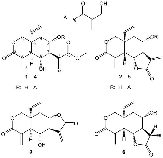
Figure 1.
Elemanolide sesquiterpene lactones isolated from V. lasiopus.
For compounds 1 and 4, the proton NMR data were similar to those of two compounds with the same structure reported from V. scorpioides []. Incidentally, these data were also identical to their C-10 epimers, epivernodalol and lasiopulide, earlier reported in V. lasiopus []. To eliminate this ambiguity, where the two sets of epimers are reported to display the same chemical shifts and 3JHH values of H-1 and H-2, a 1H/1H NOESY experiment for compound 1 was performed. From this, a correlation between H-1 and H-5 was noted, meaning that the two rings must be cis-fused. Therefore, by extension, the rings in compound 4, in view of the very similar NMR signals, can be expected to be cis-fused as well. Therefore, these compounds were confidently confirmed to be 8-desacylvernolide (1) and vernodalol (4) and the earlier report [] is probably to be revised. The other compounds, 2, 3, 5 and 6, are all cis-fused as well, without ambiguity in the literature data [,,,,].
2.3. Anti-Trypanosomal Activity of Isolated Elemanolides
All isolated compounds were tested for anti-trypanosomal activity and showed considerable degrees of activity. Vernolepin (2) was the most active with an IC50 value of 0.05 µg/mL (0.19 µM) and a selectivity index of 14.5. It is important to note that 2 was the major constituent of the DCM extract. This compound was approximately 3.5 times more active than the crude extract and it presents a ten-fold higher selectivity. It can therefore be concluded that vernolepin is a main contributor to the activity of extract. The other compounds showed slightly lower activity as well as lower selectivity. The full results are summarized in Table 3.
Table 3.
In vitro anti-trypanosomal and cytotoxic IC50 values in µg/mL (values in µM of pure compounds are reported in brackets) of elemanolides 1–6. For positive controls, see Table 1.
The mixture of vernodalin (5) and 11,13-Dihydrovernodalin (6) was the second most active with an IC50 value of 0.069 µg/mL. Vernodalin (5) has been previously reported to have anti-plasmodial activity with an IC50 value of 4.0 µg/mL against P. falciparum (multidrug resistance strain K1) []. The 11,13-Dihydrovernodalin (6) has also been reported to be active against P. falciparum chloroquine sensitive strain PoW and chloroquine-resistant strain Dd2 with IC50 values of 2.3 µg/mL and 1.1 µg/mL respectively [].
Vernodalol (4) was the third most potent compound with an IC50 value of 0.1 µg/mL (0.26 µM). This compound has been reported to have in vitro anti-plasmodial activity against P. falciparum (multidrug resistance strain K1) with an IC50 value of 4.2 µg/mL []. Compound 1 was the least active with an IC50 value of 0.779 µg/mL (2.5 µM) against Tbr. Comparing the activity of compounds 1 and 4, it is evident that compound 4 is more active by a factor of almost 10. This can be probably attributed to the ester moiety at C-8, which has an extra α, β-unsaturated carbonyl group. A similar trend could be expected for compounds 2 and 5. However, compound 5 was only tested in the 2:1 mixture with 6 so that such a correlation cannot be made with certainty.
Vernolepin (2) was chosen for in vivo studies due to its high activity and considerable selectivity. Its mechanism of action will also be evaluated and studies in this direction are in progress.
3. Materials and Methods
3.1. General Experimental Procedures
Analytical TLC was performed on silica gel plates 60 F254 (Merck Chemicals GmbH, Darmstadt, Germany) with various solvent systems consisting of Ethylacetate and hexane as the mobile phase. The plates were visualized under UV-light at 254/360 nm and then sprayed with anisaldehyde/sulfuric acid reagent and heated on a hot plate.
3.2. Plant Material
The aerial parts of the V. lasiopus O. Hoffm. plants were collected from Egerton University botanic garden in Njoro Kenya (0°21′44.8″ S 35°55′26.9″ E). The plants were identified by a taxonomist at the Biological Sciences Department at Egerton University. They were then air dried under shade at ambient temperature to constant weight. The dried materials were then powdered with a mill to 1 mm mesh size.
3.3. Extraction and Isolation
3.3.1. Small-Scale Extraction for Initial Bioassays
For the initial bioassays, the ground air-dried materials (100 g) were macerated sequentially with solvents (350 mL each) of increasing polarity (hexane, dichloromethane (DCM), ethyl acetate and methanol) for 48 h at room temperature with intermittent shaking. The extracts were filtered over whatman no. 1 filter paper, the solvent was removed under reduced pressure at 40 °C and the extracts were dried further in vacuo. The extracts were then tested for in vitro activity against T. b. rhodesiense, T. cruzi, L. donovani and P. falciparum (see Table 1). The DCM extract showed high potency against T. b. rhodesiense and was thus selected for bioassay-guided fractionation and isolation at a larger scale.
3.3.2. Soxhlet Extraction for Preparative Isolation
V. lasiopus dry powdered plant material (750 g, in portions of 250 g) was exhaustively extracted in a Soxhlet-apparatus for approximately 13 h with dichloromethane (total 5 L). The solvent was evaporated in vacuo at 40 °C to 76.45 g (10.1%) of extract. The extract (20 g) was fractionated by column chromatography on silica gel 60 (1250 g). The column was eluted with solvents of increasing polarity (100% hexane−100% ethyl acetate) of hexane–EtOAc mixture (v/v) as follows: 3 L hexane, 5 L (7:3), 2.5 L (1:1), 2.5 L (3:7), and 2.5 L EtOAc. The flow was maintained at about 1 mL/min. After collection of about 2.5 L of pre-eluate, 320 eluates of around 30 mL were collected and pooled into twelve fractions (F1–F12) according to their TLC profiles. Representative fractions were tested for anti-trypanosomal activity (see Table 2). After LC-Ms analysis of fractions F1–F12, fractions F6–F9 were recombined (4.8 g) and re-chromatographed over 250 g silica gel 60. The column was eluted with 4.5 L hexane–EtOAC–methanol mixture (7:4:1) at a flow rate of about 1 mL/min. A hundred and forty eluates of about 30 mL were collected. Compound 2 crystallized from eluates 45–65. The crystals were washed in hexane and re-crystallized in hexane: Ethyl acetate mixture (7:3). According to the TLC profiles of the rest of the eluates, they were combined in three sub-fractions F7a (980 mg), F7b (1.1 g) and F7c (995.9 mg). Sub fraction F7b was further separated on preparative HPLC to yield compounds 1 (6.2 mg) and 2 (38.6 mg). Sub fraction F7a was also separated by preparative HPLC to obtain compounds 3 (5.2 mg), 4 (5.6 mg) and a mixture of 5 and 6 (23.4 mg).
3.3.3. Purification by Preparative High Performance Liquid Chromatography (HPLC/UV-DAD)
The preparative HPLC isolation was performed on a Jasco (Groß-Umstadt, Germany) preparative HPLC system (pump: PU-2087 plus; diode array detector MD 2018 plus; column thermostat CO 2060 plus; autosampler AS 2055 plus; LC Net II ADC Chromatography Data Solutions; sample injection loop: 2000 µL) on a preparative reverse phase column Reprosil 100 C-18 (5 µm, 250 mm × 20 mm, Macherey-Nagel, Düren, Germany) with binary gradients of the mobile phase. The optimized mobile phase was composed of water (A) and methanol (B) using the following gradient conditions: 50%–55% of B (0 to 10 min) which was held for five minutes, 55%–65% of B (15 to 20 min), 65%–80% of B (20–25 min), 80%–100% of B (25 to 30 min), 100% of B (30 to 34 min) and another four minutes to return to the initial conditions. A flow rate of 9 mL/min and a column temperature of 40 °C were used in all separations. Chromatograms were recorded at 220, 254 and 265 nm. The retention times of compounds 1–6 were 8.42 min (1), 9.75 min (2), 10.82 min (3), 12.92 min (4), and 14.96 min (5 + 6), respectively.
3.4. Analysis of the Plant Extracts and Isolated Compound by UHPLC/+ESI-QTOF-MS/MS
The DCM extract, collected fractions and isolated compounds were dissolved in methanol at concentrations of 10 mg/mL and 0.1 mg/mL for crude extract or fractions, and for pure compounds, respectively. Chromatographic separations were performed on a Dionex Ultimate 3000 RS Liquid Chromatography System (Idstein, Germany) with a Dionex Acclaim RSLC 120, C18 column (2.1 × 100 mm, 2.2 µm) using a binary gradient (A: water with 0.1% formic acid; B: acetonitrile with 0.1% formic acid) at 0.8 mL/min: 0–9.5 min: linear from 5% B–100% B; 9.5–12.5 min: isocratic 100% B; 12.5–12.6 min: linear from 100% B down to 5% B; 12.6–15 min: isocratic 5% B. The injection volume was 5 µL. Eluted compounds were detected using a Dionex Ultimate DAD-3000 RS over a wavelength range of 200–400 nm and a Bruker Daltonics micrOTOF-QII quadrupole/time-of-flight mass spectrometer (Bremen, Germany) equipped with an Apollo electrospray ionization source in positive mode at 5 Hz over a mass range of m/z 50–1000 using the following instrument settings: nebulizer gas nitrogen, 5 bar; dry gas nitrogen, 9 L/min, 220 °C; capillary voltage 4500 V; end plate offset −500 V; transfer time 70 µs; collision gas nitrogen; collision energy and collision radio frequency (RF) settings were combined for each single spectrum of 1000 summations as follows: 250 summations with 20% base collision energy; 130 Vpp+ 250 summations with 100% base collision energy; 500 Vpp+ 250 summations with 20% base collision energy; and 130 Vpp + 250 summations with 100% base collision energy and 500 Vpp. Base collision energy was 50 eV for precursor ions with a m/z less than 500 and then linearly interpolated against m/z up to a maximum of 70 eV for precursor ions with a m/z of up to 1000. Internal dataset calibration (HPCmode) was performed for each analysis using the mass spectrum of a 10 mM solution of sodium formate in 50% isopropanol that was infused during LC re-equilibration using a divert valve equipped with a 20-µL sample loop. The retention times of compounds 1–6 are reported below (also see Supplementary materials).
3.5. Structural Analysis of Isolated Compounds
NMR spectra (1H, 13C, 1H/1H COSY, 1H/1H NOESY, 1H/13C HSQC, and 1H/13C HMBC) were recorded on a 600 MHz Agilent DD2 spectrometer. The spectra were obtained at 298 K in CDCl3. The CDCl3 solvent signals (1H; 7.260 ppm and 13C; 77.000 ppm) were used to reference the spectra. MestReNOVA v. 11 (Mestrelab Research, Chemistry Software Solutions, Santiago de Compostela, Spain) software was used to process and evaluate the spectra.
3.6. Analytical Data
8-Desacylvernodalol 1 UHPLC/+ESI-QTOF MS: Rt 2.16 min, MS (m/z): 309.1356 [M + H]+; calcd. for C16H21O6+: 309.1338); 1H-NMR (600 MHz, CDCl3; δ (ppm), intensity, mult., J (Hz)): 6.57 (1H, d, 1.7, H-15a); 6.40 (1H, d, 1.0, H-13a); 5.79 (1H, d, 1.0 Hz, H-13b); 5.71 (1H, br. dd, 1.7, 1.0, H-15b); 5.68 (1H, ddd, 17.5, 10.9, 0.9, H-1); 5.21 (1H, dd, 17.5, 0.9, H-2a); 5.18 (1H, dd, 10.9, 0.9, H-2b); 4.54 (1H, dd, 11.8, 0.9, H-14β); 4.26 (1H, dd, 11.9, 2.1, H-14α); 4.05 (1H, m, H-8); 3.97 (1H, td, 10.5, 2.5, H-6); 3.74 (3H, s, H-12-OMe); 2.42 (2H, m; H-5,H-7); 1.93 (1H, dd, 14.0, 4.7, H-9β); 1.51 (1H, dd, 14.0, 11.5, H-9α).
Vernolepin 2 UHPLC/+ESI-QTOF MS: Rt 3.65 min, MS (m/z): 277.1111 [M + H]+; calcd. for C15H17O5+: 277.1076); 1H-NMR (600 MHz, CDCl3; δ (ppm), intensity, mult., J (Hz)): 6.75 (1H, d, 0.9, H-15a); 6.25 (1H, d, 3.2, H-13a); 6.03 (1H, d, 3.0, H-13b); 5.96 (1H, d, 1.1, H-15b); 5.75 (1H, dd, 17.5, 11.0, H-1); 5.30 (1H,d, 11.0, H-2a); 5.27 (1H, d, 17.4, H-2b); 4.40 (1H, d, 12.1, H-14β); 4.23 (1H, dd, 1.8, 12.2, H-14α); 4.08 (1H, tdd, 10.3, 5.5, 4.7, H-8); 3.93 (1H, t, 11.3, H-6); 2.98 (1H, dd, 11.3,1.9, H-5); 2.68 (1H, tt, J = 11.1, 10.3, 3.1, H-7); 1.99 (1H, dd, 14.2, 4.7, H-9β); 1.81 (1H, d, 5.5, H-8-OH);1.68 (1H, dd, 14.2, 10.4, H-9α).
Vernomenin 3 UHPLC/+ESI-QTOF MS: Rt 3.91 min, MS (m/z): 277.1093 [M + H]+; calcd. for C15H17O5+: 277.1076); 1H-NMR (600 MHz, CDCl3; δ (ppm), intensity, mult., J (Hz)): 6.70 (1H, dd, 1.6, 0.5, H-15a); 6.67 (1H, ddt, 11.4, 10.3, 3.0, H-7); 6.23 (1H, dd, 3.1, 0.7, H-13a); 6.04 (1H, dd, 3.0, 0.7, H-13b); 5.81 (1H, dd, 1.6, 0.9, H-15b); 5.73 (1H, ddd, 17.4, 10.9, 0.8, H-1); 5.29 (1H, d, 10.9, H-2a); 5.25 (1H, dd, 17.4, 0.6 Hz, H-2b); 4.44 (1H, dd, 12.0, 0.9, H-14β); 4.30 (1H, dd, 12.0, 2.0, H-14α); 4.00 (1H, ddd, 12.4, 11.3, 4.1, H-8); 3.90 (1H, ddd, 9.5,2.2, H-6); 2.59 (1H, dd, 1H, 9.5,2.2, H-5); 2.44 (1H, d, 3.7, H-6-OH); 2.17 (1H, dd, 13.2,4.1, H-9β); 1.89 (1H, t, 12.8, H-9α).
Vernodalol 4 UHPLC/+ESI-QTOF MS: Rt 4.63 min, MS (m/z): 393.1571 [M + H]+; calcd. for C20H25O8+: 393.1549); 1H-NMR (600 MHz, CDCl3; δ (ppm), intensity, mult., J (Hz)): 6.64 (1H, d, 0.6, H-15a); 6.33 (1H, d, 0.9, H-13a); 6.17 (1H, q, 1.1, H-4′a); 5.81 (1H, q, 1.4, H-4′b); 5.74 (2H, m, H-13b,15b); 5.68 (1H, ddd, 17.6, 11.0, 0.9, H-1); 5.35 (1H, td, 11.4, 4.9, H-8); 5.26 (1H, d, 17.5, H-2a); 5.23 (1H, d, 10.9, H-2b); 4.67 (1H, dd, 12.0, 1.0, H-14β ); 4.35 (1H, dd, 12.0, 2.2, H-14α); 4.26 (2H, s, H-3’); 4.11 (1H, q, 7.1, H-6); 3.76 (3H, s, 12-OMe); 2.73 (1H, t, 10.8, H-7); 2.50 (1H, dd, 10.2, 2.1 Hz, H-5); 2.07 (1H, dd, 13.8, 4.9, H-9β); 1.61 (1H, dd, 13.8, 2.8, H-9α).
Vernodalin 5 UHPLC/+ESI-QTOF MS: Rt 4.99 min, MS (m/z): 361.1324 [M + H]+; calcd. for C19H21O7+: 361.1287); 1H-NMR (600 MHz, CDCl3; δ (ppm), intensity, mult., J (Hz)): 6.70 (1H, t, 0.9, H-15a); 6.26 (1H, d, J = 1.1, H-4’a); 6.16 (1H, d, 3.2, H-13a); 5.94 (1H, d, 1.4, H-4′b); 5.91 (1H, t, 1.2, H-15b); 5.68 (1H, m, H-1); 5.62 (1H, d, 2.9, H-13b); 5.26 (2H, m, H-2); 5.15 (1H, dt, 10.5, 5.3, H-8); 4.48 (1H, d, 12.3, H-14β); 4.32 (2H, s, H-3’ ); 4.24 (1H, m, H-14α); 4.06 (1H, m, H-6); 3.01 (1H, dd, 11.2, 1.5, H-5); 2.97 (1H, m, H-7); 2.19 (1H, dd, 14.2,4.7, H-9β); 1.64 (1H, m, H-9α).
11,13-Dihydrovernodalin 6 UHPLC/+ESI-QTOF MS: Rt 5.04 min, MS (m/z): 363.1481[M + H]+; calcd. for C19H23O7+: 363.1444); 1H-NMR (600 MHz, CDCl3; δ (ppm), intensity, mult., J (Hz)): 6.68 (1H, t, 0.9, H-15a); 6.23 (1H, d, 1.1, H-4’a); 5.92 (1H, br. d, 1.0, H-4′b); 5.88 (1H, br. d, 1.3, H-15b); 5.69 (1H, m, H-1); 5.25 (2H, m, H-2); 5.09 (1H, td, 10.9, 4.6, H-8); 4.51 (1H, d, 12.2, H-14β); 4.29 (2H, s, H-3’); 4.24 (1H, m, H-14α); 4.10 (1H, m, H-6); 2.86 (1H, d, 11.2, H-5); 2.06 (1H, dd, 14.2, 4.6, H-7); 2.64 (1H, dq, 12.0,6.9, H-11); 2.19 (1H, dd, 14.2,4.7, H-9β); 1.64 (1H, m, H-9α); 1.22 (3H, d, 0.6, H-13).
All spectral data were in full agreement with literature data [,,,,,].
3.7. In Vitro Bioassays
In vitro assays for the bioactivity of crude extracts and elemanolide sesquiterpene lactones against Trypanosoma brucei rhodesiense (bloodstream trypomastigotes, STIB 900 strain), T. cruzi (amastigotes, Tulahuen C4 strain), Leishmania donovani (amastigotes, MHOM-ET-67/L82 strain), Plasmodium falciparum (intra erythrocytic forms, NF54 IEF strain), and cytotoxicity test against mammalian cells (L6-cell-line from rat-skeletal myoblasts) were performed at the Swiss Tropical and Public Health Institute (Swiss TPH, Basel, Switzerland) according to established protocols as described earlier [].
5. Conclusions
The bioassay-guided fractionation of the dichloromethane extract of V. lasiopus leaves led to the isolation and characterization of six elemanolide sesquiterpene lactones (1–6). Compounds 5 and 6 were obtained as an inseparable mixture. Vernolepin (2) was the main constituent of the DCM extract and incidentally the most potent against Tbr with an in vitro IC50 value of 0.19 µM and a SI of 14.5. The 8-Desacylvernodalol 1 and vernodalol 4 also showed considerable in vitro activity against Tbr with SI values ˃10. Vernolepin (2) is an interesting candidate for in vivo and mechanism of action studies. These studies are already in progress. The bioactivity data obtained in this study complements previous data on sesquiterpene lactones obtained in our lab. Elemanolide type sesquiterpene lactones have not previously been reported to have anti-trypanosomal activity, therefore that the data reported here also broaden the knowledge on structure–anti-trypanosomal activity relationships of sesquiterpene lactones [,].
Supplementary Materials
The NMR and mass spectra of all isolates are provided as supplementary Figures S1–S32 available online.
Acknowledgments
The authors thank the Kenyan government, through National Commission of Science and Technology, in cooperation with the German Academic Exchange Service (NACOSTI-DAAD) for a doctoral fellowship to Njogu M. Kimani at the University of Muenster, Germany. The authors are grateful to S.T. Kariuki of Egerton University (Kenya) for the identification of the plant species. This study is part of the collaborative work within the Research Network Natural Products against Neglected Diseases (ResNetNPND, http://www.resnetnpnd.org/).
Author Contributions
N.M.K collected the plant materials, isolated and characterized the compounds, and wrote the manuscript. R.B and M.K. performed the biological activity tests. T.J.S. and J.C.M. conceived and initiated the study. T.J.S. finalized the manuscript and supervised the entire work.
Conflicts of Interest
The authors declare no conflict of interest.
References
- World Health Organization (WHO) Neglected Tropical Diseases. Available online: http://www.who.int/neglected_diseases/diseases/summary/en/ (accessed on 19 October 2016).
- Barrett, M.P.; Boykin, D.W.; Brun, R.; Tidwell, R.R. Human African trypanosomiasis: Pharmacological re-engagement with a neglected disease. Br. J. Pharm. 2007, 152, 1155–1171. [Google Scholar] [CrossRef] [PubMed]
- Schmidt, T.J.; Khalid, S.A.; Romanha, A.J.; Alves, T.M.; Biavatti, M.W.; Brun, R.; Da Costa, F.B.; de Castro, S.L.; Ferreira, V.F.; de Lacerda, M.V. The Potential of Secondary Metabolites from Plants as Drugs or Leads against Protozoan Neglected Diseases—Part I. Curr. Med. Chem. 2012, 19, 2128–2175. [Google Scholar] [PubMed]
- Hotez, P.J.; Yamey, G. The evolving scope of PLoS Neglected Tropical Diseases. PLoS Negl. Trop. Dis. 2009, 3, 2–3. [Google Scholar] [CrossRef]
- World Health Organization Trypanosomiasis, Human African (Sleeping Sickness). Available online: http://www.who.int/mediacentre/factsheets/fs259/en/ (accessed on 19 October 2016).
- Franco, J.R.; Simarro, P.P.; Diarra, A.; Jannin, J.G. Epidemiology of human African trypanosomiasis. Clin. Epidemiol. 2014, 6, 257–275. [Google Scholar] [PubMed]
- Hoet, S.; Opperdoes, F.; Brun, R.; Quetin-Leclercq, J. Natural products active against African trypanosomes: A step towards new drugs. Nat. Prod. Rep. 2004, 21, 353–364. [Google Scholar] [CrossRef] [PubMed]
- Schmidt, T.J.; Khalid, S.A.; Romanha, A.J.; Alves, T.M.; Biavatti, M.W.; Brun, R.; Da Costa, F.B.; de Castro, S.L.; Ferreira, V.F.; de Lacerda, M.V. The potential of secondary metabolites from plants as drugs or leads against protozoan neglected diseases—Part II. Curr Med. Chem. 2012, 19, 2176–2228. [Google Scholar] [CrossRef] [PubMed]
- Althaus, J.B.; Jerz, G.; Winterhalter, P.; Kaiser, M.; Brun, R.; Schmidt, T.J. Antiprotozoal activity of Buxus sempervirens and activity-guided isolation of O-Tigloylcyclovirobuxeine-B as the main constituent active against Plasmodium falciparum. Molecules 2014, 19, 6184–6201. [Google Scholar] [CrossRef] [PubMed]
- Schmidt, T.J.; Da Costa, F.B.; Lopes, N.P.; Kaiser, M.; Brun, R. Silico prediction and experimental evaluation of furanoheliangolide sesquiterpene lactones as potent agents against Trypanosoma brucei rhodesiense. Antimicrob. Agents Chemother. 2014, 58, 325–332. [Google Scholar] [CrossRef] [PubMed]
- Nogueira, M.; Da Costa, F.; Brun, R.; Kaiser, M.; Schmidt, T. ent-Pimarane and ent-Kaurane Diterpenes from Aldama discolor (Asteraceae) and Their Antiprotozoal Activity. Molecules 2016, 21, 1237. [Google Scholar] [CrossRef] [PubMed]
- Althaus, J.B.; Kaiser, M.; Brun, R.; Schmidt, T.J. Antiprotozoal activity of Achillea ptarmica (Asteraceae) and its main alkamide constituents. Molecules 2014, 19, 6428–6438. [Google Scholar] [CrossRef] [PubMed]
- Dharani, N.; Rukunga, G.; Yeneser, A.; Mbora, A.; Mwaura, L.; Dawson, I.; Jamnadass, R. Common Antimalarial Trees and Shrubs of East Africa: A Description of Species and a Guide to Cultivation and Conservation Through Use; Dawson, I., Ed.; The World Agroforestry Centre (ICRAF): Nairobi, Kenya, 2010. [Google Scholar]
- Koul, J.L.; Koul, S.; Singh, C.; Taneja, S.C.; Shanmugavel, M.; Kampasi, H.; Saxena, A.K.; Qazi, G.N. In vitro cytotoxic elemanolides from Vernonia lasiopus. Planta Med. 2003, 69, 164–166. [Google Scholar] [CrossRef] [PubMed]
- Njenga, D.; Irungu, B.; Mbaria, J.; Mutai, C. Antiplasmodial, Cytotoxic and Acute Toxicity Activities of Vernonia lasiopus O. Hoffman. Afr. J. Pharmacol. Ther. 2015, 4, 16–20. [Google Scholar]
- Katuura, E.; Waako, P.; Ogwal-Okeng, J.; Bukenya-Ziraba, R. Traditional treatment of malaria in Mbarara District, western Uganda. Afr. J. Ecol. 2007, 45, 48–51. [Google Scholar] [CrossRef]
- Muregi, F.W.; Ishih, A.; Miyase, T.; Suzuki, T.; Kino, H.; Amano, T.; Mkoji, G.M.; Terada, M. Antimalarial activity of methanolic extracts from plants used in Kenyan ethnomedicine and their interactions with chloroquine (CQ) against a CQ-tolerant rodent parasite, in mice. J. Ethnopharmacol. 2007, 111, 190–195. [Google Scholar] [CrossRef] [PubMed]
- Rachuonyo, H.O.; Ogola, P.E.; Arika, W.; Nyamai, D.; Wambani, J. In Vitro Antimicrobial Activity of Crude Leaf Extracts from Aloe secundiflora, Bulbine frutescens, Vernonia lasiopus and Tagetes minuta against Salmonella typhi. J. Tradit. Mad. Clin. Naturop. 2016, 5, 2–4. [Google Scholar]
- Irungu, B.N.; Rukunga, G.M.; Mungai, G.M.; Muthaura, C.N. In vitro antiplasmodial and cytotoxicity activities of 14 medicinal plants from Kenya. S. Afr. J. Bot. 2007, 73, 204–207. [Google Scholar] [CrossRef]
- Katuura, E.; Waako, P.; Tabuti, J.R.S.; Bukenya-Ziraba, R.; Ogwal-Okeng, J. Antiplasmodial activity of extracts of selected medicinal plants used by local communities in western Uganda for treatment of malaria. Afr. J. Ecol. 2007, 45, 94–98. [Google Scholar] [CrossRef]
- Ganjian, I.; Kubo, I.; Fludzinski, P. Insect antifeedant elemanolide lactones from Vernonia amagydalina. Phytochemistry 1983, 22, 2515–2526. [Google Scholar] [CrossRef]
- Jakupovic, J.; Baruah, R.N.; Thi, T.V.; Bohlmann, F.; Msonthi, J.D.; Schmeda-Hirschmann, G. New Vernolepin Derivatives from Vernonia glabra and Glaucolides from Vernonia scorpioides. Planta Med. 1985, 51, 378–380. [Google Scholar] [CrossRef] [PubMed]
- Looi, C.Y.; Arya, A.; Cheah, F.K.; Muharram, B.; Leong, K.H.; Mohamad, K.; Wong, W.F.; Rai, N.; Mustafa, M.R. Induction of Apoptosis in Human Breast Cancer Cells via Caspase Pathway by Vernodalin Isolated from Centratherum anthelminticum (L.) Seeds. PLoS ONE 2013, 8, e56643. [Google Scholar] [CrossRef] [PubMed]
- Laekeman, G.M.; Mertens, J.; Totté, J.; Bult, H.; Vlietinck, A.J.; Herman, A.G. Isolation and Pharmacological Characterization of Vernolepin. J. Nat. Prod. 1983, 46, 161–169. [Google Scholar] [CrossRef] [PubMed]
- Chukwujekwu, J.C.; Lategan, C.A.; Smith, P.J.; Van Heerden, F.R.; Van Staden, J. Antiplasmodial and cytotoxic activity of isolated sesquiterpene lactones from the acetone leaf extract of Vernonia colorata. S. Afr. J. Bot. 2009, 75, 176–179. [Google Scholar] [CrossRef]
- Ohigashi, H.; Huffman, M.A.; Izutsu, D.; Koshimizu, K.; Kawanaka, M.; Sugiyama, H.; Kirby, G.C.; Warhurst, D.C.; Allen, D.; Wright, C.W.; et al. Toward the chemical ecology of medicinal plant use in chimpanzees: The case of Vernonia amygdalina, a plant used by wild chimpanzees possibly for parasite-related diseases. J. Chem. Ecol. 1994, 20, 541–553. [Google Scholar] [CrossRef] [PubMed]
- Kraft, C.; Jenett-Siems, K.; Siems, K.; Jakupovic, J.; Mavi, S.; Bienzle, U.; Eich, E. In vitro antiplasmodial evaluation of medicinal plants from Zimbabwe. Phytother. Res. 2003, 17, 123–128. [Google Scholar] [CrossRef] [PubMed]
- Schmidt, T.J.; Nour, A.M.M.; Khalid, S.A.; Kaiser, M.; Brun, R. Quantitative structure—Antiprotozoal activity relationships of sesquiterpene lactones. Molecules 2009, 14, 2062–2076. [Google Scholar] [CrossRef] [PubMed]
Sample Availability: Samples of the compounds 2 and 5 + 6 are available from the authors. |
© 2017 by the authors. Licensee MDPI, Basel, Switzerland. This article is an open access article distributed under the terms and conditions of the Creative Commons Attribution (CC BY) license (http://creativecommons.org/licenses/by/4.0/).